Article

Dichlororesorcinols Produced by a Rhizospheric Fungi of Panax notoginseng as Potential ERK2 Inhibitors

1
Department of biopharmaceutical Sciences, School of Life Science and Biopharmaceutics, Shenyang Pharmaceutical University, Shenyang 110016, China
2
Zhen Cui (Jiangsu) Enzyme Biology Ltd., Suqian 223800, China
*
Author to whom correspondence should be addressed.
These authors contributed equally to this work.
Fermentation 2023, 9(6), 517; https://doi.org/10.3390/fermentation9060517
Submission received: 31 March 2023 / Revised: 20 May 2023 / Accepted: 24 May 2023 / Published: 27 May 2023
(This article belongs to the Special Issue New Research on Fungal Secondary Metabolites)

Abstract

:
Rhizospheric fungi of medicinal plants are important sources for discovering novel and valuable secondary metabolites with potential pharmaceutical applications. In our research, five new dichlororesorcinols (1–5) and five known metabolites (6–10) were separated from the secondary metabolites of Chaetomium sp. SYP-F6997, which was isolated from the rhizospheric soil of Panax notoginseng. The identification of these compounds was confirmed using various spectroscopic techniques including ESI-MS, UV, IR, NMR and ECD analyses. These findings highlight the potential of rhizospheric fungi as a rich source of novel bioactive compounds. In addition, chiral HPLC was used to successfully separate the enantiomers compound 4 and compound 5, and TDDFT-ECD/optical rotation calculations were used to test their absolute configurations. This is the first report of compounds 1–10 from the genus Chaetomium, and the first report of compounds 1–5 and 7 from the family Chaetomiaceae. We proposed plausible biosynthetic pathways for dichlororesorcinols 1–6 based on their analogous carbon skeleton. These findings provide insights into the biosynthesis of these compounds and expand our understanding of the secondary metabolites produced by Chaetomium sp. SYP-F6997. To evaluate their potential as therapeutic agents, we investigated the cytotoxic activity of all the isolated metabolites against cell lines H9, HL-60, K562, THP-1 and CEM using the MTT method. The new compounds 1 and 2 exhibited significant cytotoxic activities against H9 and CEM, with IC50 values lower than 10 µM. To further explore the potential mechanisms of action, we performed molecular docking studies to investigate the interactions between compounds 1 and 2 with the potential target ERK2. Our results demonstrate that the compounds exhibited strong binding abilities and formed H-bond interactions with ERK2, providing support for their potent antitumor activities and promising potential as lead molecules for the development of antitumor therapeutics.

1. Introduction

Panax notoginseng F. H. Chen (Araliaceae) has been an important herbal drug for centuries in Asia [1]. It has been widely used for its antidiabetic, antioxidant and antitumor biological activities [2,3]. Medicinal plants usually harbor endophytic or rhizospheric fungi which are regarded as sources of important pharmaceutical products [4,5,6,7]. Considering the symbiosis between the host plant P. notoginseng and its related endophytic or rhizospheric fungi, we conducted research to discover novel substances with anti-cancer or antimicrobial abilities through microbial fermentation [5,8,9].
ERKs are members of protein serine/threonine kinases. ERKs play a mediating and amplifying role in tumor invasion and metastasis [10]. They are key targets belonging to the mitogen-activated family. ERK1 and ERK2 are subtypes of ERKs, and they have been studied in many malignant tumors such as breast, lung, bladder and ovarian cancer [11,12]. ERK1 and ERK2 are components of the Ras/Raf/MEK/ERK pathway and they are involved in triggering many cellular reactions in the cancer process [13]. There have been various inhibitors of ERK1/2 reported in the past several decades [11,12,14]. However, most of them are still in clinical trials without FDA approval. The numbers of ERK1/2 inhibitors currently in clinical trials is still less than those targeting other pathways [14]. Due to the clinical significance of ERK1/2 inhibitors, they are continuously explored in resistant tumor cell lines by researchers. The inhibition of ERKs can restrain cell proliferation, differentiation and survival [15,16]. Therefore, compounds that can bind to ERKs might be candidates for potent ERK inhibitors. Despite an 84% sequence similarity between ERK1 and ERK2, ERK2 plays a key role in driving cell proliferation [17,18,19]. Thus, a molecular docking approach was used to explore the probable mechanism between the active substances and ERK2 in this study. Secondary metabolites with novel structures and significant biological activities from endophytic fungi have always been research hotspots [20,21,22].
In this study, five new dichlororesorcinol compounds (1–5) and five known compounds (6–10) were isolated and identified from microbial fermentation of Chaetomium sp. All the substances (1–10) were tested for cytotoxicity. Further, a molecular docking analysis was carried out for mechanism investigation.

2. Materials and Methods

2.1. General Experimental Procedures

The UV and optical rotation data were obtained using a P-2000 digital polarimeter (JASCO, Kyoto, Japan). A MOS-450 spectrometer (Bio-Logic Science, Claix, France) was used to record the circular dichroism (CD) spectrum. Bruker NMR spectrometers (Rheinstetten, Bremen, Germany) were used for 1H (400 MHz), 13C (100 MHz) and 2D NMR spectra. A Bruker Customer TOF-Q mass spectrometer (MA, Daltonics, Germany) was used to measure the mass spectra (HR-ESI-MS). CH3CN, MeOH, n-Hexane and isopropyl alcohol of chromatographic grade (Merck, Darmstadt, Germany) were used for HPLC analysis. All solvents used were bought from Tianjin Kemiou Chemical Reagent Company (Tianjing, China). Silica gel, including 100–200 mesh and 200–300 mesh, was bought from Qingdao Marine Chemical Factory (Qingdao, China). Semi-preparative and RP-HPLC isolations were performed using a Shimadzu LC-20AB instrument equipped with a YMC C18 column (10 mm × 250 mm, 5 µm) and an SPD-20A detector. Chiral HPLC separation was achieved with a column of CHIRALCEL-OJ-H (4.6 mm × 250 mm, 5 µm) equipped with a Shimadzu LC-20AB facility using a Shimadzu SPD-10A UV detector.

2.2. Fungal Material and Fermentation

The fungal strain in this research was isolated from the rhizospheric soil of Panax notoginseng, collected from Wenshan, Yunnan, China, and identified based on the DNA sequence (ITSI-5.8S-ITS2-28S), which has been submitted to GenBank with accession number OQ945315. The BLAST search result revealed that the isolate had the highest identity (99.24%) with the species Chaetomium udagawae. The colony morphology characteristics on a PDA medium are shown in Figure S1A,B. The microstructure was observed under an optical microscope, as shown in Figure S1C,D. A mixture of water and rice at a ratio of 55 mL/40 g was added to a conical flask (250 mL) and sterilized for 30 min at 121 °C. The spores of Chaetomium sp. were then added. One hundred and twenty solid fermentation flasks were incubated for 30 days at a temperature of 26 °C. The fermentation mixture was then crushed and extracted with ethyl acetate (30 L) overnight. This operation was repeated four times to obtain crude substances (110 g).

2.3. Isolation of Secondary Metabolites

The crude substances (110 g) were dissolved in 90% MeOH–H2O (500 mL), and the mixture was extracted with hexane (500 mL) three times to obtain a residue (70 g). Two silica gel columns with pore diameters of 100–200 mesh and 200–300 mesh were used to separate the extract (70 g) by column chromatography (CC). Petroleum ether-EtOAc was used as the eluent with a gradient from 80:1 to 3:1.
As results, six fractions (A–F) were obtained. The ODS in gradient from MeOH–H2O 10:90 to 90:10 was used for isolating Fraction B (22.9 g), and eight fractions (B1–B8) were obtained. Fraction B3 (1.8 g) was added to a preparative HPLC (MeOH–H2O, 70%) as an eluent, yielding substances 1 (4.6 mg, tR = 30.0 min) and 2 (3.5 mg, tR = 32.0 min). Subfraction B3 (3.7 g) was isolated by ODS using MeOH-H2O in gradient from 10:90 to 60:40. Then, Sephadex LH-20 (isocratic elution in MeOH) and preparative HPLC (isocratic elution in MeOH–H2O, 60%) were used to receive substances 3 (3.9 mg, tR = 26.1 min), 4 (4.2 mg, tR = 28.8 min) and 5 (4.8 mg, tR = 30.4 min). Compounds 6 (4.8 mg, tR = 33.5 min), 7 (6.3 mg, tR = 34.8 min) and 8 (7.7 mg, tR = 35.7 min) were separated from subfraction C (6.7 g) by ODS using MeOH-H2O in gradient from 10:90 to 60:40. Then Sephadex LH-20 (isocratic elution in MeOH) and preparative HPLC (gradient elute in MeOH–H2O, 70–30%) were used for further isolation. Preparative HPLC was used to separate subfraction C 3–6 (220 mg) using 71% MeOH-H2O to obtain substances 9 (8 mg, tR = 38.5 min) and 10 (9 mg, tR = 39.5 min).
Furthermore, compound 4 and 5 were further isolated into the pure enantiomers 4a (1.0 mg, tR = 14.3 min), 4b (1 mg, tR = 27.7 min), 5a (1 mg, tR = 25.8 min) and 5b (1 mg, tR = 34.2 min), respectively, by chiral HPLC column using a CHIRALCEL-OJ-H (4.6 mm × 250 mm, 5 µm, n-hexane: isopropyl alcohol 90: 10, 0.8 mL/min).
Cosmochlorin F (1): Amorphous colorless and white powder; UV: 210 nm, 292 nm IR (KBr): 3439, 2922, 1616, 1346 cm−1; [α] 20D −62.1 (c 0.1, MeOH); HR-ESI-MS m/z 291.0691 [M+H]+ (calcd for 291.0689, C13H1735Cl2O3), m/z 293.0662 [M+H]+ (calcd for 293.0660, C13H1735Cl37ClO3); 1H-NMR (400 MHz, DMSO-d6) data listed in Table 1; 13C-NMR (100 MHz, DMSO-d6) data listed in Table 2.
Cosmochlorin G (2): Amorphous colorless and white powder; UV: 210 nm, 295 nm IR (KBr): 3421, 2921, 1630, 1335 cm−1; HR-ESI-MS m/z 249.0081 [M+H]+ (calcd for 249.0079, C10H1135Cl2O3), m/z 251.0049 [M+H]+ (calcd for 251.0046, C10H1135Cl37ClO3); 1H (400 MHz, DMSO-d6) data listed in Table 1; 13C NMR (100 MHz, DMSO-d6) data listed in Table 2.
Cosmochlorin H (3): Amorphous colorless and white powder; UV: 210 nm, 295 nm IR (KBr): 3393, 2947, 1658, 1413, 1031 cm−1; HR-ESI-MS m/z 388.1196 [M + Na]+ (calcd for 388.1194, C16H23Na35Cl2NO4); 1H (400 MHz, DMSO-d6) data listed in Table 1; 13C NMR (100 MHz, DMSO-d6) data listed in Table 2.
Cosmochlorin I (4): Amorphous colorless and white powder; UV: 210 nm, 296 nm IR (KBr): 3386, 2948, 1652, 1413, 1031 cm−1; HR-ESI-MS m/z 360.3237 [M-H] (calcd for 360.3235, C16H2035Cl2NO4); 1H (400 MHz, DMSO-d6) data listed in Table 1; 13C NMR (100 MHz, DMSO-d6) data listed in Table 2.
Cosmochlorin J (5): Amorphous colorless and white powder; UV: 210 nm, 295 nm IR (KBr): 3395, 2921, 1645, 1429, 1046 cm−1; HR-ESI-MS m/z 347.0779 [M-H] (calcd for 347.0783, C15H1735Cl2O5), m/z 349.0762 [M-H] (calcd for 249.0760, C15H1735Cl37ClO5), m/z 351.0838 [M-H] (calcd for 351.0840, C15H1737Cl2O5); 1H (400 MHz, DMSO-d6) data listed in Table 1; 13C NMR (100 MHz, DMSO-d6) data listed in Table 2.

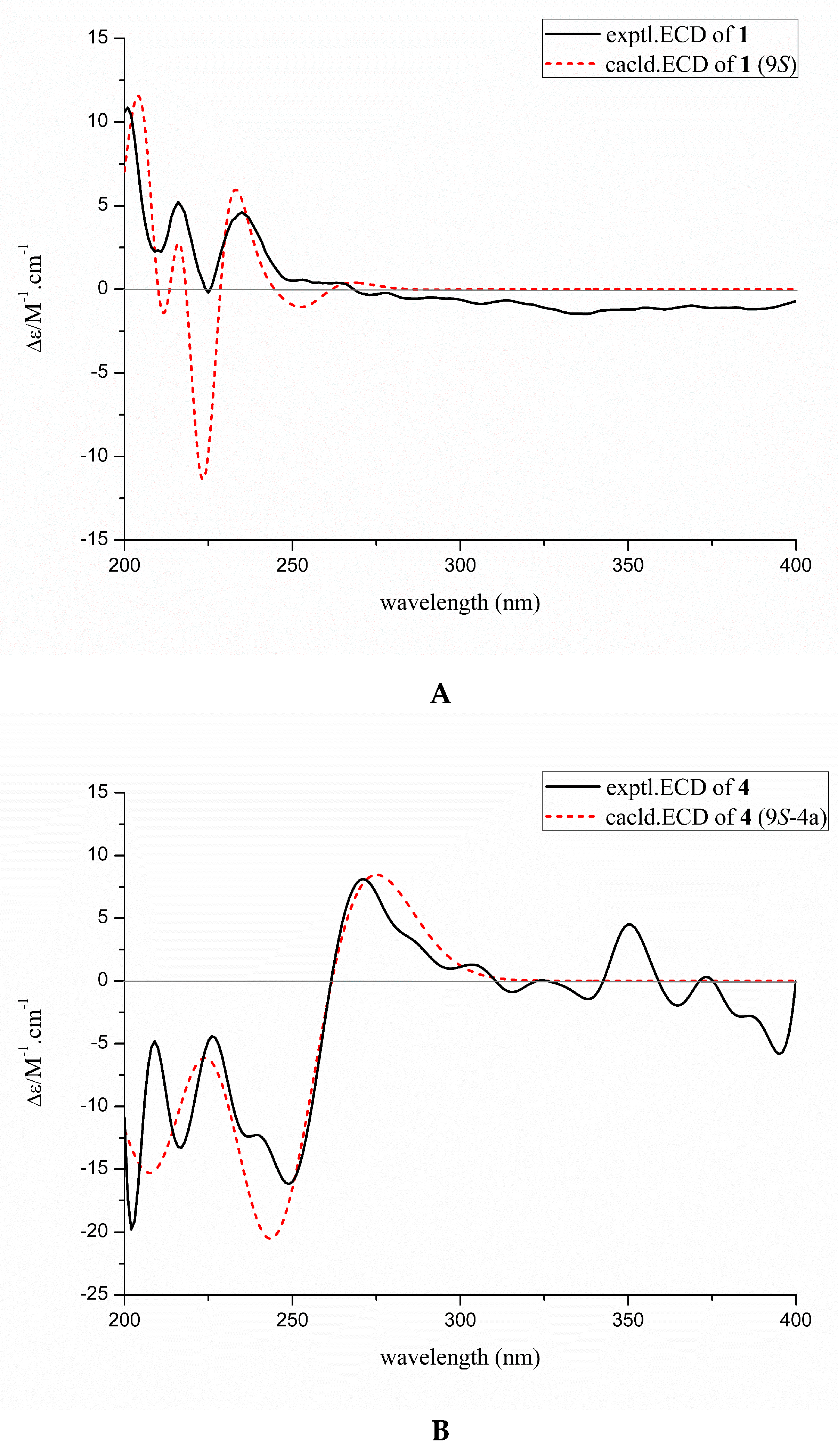
2.4. ECD Calculations

Spartan was employed to carry out the absolute configurations of substances 1 and 4.
For further optimization, the conformational analysis was performed using the density functional theory method at the B3LYP/6-31G(d) level in the Gaussian 09 program package [23]. Time-dependent density functional theory (TDDFT) was used for the theoretical electronic circular dichroism (ECD) calculations. The parameters were selected at the B3LYP/6-31 + G(d,p) level with Boltzmann processing in MeOH using the conductor-like polarizable continuum model (CPCM). SpecDis 1.51 was used for the calculated ECD curves.

2.5. Optical Rotation Calculations

Sybyl-X 2.0 was used for conformational analyses of compound 5. A MMFF94S force field was used with an energy cut-off of 2.5 kcal/mol [7]. A GAUSSIAN 09 program was performed at the B3LYP-D3(BJ)/6–31G* level in PCM MeOH [7]. DFT was optimized for all the conformers. To obtain the Boltzmann-weighted conformer population, the Gibbs free energy equation (ΔG = −RT ln K) was carried out. Gaussian 09 using the DFT method was performed for the optical rotation calculations at the CAM-B3LYP/6-311 + G (2d, p) level in polarizable continuum model (PCM) in MeOH solvent [7].

2.6. Cytotoxic Assay

The cytotoxicities of compounds 1−10 toward H9 (Human T lymphoid cell lines), HL-60 (Human promyelocytic acute leukemia cell lines), K562 (Human chronic myelogenous leukemia cancer cell lines), THP-1 (mononuclear macrophage cell lines) and CEM (leukemia cell lines) were assayed using the MTT method [24]. The cell lines were all bought from the Chinese Academy of Sciences Committee on Type Culture Collection Cell Bank (Shanghai, China).

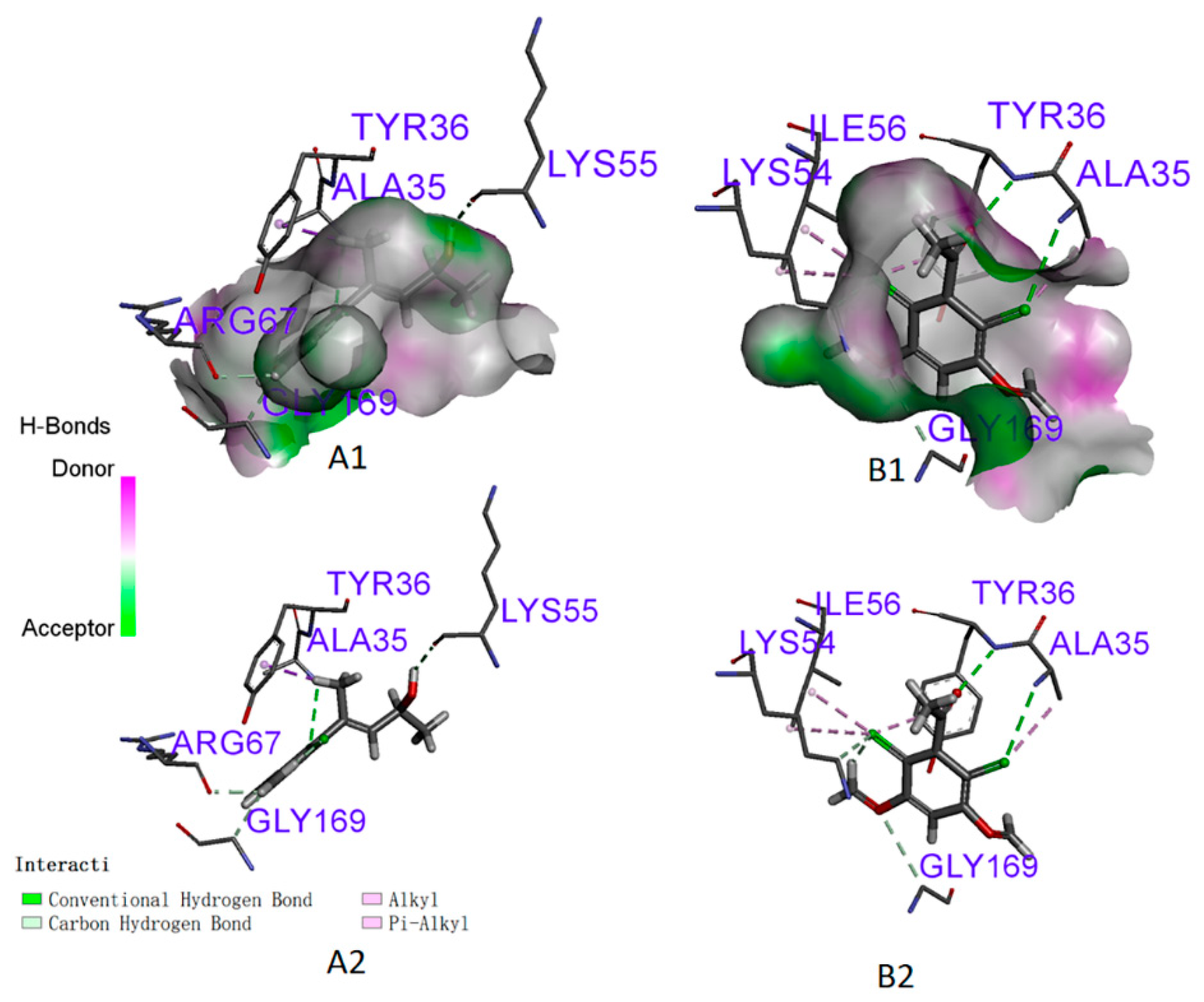
2.7. Molecular Docking

The Protein Data Bank RCSB (https://www.rcsb.org/#Category-search, accessed on 11 April 2023) was used to obtain the crystal structure of ERK2 with PDB Code 6GDQ [14]. The molecular docking method was used on the basis of our former description [25]. Discovery Studio 4.3 (Accelrys Inc., San Diego, CA, USA) was used to prepare the protein structure. For docking studies, water molecules were deleted and hydrogen atoms were appended, and the chemical structures were handled in the format of PDB. Sybyl software (Tripos, St Louis, USA) was used for the 3D structures of substances 1 and 2 by adding Gasteiger–Hückel charges. The ligand was submitted to minimize energy using the Tripos force field parameters [26]. A Molegro Virtual Docker 4.0 (Molegro ApS, Aarhus, Denmark) was put into effect by blind docking. The protein can be covered by the 3D docking grid sufficiently.

3. Results and Discussion

3.1. Structure Elucidation of Secondary Metabolites

Substance 1 was obtained as an amorphous colorless and white powder. The molecular formula of compound 1 was determined to be C13H17Cl2O3 based on HR-ESI-MS ([M+H]+ at m/z 291.0691, calcd 291.0689, C13H17Cl2O3 and m/z 293.0662, calcd 293.0660, C13H1735Cl37ClO3). In the spectrum of 1H NMR (Table 1), there were feature semaphores for a phenyl ring at δH 6.84 (1H, s), two methoxyls at δH 3.90 (6H, d), two methine signals at δH 5.16 (1H, dd, J = 4.0, 8.0 Hz) and δH 4.53 (1H, t, J = 4.0 Hz), as well as two feature semaphores of methyl proton at δH 1.80 (3H, d) and δH 1.18 (3H, d, J = 4.0 Hz). The spectra of 13C NMR (Table 2) and HSQC of 1 offered twelve aromatic carbons. The resonances at δC 98.0 (C-1), 154.1 (C-2), 154.2 (C-6), 111.8 (C-3), 112.1 (C-5) and 142.7 (C-4) were assigned similar to the carbons of Cosmochlorin D [21], which were also replaced by two methyls and two chlorines. These partial structures were further acknowledged by means of long-range correlations with the experiments of HMBC as listed in Figure 1 and Figure 2. The correlations received from HMBC such as H-1(δH 6.84) to C-3 (δC 111.8), C-5 (δC 112.1) and C-2,6 (δC 154.1) confirmed the existence of a benzene ring. In the spectrum of H-H COSY (Figure S2-4), there had been correlations between H-1 (δH 6.84) and 2,6-OCH3(δH 3.90 d). In the HMBC signals from H-1′ (δH 1.80) to C-4 (δC 142.7) and H-8 (δH 5.16) to C-4 (δC 142.7), C-7 (δC 129.6), C-9 (δC 63.1) and C-10 (δC 23.7) demonstrated that a substituent group was at the position of C-4 linking to the benzene ring, which further verified the planar construction configuration of 1 (Figure 2). The absolute configurations were further confirmed by contrasting the experimental ECD with calculated ECD by employing DFT calculations (Figure 3A). Compound 1 gave positive cotton effects at 200–220 nm and 240 nm, and anastomosed well with the calculated configuration of 9S. Thus, the absolute configuration of substance 1 was elucidated as 9S and given the name Cosmochlorin F.
Compound 2 was acquired as an amorphous colorless and white powder. The chemical structural formula of compound 2 was C10H10Cl2O3, deducing from HR-ESI-MS ([M+H]+ at m/z 249.0081, calcd 249.0079, C13H1735Cl2O3 and m/z 251.0049, calcd 251.0046, C10H1135Cl37ClO3). In the spectrum of 1H NMR (Table 1), there were feature semaphores for a phenyl ring at δH 6.96 (1H, s), two methoxyls at δH 3.94 (6H, s) and one methyl proton signal at δH 2.49 (3H, s, H-8). In the spectra of 13C NMR (Table 2) and HSQC (Figure S3-3), there were feature semaphores for a ketone carbonyl carbon at δC 199.4 (C-7) and seven aromatic carbons. The signals at δC 98.7 (C-1), 154.7 (C-2, 6), 107.2 (C-3, 5) and 140.7 (C-4) were assigned similar to the carbons of compound 1, which is also replaced by two methyls and two chlorines. These partial structures were also found by HMBC experiments as listed in Figure 2. In the data of HMBC, there were signals from H-1(δH 6.86) to C-3,5 (δC 107.2), H-1 (δH 6.86) to C-2, 6 (δC 154.7) and H-2, 6-OMe (δH 3.94) to C-2,6 (δC 154.7) confirming the existence of a benzene ring. In the HMBC, there had been correlations from H-8 (δH 2.49) to C-7 (δC 199.4) and H-8 (δH 2.49) to C-4 (δC 140.7), which confirmed the side chain of compound 2. Thus, compound 2 was confirmed (Figure 1 and Figure 2) and given the name Cosmochlorin G.
Compound 3 was acquired as an amorphous colorless and white powder. The chemical structural formula of compound 3 was C16H23NaCl2NO4, deducing from HR-ESI-MS ([M+Na]+ at m/z 388.1196, calcd 388.1194, C16H23NaCl2NO4). In the spectrum of 1H NMR (Table 1), there were feature semaphores for a phenyl ring at δH 6.89 (1H, s), an active hydrogen signal at δH 8.32 (1H, s), two methoxyls at δH 3.91 (6H, d), three methyl signals at δH 1.81 (3H, s), δH 1.48 (3H, s) and δH 1.45 (3H, s), one methylene signal at δH 3.52 (2H, s) as well as three methine signals at δH 5.49 (1H, d), δH 4.85 (1H, m) and δH 3.51 (1H, s). In the data of 13C NMR (Table 2), there were fifteen carbons including δC 98.7 (C-1), 154.7 (C-2), 154.0 (C-6), 112.5 (C-3), 111.6 (C-5) and 138.2 (C-4). They were assigned similar to the carbons of Cosmochlorin D [21], which were also replaced by two methyls and two chlorines. In the spectrogram of HMBC, there were correlations (Figure 2) from H-8 (δH 3.52) to C-9 (δC 63.4), H-8 (δH 3.52) to C-10 (δC 127.0), H-9 (δH 3.51) to C-8 (δC 63.1), H-9 (δH 3.51) to C-10 (δC 127.0), H-2′ (δH 1.81) to C-10 (δC 127.0), H-2′ (δH 1.81) to C-9 (δC 63.4) and from H-13 (δH 1.48) to C-12 (δC 61.6). The HMBC cross-peaks from OCH3 (δH 3.9) to C-2,6 (δC 154.7, 154.0) indicated the presence of two OCH3 linking to C-2 and C-6 of the phenyl in compound 3. The HMBC spectrum showed correlations of N-CH3 (δH 1.48) with C-4 (δC 138.2), which indicated evidence for the nitrogen-atom connecting to the phenyl in compound 3. The key NOESY (Figure S4-5) of compound 3 were observed between H-8 (δH3.94), H-1′ (δH1.43) and H-2′ (δH1.96), demonstrating that 7-CH3/8-OH were on a trans-orientation of the substance, which further ensured the planer structure of 3 (Figure 2), which was given the name Cosmochlorin H.
Compound 4 was acquired as an amorphous colorless and white powder. The chemical structural formula of compound 4 was C16H21Cl2NO4, deducing from HR-ESI-MS ([M-H] at m/z 360.3237, calcd 360.3235, C16H20Cl2NO4). In the spectrum of 1H NMR (Table 1), there were signals of feature semaphores for a phenyl ring at δH 6.92 (1H, s), two methoxyl protons at δH 3.92 (6H, d), a vibrant hydrogen signal at δH 8.32 (1H, s), three methyls at δH 2.24 (3H, d), δH 2.22 (3H, s) and δH 1.39 (3H, s), one methine signal at δH 1.39 (3H, s) as well as one methylene proton at δH 3.63 (2H, s). In the data of 13C NMR (Table 1), there were signals of sixteen carbons. The resonances at δC 98.0 (C-1), 154.1 (C-2), 154.6 (C-6), 113.2 (C-3), 111.5 (C-5) and 137.8 (C-4) were also assigned similar to the carbons of Cosmochlorin D [21], which were also replaced by two methoxys (δC 56.7 and 56.8, respectively) and two chlorines. The DEPT135° spectrum of 4 (Figure S5-3) showed methylene signals at δC 63.2 (C-8), methine signals at δC 65.1 (C-9), double bonds of carbon–carbon at δC 148.3 (C-10) and δC 124.3 (C-11), three methyl feature semaphores at δC 32.0 (C-13), δC 16.7 (C-2′) and δC 15.4 (C-1′). The 13C NMR signals above were similar to the carbons of compound 3, the difference being the carbonyl signals at δC 198.3 (C-12). In the data of HMBC (Figure 2), there were semaphores from H-2′ (δH 2.24) to C-10 (δC 148.3), H-2′ (δH 2.24) to C-9 (δC 65.1), H-2′ (δH 2.24) to C-8 (δC 63.2), H-2′ (δH 2.24) to C-11 (δC 124.3), H-8 (δH 3.63) to C-10 (δC 148.3), H-9 (δH 3.63) to C-10 (δC 148.3), H-13 (δH 2.22) to C-12 (δC 198.3), H-1′ (δH 1.39) to C-4 (δC 137.8), H-1′ (δH 1.39) to C-8 (δC 63.2), H-1′ (δH 1.39) to C-9 (δC 65.1), H-11 (δH 6.25) to C-9 (δC 65.1), H-11 (δH 6.25) to C-10 (δC 148.3) and H-11 (δH 6.25) to C-12 (δC 198.3), revealing the existence of a benzoyloxy component with a chain fragment team seated at C-4. The HMBC cross-peaks from OCH3 (δH 3.92) to C-2,6 (δC 154.1, 154.6) indicated the presence of two OCH3 linking to C-2 and C-6 of the phenyl in compound 4. The HMBC spectrum also showed correlations of N-CH3 (δH 1.45) with C-4 (δC 137.8), which indicated evidence for the nitrogen-atom connecting to the phenyl in compounds 4. Above all, the planar construction of compound 4 was confirmed and given the name Cosmochlorin I. Further, chiral analytical HPLC (reversed-phase) was used to separate compound 4 to yield 4a and 4b. The spectra of experimental ECD of 4a matched well with the quantum mechanically calculated ECD spectra of 9 S (Figure 3B). Thus, 9S and 9 R were confirmed as the absolute configurations of substances 4a and 4b, respectively.
Compound 5 was acquired as an amorphous colorless and white powder. The chemical structural formula of compound 5 was C15H18Cl2O5, deducing from HR-ESI-MS ([M-H] at m/z 347.0779, calcd 347.0783, C15H17Cl2O5 and m/z 351.0838, calcd 351.0840, C15H1737Cl2O5). In the spectrum of 1H NMR (Table 1), there were feature semaphores for a ring of phenyl structures at δH 6.90 (1H, s), two methoxyl protons at δH 3.91 (6H, d), one alkene proton at δH 6.23 (1H, s) for H-10, and two signals of methyl structures at δH 1.96 (3H, s, H-2′) and δH 1.43 (3H, s, H-1′). In the data of 13C NMR (Table 2), there were signals of fifteen carbons. The resonances at δC 97.9 (C-1), 154.0 (C-2), 154.6 (C-6), 113.2 (C-3), 111.7 (C-5), 137.8 (C-4) and 56.7 (C-2, 6-OMe) were also assigned similar to the carbons of Cosmochlorin D [21], which were also replaced by two methoxyls and two chlorines. The DEPT135° spectrum of 5 (Figure S6-3) showed positive peaks at δC 62.5 (C-8), δC 131.2 (C-10), δC 16.7 (C-1′) and 20.5 (C-2′), combined with 13C NMR signals at δC 61.8 (C-7) and δC 62.5 (C-8), revealing the presence of quaternary carbons and tertiary carbons with an electron-withdrawing group -OH at C-7 and C-8, respectively. In the data of HMBC, there were correlations (Figure 2) from H-8 (δH 3.94) to C-7 (δC 61.8), H-8 (δH 3.94) to C-9 (δC 142.2), H-8 (δH 3.94) to C-10 (δC 131.2), H-2′ (δH 1.96) to C-9 (δC 62.5), H-2′ (δH 1.96) to C-9 (δC 142.2), H-2′ (δH 1.96) to C-10 (δC 131.2), H-10 (δH 6.23) to C-9 (δC 142.4), H-10 (δH 6.23) to C-11 (δC 200.4), H-10 (δH 6.23) to C-2′ (δC 20.5), H-10 (δH 6.23) to C-8 (δC 62.5), H-1′ (δH 1.43) to C-7 (δC 61.8) and H-1′ (δH 1.43) to C-4 (δC 137.8), revealing an existing fragment of a benzoyloxy structure with a chain fragment group located at C-4 with C7 (C1′)-C8-C9 (C2′)-C10-C11. The key NOESY (Figure S6-6) of compound 5 were observed between H-8 (δH3.94), H-1′ (δH1.43) and H-2′ (δH1.96), demonstrating that 7-CH3/8-OH were on a trans-orientation of the molecule, and so the compound was given the name Cosmochlorin J. Further, HPLC equipped with a chiral normal-phase analytical column was used for compound 5 to afford enantiomers 5a and 5b. However, the calculated ECD curve of 5a and 5b did not agree very well with their experimental ECD curves. Thus the optical rotation of 5a (−0.0058) and 5b (+0.0056) were tested. The optical rotation calculations of 7S, 8R (5a, −87.1) and 7R, 8R (5b, +40.4) were implemented employing a gauge-invariant atomic orbital (GIAO)-based method in the solvent of DMSO-d6. The polarizable continuum model (PCM) at the B3LYP/6-311 + G(2d, p) level was selected [23]. Thus, 7S, 8R and 7R, 8R were defined as the absolute configurations of 5a and 5b, respectively.
In addition, the separation of the metabolites from Chaetomium sp. SYP-F6997 has led to the isolation of five known substances (6–10), covering Cosmochlorin D (6) (Figure S7) [21], Javanicunines A (7) (Figure S8) [27], 10-butylamino-Adenosine (8) (Figure S9) [28], Onydecalin B (9) (Figure S10) [29] and Adenosine (10) (Figure S11) [30]. It is the first time that substances 1–10 from the genus Chaetomium have been reported, and the first report of compounds 1–5 and 7 from the family Chaetomiaceae.
The basic carbon skeletons of compounds 16 are similar. Thus, their possible biosynthetic pathways are proposed according to previous research results. Cosmochlorin D (6), known as a derivative of dichlorophenols, appertains to the polyketide group. Polyketides are groups of a large number of natural products with analogous biosynthetic pathways [31]. The compounds (1–6) might be derived starting from a group of acetates and extended chains by malonate reacting [32].
The molecules might be made by aldol reaction/hydroxylation/hydrolysis reactions and decarboxylation/oxidation/chlorination/alkylation reactions, which might be the speculated precursors of Cosmochlorin series compounds. The exploratory biosynthetic pathways of 1–6 are shown in Figure S12.

3.2. Activities Assay

All of the substances were tested for their cytotoxic activities using H9, HL-60, K562, THP-1 and CEM cell lines by using the method of MTT (Table 3). Compound 1 displayed good cytotoxicity against H9 and CEM with IC50 7.9 and 9.0 µM, while showing moderate strength against HL-60, THP-1 and K562 with IC50 13.0, 13.6 and 27.2 µM. Compound 2 showed good activities against CEM and H9 with IC50 8.5 and 7.9 µM, while showing moderate strength against THP-1and HL-60, with IC50 26.0 and 30.0 µM, respectively.

3.3. Molecular Docking

Because compounds 1 and 2 possessed good antitumor effects, ERK2 (PDB ID: 6GDQ) was selected as the target for docking to further analyze a feasible antitumor mechanism with them. The results acquired are shown in Figure 4 and Table 4. The docking results showed that compounds 1 and 2 displayed high binding energies, strong H-bond interactions and hydrophobic interactions with ERK2, validating the observed antitumor activities. Compound 1 formed four conventional hydrogen bondings with the LYS55, ALA35, ARG67 and GLY169 residues of ERK2. In addition, hydrophobic bonds were observed between the TYR36 of ERK2 with compound 1. Compound 2 displayed two conventional hydrogen bindings with TYR36 and LYS55 of ERK2 (Table 4). Thus, the extra hydrogen and hydrophobic bonds were deemed to give more strength to the integration. Based on the antitumor activities and the docking results, substances 1 and 2 might be candidates for ERK2 inhibitors.

4. Conclusions

In conclusion, five new dichlororesorcinols, Cosmochlorin F (1), Cosmochlorin G (2), Cosmochlorin H (3), Cosmochlorin I (4), Cosmochlorin J (5), were separated from the endophytic fungus Chaetomium sp. SYP-F6997. The new substances have different cytotoxic activities against cell lines H9, HL-60, K562, THP-1 and CEM. New compounds 1 and 2 showed good antitumor activities against H9 and CEM with IC50 lower than 10 µM. Furthermore, molecular docking results revealed that the new compounds 1 and 2 exhibited high binding energies, strong hydrogen-bond interactions and hydrophobic interactions with ERK2, which may explain their potent antitumor activities. According to the antitumor activities and docking results, new compounds 1 and 2 might be promising antitumor lead molecules as ERK2 inhibitors.

Supplementary Materials

The following supporting information can be downloaded at: https://www.mdpi.com/article/10.3390/fermentation9060517/s1, Figure S1: Morphological images of strain SYP-F6997; Figures S2–S11: NMR Data; Figure S12: The Hypothesis of biosynthetic pathway of compounds 16.

Author Contributions

Experimental design, Y.W., M.Z. and Y.Z.; experiment implementation, Y.W., J.C. and M.X.; data processing, J.X.; writing original draft preparation, Y.W., X.Z. and M.Z. All authors have read and agreed to the published version of the manuscript.

Funding

This research project is financially supported by the National Natural Science Foundation of China (No. 81703397), Science & Technology Fundamental Resources Investigation Program (Grant No. 2019FY100700), Liao Ning Revitalization Talents Program (XLYC1902072), Liaoning Nature Foundation (2019MS298).

Institutional Review Board Statement

Not applicable.

Informed Consent Statement

Not applicable.

Data Availability Statement

Not applicable.

Acknowledgments

We thank Yungchi Cheng for testing activities of our compounds. Department of Pharmacology, Yale University School of Medicine, New Haven, CT, United States.

Conflicts of Interest

Author Xunyong Zhou was employed by the company Zhen Cui(jiangsu) enzyme biology limited., Ltd. The remaining authors declare that the research was conducted in the absence of any commercial or financial relationships that could be construed as potential conflicts of interest.

References

  1. Uzayisenga, R.; Ayeka, P.A.; Wang, Y. Anti-diabetic potential of Panax notoginseng saponins (PNS): A review. Phytother. Res. 2014, 28, 510–516. [Google Scholar] [CrossRef]
  2. He, N.W.; Zhao, Y.; Guo, L.; Shang, J.; Yang, X.B. Antioxidant, antiproliferative, and pro-apoptotic activities of a saponin extract derived from the roots of Panax notoginseng (Burk.) F.H. Chen. J. Med. Food 2012, 15, 350–359. [Google Scholar] [CrossRef]
  3. Wang, P.; Cui, J.; Du, X.; Yang, Q.; Jia, C.; Xiong, M.; Yang, Q.; Yu, X. Panax notoginseng saponins (pns) inhibits breast cancer metastasis. J. Ethnopharmacol. 2014, 154, 663–671. [Google Scholar] [CrossRef] [PubMed]
  4. Zhang, H.W.; Song, Y.C.; Tan, R.X. Biology and chemistry of endophytes. Nat. Prod. Rep. 2006, 23, 753–771. [Google Scholar] [CrossRef] [PubMed]
  5. Zhu, H.; Li, D.; Yan, Q.; An, Y.; Huo, X.; Zhang, T.; Zhang, M.W.C.; Xia, M.; Ma, X.; Zhang, Y. α-pyrones, secondary metabolites from fungus cephalotrichum microsporum and their bioactivities-sciencedirect. Bioorg. Chem. 2019, 83, 129–134. [Google Scholar] [CrossRef]
  6. Ma, G.L.; Xi, N.G.; Wang, X.L.; Li, J.; Jin, Z.X.; Han, Y.; Su, Z.D.; Jin, J.X.; Hu, J.F. Cytotoxic secondary metabolites from the vulnerable conifer cephalotaxus oliveri and its associated endophytic fungus alternaria alternate y-4-2. Bioorg. Chem. 2020, 105, 104445. [Google Scholar] [CrossRef] [PubMed]
  7. Yu, C.; Nian, Y.; Chen, H.; Liang, S.; Sun, M.; Pei, Y.; Wang, H. Pyranone Derivatives With Antitumor Activities, From the Endophytic Fungus Phoma sp. YN02-P-3. Front. Chem. 2022, 10, 950726. [Google Scholar] [CrossRef]
  8. Xie, J.; Wu, Y.Y.; Zhang, T.Y.; Zhang, M.Y. New and bioactive natural products from an endophyte of Panax notoginseng. RSC Adv. 2017, 7, 38100–38109. [Google Scholar] [CrossRef]
  9. Zhao, J.C.; Wang, Y.L.; Zhang, T.Y. Indole diterpenoids from the endophytic fungus Drechmeria sp. as natural antimicrobial agents. Phytochemistry 2018, 148, 21–28. [Google Scholar] [CrossRef] [PubMed]
  10. Boulton, T.G.; Yancopoulos, G.D.; Gregory, J.S.; Slaughter, C.; Moomaw, C.; Hsu, J.; Cobb, M.H. An insulin-stmulated Protein kinade similar to yeast kinases involved in cell cycle control. Science 1990, 249, 64–67. [Google Scholar] [CrossRef] [PubMed]
  11. Samatar, A.A. Extracellular signal-regulated kinase (ERK1 and ERK2) Inhibitors. In Conquering RAS; Elsevier: Amsterdam, The Netherlands, 2017; pp. 233–249. [Google Scholar] [CrossRef]
  12. Savoia, P.; Fava, P.; Casoni, F.; Cremona, O. Targeting the ERK signaling pathway in melanoma. Int. J. Mol. Sci. 2019, 20, 1483. [Google Scholar] [CrossRef]
  13. Marampon, F.; Ciccarelli, C.; Zani, B.M. Biological Rationale for Targeting MEK/ERK Pathways in Anti-Cancer Therapy and to Potentiate Tumour Responses to Radiation. Int. J. Mol. Sci. 2019, 20, 2530. [Google Scholar] [CrossRef]
  14. Pathania, S.; Singh, P.K.; Narang, R.K.; Rawal, R.K. Identifying novel putative erk1/2 inhibitors via hybrid scaffold hopping–fbdd approach. J. Biomol. Struct. Dyn. 2021, 40, 6771–6786. [Google Scholar] [CrossRef] [PubMed]
  15. Asati, V.; Mahapatra, D.K.; Bharti, S.K. PI3K/Akt/mTOR and Ras/Raf/MEK/ERK signaling pathways inhibitors as anticancer agents: Structural and pharmacological perspectives. Eur. J. Med. Chem. 2016, 109, 314–341. [Google Scholar] [CrossRef]
  16. Boulton, T.G.; Nye, S.H.; Robbins, D.J.; Ip, N.Y.; Yancopoulos, G.D. ERK: A family of Protein serine/threonine kinases that are activated and tyrosine PhosPhorylated in response to insulin and NGF. Cell 1991, 64, 663–675. [Google Scholar] [CrossRef] [PubMed]
  17. Lefloch, R.; Pouysségur, J.; Lenormand, P. Single and Combined Silencing of ERK1 and ERK2 Reveals Their Positive Contribution to Growth Signaling Depending on Their Expression Levels. Mol. Cell. Biol. 2008, 28, 511–527. [Google Scholar] [CrossRef]
  18. Qin, J.; Xin, H.; Nickoloff, B.J. Specifically targeting erk1 or erk2 kills melanoma cells. J. Transl. Med. 2012, 10, 15. [Google Scholar] [CrossRef]
  19. Shin, M.; Franks, C.E.; Hsu, K.L. Isoform-Selective Activity-Based Profiling of ERK Signaling. J. Chem. Sci. 2018, 9, 2419–2431. [Google Scholar] [CrossRef] [PubMed]
  20. Fatima, N.; Muhammad, S.A.; Khan, I.; Qazi, M.A.; Shahzadi, I.; Mumtaz, A.; Hashmi, M.A.; Khan, A.K.; Ismail, T. Chaetomium endophytes: A repository of pharmacologically active metabolites. Acta Physiol. Plant. 2016, 38, 136. [Google Scholar] [CrossRef]
  21. Shiono, Y.; Muslihah, N.I.; Suzuki, T.; Ariefta, N.R.; Anwar, C.; Nurjanto, H.H.; Aboshi, T.; Murayama, T.; Tawaraya, K.; Koseki, T.; et al. New eremophilane and dichlororesorcinol derivatives produced by endophytes isolated from ficus ampelas. J. Antibiot. 2017, 70, 1133–1137. [Google Scholar] [CrossRef]
  22. Li, H.Q.; Li, X.J.; Wang, Y.L.; Zhang, Q.; Zhang, A.L.; Gao, J.M.; Zhang, X.C. Antifungal metabolites from chaetomium globosum, an endophytic fungus in ginkgo biloba. Biochem. Syst. Ecol. 2011, 39, 876–879. [Google Scholar] [CrossRef]
  23. Wang, Y.X.; Zhou, L.; Lin, B.; Wang, X.B.; Huang, X.X.; Song, S.J. Anti-β-amyloid aggregation activity of enantiomeric furolactone-type lignans from Archidendron clypearia (Jack) I.C.N. Bioorg. Chem. 2018, 34, 1478–6419. [Google Scholar] [CrossRef]
  24. Dai, Y.H.; Wang, A.D.; Chen, Y.L.; Xia, M.Y.; Shao, X.Y.; Liu, D.C.; Wang, D. A new indole alkaloid from the traditional chinese medicine chansu. J. Asian Nat. Prod. Res. 2017, 20, 581–585. [Google Scholar] [CrossRef] [PubMed]
  25. Wu, Y.Y.; Zhang, T.Y.; Zhang, M.Y.; Cheng, J.; Zhang, Y.X. An endophytic fungi of Ginkgo Biloba L. produces antimicrobial metabolites as potential inhibitors of ftsz of staphylococcus aureus. Fitoterapia 2018, 128, 265–271. [Google Scholar] [CrossRef]
  26. Fritsche, E.; Humm, A.; Huber, R. The ligand-induced structural changes of humanl-arginine:glycine amidinotransferase a mutational and crystallographic study. J. Biol. Chem. 1999, 274, 3026–3032. [Google Scholar] [CrossRef] [PubMed]
  27. Nakadate, S.; Nozawa, K.; Horie, H.; Fujii, Y.; Nagai, M.; Komai, S.; Hosoe, T.; Kawai, K.; Yaguchi, T.; Fukushima, K. New dioxomorpholine derivatives, javanicunine a and b, from eupenicillium javanicum. Heterocycles 2006, 9, 1969–1972. [Google Scholar] [CrossRef]
  28. Casati, S.; Manzocchi, A.; Ottria, R.; Ciuffreda, P. 1H, 13C and 15N NMR assignments of adenosine derivatives with different amino substituents at C6-position. Magnetic Resonance in Chemistry. MRC Lett. 2011, 49, 279–283. [Google Scholar] [CrossRef]
  29. Lin, Z.; Phadke, S.; Lu, Z.; Beyhan, S.; Aziz, M.A.; Reilly, C.; Schmidt, E.W. Onydecalins, fungal polyketides with anti-histoplasma and anti-TRP activity. J. Nat. Prod. 2018, 81, 2605–2611. [Google Scholar] [CrossRef]
  30. Song, M.C.; Yang, H.J.; Jeong, T.S.; Kim, K.T.; Baek, N.I. Heterocyclic compounds from Chrysanthemum coronarium L. and their inhibitory activity on hACAT-1, hACAT-2, and LDL-oxidation. Arch. Pharmacal Res. 2008, 31, 573–578. [Google Scholar] [CrossRef] [PubMed]
  31. Shiono, Y.; Miyazaki, N.; Murayama, T.; Koseki, T.; Harizon; Katja, D.G.; Supratman, U.; Nakata, J.; Kakihara, Y.; Saeki, M.; et al. GSK-3β inhibitory activities of novel dichroloresorcinol derivatives from Cosmospora vilior isolated from a mangrove plant. Phytochem. Lett. 2016, 18, 122–127. [Google Scholar] [CrossRef]
  32. Dewick, P.M. Medicinal Natural Products: A Biosynthetic Approach, 2nd ed.; John Wiley Sons Ltd.: Chichester, UK, 2002; pp. 35–96. [Google Scholar]
Figure 1. Metabolites 1–10 isolated from the cultivated culture of Chaetomium sp. SYP-F6997.
Fermentation 09 00517 g001
Figure 2. Key HMBC (H → C) correlations of compounds 1–5.
Fermentation 09 00517 g002
Figure 3. Calculated and experimental ECD spectra of compound 1 (A) and 4a (B).
Fermentation 09 00517 g003
Figure 4. In silico docking simulation of compounds 1 and 2 to ERK2 (PDB ID: 6GDQ). A1: H-bond interactions between 1 and ERK2 pocket in a 3D docking model. A2: H-bond and hydrophobic interaction between 1 and ERK2. B1: H-bonds and hydrophobic interactions between 2 and ERK2 pocket in a 3D docking model. B2: H-bonds and hydrophobic interactions between 2 and ERK2.
Fermentation 09 00517 g004
Table 1. 1H NMR data (400 MHz) for compounds 1, 2, 3, 4 and 5 in DMSO-d6.
No.δH (J in Hz)
12345
16.84 s6.96 s6.89 s6.92 s6.90 s
7-----
85.16 dd (4.0, 8.0)2.49 s3.52 s3.63 s3.94 s
94.53 t (6.3)-3.51 s3.6 s-
101.18 d (6.3)---6.23 s
11--5.49 dt (1.1, 8.4)6.25 s-
12--4.85 s--
13--1.48 s2.22 s-
1′1.80 d (6.3)-1.45 t (7.1)1.39 s1.43 s
2′--1.81 s2.24 d (0.7)1.96 s
2, 6-OMe3.90 d (0.9)3.94 s3.90 d (1.3)3.92 d (3.38)3.91 d (3.89)
N-CH3--1.481.45-
Table 2. 13C NMR data (100 MHz) for compounds 1, 2, 3, 4 and 5 in DMSO-d6.
No.δC
12345
198.098.797.898.097.9
2, 6154.2154.7154.7, 154.0154.1, 154.6154.6, 154.0
3, 5111.8, 112.1107.2113.5, 111.6113.2, 111.5113.2, 111.7
4142.7140.7138.2137.8137.8
7129.6199.7--61.8
8136.830.863.163.262.5
963.1-63.465.1142.2
1023.7-127.0148.3131.2
11--137.6124.3200.4
12--61.6198.3-
13--16.732.0-
1′16.3-24.515.516.7
2′--21.416.720.5
2, 6-OMe56.656.956.756.856.7
Table 3. Inhibition effects of fungal metabolites 1–10 a on the growth of tumor cells in vitro b.
CompoundsH9HL-60K562THP-1CEM
17.913.027.213.69.0
28.530.08026.07.9
38693797283
46487767088
57994827489
66873878992
79387968279
889>10068>10081
9>100>10088>100>100
10>100>100>100>100>100
AraC0.0160.0150.0360.0400.0048
a Results are expressed as IC50 values in µM. AraC were used as a positive control. b Key: H9, Human T lymphoid cell lines; HL-60, Human promyelocytic acute leukemia cell lines; K562, Human chronic myelogenous leukemia cancer cell lines; THP-1, mononuclear macrophage cell lines; CEM, leukemia cell lines.
Table 4. Binding residues involved in the formation of hydrophobic bonds and hydrogen bonds with compounds 1 and 2.
Com.PDB (ID)Docking Score
(Kcal/mol)
Residues
Conventional Hydrogen BondHydrophobic Bond
16GDQ−108LYS55, ALA35, ARG67, GLY169TYR36
26GDQ−100TYR36, LYS55VAL39, LYS54
Disclaimer/Publisher’s Note: The statements, opinions and data contained in all publications are solely those of the individual author(s) and contributor(s) and not of MDPI and/or the editor(s). MDPI and/or the editor(s) disclaim responsibility for any injury to people or property resulting from any ideas, methods, instructions or products referred to in the content.

