Article

Exploration and Biorefinery Antimicrobial Agent through Solid State Fermentation from Indonesia’s Marine Actinomycetes

1
Department of Chemistry, Faculty of Sciences, Lampung University, Bandar Lampung 35145, Indonesia
2
Doctoral Program Faculty of Science, Lampung University, Bandar Lampung 35145, Indonesia
3
Faculty of Medicine, Lampung University, Bandar Lampung 35145, Indonesia
4
Research Center for Vaccine and Drug, Research Organization for Health, National Research and Innovation (BRIN), Jl Raya Bogor Km. 46, Cibinong 16911, Indonesia
5
Graduate School of Pharmaceutical Sciences, Osaka University, 1-6 Yamada-oka, Suita, Osaka 565-0871, Japan
*
Author to whom correspondence should be addressed.
Fermentation 2023, 9(4), 334; https://doi.org/10.3390/fermentation9040334
Submission received: 20 February 2023 / Revised: 15 March 2023 / Accepted: 21 March 2023 / Published: 28 March 2023
(This article belongs to the Special Issue Biodegradation and Fermentation in Biorefinery)

Abstract

:
This study aimed to obtain novel bioactive compounds derived from actinomycetes associated with marine biota from the coast of Indonesia. Actinomycetes have been identified as a potential source of bioactive compounds through enzymatic fermentation. In order to obtain bioactive compounds from the results of the biorefinery process, cultivation was performed by solid state fermentation (SSF) on shrimp shell waste medium. The inhibitory activity against pathogenic microorganisms was measured based on the optical density of samples from Gorontalo and Buleleng, Bali, Indonesia. Six isolates had a clear zone as an indicator of the chitinase activity of chitinase enzymes. The SSF extract, obtained after 14 days of incubation, was assayed for its antimicrobial activity by the 96-well plate microtiter method. Among the six isolates, isolate 19B19A1 had antibacterial and antifungal activity against Staphylococcus aureus and Malassezia globosa, respectively. DNA analysis indicated that the 19B19A1 isolate was Streptomyces tritolerans 19B19A1 and that its extract contained an alkaloid component that played a role in antimicrobial activity. These results indicate that shrimp shells can be used as a nutrient-rich alternative culture medium for actinomycetes. This study is expected to become a source of information related to biorefinery, especially in the exploration of bioactive compounds produced by actinomycetes.

1. Introduction

Infectious diseases caused by pathogens adversely affect human health worldwide [1]. One of the deadliest problems associated with infectious diseases is antibiotic resistance. This poses a major threat to human health and is one of the biggest health threats of the 21st century. The top six pathogenic bacteria responsible for antibiotic resistance-related deaths are ESKAPE bacteria, namely, Enterococcus faecium, Staphylococcus aureus, Klebsiella pneumoniae, Acinetobacter baumannii, Pseudomonas aerugunosa, and Enterobacter sp. [2]. Besides bacteria, invasive fungal diseases have emerged as one of the causes of human disease during the last three decades [3]. Invasive fungal infections are mostly caused by Candida spp., Aspergillus spp., Cryptococcus spp. [4], Malassezia spp. [5], Trichophyton spp. [6], and Fusarium spp. [7]. Pathogens such as Staphylococcus sp. and Malassezia sp. can cause various infectious diseases in humans, ranging from skin infec-tions to pneumonia, endocarditis, meningitis, surgical infections, septicemia, and toxic shock syndrome [8]. Moreover, the diseases caused by microbial infections are predicted to result in approximately 10 million deaths worldwide annually by 2050 [9]. Therefore, the discovery of new medicinal product compounds from natural sources is very important for the development of antimicrobial agents [10]. Actinomycetes are an excellent source of bioactive compounds and enzymes [11].
In general, actinomycetes are commonly reported in terrestrial habitats, but only 1% of new reports are from marine habitats [12]. Actinomycete isolation methods derived from soil often have an impact on the similarity (re-discovery) of the metabolite com-pounds obtained; this is a shortcoming of approaches involving the isolation of novel natural product compounds. Indonesia is the first-known marine biodiversity in the world, especially its coral reef habitat that is host to 590 species, totaling 50,000 hectares [13].
Marine actinomycetes live symbiotically under extreme conditions with invertebrates, thus providing an opportunity to acquire new rare actinomycetes that synthesize new secondary metabolites and may prevent bacterial infections. One of the genera producing metabolite compounds is Streptomyces sp., which is still an expected source of medicinal materials. Some recent reports have shown that Chrysomycin A isolated from Streptomyces smyrnaeus DSM 4210 [14] and Hygrocins K-U and Streptophenylpropanamide A from Streptomyces sp. ZZ1956 are antimicrobial [15].
Based on genetic studies, the genus Streptomyces has 25–70 biosynthetic gene clusters (BGCs), most of which are silent (BGC silent) and show no antimicrobial activity [16], but with culture conditions and selective media such as shrimp shells that can cause silent genes to become active [17]. In addition to metabolites, actinomycetes are also mi-croorganisms known to degrade biomaterials using enzymes, including protease [18], lignocellulase [19], and chitinase [20], through submerged and solid state fermentation.
However, research on optimizing the use of actinomycete chitinase in solid state fermentation is limited, becoming a solution to improve the added value of waste utili-zation, especially in the recycling of agricultural waste [21] and crustacean waste such as shrimp shell waste [22]. According to the Food and Agriculture Organization of the United Nations (FAO) [23], crustacean production in this region reaches 14 million tons, and almost half of this is wasted and not yet widely used as a food source of new mate-rials [24]. Shrimp shells constitute an underappreciated waste. Furthermore, fishery products are very important, especially in Asia, as a source of State budget revenue [25]. The use of solid state fermentation has many advantages over submerged fermentation; the extracellular product is stable and has reduced energy consumption for mixing and reducing waste generation, making it more durable and economic for industrial applications [26].
The chitin content contained in crustaceans is different; for example, shrimp shells contain 15%–40% chitin [27]. Shrimps have thinner shell walls than lobsters and crabs [28], so the process of degradation into its by-products is more efficient [29], where chitin can be processed into derivative products such as chitosan, glucosamine, and oligomers in the form of chitooligosaccharide, which are used as biopharmaceutical ingredients. Thus, shrimp shells are a potential raw material in the biorefinery process based on the eco-friendly concept. This research was carried out using solid state fermentation tech-niques for the development of the antimicrobial compound for various types of industries, such as pharmaceutics, food, and cosmetics.
In this paper, Streptomyces tritolerans (Isolate 19B19A1), collected from costal Gorontalo, was obtained and exhibited great potential as an antimicrobial activity against multidrug-resistant Staphylococcus aureus and as an antifungal against Malassezia globosa. The isolates and products of shrimp shell degradation are capable of producing antimi-crobial compounds via solid state fermentation and can be a strategy for the discovery of bioactive compounds from marine actinomycetes.

2. Materials and Methods

2.1. Biomaterial and Maintenance

The actinomycetes were obtained from the Technical Service Unit, Integrated Laboratory of Innovation and Technology Center (UPT LTSIT) deposit, previously been isolated from marine organisms such as sponges and tunicates from Gorontalo and Bali, Indonesia. The actinomycetes were grown in a 1% colloidal chitin agar medium (1 g of colloidal chitin and 2 g of nutrient agar in 100 mL of artificial sea water). Multidrug-resistant Staphylococcus aureus bacteria were obtained from Abdul Moeloek Hospital, Bandar Lampung, and maintained on tryptic soy agar medium (2 g of tryptic soy Broth and 2 g of nutrient agar in 100 mL of distilled water). The pathogenic fungus Malassezia globosa was obtained from the Laboratory of Parasitology, University of Indonesia. The pathogenic fungi were maintained on potato dextrose agar (PDA) medium.

2.2. Chitinolytic Assay and Analysis of Degradation Products

A chitinolytic activity assay was carried out based on the slight modification method of Agrawal et al. [30]. The actinomycete isolates were inoculated on 1% colloidal chitin agar and incubated for 14 days. Chitinolytic activity was indicated by the formation of a clear zone around the isolate. An enzyme activity test was conducted, referring to the method of Zhu et al. [31] with some modifications. To 1 mL of the crude extract of the sample, 1 mL of 1% chitin colloidal substrate was added. This was then incubated at 40 °C for 30 min, and then 1 mL of 3,5-dinitrosalicilic acid (DNS) reagent was added before heating at 100 °C for 10 min and cooling. A standard glucosamine solution and blanks with various concentrations were used as standards. Afterward, the absorbance value was read using a UV-vis spectrophotometer at a wavelength of 540 nm. One unit (1 U) of chitinase activity represents the amount of enzyme required to produce 1 µmol of glucosamine for 1 min.
Furthermore, determination of the glucosamine concentration was conducted, referring to Zbircea et al.’s method with some modifications [32]. First, 1.5 mL of crude extract was added to 1.5 mL of DNS reagent, then heated at 100 °C for 10 min and cooled. A standard glucosamine solution and blanks with various concentrations were used as standards. The absorbance value was read using a UV-vis spectrophotometer at a wave-length of 540 nm.

2.3. Screening of Antimicrobial Assay

The antibacterial activity test was carried out using the 96-well plate method [33]. The extract at a concentration of 2 mg/mL was tested against Staphylococcus aureus bacteria, the bacterial suspension according to 0.5 Mc Farland Standard to reach approximately 1 × 10⁸ CFU/mL, each extract was pipetted into the first three wells of the microplate well as a positive control, the positive control was added 145 μL TSB medium, 50 μL chloramphenicol (CHL) (2 mg/mL), and 25 μL bacterial inoculum. The negative control was added 145 μL of TSB medium, 50 μL of 12.5% MeOH Pro Analysis (PA), and 25 μL of bacterial inoculum. Growth control (blank) was added with 220 μL of TSB medium. The plates were then incubated for 24 h and checked for turbidity using the Hospitex Plate Reader at wavelength 635 nm. Antibacteria inhibition percentage activity were measured by using the formula % inhibition = GC − GT/GC × 100% (where GC is growth control and GT is growth treatment with sample).
Additionally, to screen for antifungal activity against Malassezia globosa, then test for Malassezia globosa using the 96-well microtitter plate method using the Hospitex Plate Reader according to modifications by Leong et al. [34] was used with ketoconazole (KTZ) as positive control and DMSO 2% as negative control. Antifungal inhibition percentage activity were measured by using the formula % inhibition = GC − GT/GC × 100% (where GC is growth control and GT is growth treatment with sample).

2.4. Morphological Identification of Actinomycetes Isolate

Morphological identification was carried out for potent isolates with the following procedure. Observations were made using scanning electron microscope (SEM) based on the identification of mycelium and spore ornamentation. Selected actinomycetes inoculums were prepared in 1% colloidal chitin liquid medium in artificial seawater and incubated under static conditions. After 7 days, the shrimp shells were placed on clean petri dishes. One milliliter of actinomycetes suspension is added to moisten one gram of shrimp shells. Then, the cultures were incubated for additional 7 days under static conditions. After incubation, a small piece of shrimp shell was cut using the SLEE Disposable Blades microtome. Samples were prepared by placing them on aluminum stubs, which were fixed with carbon adhesive tabs. Then, the gold plating was completed in 20 min and observed using SEM EVO with 10 kV high voltage electrons, Carl Zeiss EVO MA 10, Oberkochen, Germany [35].

2.5. Phylogenetic Analysis

The potent isolates were phylogenetically analyzed by the following procedure: genomic DNA was extracted following the Genome Wizard® genomic DNA KIT protocol (cat. no. A1120, Promega, Madison, WI, USA). PCR of 16S rDNA sequences was completed using the Sensoquest Sensodirect thermocycler from Germany. PCR was performed using a forward primer: 5′-AGA GTT TGA TCM TGG CTC AG-3′ [36]. In addition, the reverse primer: 5′-CCG TAC TCC CCA GGC GGG G-3′ [37] was also conducted. PCR reactions were completed using a 2 G Fast ReadyMix Kit (cat. no. KK5102, Merck, Taufkirchen, Germany). PCR reactions were carried out at a total volume of 25 μL, containing 5 μL template DNA (50 ng/L), 12.5 μL 2 G Fast ReadyMix, 6.5 μL RNAse-free water, 0.5 μL forward primer, and 0.5 μL reverse primary. Amplification was carried out in 35 cycles as follows: denaturation for 60 s at 92 °C, primary annealing for 60 s at 54 °C, and polymerization for 90 s at 72 °C. The PCR results that produced the amplicon were electrophoresed using the Sanger method. The sequencing results were analyzed phylogenetically using Mega software version 11.

2.6. Cultivation and Extraction

Actinomycetes isolate was grown in 1% colloidal chitin agar medium for 7 days at room temperature. Then, the inoculum was made containing 50 mL of 1% colloidal chitin liquid medium in a 250 mL Erlenmeyer and incubated for 7 days. Furthermore, the inoculum was transferred to a 100 g moist shrimp shell cultivation vessel which had been autoclaved at 121 °C, 1.2 atm pressure for 20 min, then cultivated for 14 days at room temperature in a static state. On the other hand, the isolate was grown on the cultivation media International Streptomyces Project-2 (ISP-2) containing 1% Malt extract, 0.4% yeast extract, and 0.4% Glucose in artificial seawater. The results of 14 days of cultivation were extracted using Ethyl Acetate (EtOAc). The extract was concentrated using a rotary evaporator at 40 °C and the weight obtained was weighed. The same treatments were carried out using water extraction to obtain oligomeric compounds resulting from the degradation of shrimp shells.

3. Results and Discussion

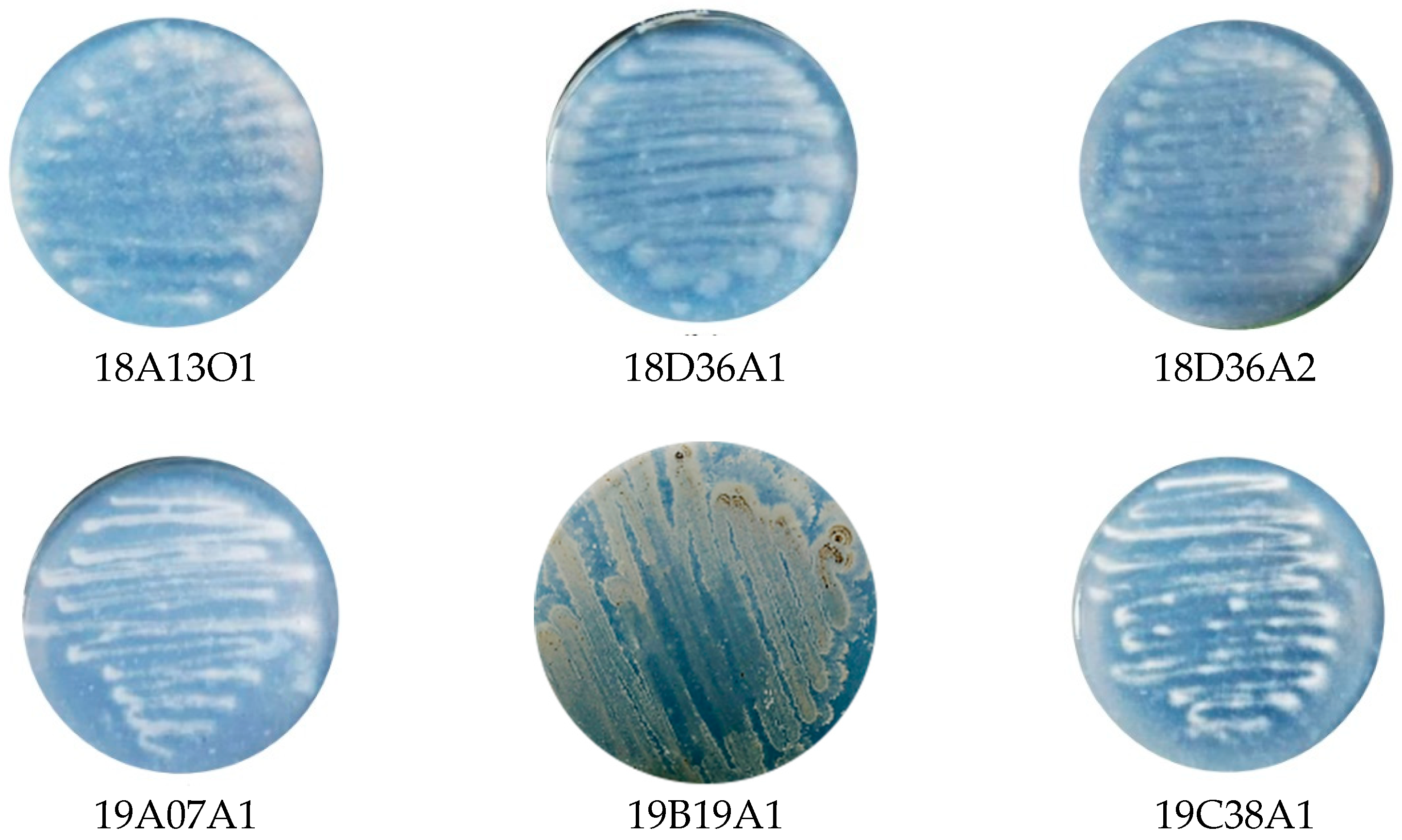
3.1. Isolation of Actinomycetes from Bali and Gorontalo Marine Biota

In this study, six actinomycetes isolates were isolated from marine sponge and tu-nicates were collected, three from the coast of Buleleng, Bali (8°07′20.9″ S 114°34′03.8″ E) (18A13O1, 18D36A1, 18D36A2), and three from Tomini Bay, Gorontalo (0°25′11.9″ N 123°08′31.8″ E) (19A07A1, 19B19A1, 19C38A1). The collection of marine biota was based on two different areas: Bali waters located in the Indian Ocean [38] and Gorontalo located in the Pacific Ocean [39]. At different locations, it is possible to learn more about the bio-diversity of actinomycetes for different metabolizing compounds and to characterize their enzymatic activities [40]. The results of the isolation can be seen in Figure 1. The isolates were white and cream in color on 1% colloidal chitin agar medium. As shown in the picture, the homogeneity of the actinomycete colonies indicates the purity of the isolates. Colloidal chitin medium is a selective medium for isolating marine actinomycetes, because not all microorganisms are capable of converting chitin into a nutrient source [41]. According to Buchanan and Gibbons [42], actinomycetes are identified in terms of their morphology with the characteristics of rough and hard textured colonies, branched substrate mycelia, and aerial mycelium of various shades of grey, white, red, yellow, green, cream, brown, and orange.
Actinomycete strains with codes 18A13O1, 19A07A1, and 19C38A1 were obtained, associated with sponges, while isolates with codes 18D36A1, 18D36A2, and 19B19A1 associated with envelope membranes were obtained. Both marine organisms are classi-fied as filter feeders; actinomycetes are a type of microorganism found in the inner layer of sponges, and in their ecosystem, filter feeders consume prokaryotes. As a food source, these microorganisms are retained in the cell compartment, then transferred to the in-ternal mesohyl and digested by phagocytosis. Many filter feeders are symbiotic, with the microbial colony in the mesohyl matrix appearing as a homogeneous mixture, even ac-counting for up to half of the biomass [43].

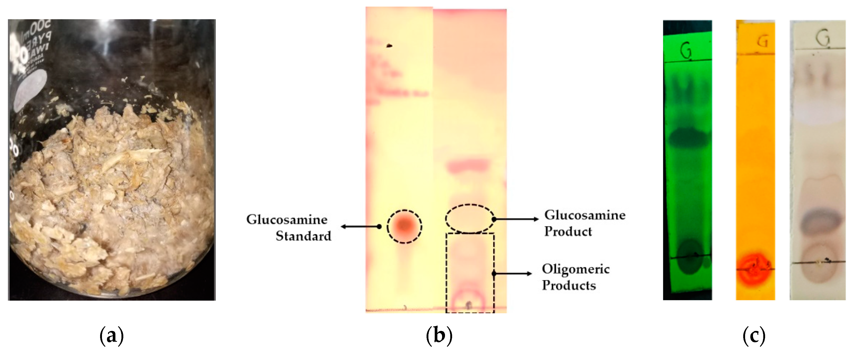
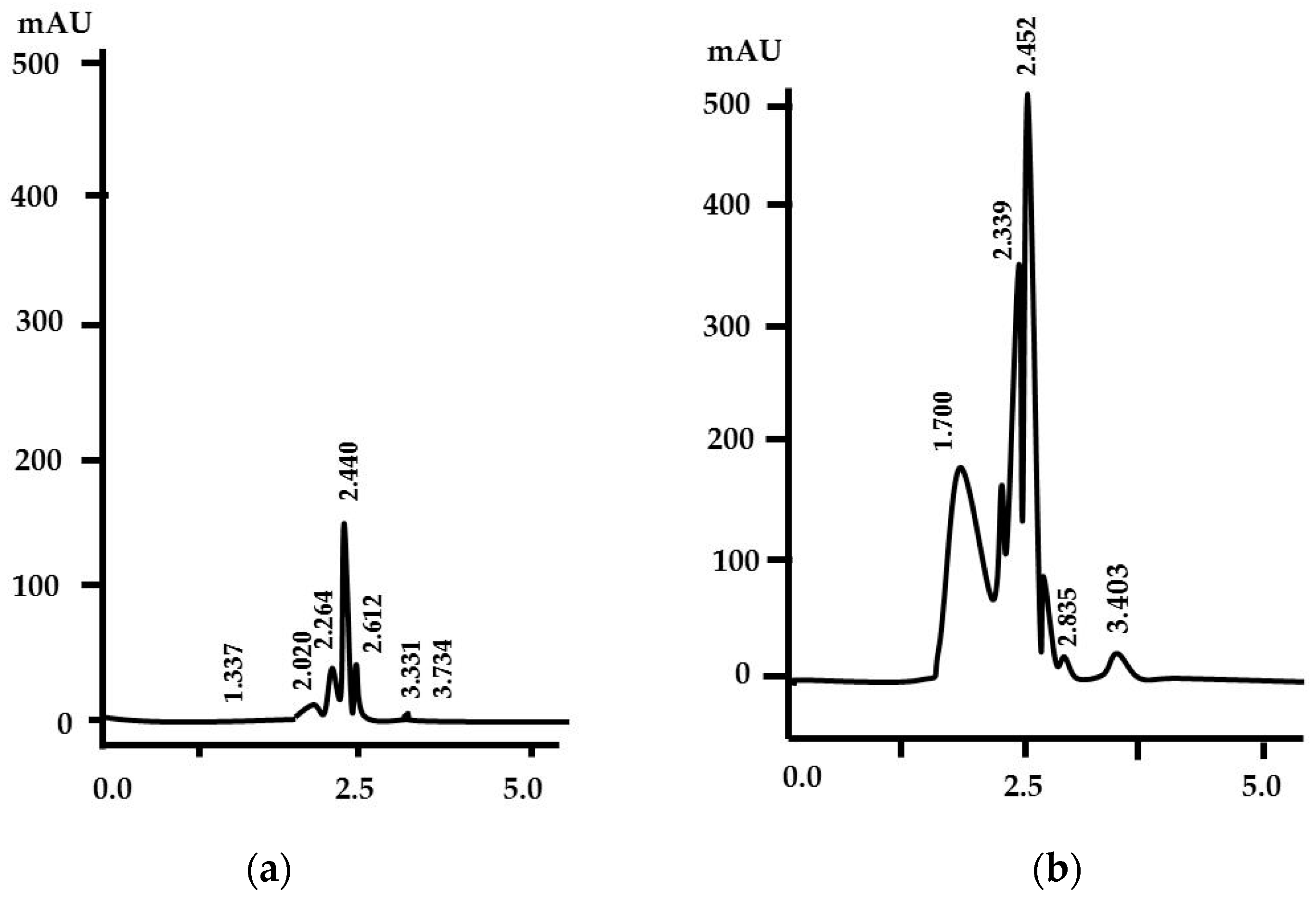
3.2. Chitinolytic Assay and Analysis of Degradation Products

Prior to cultivation on shrimp shell waste medium, which contained chitin as the main component, the six isolates were tested for chitinolytic activity with 1% colloidal chitin agar medium. The chitinolytic clear zone was then observed after seven days of incubation. The results indicate that each isolate has various chitinolytic activities (Figure 2). Among the tested isolates, 19B19A1 exhibited the largest clear zone, with a chitinolytic index of 1.328. This is the basis for SSF fermentation on chitin-based shrimp shell me-dium. The appearance of the clear zone showed that the isolate of 19B19A1 was able to obtain nutrients in the form of carbon and nitrogen from the colloidal chitin medium; this indicates that the isolate has the chitinase enzyme [44]. The diversity of chitin degradation is influenced by the type of extracellular chitinase in actinomycetes, as well as other types of enzymes such as lytic polysaccharide monooxygenases (LPMOs) [45].

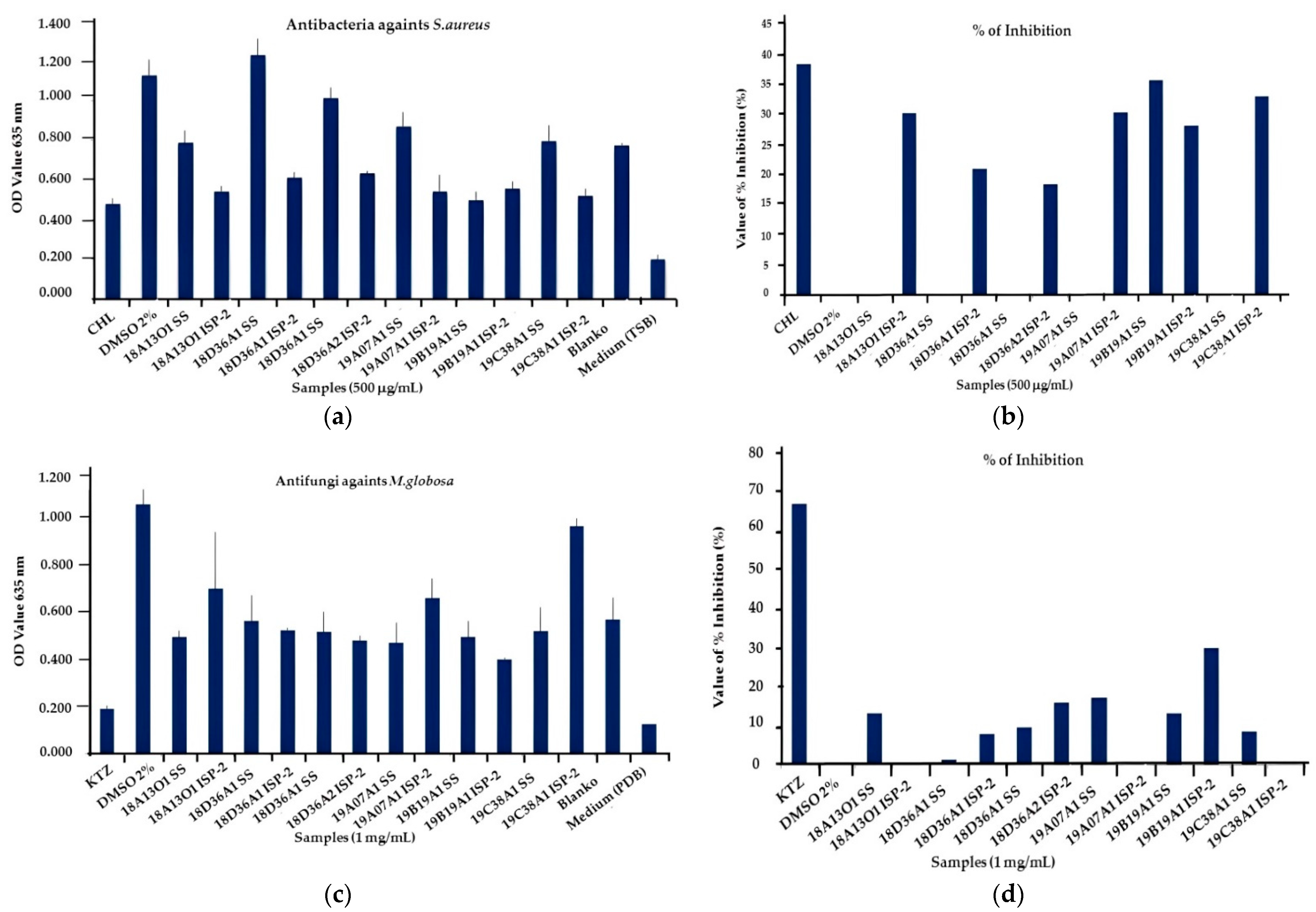
3.3. Screening of Antimicrobial Assay

The actinomycete extracts obtained from fermentation on shrimp shell medium and ISP-2 were screened against multidrug resistant S. aureus, which was resistant to clindamycin (CDM), ciprofloxacin (CFX), erythromycin (ERH), lincomycin (LC), and amoxicillin (AMX) based on previous work Setiawan et al. [33]. As shown in Table 1, the EtOAc extract from isolate 19B19A1 on shrimp shells showed the strongest growth inhibition of multidrug resistant S. aureus at a concentration of 500 μg/mL, whereas all EtOAc extracts from ISP-2 medium elicited inhibition at a concentration of 500 μg/mL against MDR S. aureus.
On the contrary, the results of the actinomycete extract bioactivity test against M. globosa are shown in Figure 3c,d. The EtOAc extract of 19B19A1, which was cultivated from shrimp shells, showed the greatest inhibitory activity compared to ISP-2 medium. It can be concluded that actinomycetes cultivated in shrimp shell waste medium are capable of producing antifungal metabolites. This indicates that shrimp shell fermentation is a selective medium and can potentially be used as an actinomycete inducer in the production of bioactive compounds. Actinomycetes use chitin as a carbon and nitrogen source. Chitin is highly compatible for the growth of actinomycetes, and it is an environmental marker for Streptomycetes [46]. This is in line with Craig et al.’s [47] and ven der Meij et al.’s [48] results, in that chitin sources are metabolized by actinomycetes into glucosamine 6-P in antibiotic biosynthesis and siderophore production.

3.4. Scale up Solid State Fermentation and Extraction

The results of 19B19A1 on the 14th day of cultivation showed that the actinomycetes grew well in the shrimp shell waste medium, and in this study, the cultivation was compared to standard ISP-2 medium as a source of nutrition. Based on its biomass (Figure 4a), it can be seen as the grayish-white aerial mycelium growing on the surface of the shrimp shell, and the earthy aroma is characteristic of actinomycetes. In addition, the results of SSF degradation could be observed using the TLC method, where ninhydrin was the specific reagent. The TLC test indicated the presence of monomers in the form of glucosamine and oligomers on day 14 (Figure 4b), in accordance with previous results reported by Ma et al. [49] that stated that microorganisms can convert chitin (shrimp shell) into glucosamine through the chitinase enzyme. Moreover, the extract from the isolate 19B19A1 contained alkaloid compounds, which are marked in orange in Figure 4c. These alkaloids might be indoles, diketopiperazines, glutarimides, indolizidines, or pyrroles, which are known to have antimicrobial activity [50].
The selection of selective shrimp shell medium can activate genes that play a role in the production of secondary metabolite compounds. In accordance with the study of Se-tiawan et al. [17], the increase in metabolite compounds in media with a chitin content can increase the production of secondary metabolite compounds up to 25 times after being initiated by chitinase [51]. Based on the study of Umar et al. [52], chitinolytic activity is not observed in medium containing sucrose or glucose as carbon and nitrogen sources. This suggests that colloidal chitin medium can induce chitinase-producing genes [53].
In addition, analysis of the degradation products of the SSF process at the 14-day incubation exhibited chitinase activity of 0.053 U/mL and glucosamine levels of 0.619 mg/mL. The levels of glucosamine using the HPLC method are shown in Figure 5.
The results of the HPLC analysis showed that the peak of the glucosamine chro-matogram was at a retention time (RT) of 2.452 min with an intensity of 492 mAU and a glucosamine concentration of 1.458 mg/mL. There were also several other chromatogram peaks at RT 2.617; 2.835; 3.403 min which were identified as a derivative products in the form of oligomers such as chitooligosaccharide [35].
Previous research from our group, by Widyastuti et al. [35], isolated Pseudonocardia antitumoralis 18D36A1 from Buleleng Waters, Bali, Indonesia, showing that it has the ability to degrade shrimp shell into chitooligosaccharide DP 6 as an antifungal. When compared to its oligomeric product, Pseudonocardia antitumoralis 18D36A1 had sharper HPLC chromatogram peaks at retention times of 3.1 and 3.4 min, indicating the presence of different chitin cleavages between Streptomyces tritolerans 19B19A1 and Pseudonocardia antitumoralis 18D36A1. This indicates that the diversity of actinomycetes affects the degradation of shrimp shell products into derivatives.
According to Xu et al. [54], chitinase from Streptomyces strains can be used for chitibiose products, which is a potential antioxidant commonly applied in biomedicine. For the hydrolysis of chitin oligosaccharides that have the potential to be used as antioxidant, antimicrobial, anticancer, anticoagulant, and antitumor agents, as well as in the food industry, chitinases derived from actinomycetes other than Streptomyces, such as Nocardiopsis prasine, are used.

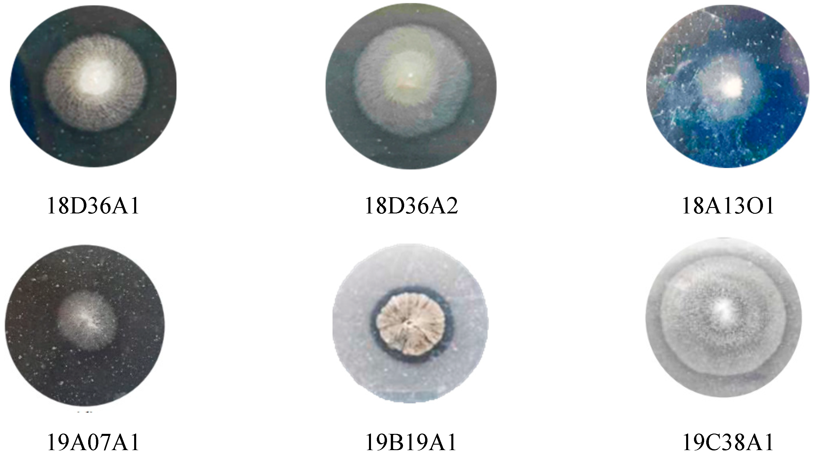
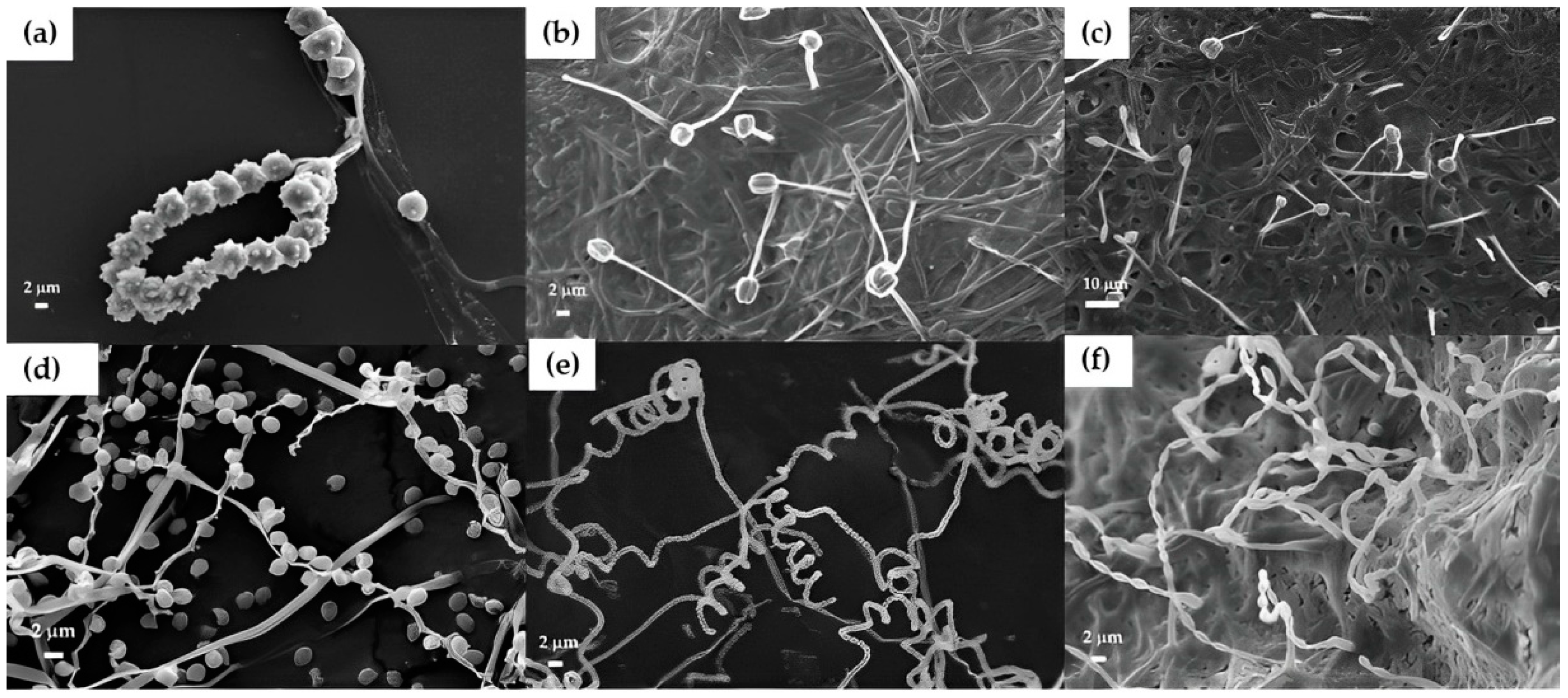
3.5. Morphological Identification of Actinomycetes

Morphological observation using shrimp shell as a matrix and SEM of six strains (Figure 6) clearly showed different shapes of spore decoration and variation of the aerial mycelium. Pseudonocardia carboxydivorans 18A13O1 (Figure 6a) has a rounded spore shape, with apexes on the surface of the spore and smooth, branched filaments. Pseudonocardia antumoralis 18D36A1 (Figure 6b) and Micrococcus sp. 18D36A2 (Figure 6c) possess spherical sporangia containing a single hypha and fine-textured substrate mycelium. Next, Brevibacterium linens 19A07A1 (Figure 6d) has single spores arranged alternately within the filament. Then, Streptomyces tritolerans 19B19A1 (Figure 6e) has long, flexible, spiral spores with a smooth surface. The other isolate, Kocuria palustris 19C38A1 (Figure 6f), forms oval spore chains, and the spores and spore chains can branch, while the substrate mycelium tends to fragment.
This could be used as a basis for the classification of actinomycetes. Observations were made based on the method of Widyastuti et al. [35], as a determination of the type of spore and the size of actinomycetes, which was relatively simple, environmentally friendly, and fast. It is expected that this method will become a reference that can be used routinely as a clarification method in addition to the phylogenetic method.

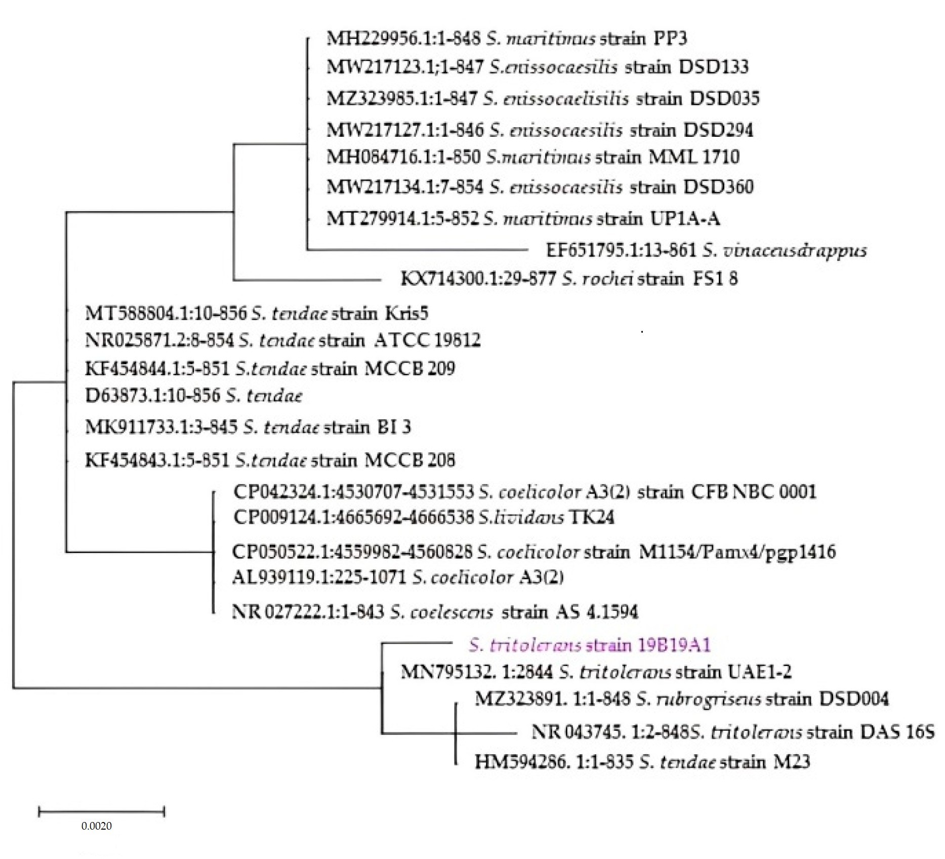
3.6. Phylogenetical Analysis

Sequence analysis of the 16S rRNA gene showed that isolate 19B19A1 is affiliated with the genus of Streptomyces, and isolate 19B19A1 showed 99.41% identity to Streptomyces tritolerans (Figure 7). This strain was registered with access number LC82288. The isolate can be further exploited for lead compound isolation. Following on from previous research, each isolate has a different character (Table 2). Meanwhile, isolate 18A13O1 was identified based on a sequencing similarity of 99.65% as a new strain of Pseudonocardia carboxydivorans 18A13O1 [17]. The 16S rDNA gene sequence indicated the genus Kocuria. In addition, isolate 19C38A1 was identified as Kocuria palustris 19C38A1 with a similarity rate of 99.77% [55]. Furthermore, isolate 18D36A1 was identified as Pseudonocardia antitumoralis, which showed a similarity of 99.93% [35], while two isolates—Brevibacterium linens 19A07A1 and Micrococcus sp. 18D36A2—are still being investigated.
Generally, chitinase-producing Streptomyces are still widely used as a source of me-tabolite compounds with diverse activities. Recent studies have reported over 70 novel compounds used as antibacterial, anticancer, and antifungal agents [56]. Several studies have found that actinobacteria spores are generally dormant, washed out of terrestrial ecosystems in drains and rivers to the seafloor, where they remain dormant [57]. The seafloor is often exposed to harsh environments such as high pressure, high salinity, and nutrient deficiencies. As a result, it has evolved genetically over time to produce secondary metabolite profiles distinct from terrestrial actinomycetes [58]. However, knowledge of the chemistry, distribution, and biodiversity of marine actinomycetes and other marine genera is still limited [59]. In addition, marine ecosystems are so dynamic that it is difficult to access different depths of the sea for sampling. Marine actinomycetes are found not only in seawater and sediments, but also in various biological sources such as sponges, tunicates, and mangroves [60].
To the best of our knowledge, this study is the first preliminary report of actino-mycetes of Streptomyces tritolerans 19B19A1 from tunicate, and in the utilization of Strep-tomyces tritolerans 19B19A1 as actinomycetes with the concept of biorefinery, both in the production of metabolite compounds as antimicrobials and in actinomycetes degrading shrimp shell medium as substrate into glucosamine and oligomer (COS) products.

4. Conclusions

The extract of Streptomyces tritolerans 19B19A1 is able to inhibit the growth of the multidrug-resistant Staphylococcus aureus and the fungus Malassezia globosa. This study concludes that biorefinery actinomycetes in shrimp shell medium using the solid state fermentation concept has potential in the production of antimicrobial compounds, and the utilization of by-products from this fermentation in the form of oligomers and glu-cosamine has benefits in the pharmaceutical industry. This information is very important for further research related to developing novel bioactive compounds from actinomycetes through biorefinery, being environmentally friendly and sustainable in the industrial field.

Author Contributions

Conceptualization, A.L., A.S. and J.H.; methodology, A.L., A.S., E.A., F.S. and J.H; formal analysis, P.A., W.W. and W.A.S.; investigation, F.S., W.W. and W.A.S.; writing—original draft preparation, A.L., J.H., E.A., F.S., A.L. and M.R.F.; writing—review and editing, J.H., A.S., A.L., M.A. and N.L.G.R.J.; supervision, A.L., J.H., A.S. and P.A.; project administration, W.W. All authors have read and agreed to the published version of the manuscript.

Funding

This research was funded by the Higher Education Technology Innovation Lampung University (HETI UNILA), Asian Development Bank, Directorate General of Higher Education, Research and Technology, the Ministry of Education, Culture, Research and Technology, Republic of Indonesia, for the Innovation Research Scheme and Domestic Cooperation award with grant no. 6044/UN26/HK.01.03/2022 and the Directorate General of Higher Education, Research and Technology, The Ministry of Education, Culture, Research and Technology, Republic of Indonesia, with a Basic Research award with grant no. 2143/UN26.21/PN/2022 to J.H. This research was also partially supported by the Research Support Project for Life Science and Drug Discovery (Basic for Supporting Innovative Drug Discovery and Life Science Research (BINDS)) from the Japan Agency for Medical Research and Development (AMED) (grant no. JP22ama121054), the Kobayashi Inter-national Scholarship Foundation, and a Grant-in-Aid for Scientific Research B (grant nos. 18H02096, 17H04645, and 21H02069) from the Japan Society for the Promotion of Science (JSPS) to M.A.

Institutional Review Board Statement

Not applicable.

Informed Consent Statement

Not applicable.

Data Availability Statement

The data presented in the study are available on reasonable request from the corresponding author.

Acknowledgments

The authors would like to thank the HETI Project UNILA, the Asian Development Bank, the Directorate General of Higher Education, Research and Technology, the Ministry of Education, Culture, Research and Technology, Republic of Indonesia, for the Innovation Research Scheme and the Domestic Cooperation funding. This research was also partially supported by the Research Support Project for Life Science and Drug Discovery (Basis for Supporting Innovative Drug Discovery and Life Science Research (BINDS)) from the Japan Agency for Medical Research and Development (AMED), the Kobayashi International Scholarship Foundation, and a Grant-in-Aid for Scientific Research B from the Japan Society for the Promotion of Science (JSPS) to M.A. The authors also thank the Technical Service Unit, Integrated Laboratory of Innovation and Technology Center (UPT LTSIT), Lampung University, for providing the scanning electron microscopy facilities.

Conflicts of Interest

The author declare no conflict of interest.

References

  1. Spernovasilis, N.; Tsiodras, S.; Poulakou, G. Emerging and Re-Emerging Infectious Diseases: Humankind’s Companions and Competitors. Microorganisms 2022, 10, 98. [Google Scholar] [CrossRef] [PubMed]
  2. De Oliveira, D.M.P.; Forde, B.M.; Kidd, T.J.; Harris, P.N.A.; Schembri, M.A.; Beatson, S.A.; Walker, M.J. Antimicrobial Resistance in ESKAPE Pathogens. Clin. Microbiol. Rev. 2020, 33, e00181-19. [Google Scholar] [CrossRef] [PubMed]
  3. Von Lilienfeld-Toal, M.; Wagener, J.; Einsele, H.; Cornely, O.A.; Kurzai, O. Invasive Fungal Infection: New Treatments To Meet New Challenges. Dtsch. Ärzteblatt Int. 2019, 116, 271. [Google Scholar]
  4. Rathore, S.S.; Sathiyamoorthy, J.; Lalitha, C.; Ramakrishnan, J. A holistic review on Cryptococcus neoformans. Microb. Pathog. 2022, 166, 105521. [Google Scholar] [CrossRef]
  5. Huang, C.Y.; Peng, C.C.; Hsu, C.H.; Chang, J.H.; Chiu, N.C.; Chi, H. Systemic Infection Caused by Malassezia pachydermatis In Infants: Case Series and Review of The Literature. Pediatr. Infect. Dis. J. 2020, 39, 444–448. [Google Scholar] [CrossRef]
  6. Mularoni, A.; Graziano, E.; Todaro, F. Invasive Trichophyton Infection In A Liver Transplant Recipient. Transpl. Infect. Dis. 2022, 24, e13794. [Google Scholar] [CrossRef]
  7. Gong, J.; Hou, X.; Geng, Y.; Dai, R.C.; Zhao, F.; He, L. Rapid Identification of Four Fusarium spp. Complex by High-Resolution Melting Curve Analysis and Their Antifungal Susceptibility Profiles. Mycopathologia 2022, 187, 345–354. [Google Scholar]
  8. Kawabe, Y.; Sakurai, A.; Sasaki, T.; Hasegawa, M.; Suzuki, M.; Hoshino, N.; Doi, Y. Native Valve Infective Endocarditis Due To Sequence Type 97 Community-Associated Methicillin-Resistant Staphylococcus aureus Complicated by Meningitis and Multiple Septic Emboli In A Young Healthy Adult. J. Infect. Chemother. 2022, 28, 828–832. [Google Scholar] [CrossRef]
  9. Mancuso, G.; Midiri, A.; Gerace, E.; Biondo, C. Bacterial Antibiotic Resistance: The Most Critical Pathogens. Pathogens 2021, 10, 1310. [Google Scholar] [CrossRef]
  10. Roncarati, D.; Scarlato, V.; Vannini, A. Targeting of Regulators as a Promising Approach in the Search for Novel Antimicrobial Agents. Microorganisms 2022, 10, 185. [Google Scholar] [CrossRef]
  11. De Simeis, D.; Serra, S. Actinomycetes: A Never-Ending Source of Bioactive Compounds—An Overview on Antibiotics Production. Antibiotics 2021, 10, 483. [Google Scholar] [CrossRef] [PubMed]
  12. Bérdy, J. Thoughts and facts about antibiotics: Where we are now and where we are heading. J. Antibiot. 2012, 65, 385–395. [Google Scholar] [CrossRef] [PubMed] [Green Version]
  13. Do Khac, A.L. The Political Sea: Conservation Policies, State Power, and Symbolic Violence: The Case of the Bajau in the Wakatobi Marine National Park. Explorations 2018, 14, 37–50. [Google Scholar]
  14. Hu, X.; Tang, Y.; Liu, Y.; Pei, X.; Huang, Z.; Song, F.; Zhang, H. Comprehensive Genomic Analysis of Marine Strain Streptomyces sp. 891, an Excellent Producer of Chrysomycin A with Therapeutic Potential. Mar. Drugs 2022, 20, 287. [Google Scholar] [CrossRef] [PubMed]
  15. Yi, W.; Newaz, A.W.; Yong, K.; Ma, M.; Lian, X.-Y.; Zhang, Z. New Hygrocins K–U and Streptophenylpropanamide A and Bioactive Compounds from the Marine-Associated Streptomyces sp. ZZ1956. Antibiotics 2022, 11, 1455. [Google Scholar] [CrossRef]
  16. Donald, L.; Pipite, A.; Subramani, R.; Owen, J.; Keyzers, R.A.; Taufa, T. Streptomyces: Still the biggest producer of new natural secondary metabolites, a current perspective. Microbiol. Res. 2022, 13, 418–465. [Google Scholar] [CrossRef]
  17. Setiawan, A.; Widyastuti, W.; Irawan, A.; Wijaya, O.S.; Laila, A.; Setiawan, W.A.; Juliasih, N.L.G.R.; Nonaka, K.; Arai, M.; Hendri, J. Solid State Fermentation of Shrimp Shell Waste Using Pseudonocardia carboxydivorans 18A13O1 to Produce Bioactive Metabolites. Fermentation 2021, 7, 247. [Google Scholar] [CrossRef]
  18. Raju, K.J.; Rittapalli, T. Production and Optimization Studies of Protease by Soil Isolate Actinomycetes Using Mixed Substrate of Pomegranate Peel Powder and Banana Peel Powder under Solid State Fermentation. Asian J. Biotechnol. Bioresour. Technol. 2022, 8, 39–47. [Google Scholar]
  19. Wu, D.; Wei, Z.; Mohamed, T.A.; Zheng, G.; Qu, F.; Wang, F.; Zhao, Y.; Song, C. Lignocellulose biomass bioconversion during composting: Mechanism of action of lignocellulase, pretreatment methods and future perspectives. Chemosphere 2022, 286, 131635. [Google Scholar] [CrossRef]
  20. Swiontek Brzezinska, M.; Jankiewicz, U.; Kalwasińska, A.; Świątczak, J.; Żero, K. Characterization of chitinase from Streptomyces luridiscabiei U05 and its antagonist potential against fungal plant pathogens. J. Phytopathol. 2019, 167, 404–412. [Google Scholar] [CrossRef]
  21. Alzahrani, O.M.; Sohail, M.; Mahmoud, S.F.; Alswat, A.S.; El-Halmouch, Y. Preparation of a Pectinase-Enriched Multienzyme under Solid State Fermentation of Sugarcane Bagasse. Fermentation 2023, 9, 141. [Google Scholar] [CrossRef]
  22. Vicente, F.A.; Ventura, S.P.; Passos, H.; Dias, A.C.; Torres-Acosta, M.A.; Novak, U.; Likozar, B. Crustacean waste biorefinery as a sustainable cost-effective business model. Chem. Eng. J. 2022, 442, 135937. [Google Scholar] [CrossRef]
  23. FAO. The State of World Fisheries and Aquacultures: Sustainability in Action; FAO: Rome, Italy, 2020. [Google Scholar]
  24. Lu, D.L.; Zhang, M.S.; Deng, J.J.; Lu, W.J.; Yang, Z.D.; Li, Z.W.; Chen, L.-C.; Luo, X.C. Highly Efficient Shrimp Shell Recovery by Solid-State Fermentation with Streptomyces sp. SCUT-3. Chem. Eng. J. 2023, 458, 141256. [Google Scholar] [CrossRef]
  25. Ofori, S.A.; Kodikara, S.K.; Jayatissa, L.P.; Madarasinghe, S.K.; Nijamdeen, T.M.; Dahdouh-Guebas, F. What is the ecological footprint of aquaculture after 5 decades of competition between mangrove conservation and shrimp farm development? Aquat. Conserv. Mar. Freshw. Ecosyst. 2023, 33, 15–28. [Google Scholar] [CrossRef]
  26. Ruslan, N.F.; Ahmad, N.; Abas, A.; Sanfilippo, A.; Mahmoud, K.; Munaim, M.S.A.; Nour, A.H. Sustainable Bioethanol Production by Solid State Fermentation: A Systematic Review. Res. Sq. 2023; preprint. [Google Scholar]
  27. Yan, N.; Xi, C. Sustainability: Don’t waste seafood waste. Nature 2015, 524, 155–157. [Google Scholar] [CrossRef] [Green Version]
  28. Tan, Y.N.; Lee, P.P.; Chen, W.N. Microbial extraction of chitin from seafood waste using sugars derived from fruit waste-stream. AMB Expr. 2020, 10, 17. [Google Scholar] [CrossRef]
  29. Suryawanshi, N.; Eswari, J.S. Chitin from seafood waste: Particle swarm optimization and neural network study for the improved chitinase production. J. Chem. Technol. Biotechnol. 2022, 97, 509–519. [Google Scholar] [CrossRef]
  30. Agrawal, T.; Kotasthane, A.S. Chitinolytic Assay of Indigenous Trichoderma Isolates Collected from Different Geographical Locations of Chhattisgarh in Central India. SpringerPlus 2012, 1, 73. [Google Scholar] [CrossRef] [Green Version]
  31. Zhu, X.F.; Zhou, Y.; Feng, J.L. Analysis of both chitinase and chitosanase produced by Sphingomonas sp. CJ-5. J. Zhejiang Univ. Sci. B 2007, 8, 831–838. [Google Scholar] [CrossRef] [Green Version]
  32. Zbîrcea, R.I.; Menghiu, G.; Matica, A.; Ostafe, V. Use of 3, 5-dinitrosalicylic Acid Reaction To Study The Chitosan Hydrolysis. New Front. Chem. 2016, 25, 145–153. [Google Scholar]
  33. Setiawan, A.; Lutfiah, R.; Juliasih, N.L.G.R.; Setiawan, W.A.; Hendri, J.; Arai, M. Antibacterial activity of EtOAc extract from marine-derived fungus Aspergillus nomiae A12-RF against clinical pathogen bacteria, Staphylococcus aureus. AACL Bioflux 2022, 15, 1413–1421. [Google Scholar]
  34. Leong, C.; Buttafuoco, A.; Glatz, M.; Bosshard, P.P. Antifungal Susceptibility Testing of Malassezia spp. With an Optimized Colorimetric Broth Microdilution Method. J. Clin. Microbiol. 2017, 55, 1883–1893. [Google Scholar] [CrossRef] [PubMed] [Green Version]
  35. Widyastuti, W.; Setiawan, F.; Al Afandy, C.; Irawan, A.; Laila, A.; Juliasih, N.L.G.R.; Setiawan, W.A.; Arai, M.; Hendri, J.; Setiawan, A. Antifungal Agent Chitooligosaccharides Derived from Solid-State Fermentation of Shrimp Shell Waste by Pseudonocardia antitumoralis 18D36-A1. Fermentation 2022, 8, 353. [Google Scholar] [CrossRef]
  36. Heuer, H.; Krsek, M.; Baker, P.; Smalla, K.; Wellington, E.M.H. Analysis of Actinomycete Communities by Specific Amplification of Genes Encoding 16S rRNA and Gel-Electrophoretic Separation In Denaturing Gradients. Appl. Environ. Microbiol. 1997, 63, 3233–3241. [Google Scholar] [CrossRef] [Green Version]
  37. Baskaran, R.; Mohan, P.M.; Sivakumar, K.; Kumar, A. Antimicrobial Activity and Phylogenetic Analysis of Streptomyces Parvulus Dosmb-D105 Isolated from the Mangrove Sediments of Andaman Islands. Acta Microbiol. Immunol. Hung. 2016, 63, 27–46. [Google Scholar] [CrossRef] [PubMed] [Green Version]
  38. Setyohadi, D.; Zakiyah, U.; Sambah, A.B.; Wijaya, A. Upwelling impact on sardinella lemuru during the indian ocean dipole in the bali strait, Indonesia. Fishes 2021, 6, 8. [Google Scholar] [CrossRef]
  39. Djunarsjah, E.; Julian, M.M.; Muhammad, F.; Putra, A.P.; Lubis, N.S.; Welly, T.K.; Wahab, W.A.; Pamungkas, B. Utilization of Marine Geospatial Data for Determining Optimal FAD Locations in Improving the Living Standards of the North Gorontalo Coastal Community. Sustainability 2023, 15, 2242. [Google Scholar] [CrossRef]
  40. Edlund, A.; Loesgen, S.; Fenical, W.; Jensen, P.R. Geographic distribution of secondary metabolite genes in the marine actinomycete Salinispora arenicola. Appl. Environ. Microbiol. 2011, 77, 5916–5925. [Google Scholar] [CrossRef] [Green Version]
  41. Ekundayo, F.O.; Folorunsho, A.E.; Ibisanmi, T.A.; Olabanji, O.B. Antifungal activity of chitinase produced by Streptomyces species isolated from grassland soils in Futa Area, Akure. Bull. Natl. Res. Cent. 2022, 46, 95. [Google Scholar] [CrossRef]
  42. Buchanan, R.E.; Gibbons, N.E. (Eds.) Bergey’s Manual of Determinative Bacteriology, 8th ed.; Williams & Wilkins Co.: Baltimore, MD, USA, 1974; pp. xxvi + 1246. [Google Scholar]
  43. Jahn, M.T. Physiology, Syntrophy and Viral Interplay in the Marine Sponge Holobiont. Ph.D. Dissertation, Christian-Albrechts-Universität Kiel, Kiel, Germany, 2019. [Google Scholar]
  44. Paulsen, S.S.; Andersen, B.; Gram, L.; Machado, H. Biological Potential of Chitinolytic Marine Bacteria. Mar. Drugs 2016, 14, 230. [Google Scholar] [CrossRef] [PubMed] [Green Version]
  45. Itoh, T.; Kimoto, H. Bacterial chitinase system as a model of chitin biodegradation. Target. Chitin-Contain. Org. 2019, 26, 131–151. [Google Scholar]
  46. Wang, S.; Fang, X.; Liang, K.; Li, S.; Han, S.; Zhu, T. Cloning, expression and antifungal effect of the recombinant chitinase from Streptomyces sampsonii KJ40. Ciência Rural 2023, 53, e20210663. [Google Scholar] [CrossRef]
  47. Craig, M.; Lambert, S.; Jourdan, S.; Tenconi, E.; Colson, S.; Maciejewska, M.; Ongena, M.; Martin, J.F.; van Wezel, G.; Rigali, S. Unsuspected control of siderophore production by N-acetylglucosamine in streptomycetes. Environ. Microbiol. Rep. 2012, 4, 512–521. [Google Scholar] [CrossRef] [PubMed]
  48. Van der Meij, A.; Worsley, S.F.; Hutchings, M.I.; van Wezel, G.P. Chemical ecology of antibiotic production by actinomycetes. FEMS Microbiol. Rev. 2017, 41, 392–416. [Google Scholar] [CrossRef] [PubMed] [Green Version]
  49. Ma, X.; Gözaydın, G.; Yang, H.; Ning, W.; Han, X.; Poon, N.Y.; Liang, H.; Ning, Y.; Zhou, K. Upcycling Chitin-Containing Waste Into Organonitrogen Chemicals Via An Integrated Process. Proc. Natl. Acad. Sci. USA 2020, 117, 7719–7728. [Google Scholar] [CrossRef]
  50. De Rop, A.S.; Rombaut, J.; Willems, T.; De Graeve, M.; Vanhaecke, L.; Hulpiau, P.; De Maeseneire, S.L.; De Mol, M.L.; Soetaert, W.K. Novel Alkaloids From Marine Actinobacteria: Discovery and Characterization. Mar. Drugs 2022, 20, 6. [Google Scholar] [CrossRef]
  51. Manucharova, N.A.; Trosheva, E.V.; Kol’tsova, E.M.; Demkina, E.V.; Karaevskaya, E.V.; Rivkina, E.M.; Mardanov, A.V.; El’-Registan, G.I. Characterization of the structure of the prokaryotic complex of antarctic permafrost by molecular genetic techniques. Mikrobiologiya 2016, 85, 83–91. [Google Scholar] [CrossRef]
  52. Umar, A.A.; Hussaini, A.B.; Yahayya, J.; Sani, I.; Aminu, H. Chitinolytic and Antagonistic Activity of Streptomyces Isolated from Fadama Soil against Phytopathogenic Fungi. Trop. Life Sci. Res. 2021, 32, 25–38. [Google Scholar] [CrossRef]
  53. Fan, Z.; Wang, L.; Qin, Y.; Li, P. Activity of chitin/chitosan/chitosan oligosaccharide against plant pathogenic nematodes and potential modes of application in agriculture: A review. Carbohydr. Polym. 2023, 306, 120592. [Google Scholar] [CrossRef]
  54. Xu, T.; Qi, M.; Liu, H.; Cao, D.; Xu, C.; Wang, L.; Qi, B. Chitin degradation potential and whole-genome sequence of Streptomyces diastaticus strain CS1801. AMB Express 2020, 10, 1–12. [Google Scholar] [CrossRef] [PubMed] [Green Version]
  55. Setiawan, A.; Setiawan, F.; Juliasih, N.L.G.R.; Widyastuti, W.; Laila, A.; Setiawan, W.A.; Djailani, F.M.; Mulyono, M.; Hendri, J.; Arai, M. Fungicide Activity of Culture Extract from Kocuria palustris 19C38A1 against Fusarium oxysporum. J. Fungi 2022, 8, 280. [Google Scholar] [CrossRef] [PubMed]
  56. Lacey, H.J.; Rutledge, P.J. Recently Discovered Secondary Metabolites from Streptomyces Species. Molecules 2022, 27, 887. [Google Scholar] [CrossRef]
  57. Moran, M.A.; Rutherford, L.T.; Hodson, R.E. Evidence for indegenous Streptomyces population in a marine environment determined with a 16S rRNA probe. Appl. Environ. Microbiol. 1995, 61, 3695–3700. [Google Scholar] [CrossRef] [PubMed] [Green Version]
  58. Tiwari, K.; Gupta, R.K. Diversity and isolation of rare actinomycetes: An overview. Crit. Rev. Microbiol. 2013, 39, 256–294. [Google Scholar] [CrossRef] [PubMed]
  59. Siro, G.; Pipite, A.; Christi, K.; Srinivasan, S.; Subramani, R. Marine Actinomycetes Associated with Stony Corals: A Potential Hotspot for Specialized Metabolites. Microorganisms 2022, 10, 1349. [Google Scholar] [CrossRef] [PubMed]
  60. Kathiresan, K.; Nabeel, M.A.; Manivannan, S. Bioprospecting of marine organisms for novel bioactive compounds. Sci. Trans. Environ. Technovation 2008, 1, 107–120. [Google Scholar] [CrossRef]
Figure 1. Six actinomycete isolates from marine organisms collected from the coastal regions of Bali and Gorontalo.
Fermentation 09 00334 g001
Figure 2. The chitinolytic zone of the isolates collected from Bali (18D36A1, 18D36A2, and 18A13O1) and Gorontalo (19A07A1, 19B19A1, and 19C38A1).
Fermentation 09 00334 g002
Figure 3. (a) OD635 nm results of antibacterial activity against S. aureus; (b) % of Inhibition of antibacteria; (c) OD635 nm results of antifungal activity against M. globosa; (d) % of Inhibition of antifungal activity.
Fermentation 09 00334 g003
Figure 4. Isolate 19B19A1: (a) Shrimp shell medium; (b) TLC of the degradation product from 19B19A1; (c) TLC of crude EtOAc from 19B19A1.
Fermentation 09 00334 g004
Figure 5. HPLC Chromatogams; (a) Standard glucosamine; (b) Extract of degradation product day 14.
Fermentation 09 00334 g005
Figure 6. Observation of Spores using SEM (a) Spikes; (b) Sphrecial; (c) Spherical; (d) Smooth; (e) Spiral and; (f) Ovals.
Fermentation 09 00334 g006
Figure 7. Phylogenetic tree using maximum likelihood method and Hasegawa–Kishino–Yano model. It can be concluded that 19B19A1 is Streptomyces tritolerans.
Fermentation 09 00334 g007
Table 1. Antimicrobial Assay EtOAc Extract against MDR S.aureus dan M.globosa.
Isolate CodeAntibacteria (0.5 mg/mL)Antifungi (1 mg/mL)
ISP-2Shrimp ShellISP-2Shrimp Shell
18A13O1+++-
18D36A1+-+-
18D36A2+-++
19A07A1+-+-
19B19A1++++
19C38A1+++-
Table 2. Result of 16S rRNA Analysis.
Analysis of 16S rRNAProductPotential Use
Pseudonocardia carboxydivorans 18A13O1Branymicin BAntibacteria
Pseudonocardia antitumoralis 18D36A1ChitinaseAntifungi
Micrococcus sp 18D36A2ChitinaseChitinase producing
Brevibacterium linens 19A07A1AlkaloidAntibiofilm
Streptomyces tritolerans 19B19A1AlkaloidAntibacteria, Antifungi
Kocuria palustris 19C38A1BenzimidazoleAntifungi
Disclaimer/Publisher’s Note: The statements, opinions and data contained in all publications are solely those of the individual author(s) and contributor(s) and not of MDPI and/or the editor(s). MDPI and/or the editor(s) disclaim responsibility for any injury to people or property resulting from any ideas, methods, instructions or products referred to in the content.

