Article

Characterization of the Red Biochromes Produced by the Endophytic Fungus Monascus purpureus CPEF02 with Antimicrobial and Antioxidant Activities

1
Sustainable Agriculture Program, The Energy and Resources Institute, TERI-Gwal Pahari, Gurugram 122001, Haryana, India
2
Centre for Sustainable Bioproducts, Deakin University Geelong, Geelong, VIC 3216, Australia
3
Analytical Chemistry Laboratory, Regulatory Toxicology Group, CSIR-Indian Institute of Toxicology Research (CSIR-IITR), Vishvigyan Bhawan, 31, Mahatma Gandhi Marg, Lucknow 226001, Uttar Pradesh, India
4
Academy of Scientific and Innovative Research (AcSIR), Ghaziabad 201002, Uttar Pradesh, India
*
Author to whom correspondence should be addressed.
Fermentation 2023, 9(4), 328; https://doi.org/10.3390/fermentation9040328
Submission received: 2 February 2023 / Revised: 21 March 2023 / Accepted: 21 March 2023 / Published: 25 March 2023
(This article belongs to the Special Issue Pigment Production in Submerged Fermentation)

Abstract

:
Food acceptability and appeal are significantly influenced by colour. Harmful effects associated with synthetic colorants are well established, and research is currently focused on developing natural, synthetic chemical-free substitutes from fungal sources, with broad applications in food, medicine, textiles and agriculture. Additionally, the market’s dearth of natural red colour substitutes requires the creation of novel red pigment alternatives from secure and scalable sources. The goal of the current research was to establish new endophytic marine fungi that are naturally occurring bio-sources of the red pigment. Based on its profuse extracellular red pigment-producing capacity, the fungus CPEF02 was selected and identified as Monascus purpureus CPEF02 via internal transcribed spacer (ITS) sequences and phylogenetic analysis. The chemical moieties of the pigmented extracts were identified by liquid chromatography-high resolution mass spectrometry (LC-HRMS). The optimal culture conditions for maximum pigment production were investigated by surveying various media compositions. The methanolic fungal colourant extract was shown to have substantial antibacterial and antifungal activities against anthropogenic pathogens, Staphylococcus aureus (MTCC 1430), methicillin-resistant Staphylococcus aureus (ATCCBAA811), Salmonella typhimurium (MTCC 3241) and Vibrio cholerae (N16961) at a 100 µg/mL concentration and at a 1 mg/mL concentration for Alternaria solani (ITCC 4632) and Rhizoctonia solani (AG1-IA). This extract also exhibited antioxidant activity against the 2,2′-azino-bis (3-ethylbenzothiazoline-6-sulfonic acid) radical with an IC50 of 14.42 µg/mL and a Trolox equivalent antioxidant capacity of 0.571 µM Trolox/µg of the methanolic colourant extract. The findings suggested that M. purpureus’s pigment could be a source of an industrially useful natural red colourant.

1. Introduction

Increased awareness of the potential negative environmental and health effects of synthetically produced pigments has encouraged research towards natural alternatives for the food, textile, cosmetic, pharmaceutical and other sectors. Pigments can enhance the acceptability of and increase the demand for certain products, particularly food. Commonly used synthetic food colourants, for example, allura red, carmoisine, ponceau 4R, quinoline yellow WS, sunset yellow and tartrazine, come with label warnings in some countries such as “may have an adverse effect on activity and attention in children”, which discourages their use [1].
Amongst all the natural bioresources available for natural pigments, microbes have advantages over other sources due to their high stability, better yield, water solubility, year-round availability and cost-effective scalability. Endophytic fungi, which live inside plant tissues without causing harm to them, have been found to be prolific producers of a myriad of bio-pigments with therapeutic benefits [2]. Fungal pigments are secondary metabolites that are chemically diverse in nature, including polyketides, azophilones, quinones, melanin, carotenoids, xanthophylls, flavins, monascins, violacein and indigo [3]. These metabolites are produced under stress conditions as a defence response and have a range of bioactivities including antimicrobial, cytotoxicity, anti-inflammatory, anticancer, antioxidant and anti-atherosclerotic activities.
Monascus are filamentous ascomycetes, well studied for their red and yellow bioactive pigments with a wide range of applicability in nutraceuticals, pharmaceutics and textile industries. A total of 65 pigmented compounds have been isolated from Monascus. These pigmented bioactive secondary metabolites are primarily azaphilonic or polyketide in chemical nature [4]. The absorption maxima studied at distinct wavelengths have identified major hues produced by this genus, for example, yellow colourants at 330–450 nm, red colourants at 490–530 nm and orange colourants at 460–480 nm [5,6]. Monascus purpureus is a commercially important strains and is used in food and beverages for preservation, pigment, flavour, antimicrobial, anticancer, antioxidant, anti-obesity, human health promotion and immune-enhancing properties [7]. Its pigments are thermally stable and can be cultivated via submerged or solid-state fermentations [8]. Red yeast rice is a well-known east-Asian food that has been grown by fermenting Monascus purpureus on rice and has cholesterol-lowering and antimicrobial properties [9].
In the present study, the endophytic fungus Monascus purpureus isolated from Avicennia marina roots from Alibaug coastal area, Maharashtra, India [10], was optimized in different growth media and extracted with different solvents for studying maximum extracellular red pigment production. The antibacterial and antifungal activities of the pigments were tested against human pathogenic bacteria and phytopathogenic fungi. Chemical characterization was carried out to estimate the compounds responsible for the colour, bioactivity and antioxidant potential of the colourant. The colourant with the highest quantity of extracellular red pigment was tested for antioxidant activity. The methanol extracted colourant exhibited biological activity against the tested systems.

2. Materials and Methods

2.1. Isolation, Cultivation and Identification of Pigment-Producing Endophytic Fungi

The strain M. purpureus CPEF02 (NCBI accession number MT013404.1) had been isolated earlier in the laboratory from the roots of A. marina. Using a light (EVOS XL Core digital inverted microscope, Life Technologies, Thermo Fisher Scientific, Maharashtra, India) and scanning electron microscopy (Carl Zeiss, Jena, Thuringia, Germany), the strain was identified morphologically and molecularly by following the protocols of Mishra and group [10]. The culture was kept on potato dextrose agar (PDA, HiMedia, Maharashtra, India) plates at 4 °C, and it was sub-cultured every 25 days. Activated fungal agar discs from seven-day-old culture plates were used for further inoculations.

2.2. Screening of Culture Media for Pigment Production and Extraction Optimization of CPEF02

In order to maximize extracellular pigment production by the CPEF02 strain, different commercially viable media compositions were studied; 500 mL flasks containing 100 mL of five different media broths were prepared. There were Czapek’s extract dox Broth (CDB), malt extract broth (MEB), potato dextrose broth (PDB), Sabouraud dextrose broth (SDB) and yeast extract peptone dextrose broth (YEPD). All the chemicals were procured from HiMedia (HiMedia, Maharashtra, India). Inoculation was carried out by adding four 6 mm, 8–10-day-old PDA fungal discs, and the flasks were maintained at 28 °C for 14 days in the dark under static conditions. After about two weeks of incubation, the different medium culture broths and the mycelia were separated using a Büchner funnel and Whatman filter paper. The separated culture filtrates were dried and lyophilized for extracellular biochrome estimation. These lyophilized powders were further used for studying the effect of medium composition on bio-hue production and spectrophotometric analysis.

2.3. Quantification of Extracellular Polyketide Red Pigment

The culture filtrates obtained from different medium broths were UV-vis spectrophotometrically analysed using an established method [10]. Briefly, the culture filtrates were diluted in deionized water and the absorption profile was examined in the wavelength range of 200–800 nm. Carmine (Sigma Aldrich India Pvt. Ltd., Karnataka, India) was used as a standard red pigment because of its importance as a natural food colorant and stable nature [11]. The pigment production in different media was compared through the quantification of polyketide-based pigment metabolites, measured at λmax of Carmine red 494 nm and expressed as milligram equivalents of Carmine per litre of the medium. The extracellular pigment yield was also measured in the form of absorbance unit at 490 nm/mL (AU490/mL) as 490 nm is λmax of red pigments. Additionally, to determine the best solvent for the extraction of a maximum bio-pigment, 200 mL of PDB in a 1000 mL flask was inoculated with 8–10-day-old PDA fungal discs and incubated at 28 °C for 14 days in the dark under static conditions. After incubation, the media culture broths and the mycelia were separated as before using a Buchner funnel and Whatman filter paper. The separated culture filtrates were dried and lypophilized for extracellular biochrome estimates. The culture filtrate was passed through a Dianion HP20 column and eluted with methanol (MSM). Pigment extraction was performed using ethyl acetate as a solvent (MSE). The solvent extracts were rotary concentrated and lyophilized for further analysis.

2.4. Antimicrobial Assays

To investigate the antimicrobial activity of the extracted biochrome anthropogenic bacterial cultures, Staphylococcus aureus (MTCC737), methicillin-resistant Staphylococcus aureus (ATCCBAA811), Salmonella typhimurium (MTCC734) and Vibrio cholerae (N16961) were obtained from the Microbial Type Culture Collection (MTCC), Institute for Microbial Technology (IMTECH), Chandigarh, India, and maintained by sub-culturing on Muller Hinton agar (MHA) plates. Plant pathogenic fungal cultures Alternaria solani (ITCC4632), Colletotrichum capsici (ITCC6071), Fusarium oxysporum (ITCC6709) and Rhizoctonia solani (AG1-IA) were obtained from the Indian Type Culture Collection (ITCC), Indian Council of Agricultural Research–Indian Agricultural Research Institute (ICAR-IARI), New Delhi, India, and maintained on potato dextrose agar (PDA) plates.

2.4.1. Antibacterial Assay

The antibacterial activity of the culture filtrates of the strain CPEF02 was tested using a modified disc diffusion assay [12]. Primary inoculation was performed aseptically in nutrient broth (NB, HiMedia, Maharashtra, India) with 0.1% inoculum and incubated overnight at 37 °C at 180 rpm. The overnight grown cultures were used to establish the secondary cultures, to obtain logistically grown cultures at the time of the assay. Using sterile L rods, 100 µL of the secondary cultures were spread over MHA plates and five sterile 6 mm discs of Whatman no. 1 paper were aseptically placed on them; 10% dimethyl sulfoxide (Sigma Aldrich India Pvt. Ltd., Karnataka, India) was treated as a negative control, stock solutions of the samples with concentrations of 1 mg/mL (for ethyl acetate, CFDP and methanolic extracts) and 8 mg/100 µL, 4 mg/100 µL and 2 mg/100 µL (for methanolic extract) in 10% dimethyl sulfoxide were used as test samples, and 10,000 ppm stocks of antibiotics (vancomycin and chloramphenicol) were used as positive controls. Vancomycin (Sigma Aldrich India Pvt. Ltd., Karnataka, India) was used against the Gram-positive bacteria S. aureus and methicillin-resistant S. aureus, and chloramphenicol (Sigma Aldrich India Pvt. Ltd., Karnataka, India) was used against the Gram-negative bacteria S. typhimurium and V. cholerae. Five microlitres of each control and sample were loaded onto the discs, and the plates were incubated at 37 °C. After 24 h, the inhibition zones were measured. The assay was performed in triplicate to obtain statistically significant results.

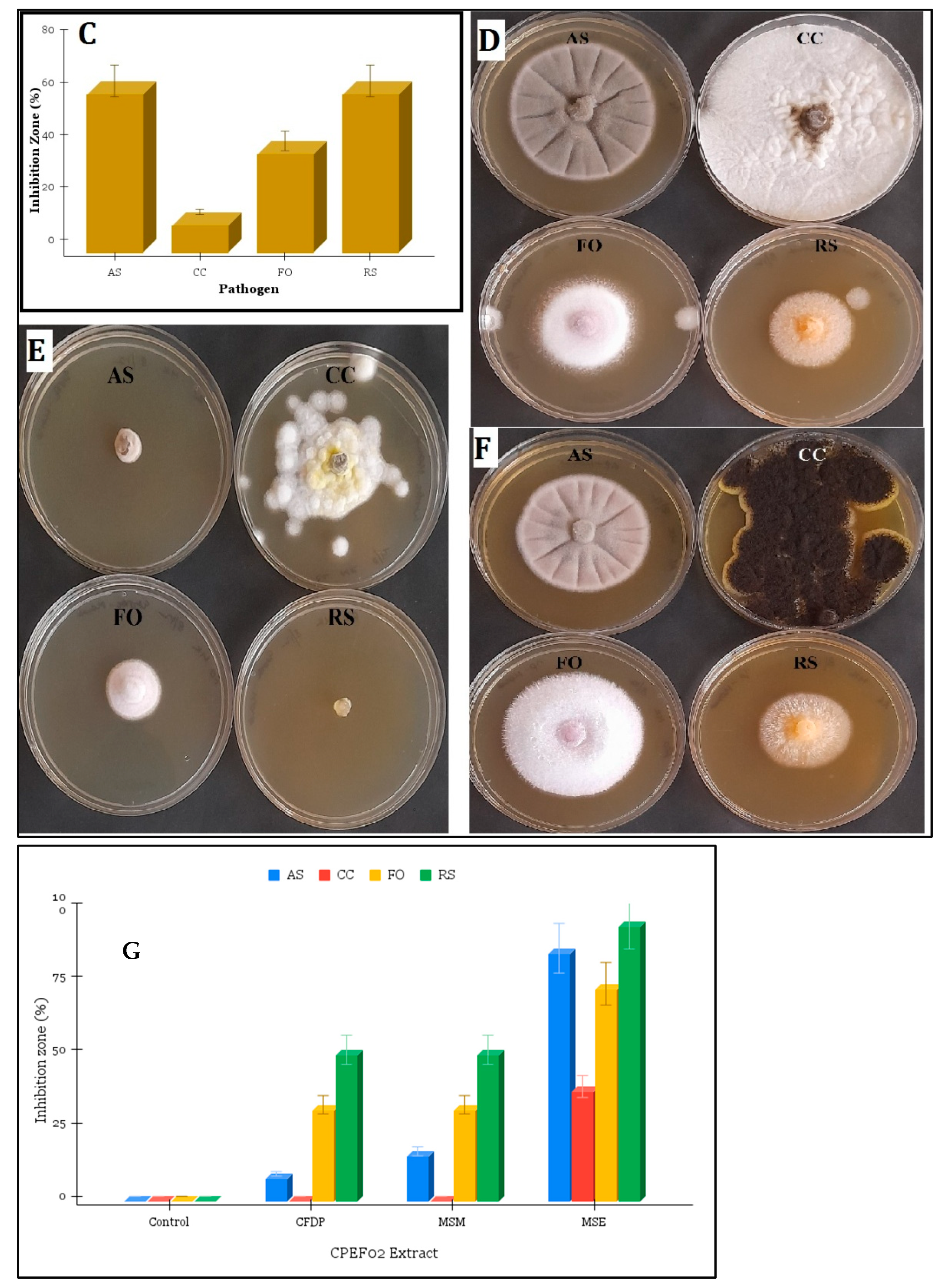
2.4.2. Antifungal Assays

A dual culture assay was used as previously described [13] to determine the antifungal activity of CPEF02. Briefly, 3 mm discs of fungal cultures of CPEF02 were aseptically placed on one corner and discs of pathogenic strains were placed on the opposite side. The negative controls were plates containing only pathogenic strains. The plates were incubated for seven days at 28 °C, and the variations between the negative control and test plates were noted. Inhibition capacity was calculated by estimating the percentage decrease in the test plates (Re) from the control plates (Rc) using [(Rc − Re)/Rc × 100]. To check the antifungal efficacy of the solvent extracts, 1 mg/mL concentrations were prepared in sterile deionized water and added to PDA media. PDA plates without any test samples were used as negative control. The test pathogens were inoculated into 3 mm discs and incubated for 7 days at 28 °C. Inhibition capacity was calculated using the same method as described above.

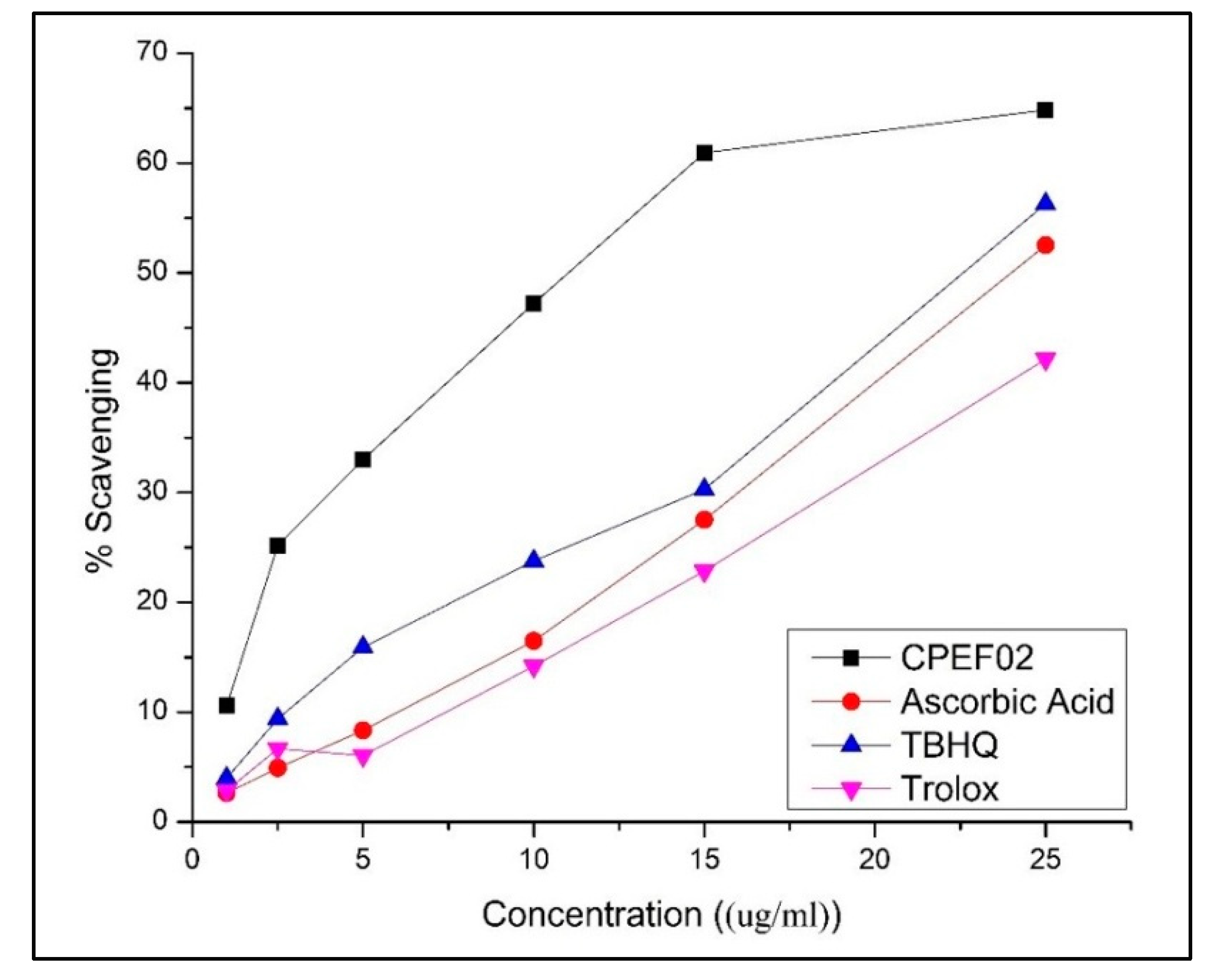
2.5. Antioxidant Assay

An antioxidant capacity assay was carried out as per the previously published method [10]. Briefly, a 7 mM ABTS (Sigma Aldrich India Pvt. Ltd., Karnataka, India) aqueous solution and a 2.45 mM potassium persulfate solution were combined and allowed to sit overnight at ambient temperature (25 °C) in the dark to produce ABTS+ radical cations with an initial absorbance of 0.70 ± 0.02 at 734 nm. In a final volume of 1 mL with the ABTS+ working standard, the gradient sample concentrations and standards of abscisic acid, tert-butyl hydroquinone (TBHQ), and Trolox (Sigma Aldrich India Pvt. Ltd., Karnataka, India) were used to assess antioxidant capacity. The absorbance at 734 nm was measured immediately after a 6 min incubation time at room temperature. Plotting the scavenging ability versus concentration yielded the sample concentration to scavenge 50% ABTS radicals (IC50). The findings were expressed as the µM Trolox/g pigment Trolox equivalent antioxidant capacity (TEAC).

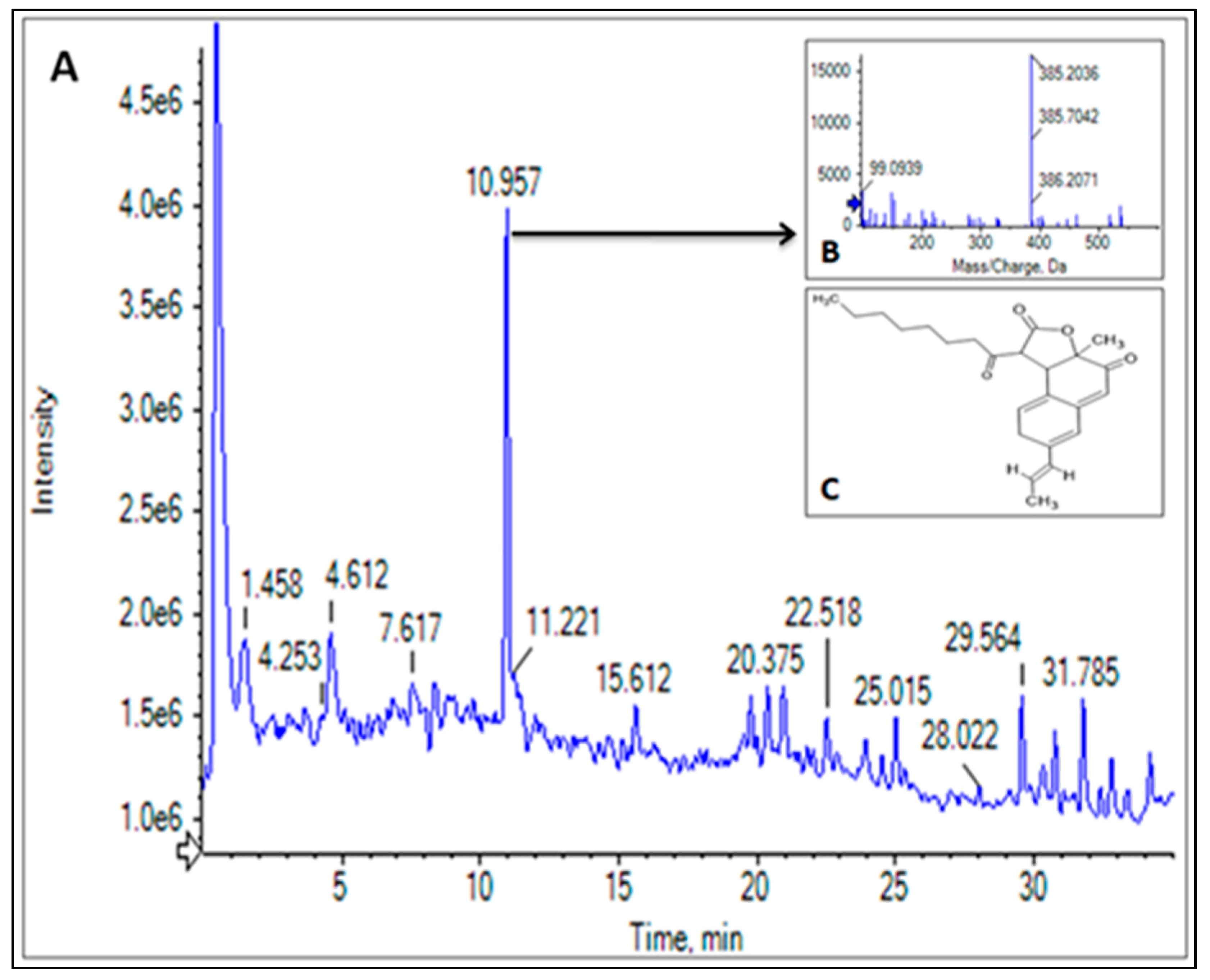
2.6. LC-HRMS Analysis

LC-MS/MS analysis was performed using an AB SCIEX TripleTOF®5600 system equipped with an Exion LC system (AB SCIEX, Framingham, MA, USA). The chromatographic separation was carried out on a reverse phase phenomenex Kinetex C18 (100 mm × 2.1 mm, 1.7 µm) column. Methanol with 0.1% formic acid and water (v/v) were used as the mobile phases and are referred here as eluents A and B, respectively. The multistep gradient elution program used for the chromatographic analysis was as follows: 0–35 min, 5–95% B; 35–36 min, 95–5%, which was kept constant for 40 min. The flow rate and sample injection volume were kept constant at 0.5 µL/min and 10 µL for all the samples with a fixed column temperature of 40 °C. The mass spectrometric source parameters were set as nebulizer (GS1), drying (GS2) and curtain (CUR) gases at 40, 40 and 25 psi; desolvation temperature 500 °C; and ion spray voltage 5500 (ESI+), while the compound-specific parameters, i.e., declustering potential (DP) and collision energy (CE), were kept at 100 V and 10 V, respectively. The generated data were analysed using Analyst 1.8, MultiQuant 2.0.

3. Results

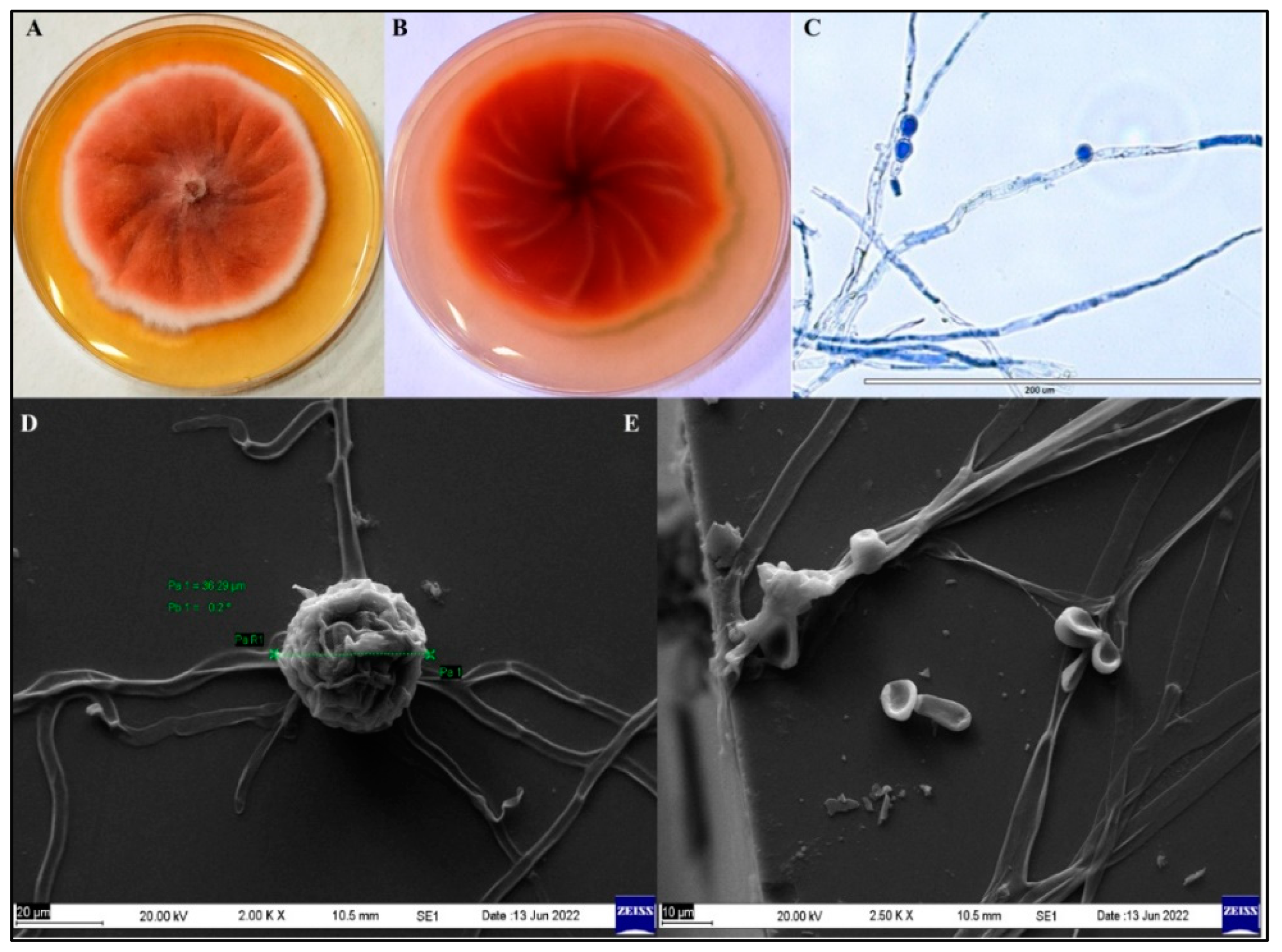
3.1. Identification and Characterization of Pigment-Producing Endophytic Fungus

Endophytic isolate CPEF02, propagated on PDA plate, was morphologically identified using light and scanning electron microscopy. The fungal hyphae displayed apical growth, with white dense and velvet mycelia initially. After 7–10 days, the agar medium started to display orange to reddish colouration owing to the secretion of pigmented metabolites extracellularly in the nutrient medium (Figure 1A,B). In visible light micrographs (Figure 1C), exogenous oval, pyriform conidia with septate hyphae were observed, whereas scanning electron micrographs (Figure 1D,E) displayed spherical ascospores originating from the conidia. Our macroscopic as well as microscopic findings were in agreement with earlier studies on M. purpureus FTCC 5391 [14,15]. Additionally, molecular characterization studies previously carried out [10] in our laboratory confirmed the endophytic strain CPEF02 as Monascus purpureus (GenBank accession number MT013404.1).

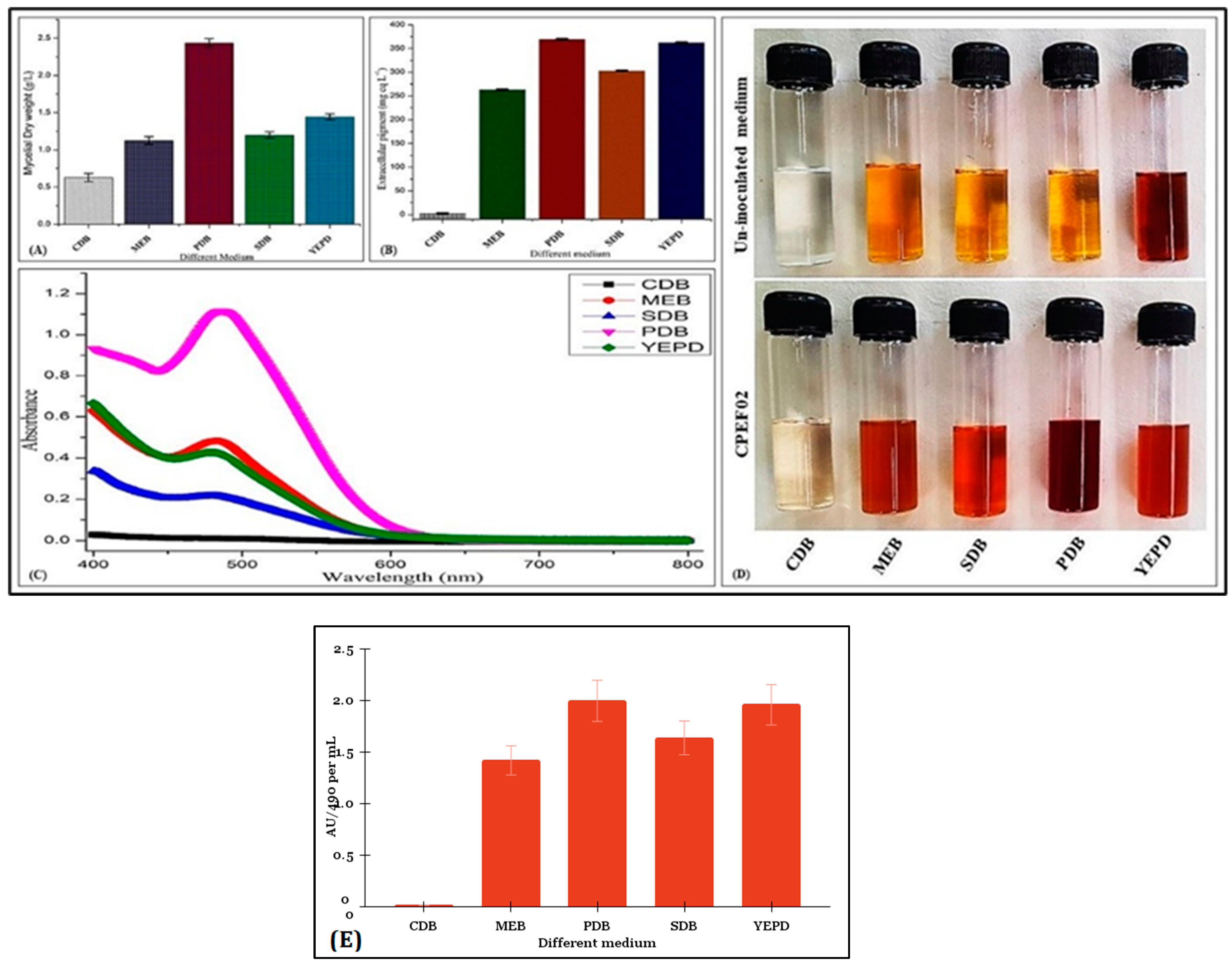
3.2. Screening of Culture Media for Pigment Production and Solvent Extraction by CPEF02

The optimal culture medium for the marine fungal endophytic isolate CPEF02’s growth and extracellular pigment was determined using five different growth media. The production of mycelial biomass ranged between 0.62 ± 0.05 and 2.43 ± 0.05 g/L. Maximum biomass was observed in PDB, by YEPD, SDB, MEB and CDB, respectively (Figure 2A). Among the six media tested, CDB, MEB, PDB, SDB and YEPDB, potato dextrose broth (PDB) was found to be the optimal media based on the maximum extracellular pigment production and absorption at 494 nm (λmax for commercial Carmine standard) and absorbance unit (AU) at 490 nm/mL (Figure 2E). The yield of extracellular polyketide red pigment ranged from 1.59 ± 0.04 to 369.38 ± 0.53 mg eq. of Carmine per litre (Figure 2B). PDB media yielded the maximum extracellular red pigment followed by YEPD, SDB, GYEM, MEB and CDB, respectively (Figure 2B,C). The increased pigment production in PDB is partly due to the complex source of carbon in it [11,16]. In the solvent extracts, maximum pigment yield was observed in MSM obtained from Dianion HP 20 (647.87 mg. equivalent of Carmine per litre and AU490 3.502) compared to MSE (107.85 mg. equivalent of Carmine per litre and AU490 0.583). It was previously reported [17] that similar yields could be achieved by utilizing demineralized whey for the production of red extracellular pigment in Monascus. Monascus pigments are polyketide and hydrophilic in nature, and thus, methanol, being a more polar solvent than ethyl acetate, yielded the maximum red pigment. Previous studies [18,19] also found similar results and deemed non-polar solvents as not suitable for Monascus pigment extraction as they yielded less extracellular red pigment when compared with methanol.

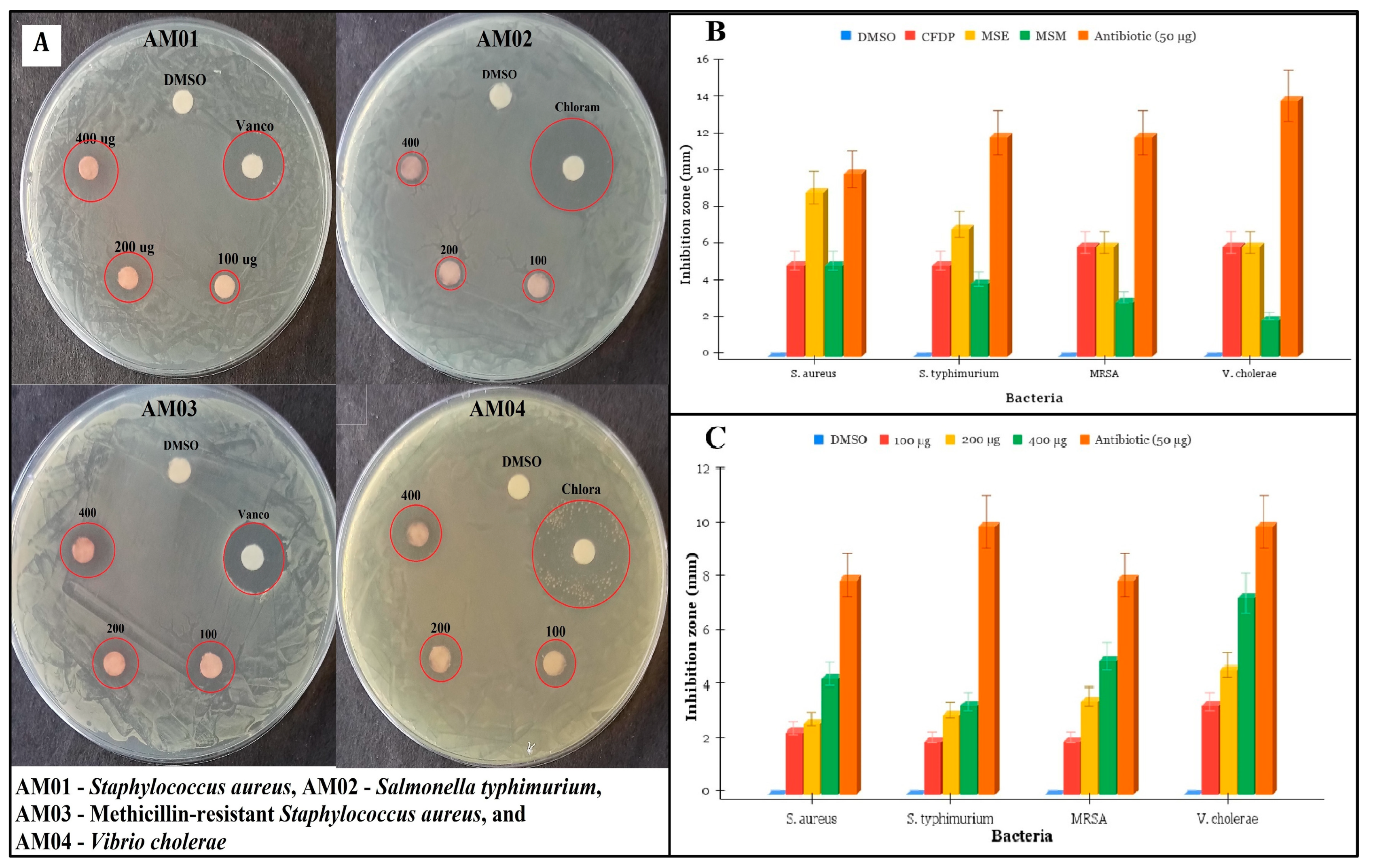
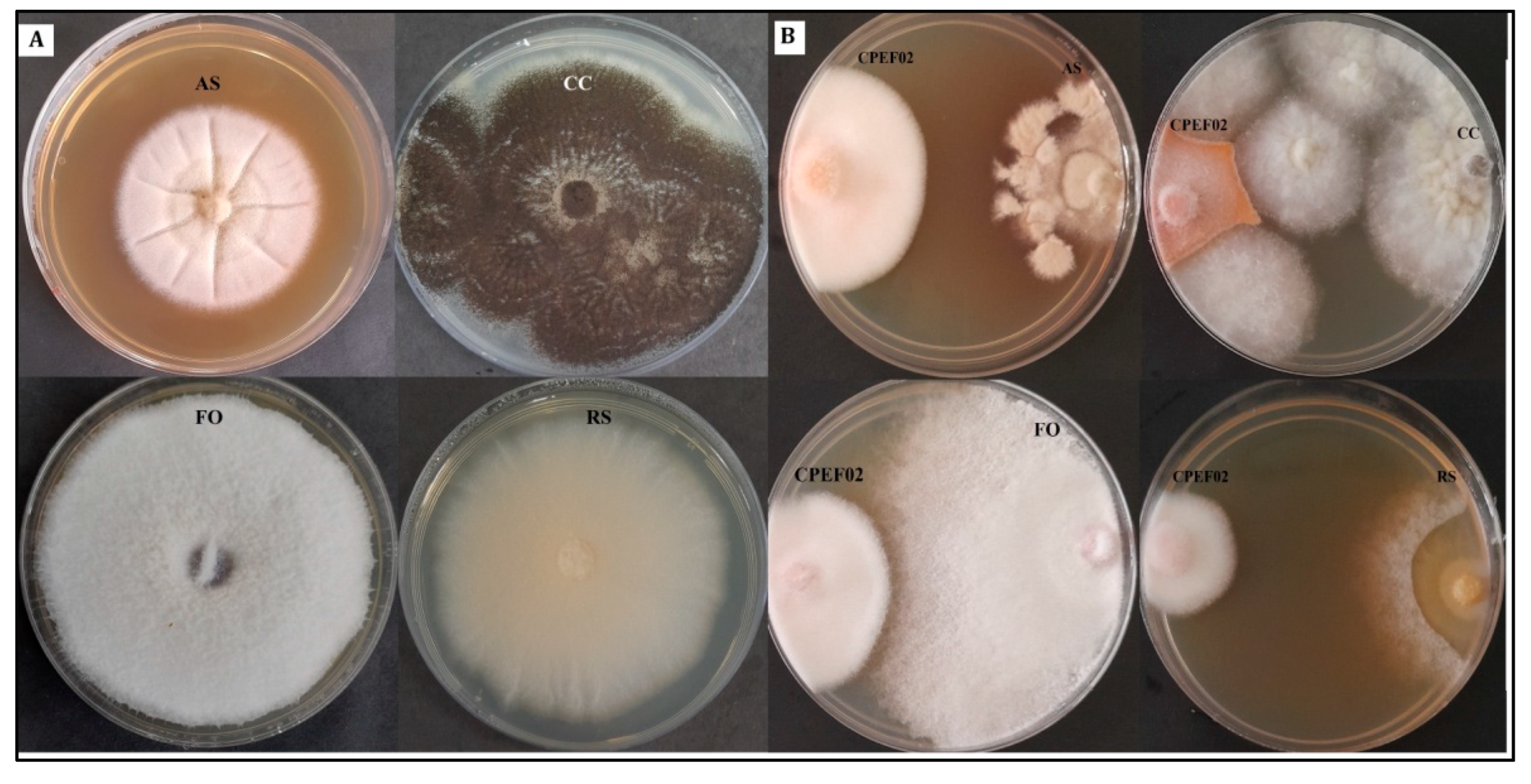
3.3. Antimicrobial Activity of CPEF02 Extracts

Antibacterial activity of CPEF02 extracts carried out using a disc diffusion assay showed notable inhibition zones (in mm) against four bacterial pathogens, namely, S. aureus, S. typhimurium, methicillin-resistant S. aureus and V. cholerae (Figure 3A–C, Table 1 and Table 2). The maximum activity was present against V. cholerae, followed by S. aureus and then S. typhimurium. MSE exhibited maximum antibacterial activity followed by CFDP and methanol extracted colourant (MSM) (Table 1). The maximum inhibition activity of MSE was against S. aureus, followed by S. typhimurium and V. cholerae. As MSE had the highest level of extracellular pigment, its extract was also tested against the bacterial pathogens at multiple concentrations (100 µg/mL, 200 µg/mL and 400 µg/mL) (Table 2), and inhibitory activity was present at the lowest concentration, i.e., 100 µg/mL. M. purpureus extracts were tested previously by [20,21] for their antibacterial activities against Bacillus subtilis and S. aureus. These extract of CPEF02 exhibited some antibacterial activity, but this was lower than that observed for other strains (e.g., CPEF04 [10]). Antimicrobial activity may not be the primary activity of this extract, and further work is required to determine if this is a useful activity, including examining the potential mechanisms against both Gram-positive and Gram-negative bacteria.
Antifungal activity of CPPEF02 against the phytopathogens was tested by two methods—dual culture assay and another previously described method [13]. Antagonistic activity of the fungus was prominent against A. solani and R. solani with more than 50% inhibition, followed by F. oxysporum and C. capsici (Figure 4A–C). Antifungal activity of MSE was maximal with 100% inhibition against R. solani, 84% inhibition against A. solani, 72% against F. oxysporum and 37% against C. capsici (Figure 4E). MSM (Figure 4F) and CFDP (Figure 4D) showed similar activity of 50% inhibition against R. solani, 31% against F. oxysporum and none against C. capsici. MSM had better (15%) activity against A. solani as compared with CFDP (7%). Due to the medium polarity of the solvent (ethyl acetate), the MSE extract may contain non-polar metabolites of antimicrobial efficacy. Purpureusone is a known antifungal pigmented metabolite that is found in M. purpureus, as identified by [22], and inhibits the growth of Candida albicans and Streptomyces cerevisiae. Furthermore, the antibacterial and antifungal activity of the Monascus purpureus colourant is similar to ones reported [21,23]. The antibacterial activity of the colourant isolated from M. purpureus has received less attention than the antimicrobial activity of fungal fermented rice powder or Monascus koji powder. The current work sought to shed light on the bioactivity of the M. purpureus colourant [24].

3.4. Antioxidant Activity of CPEF02 Colourant

As MSM gave the highest extracellular polyketide pigment output, its antioxidant efficiency was also determined. The antioxidant activity of the CPEF02 methanolic colourant (MSM) was estimated with the help of ABTS radical scavenging assay by calculating IC50 (sample concentration to scavenge 50% ABTS radicals) and Trolox equivalent antioxidant capacity (TEAC). Ascorbic acid, TBHQ and Trolox were used as positive standards (Figure 5). The IC50 value of the CPEF02 colourant (MSM) against these radicals was found to be 14.42 µg/mL, while the TEAC value was 0.571 µM Trolox/µg of the pigmented extract. These results indicated a higher antioxidant activity against ABTS radicals and were consistent with the antioxidant activity shown against ABTS radicals [21,25]. Monascus pigments such as rubropunctamine and monasfluore B are known antioxidant compounds [26,27].

3.5. LC-HRMS Profiling of CPEF02 Pigments

Colourant profiling and chemical characterization were performed after subjecting the methanolic extract (MSM) to LC-HRMS (Figure 6 and Table 3). The total ion chromatogram (TIC) (Figure 6A) and a table of the identified compounds by the molecular ion’s [M + H]+ mass/charge ratio (Table 3 and Figure 6B) with their structures are shown. The chromatogram showed a high intensity peak at retention time (Rt) 10.965 with a mass by charge ratio (m/z) of 385.2. This peak was identified as Monasfluore B with a molecular formula of C23H28O5 (Figure 6C) [28]. Monasfluore B is known for its antioxidant activity tested in mice models [27]. Other pigmented compounds identified were rubropunctine [29], PP-V [30], monaphilones B and C [31], purpureusone [22], monascin and rubropunctamine [5,32]. Monascus pigments are known for their antimicrobial and antioxidant activities, and their presence enhances the pharmaceutical potential of this material. Monascin and ankaflavin are known for their anticancer, antidiabetic, antioxidant, anti-obesity and anti-inflammatory properties and are relatively well studied pigments along with Monacolin K [33,34,35,36,37,38]. Monasfluore A and B have been tested on mice models and were found to exhibit antioxidant and anti-obesity activities [27]. The major pigments reported from Monascus purpureus strains are monascin (yellow); monascorubrin (orange); monascorubramine; monapurone A–C; monasphilone A, B and C; monasfluore A and B; monascolin K; ankaflavin; rubropunctamine; rubropunctatin; and monopilol A–D [7,28,31,39,40,41,42]. However, only a few of these, namely, monascin, ankaflavin, rubropunctatin and monascorubamine, are used in the food and dyeing industries [7,43]. The extract’s antioxidant action may be ascribed to monasfluore B, monascin and rubropunctamine, while its antibacterial activity may be related to purpureosone. These compounds identified in the methanolic extract are most likely responsible for the antibacterial and antioxidant properties of the extracts.

4. Conclusions

Microbial pigments can be good alternatives to synthetic and plant-based pigments because of their stability, bioactivity and cost-effective production. Apart from adding colour, they increase the health potential of food with bioactivities such as antibacterial, antifungal and antioxidant activities. This work investigated the optimal media and extraction conditions to obtain maximum extracellular polyketide pigment from the endophytic marine fungus Monascus purpureus CPEF02. The methanolic extract of the fungus was found to produce maximum extracellular pigment (647.87 mg. equivalent of Carmine per litre) along with exhibiting antibacterial and antioxidant activities. The antioxidant activity of 14.42 µg/mL IC50 and a TEAC value of 0.571 µM Trolox/µg was also obtained for the pigmented extract. The ethyl acetate extract was shown to have antimicrobial activity and inhibited the growth of both bacterial and fungal pathogens (more than 90% inhibition in the case of fungal pathogens R. solani and A. solani and 9.33 ± 0.33 mm zone of inhibition in the case of bacterial pathogen S. aureus). The presence of Monasfluore B, monascin, purpureosone and rubropunctamine in the methanolic extract of CPEF02 is consistent with its observed activities. CPEF02 was shown to have higher organic solvent solubility and higher antioxidant activity but lower antibacterial activity than pigment from a previous fungal strain (Talaromyces strain CPEF04 [10]). CPEF02 is derived from red yeast rice, while CPEF04 is from a non-food grade strain, and so, the pigment reported here has more potential to be developed into a food grade product. These pigments could be further developed for pharmaceutical, food, cosmeceutical, or sensitized solar cells or textile applications, particularly where their bioactivity could be used as an additional benefit to their colour. The biosynthetic pathways of the pigments being produced could be systematically explored for biotransformation of the Monascus cells into cell factories via metabolic engineering for industrial-scale production. Pigment production could be readily scaled to the pilot and manufacturing scales and formulated for pharmaceutical, textile or food use.

Author Contributions

M.K. contributed towards the execution of experiments, data curation and analysis, original draft writing, editing and incorporation of editions at various stages of reviewing. M.G. conceptualized, designed and supervised the work, critically reviewed the manuscript at every stage of the drafting, and provided resources for the project. R.C.M. isolated and identified the fungal strain used in the study and edited some sections at reviewing stage. V.L. carried out the LC-MS experiment and A.K.Y. analyzed the data and provided resources for the same. C.J.B. contributed toward study design and critically edited, reviewed and performed language corrections in the draft. All authors have read and agreed to the published version of the manuscript.

Funding

This research received no external funding.

Institutional Review Board Statement

Not applicable.

Informed Consent Statement

Not applicable.

Data Availability Statement

Data is available from the authors.

Conflicts of Interest

The authors declare no conflict of interest.

References

  1. Fried, R.; Oprea, I.; Fleck, K.; Rudroff, F. Biogenic Colourants in the Textile Industry—A Promising and Sustainable Alternative to Synthetic Dyes. Green Chem. 2022, 24, 13–35. [Google Scholar] [CrossRef]
  2. Bai, Y.; Kissoudis, C.; Yan, Z.; Visser, R.G.F.; van der Linden, G. Plant Behaviour under Combined Stress: Tomato Responses to Combined Salinity and Pathogen Stress. Plant J. 2018, 93, 781–793. [Google Scholar] [CrossRef] [Green Version]
  3. Kalra, R.; Conlan, X.A.; Goel, M. Fungi as a Potential Source of Pigments: Harnessing Filamentous Fungi. Front. Chem. 2020, 8, 369. [Google Scholar] [CrossRef]
  4. Zheng, Y.; Xin, Y.; Guo, Y. Study on the Fingerprint Profile of Monascus Products with HPLC–FD, PAD and MS. Food Chem. 2009, 113, 705–711. [Google Scholar] [CrossRef]
  5. Chen, W.; Chen, R.; Liu, Q.; He, Y.; He, K.; Ding, X.; Kang, L.; Guo, X.; Xie, N.; Zhou, Y.; et al. Orange, Red, Yellow: Biosynthesis of Azaphilone Pigments in Monascus Fungi. Chem. Sci. 2017, 8, 4917–4925. [Google Scholar] [CrossRef] [Green Version]
  6. He, J.; Jia, M.; Li, W.; Deng, J.; Ren, J.; Luo, F.; Bai, J.; Liu, J. Toward Improvements for Enhancement the Productivity and Color Value of Monascus Pigments: A Critical Review with Recent Updates. Crit. Rev. Food Sci. Nutr. 2021, 62, 7139–7153. [Google Scholar] [CrossRef]
  7. Lagashetti, A.C.; Dufossé, L.; Singh, S.K.; Singh, P.N. Fungal Pigments and Their Prospects in Different Industries. Microorganisms 2019, 7, 604. [Google Scholar] [CrossRef] [Green Version]
  8. Abdollahi, F.; Jahadi, M.; Ghavami, M. Thermal Stability of Natural Pigments Produced by Monascus purpureus in Submerged Fermentation. Food Sci. Nutr. 2021, 9, 4855–4862. [Google Scholar] [CrossRef]
  9. Turgay, O.; Azirak, S. A Review on the Red Yeast Rice (Monascus purpureus). Turk. Electron. J. Biotechnol. 2004, 2, 37–49. [Google Scholar]
  10. Mishra, R.C.; Kalra, R.; Dilawari, R.; Deshmukh, S.K.; Barrow, C.J.; Goel, M. Characterization of an Endophytic Strain Talaromyces assiutensis, CPEF04 with Evaluation of Production Medium for Extracellular Red Pigments Having Antimicrobial and Anticancer Properties. Front. Microbiol. 2021, 12, 665702. [Google Scholar] [CrossRef]
  11. Lebeau, J.; Venkatachalam, M.; Fouillaud, M.; Petit, T.; Vinale, F.; Dufossé, L.; Caro, Y. Production and New Extraction Method of Polyketide Red Pigments Produced by Ascomycetous Fungi from Terrestrial and Marine Habitats. J. Fungi 2017, 3, 34. [Google Scholar] [CrossRef] [Green Version]
  12. Weinstein, M.P. Clinical and Laboratory Standards Institute. Performance Standards for Antimicrobial Susceptibility Testing; Clinical and Laboratory Standards Institute: Wayne, PA, USA, 2021. [Google Scholar]
  13. Aguilar-Méndez, M.A.; San Martín-Martínez, E.; Ortega-Arroyo, L.; Cobián-Portillo, G.; Sánchez-Espíndola, E. Synthesis and Characterization of Silver Nanoparticles: Effect on Phytopathogen Colletotrichum gloesporioides. J. Nanoparticle Res. 2010, 13, 2525–2532. [Google Scholar] [CrossRef]
  14. Almeida, A.B.D.; de Lima, T.M.; Santos, N.H.; Santana, R.V.; dos Santos, S.C.; Egea, M.B. An Alternative for Corn Bran Byproduct: Fermentation Using M. purpureus. Nutr. Food Sci. 2019, 50, 515–527. [Google Scholar] [CrossRef]
  15. Rasheva, T.; Hallet, J.-N.; Kujumdzieva, A. Taxonomic investigation of Monascus purpureus 94-25 strain. J. Cult. Collect. 1998, 2, 51–59. [Google Scholar]
  16. Parul; Thiyam, G.; Dufossé, L.; Sharma, A.K. Characterization of Talaromyces purpureogenus Strain F Extrolites and Development of Production Medium for Extracellular Pigments Enriched with Antioxidant Properties. Food Bioprod. Process. 2020, 124, 143–158. [Google Scholar] [CrossRef]
  17. Mehri, D.; Perendeci, N.A.; Goksungur, Y. Utilization of Whey for Red Pigment Production by Monascus purpureus in Submerged Fermentation. Fermentation 2021, 7, 75. [Google Scholar] [CrossRef]
  18. De Carvalho, J.C.; Oishi, B.; Woiciechowski, A.L.; Pandey, A.; Sumathy, B.; Socool, C. Effect of Substrates on the Production of Monascus Biopigments by Solid-State Fermentation and Pigment Extraction Using Different Solvents. Indian J. Biotechnol. 2007, 6, 194–199. [Google Scholar]
  19. Padmavathi, T.; Prabhudessai, T. A Solid Liquid State Culture Method to Stimulate Monascus Pigments by Intervention of Different Substrates. Int. Res. J. Biol. Sci. 2013, 2, 22–29. [Google Scholar]
  20. Chaudhary, V.; Katyal, P.; Panwar, H.; Puniya, A.K.; Poonia, A.K. Evaluating Anti-Microbial and Anti-Oxidative Potential of Red Biopigment from Monascus purpureus. Environ. Conserv. J. 2022, 23, 83–93. [Google Scholar] [CrossRef]
  21. de Almeida, A.B.; Santos, N.H.; de Lima, T.M.; Santana, R.V.; de Oliveira Filho, J.G.; Peres, D.S.; Egea, M.B. Pigment Bioproduction by Monascus purpureus Using Corn Bran, a Byproduct of the Corn Industry. Biocatal. Agric. Biotechnol. 2021, 32, 101931. [Google Scholar] [CrossRef]
  22. Cheng, M.-J.; Wu, M.-D.; Chen, I.-S.; Tseng, M.; Yuan, G.-F. Chemical Constituents from the Fungus Monascus purpureus and Their Antifungal Activity. Phytochem. Lett. 2011, 4, 372–376. [Google Scholar] [CrossRef]
  23. Gökmen, G.G.; Şılbır, M.S.; Göksungur, Y.; Kışla, D. Antimicrobial Activity of Red Pigments Derived from Monascus purpureus: A Comparison to Industrial Red Pigments. JSFA Rep. 2021, 1, 5–10. [Google Scholar] [CrossRef]
  24. Yudiarti, T.; Sugiharto, S.; Isroli, I.; Widiastuti, E.; Wahyuni, H.; Sartono, T. Effect of Fermentation Using Chrysonillia crassa and Monascus purpureus on Nutritional Quality, Antioxidant, and Antimicrobial Activities of Used Rice as a Poultry Feed Ingredient. J. Adv. Vet. Anim. Res. 2019, 6, 168. [Google Scholar] [CrossRef] [PubMed]
  25. Kim, B.; Ryu, J.; Jang, S.O.; Kim, M. Antioxidant Activity and Cell Differentiation Effects of Monascus purpureus Pigment on Osteoblast-like MC3T3-E1 Cells. J. Life Sci. 2020, 30, 468–475. [Google Scholar]
  26. Dahle, M.A.; Divakar, S.; Kumar, S.U.; Vijayalakshmi, G. Isolation and Characterization of Dihydromonacolin-MV from Monascus purpureus for Antioxidant Properties. Appl. Microb. Cell Physiol. 2007, 73, 1197–1202. [Google Scholar] [CrossRef]
  27. Yeh, Y.-H.; Hsieh, Y.-L.; Lee, Y.-T. Effect of RYR, MFA and MFB on Serum Lipids and Antioxidant Activity in Rats. e-SPEN J. 2012, 7, e176–e181. [Google Scholar] [CrossRef]
  28. Hsu, Y.-W.; Hsu, L.-C.; Chang, C.-L.; Liang, Y.-H.; Kuo, Y.-H.; Pan, T.-M. New Anti-Inflammatory and Anti-Proliferative Constituents from Fermented Red Mold Rice Monascus purpureus NTU 568. Molecules 2010, 15, 7815–7824. [Google Scholar] [CrossRef]
  29. Loret, M.-O.; Morel, S. Isolation and Structural Characterization of Two New Metabolites from Monascus. J. Agric. Food Chem. 2010, 58, 1800–1803. [Google Scholar] [CrossRef]
  30. Mapari, S.A.S.; Hansen, M.E.; Meyer, A.S.; Thrane, U. Computerized Screening for Novel Producers of Monascus-like Food Pigments in Penicillium Species. J. Agric. Food Chem. 2008, 56, 9981–9989. [Google Scholar] [CrossRef]
  31. Hsu, Y.-W.; Hsu, L.-C.; Liang, Y.-H.; Kuo, Y.-H.; Pan, T.-M. Monaphilones A−C, Three New Antiproliferative Azaphilone Derivatives from Monascus purpureus NTU 568. J. Agric. Food Chem. 2010, 58, 8211–8216. [Google Scholar] [CrossRef]
  32. Yuliana, A.; Singgih, M.; Julianti, E.; Blanc, P.J. Derivates of Azaphilone Monascus Pigments. Biocatal. Agric. Biotechnol. 2017, 9, 183–194. [Google Scholar] [CrossRef]
  33. Lin, C.-P.; Lin, Y.-L.; Huang, P.-H.; Tsai, H.-S.; Chen, Y.-H. Inhibition of Endothelial Adhesion Molecule Expression by Monascus purpureus-Fermented Rice Metabolites, Monacolin K, Ankaflavin, and Monascin. J. Sci. Food Agric. 2011, 91, 1751–1758. [Google Scholar] [CrossRef] [PubMed]
  34. Shi, Y.-C.; Liao, V.H.-C.; Pan, T.-M. Monascin from Red Mold Dioscorea as a Novel Antidiabetic and Antioxidative Stress Agent in Rats and Caenorhabditis elegans. Free Radic. Biol. Med. 2012, 52, 109–117. [Google Scholar] [CrossRef] [PubMed]
  35. Hsu, W.-H.; Lee, B.-H.; Liao, T.-H.; Hsu, Y.-W.; Pan, T.-M. Monascus-Fermented Metabolite Monascin Suppresses Inflammation via PPAR-γ Regulation and JNK Inactivation in THP-1 Monocytes. Food Chem. Toxicol. 2012, 50, 1178–1186. [Google Scholar] [CrossRef] [PubMed]
  36. Hsu, L.-C.; Liang, Y.-H.; Hsu, Y.-W.; Kuo, Y.-H.; Pan, T.-M. Anti-Inflammatory Properties of Yellow and Orange Pigments from Monascus purpureus NTU 568. J. Agric. Food Chem. 2013, 61, 2796–2802. [Google Scholar] [CrossRef]
  37. Jou, P.-C.; Ho, B.-Y.; Hsu, Y.-W.; Pan, T.-M. The Effect of Monascus Secondary Polyketide Metabolites, Monascin and Ankaflavin, on Adipogenesis and Lipolysis Activity in 3T3-L1. J. Agric. Food Chem. 2010, 58, 12703–12709. [Google Scholar] [CrossRef]
  38. Lee, C.-L.; Wen, J.-Y.; Hsu, Y.-W.; Pan, T.-M. Monascus-Fermented Yellow Pigments Monascin and Ankaflavin Showed Antiobesity Effect via the Suppression of Differentiation and Lipogenesis in Obese Rats Fed a High-Fat Diet. J. Agric. Food Chem. 2013, 61, 1493–1500. [Google Scholar] [CrossRef]
  39. Mapari, S.A.; Meyer, A.S.; Thrane, U.; Frisvad, J.C. Identification of Potentially Safe Promising Fungal Cell Factories for the Production of Polyketide Natural Food Colorants Using Chemotaxonomic Rationale. Microb. Cell Factories 2009, 8, 24. [Google Scholar] [CrossRef] [Green Version]
  40. Kaur, B.; Chakraborty, D.; Kaur, H. Production and Evaluation of Physicochemical Properties of Red Pigment from Monascus purpureus MTCC 410. Internet J. Microbiol. 2009, 7, 1–7. [Google Scholar]
  41. Li, J.-J.; Shang, X.-Y.; Li, L.-L.; Liu, M.-T.; Zheng, J.-Q.; Jin, Z.-L. New Cytotoxic Azaphilones from Monascus purpureus-Fermented Rice (Red Yeast Rice). Molecules 2010, 15, 1958–1966. [Google Scholar] [CrossRef] [Green Version]
  42. Mukherjee, G.; Singh, S.K. Purification and Characterization of a New Red Pigment from Monascus purpureus in Submerged Fermentation. Process. Biochem. 2011, 46, 188–192. [Google Scholar] [CrossRef]
  43. Feng, Y.; Shao, Y.; Chen, F. Monascus Pigments. Appl. Microbiol. Biotechnol. 2012, 96, 1421–1440. [Google Scholar] [CrossRef] [PubMed]
  44. Sato, K.; Goda, Y.; Sakamoto Sasaki, S.; Shibata, H.; Maitani, T.; Yamada, T. Identification of Major Pigments Containing D-Amino Acid Units in Commercial Monascus Pigments. Chem. Pharm. Bull. 1997, 45, 227–229. [Google Scholar] [CrossRef] [Green Version]
  45. Srianta, I.; Zubaidah, E.; Estiasih, T.; Iuchi, Y.; Harijono; Yamada, M. Antioxidant Activity of Pigments Derived from Monascus purpureus Fermented Rice, Corn, and Sorghum. Int. Food Res. J. 2017, 24, 1186–1191. [Google Scholar]
  46. Akihisa, T.; Tokuda, H.; Yasukawa, K.; Ukiya, M.; Kiyota, A.; Sakamoto, N.; Suzuki, T.; Tanabe, N.; Nishino, H. Azaphilones, Furanoisophthalides, and Amino Acids from the Extracts of Monascus pilosus-Fermented Rice (Red-Mold Rice) and Their Chemopreventive Effects. J. Agric. Food Chem. 2005, 53, 562–565. [Google Scholar] [CrossRef] [PubMed]
Figure 1. (A,B) Morphological images of CPEF02 after 10 days of growth at 28 °C; (C) light microscopic image of CPEF02 hyphae and conidia; (D,E) SEM images of CPEF02 ascospores, conidia and hyphae.
Fermentation 09 00328 g001
Figure 2. (A) Mycelial biomass of CPEF02 produced in different media, Czapek’s extract dox Broth (CDB), malt extract broth (MEB), potato dextrose broth (PDB), Sabouraud dextrose broth (SDB), and yeast extract peptone dextrose broth (YEPD); (B,D) extracellular pigment production by CPEF02 (measured in mg equivalent of Carmine per litre) in different media; and (C) UV—visible absorption spectra of different media culture filtrates. (E) AU490/mL (absorbance unit at 490 nm/mL) of colourant produced in different media.
Fermentation 09 00328 g002
Figure 3. (A) Disc diffusion assay of CPEF02 (inhibition zone measured in mm); (B) antibacterial activity of CPEF02 extracts (CFDP, MSE and MSM) at the concentration of 1 mg/mL; (C) antibacterial activity of CPEF02 methanolic extract (MSM) at different concentrations against bacterial pathogens S. aureus (AM01), S. typhimurium (AM02), methicillin-resistant S. aureus (AM03) and V. cholerae (AM04).
Fermentation 09 00328 g003
Figure 4. (AC) Antagonistic activity of CPEF02 on pathogenic fungi A. solani (AS), C. capsici (CC), F. oxysporum (FO) and R. solani (RS). (D) Antifungal activity of CFDP, (E) antifungal activity of MSE extract, (F) antifungal activity of MSM extract and (G) inhibition zone in percentage (%).
Fermentation 09 00328 g004aFermentation 09 00328 g004b
Figure 5. Antioxidant activity of methanolic CPEF02 extract and standards (ascorbic acid, TBHQ and trolox) against the ABTS radical.
Fermentation 09 00328 g005
Figure 6. (A) Liquid chromatogram of methanolic extract of CPEF02, the arrow on the y-axis indicating base (starting point) for the intensity, (B) mass spectra of Monasfluore B at retention time 10.965, (C) molecular structure of Monasfluore B.
Fermentation 09 00328 g006
Table 1. Antibacterial activity of CPEF02 extracts CFDP, MSE and MSM at the concentration of 1 mg/mL against bacterial pathogens (measured in mm).
S. aureus
(in mm)
S. typhimurium (in mm)MRSA
(in mm)
V. cholerae
(in mm)
DMSO0000
CFDP5.77 ± 0.185.62 ± 0.086.7 ± 0.126.14 ± 0.9
MSE9.33 ± 0.337.68 ± 0.26.42 ± 0.156.9 ± 0.33
MSM5.45 ± 0.054.33 ± 0.183 ± 0.672.78 ± 0.05
Antibiotic (50 µg)10121214
Table 2. Antibacterial activity (zone of inhibition) of CPEF02 methanolic colourant against bacterial pathogens (measured in mm).
S. aureus
(in mm)
S. typhimurium (in mm)MRSA
(in mm)
V. cholerae
(in mm)
DMSO0000
100 µg2.33 ± 0.572 ± 0.002 ± 0.053.33 ± 0.66
200 µg2.67 ± 0.353 ± 0.333.5 ± 0.334.67 ± 0.57
400 µg4.33 ± 0.23.33 ± 0.575 ± 0.457.33
Antibiotic (50 µg)810810
Table 3. Pigmented compounds in the methanol-extracted CPEF02 colourant determined using LC-HRMS analysis.
Analyte No. Tentative Compound AllottedChemical FormulaRetention TimeStructureMass Recorded
(m/z)
ActivityReferences
1RubropunctinC22H30O44.610Fermentation 09 00328 i001359.67-[29]
2PP-V (Penicillium purpurogenum–Violet)C23H25NO68.361Fermentation 09 00328 i002411.66-[30]
3Monasfluore BC23H28O510.965Fermentation 09 00328 i003385.20Antioxidant, anti-inflammatory activity[5,27,28]
4Red derivative 1C26H31NO618.173Fermentation 09 00328 i004453.34-[44]
5Monaphilone CC20H32O422.019Fermentation 09 00328 i005337.241Anti-proliferative[31]
6RubaropunctamineC21H23NO423.922Fermentation 09 00328 i006354.25Antioxidant, anticancer activity[5,45,46]
7MonascinC21H26O524.518Fermentation 09 00328 i007359.18Anticancer, antitumor, antidiabetic, antioxidant, anti-inflammatory, anti-obesity activity[5,33,34,35,36,37,38]
8Monaphilone BC20H28O425.028Fermentation 09 00328 i008333.21Anti-proliferative activity[31]
9PurpureusoneC23H34O525.349Fermentation 09 00328 i009391.21Antifungal activity[22]
Disclaimer/Publisher’s Note: The statements, opinions and data contained in all publications are solely those of the individual author(s) and contributor(s) and not of MDPI and/or the editor(s). MDPI and/or the editor(s) disclaim responsibility for any injury to people or property resulting from any ideas, methods, instructions or products referred to in the content.

