Article

Highly Active Astaxanthin Production from Waste Molasses by Mutated Rhodosporidium toruloides G17

1
Faculty of Applied Sciences, Ton Duc Thang University, Ho Chi Minh City 700000, Vietnam
2
Department of Chemical Engineering, Ming Chi University of Technology, New Taipei City 24301, Taiwan
3
School of Life and Environmental Sciences, Deakin University, Geelong, VIC 3216, Australia
4
Faculty of Biology and Biotechnology, University of Science, Vietnam National University-HCM, Ho Chi Minh City 700000, Vietnam
*
Authors to whom correspondence should be addressed.
Fermentation 2023, 9(2), 148; https://doi.org/10.3390/fermentation9020148
Submission received: 9 January 2023 / Revised: 23 January 2023 / Accepted: 29 January 2023 / Published: 2 February 2023
(This article belongs to the Special Issue Pigment Production in Submerged Fermentation)

Abstract

:
Astaxanthin is increasingly attracting commercial interest for its application in the nutraceutical and pharmaceutical industries. This study aimed to produce astaxanthin from molasses with our newly mutated strain of Rhodosporidium toruloides G17 and to evaluate biological activities of the produced astaxanthin. To maximize the astaxanthin yield, the response surface methodology was used so as to optimize the culture conditions. A maximum astaxanthin yield of 1262.08 ± 14.58 µg/L was achieved by growing R. toruloides G17 in a molasses-based medium containing 49.39 g/L reducing sugar, 1.00 g/L urea, 4.15 g/L MgSO4·7H2O, and 10.05% inoculum ratio. The produced astaxanthin was then purified and studied for its antioxidant and anticancer activities. This compound exhibited 123-fold higher antioxidant activity than α-tocopherol, with an IC50 value of 0.97 ± 0.01 µg/mL. The astaxanthin also showed a potent inhibitory ability against the following three cancer cell lines: HeLa, A549, and MCF7, with IC50 values of 69.07 ± 2.4 µg/mL, 55.60 ± 2.64 µg/mL, and 56.38 ± 4.1 µg/mL, respectively. This study indicates that astaxanthin derived from our newly mutated R. toruloides G17 is a promising anticancer and antioxidant agent for further pharmaceutical applications.

1. Introduction

In recent decades, bioactive compounds from natural sources have received growing interest due to their effectiveness and positive effect on human health, especially antioxidant and anticancer activities [1]. Particularly, astaxanthin, a xanthophyll carotenoid, possesses potential antioxidant and inhibitory effects against a variety of cancers including gastric, bladder, lung, colon, breast, and hepatocellular carcinoma [1,2]. In addition, astaxanthin exhibits anti-inflammatory, anti-apoptotic, antidiabetic, cardioprotective, and immune modulation characteristics [3,4]. Because of such health benefits, studies have been increasingly focusing on developing efficient methods for producing astaxanthin.
Astaxanthin is primarily produced industrially using chemical synthesis. Synthetic astaxanthin accounts for 95% of the total global market of astaxanthin products [5] and is commonly used for pigmentation in the aquaculture sector [3]. However, the production of astaxanthin using chemical synthesis has negative environmental effects and its use for human applications raises food safety concerns. Consequently, the production of astaxanthin through biological processes has received considerable attention, as a variety of microorganisms can biosynthesize astaxanthin [6,7]. Different microorganisms such as bacteria [8], microalgae [9], and yeast [10] have been investigated for their ability to biosynthesize astaxanthin. However, the industrial biosynthesis of astaxanthin remains limited due to low astaxanthin productivity and high production costs [10]. Therefore, significant efforts have been made to identify new microbial candidates for astaxanthin production and to develop efficient methods for its production [11,12].
The red yeast Rhodosporidium toruloides has been reported as one of the most important sources of carotenoids due to its high biomass accumulation and high growth rate. R. toruloides can reach a biomass productivity of up to 6.84 g/L within 4 days of cultivation while microalgae require more than 10 days of cultivation to reach the biomass yield of 0.028–0.7 g/L [13,14,15]. Recently, we isolated new R. toruloides strains for astaxanthin production [16]. To further improve astaxanthin accumulation, we successfully attained an R. toruloides mutant (G17) using a gamma irradiation mutagenesis [14]. This gamma-induced mutant is a promising candidate for astaxanthin production. However, neither the cultivation of this strain using waste materials as a cheap carbon source, nor the biological activity of astaxanthin produced by this strain, have been investigated.
Molasses (a by-product of sugar production) has been reported as a suitable low-cost medium for the cultivation of different microorganisms (e.g., microalgae and bacteria) as this waste is a rich source of micronutrients (Mg2+, Ca2+, K+), organic compounds (monosaccharides, protein, lactic acids, glutamic acids, aspartic acids), nitrogen, and phosphorous [17,18,19]. Therefore, this study investigated the use of waste molasses as a cheap carbon source for growing R. toruloides G17. The response surface methodology (RSM) was used to optimize the culture conditions (reducing sugar concentration, urea concentration, mineral concentration, inoculum ratio) used for maximizing the astaxanthin yield. The produced astaxanthin was then extracted, purified, and studied for its anticancer and antioxidant activities.

2. Materials and Methods

2.1. Materials

Molasses (73°Brix) was obtained from Bien Hoa sugar factory (Bien Hoa, Vietnam). Astaxanthin, 2,2-diphenyl-1-picrylhydrazyl (DPPH), 3-5-dinitrosalicylic acid (DNS), α-tocopherol, and 3-(4,5-dimethylthiazol-2-yl)-2,5-diphenyltetrazolium bromide (MTT) were provided by Sigma-Aldrich (Saint Louis, MO, USA). Dimethyl sulfoxide (DMSO), petroleum ether, acetone, and n-hexane were obtained from Sigma-Aldrich (Singapore). Fetal bovine serum (FBS), antibiotics, Dulbecco’s Modified Eagle Medium/Nutrient Mixture F12 (DMEM/F12), and DMEM were provided by Gibco (Waltham, MA, USA).

2.2. Inoculum Preparation

The strain used was gamma-mutated R. toruloides G17, which was obtained from our previous study [14]. It is a wild-type strain isolated from a water sample collected in Ba Ria-Vung province (Vietnam). The strain was maintained on agar plates containing Hansen medium (50 g/L glucose, 10 g/L peptone, 4 g/L MgSO4·7H2O, 3 g/L KH2PO4, and 20 g/L agar) and renewed every two weeks. The culture inoculum was prepared by growing R. toruloides G17 in Hansen broth for 24 h and used for further experiments.

2.3. Optimization of Growth Conditions

Molasses was hydrolyzed by sulfuric acid using a previously reported method [20]. The resulting hydrolysate was then used for the cultivation of R. toruloides G17. The yeast strain was grown in 250 mL Erlenmeyer flasks containing 100 mL of the molasses hydrolysate-based medium (molasses hydrolysate, urea, and MgSO4·7H2O) at 30 °C for 4 d with shaking at 200 rpm. To maximize the astaxanthin yield, a central composite design (CCD) with five levels and four factors was employed to optimize the culture conditions. The four factors (input variables) included the concentration of reducing sugar in molasses hydrolysate (X1), urea concentration (X2), MgSO4·7H2O concentration (X3), and inoculum ratio (X4). Table 1 displays the coded and actual values of four input variables. After the cultivation was completed, the culture was centrifugated at 4000× rpm for 5 min. The cell pellets were then collected, washed twice with distilled water, and dried at 60 °C to a constant weight to measure the cell mass. The dried biomass was then used for determination of the astaxanthin yield [14,16]. The relationship between the astaxanthin yield (measured response, Y, g/L) and the culture conditions was established by the following equation:
Y = β 0 + i = 1 4 β i X i + i = 1 4 β i i X i 2 + i = 1 3 j = 2 4 β i j X i X j
where β0, βi, βij, and βii are the intercept, linear, interaction, and quadratic coefficients, respectively. Minitab 19 (Minitab Inc., State College, PA, USA) was applied to perform an analysis of variance (ANOVA) and regression analysis to determine the model significance and model parameters. The developed model was then used to determine the optimal conditions for obtaining the maximum astaxanthin yield.

2.4. Determination of Reducing Sugar Content

The content of reducing sugars in molasses and molasses hydrolysate was determined using the DNS method [21,22]. Briefly, samples were mixed with an equal volume of DNS reagent, heated at 90 °C for 10 min, and then cooled to room temperature. The absorbance of the mixture was then determined at 540 nm using a V-730 UV-Vis spectrophotometer (Jasco, Portland, OR, USA). Glucose was used as a standard to calculate the reducing sugar content in the sample.

2.5. Determination of Astaxanthin Content

Astaxanthin content was determined using the methods reported by An et al. [23] and Fang and Cheng [24] with some modifications. Dried cells (0.2 g) were homogenized in 3 mL of DMSO for 30 min. The DMSO extract was then obtained by centrifuging the mixture at 4000× rpm for 5 min and removing the solid residues. The residues were then extracted 3 times with 5 mL of acetone to obtain all pigments. The acetone and DMSO extracts were combined, mixed with petroleum ether (1:2, v/v), 20% NaCl solution (5 mL), distilled water (10 mL), and placed at room temperature for phase separation. The petroleum ether extract containing astaxanthin was then collected and the absorbance was measured at 468 nm using a V-730 UV-Vis spectrophotometer (Jasco, Portland, OR, USA). Astaxanthin content was then calculated as follows [14]:
A s t a x a n t h i n   c o n t e n t   ( μ g / g ) = A 468 × V × 10 4 A 1 c m   % × M
A s t a x a n t h i n   y i e l d   ( μ g / L ) = A s t a x a n t h i n   c o n t e n t   ( μ g / g ) × M V o l u m e   o f   c u l t u r e   ( L )  
where A468, A1 cm %, V, and M are the sample absorbance at 468 nm, the absorbance of 1% (w/v) astaxanthin solution in petroleum ether (cuvette width of 1 cm, A1 cm % = 2100), the total volume of petroleum ether extract (mL), and the total dry weight of yeast biomass (g), respectively.

2.6. Astaxanthin Purification

The crude extract of astaxanthin was subjected to a petroleum ether–acetone–water system to obtain the carotenoids-rich upper layer. The carotenoids-rich extract was then loaded onto a silica gel 60 (50 g, 230–400 µm, Merck, Singapore) column and eluted with n-hexane:acetone (4:1, v/v) to fractionate the extract and purify astaxanthin. Subsequently, three fractions (A1, A2, and A3) were obtained and analyzed using thin-layer chromatography (TLC) (Silica gel F254 TLC plate, Merck, Singapore). Astaxanthin in each fraction was then identified by comparing its Rf value with the Rf value of the astaxanthin standard.

2.7. Antioxidant Activity Determination

The antioxidant activity of astaxanthin was investigated using the DPPH radical scavenging activity assay described by Sánchez-Moreno et al. [25] with slight modifications. Serial dilutions of astaxanthin (0.25–1.25 µg/mL) were prepared in DMSO. Each dilution (1 mL) was then mixed with 1 mL of the DPPH solution (0.2 mM in DMSO) and placed at 37 °C for 30 min in the dark. The absorbance of the mixture was then determined at 517 nm using a V-730 UV-Vis spectrophotometer (Jasco, Portland, OR, USA). The DPPH radical scavenging activity was calculated as follows:
D P P H   s c a v e n g i n g   a c t i v i t y   ( % ) = ( A 0 a x ) A A 0 × 100
where A, A0, and ax are the absorbances of the extracted astaxanthin, the DPPH solution (0.02 mM in DMSO) without astaxanthin, and the extracted astaxanthin without DPPH, respectively. The percentage of DPPH inhibition versus the sample concentration was then plotted to determine the concentration required for a 50% inhibition of DPPH (IC50).

2.8. Cell Culture

Normal kidney cell line HK2, human cervical cancer cell line HeLa, human lung cancer cell line A549, and human breast cancer cell line MCF7 were provided by the American Type Culture Collection (Rockville, MD, USA). Normal HK2 cells were cultivated in DMEM/F12 supplemented with 10% FBS, 5 ng/mL human recombinant epidermal growth factor (Gibco, Waltham, MA, USA), and 1% antibiotics (100 g/mL streptomycin and 100 U/mL penicillin). Cancer cells (HeLa, A549, and MCF7) were grown in DMEM supplemented with 10% FBS and 1% antibiotics (100 µg/mL streptomycin and 100 U/mL penicillin). All cell lines were incubated in a humidified 5% CO2 incubator at 37 °C.

2.9. MTT Assay

An MTT assay was used to determine cell viability [26]. To facilitate cell attachment, the cell lines (100 µL) were seeded at 8 × 10³ cells per well in a 96-well plate for 24 h at 37 °C in a humidified environment of 5% CO2. Cells were subsequently treated with different concentrations of extracted astaxanthin (10–100 µg/mL) for 72 h before the addition of 10 µL of MTT solution (5 mg/mL) and incubating for 4 h. The supernatant was decanted and 100 µL of DMSO was added to each well for formazan solubilization. The absorbance was then measured at 570 nm using an Envision microplate reader (Perkin Elmer, Waltham, MA, USA). The cell viability was then calculated as follows:
C e l l   v i a b i l i t y   ( % ) = A b s o r b a n c e   o f   t h e   t r e a t e d   s a m p l e s A b s o r b a n c e   o f   t h e   c o n t r o l   ( u n t r e a t e d   s a m p l e s ) × 100

2.10. Statistical Analysis

The antioxidant and anticancer activities were observed in triplicate and the data were presented as mean ± standard deviation (SD). ANOVA with the least significant (LSD) test was conducted to statistically analyze these data using Minitab 19 (Minitab Inc., State College, PA, USA).

3. Results and Discussion

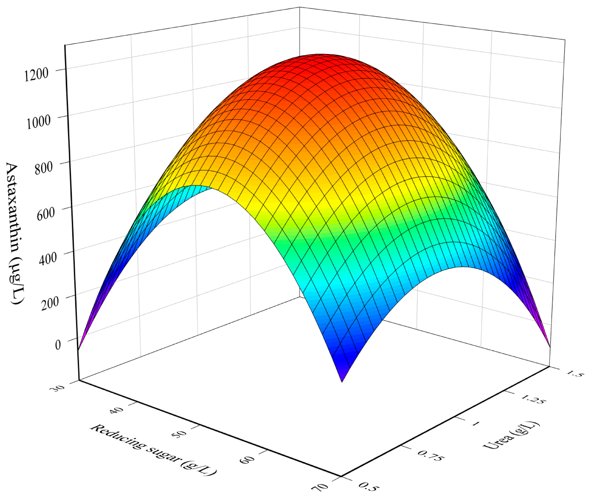
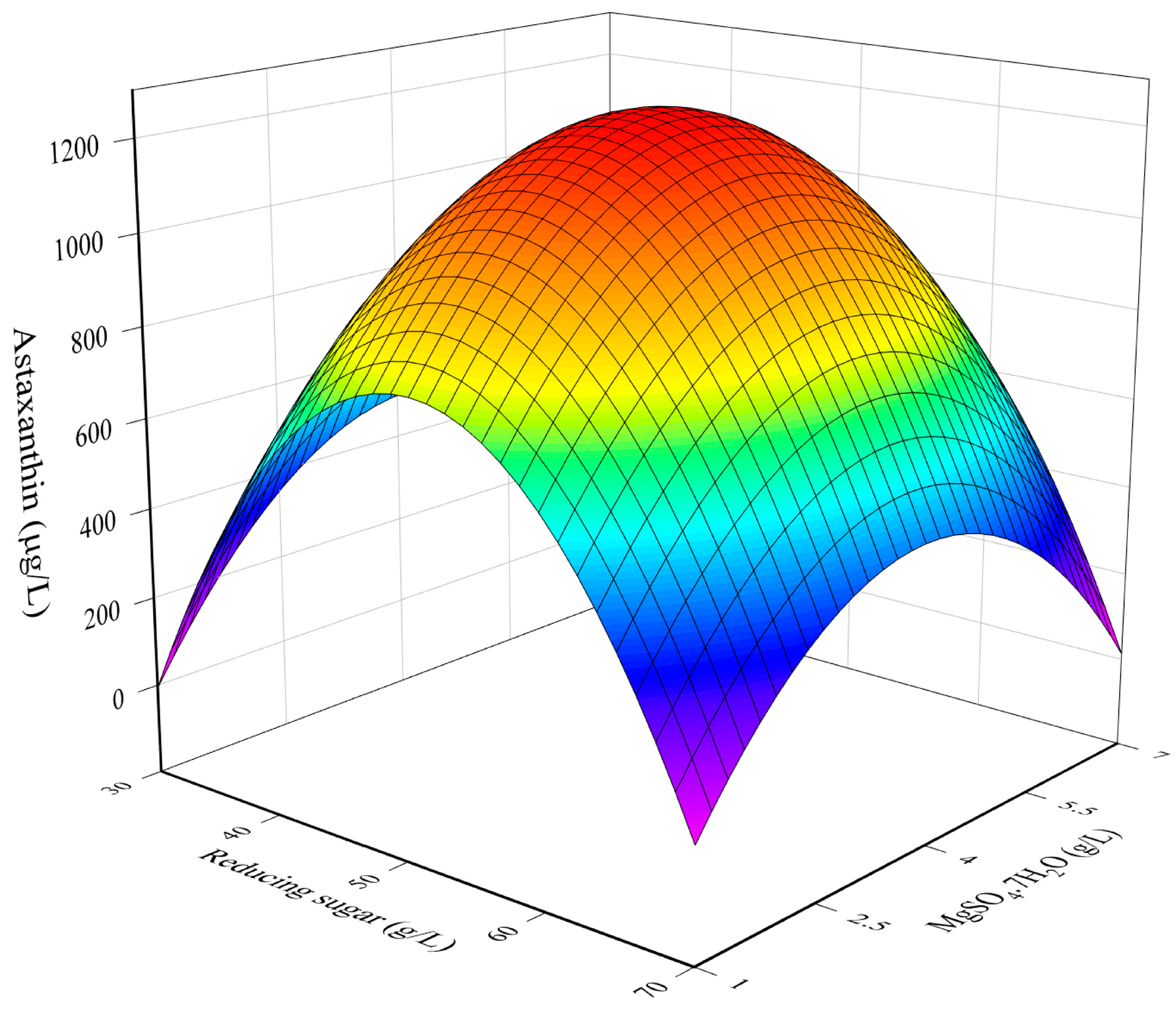
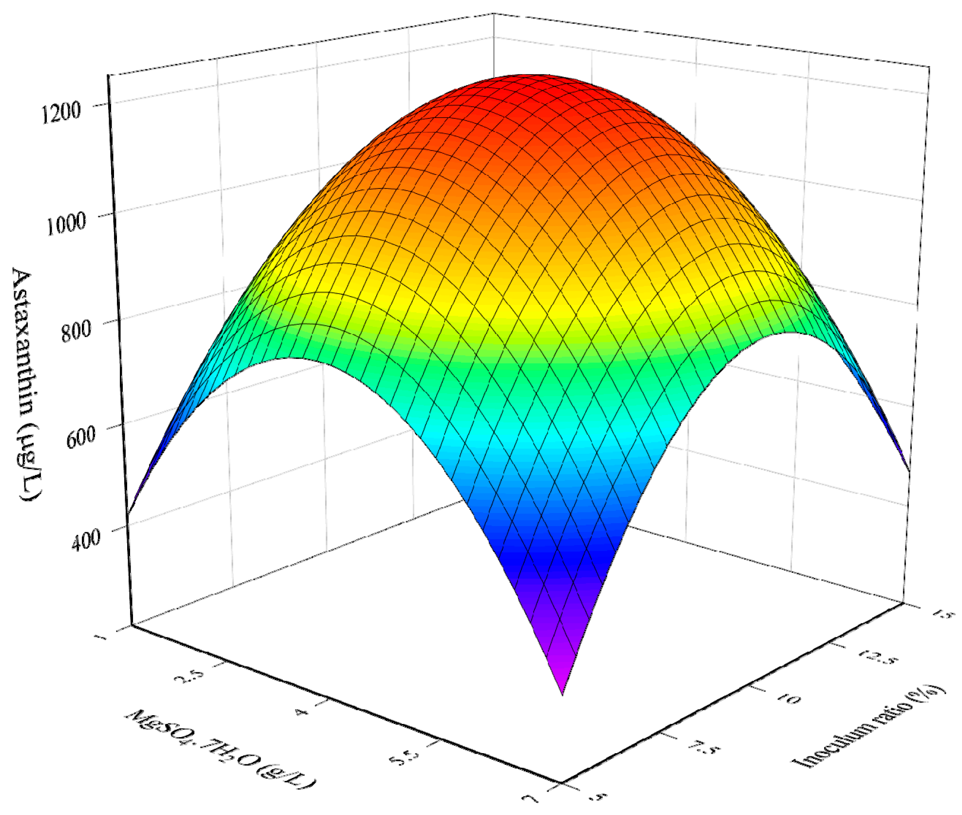
3.1. RSM Model for The Optimization of Culture Conditions

This work used molasses as a cheap carbon source for growing R. toruloides G17. To maximize the astaxanthin yield, a CCD-RSM model was used to examine the influence of the culture parameters (reducing sugar concentration, urea concentration, MgSO4·7H2O concentration, inoculum ratio) on the astaxanthin accumulation and then optimize the culture conditions. Table 2 shows the experimental design matrix for the cultivation of R. toruloides G17 and the resulting astaxanthin yield. A low coefficient of variance (CV = 2.85%) was obtained for the central experiments (runs 25–31), indicating the reproducibility and desirable accuracy of the experiments. Thus, the model was then established to express the relationship between the astaxanthin yield and culture conditions as follows:
Y = 1245 15.61 X 1 0.06 X 2 + 18.6 X 3 + 3.45 X 4 182.99 X 1 2 105.02 X 2 2 108.43 X 3 2 107.22 X 4 2 38.6 X 1 X 2 14.21 X 1 X 3 16.62 X 1 X 4 7.06 X 2 X 3 + 35.26 X 2 X 4 + 12.1 X 3 X 4
ANOVA was conducted to evaluate the established model shown in Equation (6). As shown in Table 3, the model had an extremely low p value (<0.001), indicating that the developed model was statistically significant at the 95% confidence level. Moreover, the coefficient of determination (R2) was high (98.53%), signifying excellent correlation between the predicted and experimental values. Therefore, the established model could provide a reliable prediction. The t test was also performed to evaluate the significance of model parameters. As can be seen from Table 4, small p values (<0.05) were observed for the following factors: linear coefficient of MgSO4·7H2O concentration (X3), all quadratic coefficients of reducing sugar concentration ( X 1 2 ), urea concentration ( X 2 2 ), MgSO4·7H2O concentration ( X 3 2 ), inoculum ratio ( X 4 2 ), two interaction coefficients (X1X2, and X2X4), and the intercept. These small p values indicate that these parameters were significant factors in the cultivation of R. toruloides G17. The developed model was then used to obtain optimal conditions for maximizing the astaxanthin yield.

3.2. Combined Influence of Culture Conditions

Figure 1 illustrates the mutual influence of reducing sugar concentration and urea concentration on the astaxanthin yield while other factors were maintained at a central level. At a specific concentration of urea, increasing the concentration of reducing sugar in molasses hydrolysate resulted in a significantly enhanced astaxanthin yield. This is because the yeast strain required sugars as a carbon source for its growth and metabolism [27]. Yeast cells consume and convert glucose and other reducing sugars to intermediates such as ethanol and acetic acid through their metabolic pathways. Acetyl CoA (a precursor for carotenoid biosynthesis) was then produced from ethanol by alcohol dehydrogenase [16] and facilitated the synthesis of carotenoids (including astaxanthin). However, after reaching the highest astaxanthin yield, a further increase in the reducing sugar concentration caused a reduction in astaxanthin yield. This might be because the high molasses hydrolysate concentration increased the sugar osmotic pressure, which inhibited cell growth and promoted the synthesis of other energetic substances (e.g., protein), instead of astaxanthin to resist to environmental stress, thus lowering astaxanthin production [28]. Moreover, several by-products (e.g., 5 hydroxymethylfurfural) might be formed during the hydrolysis step and their presence at a high concentration in the culture medium could inhibit cell growth and astaxanthin production [29,30]. Similarly, at a given concentration of reducing sugar, the astaxanthin yield increased with an increasing concentration of urea, reached the highest level, and then reduced [28]. Excess urea resulted in a reduction in the carbon-to-nitrogen ratio, thus suppressing the cell growth and synthesis of secondary pigments, including astaxanthin. Therefore, a sufficient carbon-to-nitrogen ratio is required for the cell growth and biosynthesis of astaxanthin by R. toruloides G17. In this work, the highest astaxanthin yield was obtained in the middle-range concentrations of both the reducing sugar and urea.
Figure 2 illustrates the combined influence of reducing sugar concentration and MgSO4·7H2O concentration on astaxanthin yield. At a given reducing sugar concentration, the astaxanthin yield increased and reached its highest level when the MgSO4·7H2O concentration increased. However, high concentrations of MgSO4·7H2O (>5.5 mg/mL) resulted in a decrease in astaxanthin yield. This was because this salt is essential for cell growth and, when present in sufficient quantities, it promotes the carotenogenesis process in the cells. However, an excess of MgSO4 suppressed cell growth and astaxanthin biosynthesis, thus reducing the astaxanthin yield [31,32].
Figure 3 displays the mutual effects of the urea concentration and inoculum ratio on the astaxanthin yield, while other factors were maintained at their central levels. At a specific urea concentration, increasing the inoculum ratio led to an enhanced astaxanthin yield. This result indicated that the initial concentration of R. toruloides G17 inoculum strongly affected astaxanthin production. The highest astaxanthin yield was obtained with the culture containing high inoculum ratios. This could be because these cultures shortened the lag phase, efficiently consuming and converting nutrients into their biomass and astaxanthin accumulation, thus enhancing astaxanthin yield. This result was similar to previous studies which demonstrated that when the inoculum ratio is under or over the limit threshold, it could lead to a poor growth rate of microorganisms [33]. Studies have reported that the lower the inoculum ratio, the longer it takes to pass the log phase [34], thus reducing the accumulation of secondary metabolites, which are mainly formed in the stationary phases [14].

3.3. Determination of Optimal Culture Conditions

The model Equation (6) was solved to predict the optimal conditions for maximizing the astaxanthin yield. The optimal conditions were determined to be a reducing sugar concentration of 49.39 g/L, urea concentration of 1.00 g/L, MgSO4·7H2O concentration of 4.15 g/L, and inoculum ratio of 10.05%, with a corresponding astaxanthin yield of 1246.19 µg/L. To validate this prediction, R. toruloides G17 was grown under the same optimized conditions to produce astaxanthin. The experimental astaxanthin content was 1262.08 ± 14.58 µg/L, which was consistent with the prediction. This finding demonstrated that the RSM model was successfully developed to establish the relationship between culture conditions and astaxanthin yield in the cultivation of R. toruloides G17 in molasses hydrolysate-based medium. The cultivation of R. toruloides G17 in waste molasses-based medium produced a higher astaxanthin yield than Phaffia rhodozyma (639.6 µg/L) [35] and Spirulina platensis (38 µg/L) [36]. Therefore, R. toruloides G17 is promising for further applications.

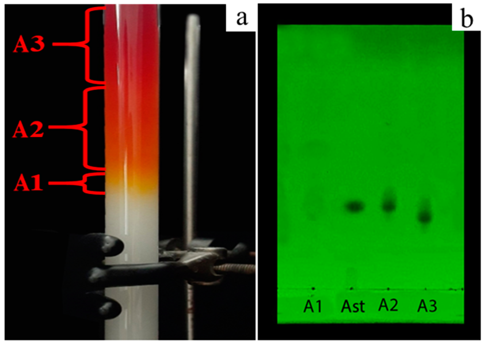
3.4. Astaxanthin Purification

Astaxanthin extract was loaded onto a silica gel column and eluted with n-hexane:acetone (4:1, v/v) for purification (Figure 4a). Subsequently, three fractions (A1, A2, and A3) were obtained and subjected to TLC analysis. As shown in Figure 4b, high purity astaxanthin was found in fraction A2 with an Rf value of 0.29, which was the same Rf value as that of the astaxanthin standard. In addition, liquid chromatography-mass spectrometry (LC-MS) was used to further identify the extracted astaxanthin. GC–MS analysis showed that the extracted astaxanthin and astaxanthin standard had the same retention time (5.849–5.865 min) and mass to charge ratio [m/z 597.39 (M+)] (Figures S1 and S2, Supplementary Material). Therefore, this fraction was used for further experiments to investigate its antioxidant and anticancer activities.

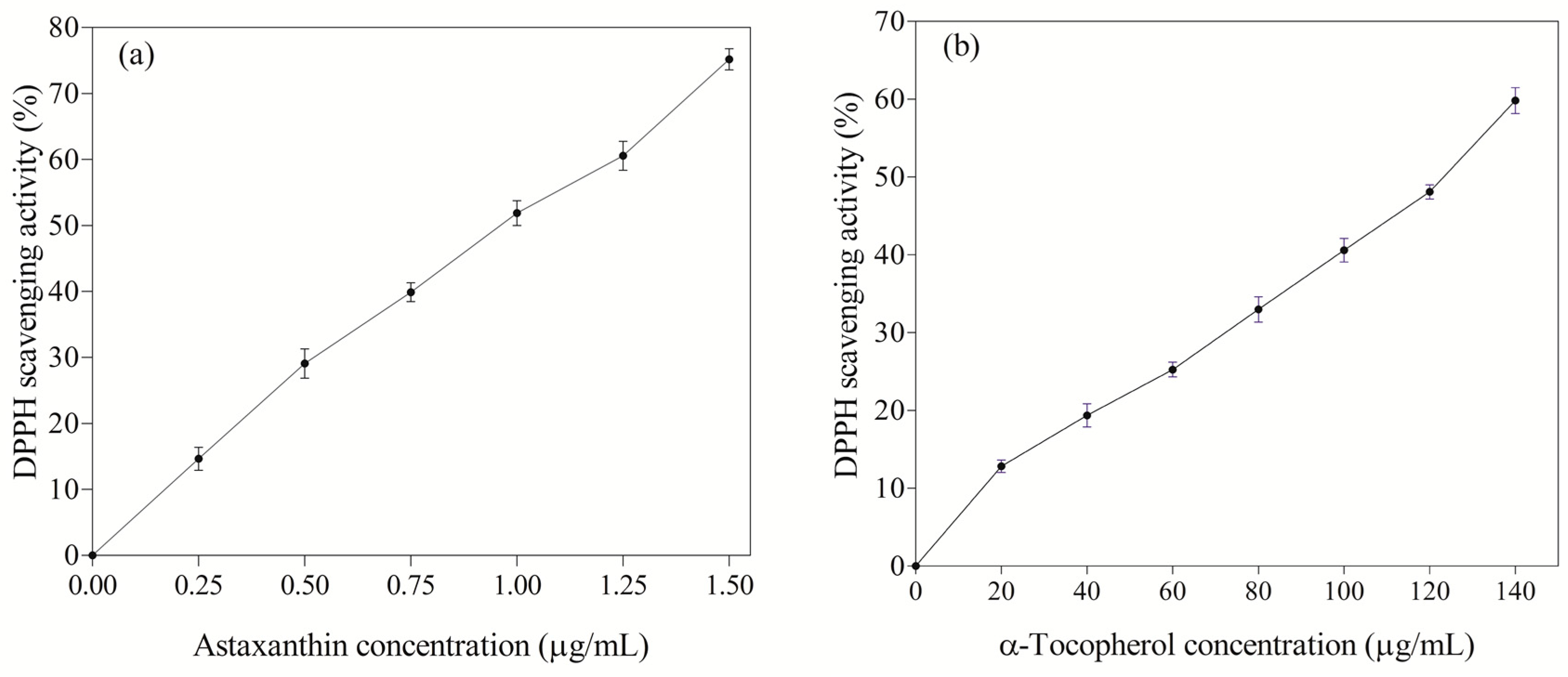
3.5. Antioxidant Activity of Astaxanthin

There has been a growing interest in using natural antioxidants for preventing diseases and promoting health [37,38]. As a result, various natural sources have been studied for producing antioxidant compounds [39,40]. In recent years, astaxanthin has increasingly received attention from academia and industry since it exhibits potent antioxidant activity. In the current study, the antioxidant activity of astaxanthin produced by mutated R. toruloides G17 was investigated using the DPPH scavenging activity assay and compared to that of α-tocopherol (a positive control). The DPPH reaction mechanism is based on hydrogen donation, whereby DPPH compounds (deep-violet) are captured by antioxidant compounds, releasing hydrogen radicals to form stable the molecules of DPPH-H (colorless or pale-yellow) [41,42]. As shown in Figure 5a, the produced astaxanthin strongly scavenged DPPH radicals at low concentrations. Notably, the astaxanthin exhibited 123.6-fold higher DPPH scavenging activity (IC50 of 0.97 ± 0.01 µg/mL) than α-tocopherol (IC50 of 119.91 ± 0.91 µg/mL). In addition, the DPPH radical scavenging activity of the astaxanthin obtained in this work was found to be higher than that obtained from other natural sources, as illustrated in Table 5. It could be because astaxanthin from different sources has variations in terms of its chiral structure, thus affecting its antioxidant activity [43]. Astaxanthin has multiple isomeric forms including geometric isomers, stereoisomers, esterified, and free forms [44]. Studies have reported that the biological activity of astaxanthin varies among its isomeric forms [43,44]. The (3S, 3′S)-isomers have been found to exhibit higher antioxidant activity than (3R, 3′R) and (3R, 3’S) astaxanthins [43]. The high antioxidant activity of astaxanthin can be explained by the keto group in astaxanthin activating the hydroxyl group and consequently promoting hydrogen transfer to the peroxyl radicals [45]. Hence, astaxanthin effectively inhibited free radicals. This finding indicated that astaxanthin produced by the mutated R. toruloides G17 is a promising antioxidant agent for further applications.

3.6. Anticancer Activity of Astaxanthin

This study examined the cytotoxicity of astaxanthin produced by R. toruloides G17 in one control (HK2) and three cancer cell lines (MCF7, A549, and HeLa). Astaxanthin significantly suppressed the proliferation of MCF7, A549, and Hela cell lines in a dose-dependent manner while exerting less damage to the HK2 cells. The anticancer activity of astaxanthin was found to be lower than that of Cisplatin, known to be one of the most effective chemotherapeutic anticancer agents, but it still exhibited cytotoxicity against A549, MCF7, and HeLa cells with IC50 values of 56.38 ± 4.1 µg/mL, 55.60 ± 2.64 µg/mL, and 69.07 ± 2.4 µg/mL, respectively (Table 6). The cytotoxicity of astaxanthin obtained in this work was comparable to that previously observed for astaxanthin from other sources. Kim et al. [51] reported the cytotoxicity of astaxanthin (Sigma-Aldrich) in two gastric cancer cell lines, KATO-III and SNU-1, with the IC50 values of >100 µg/mL. Ramamoorthy et al. [52] investigated the effect of astaxanthin (purified fraction) extracted from the microalgae H. pluvialis and observed a significant reduction in cell viability (about 50%) at an astaxanthin concentration of 50 µM. The present work suggests that astaxanthin produced by R. toruloides G17 is a potential antitumor agent.

4. Conclusions

This study reported the production of astaxanthin from waste molasses by R. toruloides G17. RSM was employed to optimize the culture conditions for maximizing the astaxanthin yield. Through RSM, the optimal culture conditions (concentration of reducing sugar of 49.39 g/L, urea concentration of 1.00 g/L, MgSO4·7H2O concentration of 4.15 g/L, and inoculum ratio of 10.05%) were obtained, with the highest astaxanthin yield of 1262.08 ± 14.58 µg/L. The produced astaxanthin exhibited potent antioxidant activity (IC50 of 0.97 ± 0.01 µg/mL) and anticancer activity against the three cancer cell lines, HeLa, A549, and MCF7, with IC50 values of 69.07 ± 2.4 µg/mL, 55.60 ± 2.64 µg/mL, and 56.38 ± 4.1 µg/mL, respectively. This study suggests that R. toruloides G17 is a promising strain for the production of bioactive astaxanthin using low-cost molasses as a source of carbon and micronutrients. The produced astaxanthin may have applications as a nutraceutical or pharmaceutical grade antioxidant or antitumor agent.

Supplementary Materials

The following supporting information can be downloaded at: https://www.mdpi.com/article/10.3390/fermentation9020148/s1, Figure S1: Liquid chromatography-mass spectrometry (LC-MS) chromatogram of astaxanthin standard; Figure S2: Liquid chromatography-mass spectrometry (LC-MS) chromatogram of extracted astaxanthin.

Author Contributions

Conceptualization, T.N.T., H.C.N., D.-N.N. and C.J.B.; methodology, T.N.T., H.C.N., C.-H.S. and D.-C.P.; validation, N.-T.T., T.-A.T., D.-C.P. and T.N.T.; formal analysis, T.N.T. and H.C.N.; investigation, T.N.T., N.-T.T. and T.-A.T.; resources, H.C.N., C.J.B. and D.-N.N.; writing—original draft preparation, H.C.N.; writing—review and editing, C.J.B.; visualization, T.N.T. and H.C.N.; supervision, D.-N.N., H.C.N. and C.J.B.; project administration, D.-N.N.; funding acquisition, D.-N.N. All authors have read and agreed to the published version of the manuscript.

Funding

This research received no external funding.

Institutional Review Board Statement

Not applicable.

Informed Consent Statement

Not applicable.

Data Availability Statement

The data are available from the corresponding author upon request.

Conflicts of Interest

The authors declare no conflict of interest.

References

  1. Faraone, I.; Sinisgalli, C.; Ostuni, A.; Armentano, M.F.; Carmosino, M.; Milella, L.; Russo, D.; Labanca, F.; Khan, H. Astaxanthin anticancer effects are mediated through multiple molecular mechanisms: A systematic review. Pharmacol. Res. 2020, 155, 104689. [Google Scholar] [CrossRef]
  2. Demirel, P.B.; Tuna, B.G. Anticancer properties of astaxanthin: A molecule of great promise. In Global Perspectives on Astaxanthin; Academic Press: Cambridge, MA, USA, 2021; pp. 427–445. [Google Scholar]
  3. Fakhri, S.; Abbaszadeh, F.; Dargahi, L.; Jorjani, M. Astaxanthin: A mechanistic review on its biological activities and health benefits. Pharmacol. Res. 2018, 136, 1–20. [Google Scholar] [CrossRef]
  4. Kurashige, M.; Okimasu, E.; Inoue, M.; Utsumi, K. Inhibition of oxidative injury of biological membranes by astaxanthin. Physiol. Chem. Phys. Med. NMR 1990, 22, 27–38. [Google Scholar]
  5. Davinelli, S.; Nielsen, M.E.; Scapagnini, G. Astaxanthin in skin health, repair, and disease: A comprehensive review. Nutrients 2018, 10, 522. [Google Scholar] [CrossRef]
  6. Higuera-Ciapara, I.; Felix-Valenzuela, L.; Goycoolea, F. Astaxanthin: A review of its chemistry and applications. Crit. Rev. Food Sci. Nutr. 2006, 46, 185–196. [Google Scholar] [CrossRef]
  7. Zhou, P.; Li, M.; Shen, B.; Yao, Z.; Bian, Q.; Ye, L.; Yu, H. Directed coevolution of β-carotene ketolase and hydroxylase and its application in temperature-regulated biosynthesis of astaxanthin. J. Agric. Food Chem. 2019, 67, 1072–1080. [Google Scholar] [CrossRef]
  8. Asker, D. Isolation and characterization of a novel, highly selective astaxanthin-producing marine bacterium. J. Agric. Food Chem. 2017, 65, 9101–9109. [Google Scholar] [CrossRef]
  9. Kamath, B.S.; Srikanta, B.M.; Dharmesh, S.M.; Sarada, R.; Ravishankar, G.A. Ulcer preventive and antioxidative properties of astaxanthin from Haematococcus pluvialis. Eur. J. Pharmacol. 2008, 590, 387–395. [Google Scholar] [CrossRef]
  10. Schmidt, I.; Schewe, H.; Gassel, S.; Jin, C.; Buckingham, J.; Hümbelin, M.; Sandmann, G.; Schrader, J. Biotechnological production of astaxanthin with Phaffia rhodozyma/Xanthophyllomyces dendrorhous. Appl. Microbiol. Biotechnol. 2011, 89, 555–571. [Google Scholar] [CrossRef]
  11. Chang, M.X.; Xiong, F. Astaxanthin and its effects in inflammatory responses and inflammation-associated diseases: Recent advances and future directions. Molecules 2020, 25, 5342. [Google Scholar] [CrossRef]
  12. Nguyen, K.D. Astaxanthin: A Comparative Case of Synthetic vs. Natural Production. Chemical and Biomolecular Engineering Publications and Other Works. Available online: http://trace.tennessee.edu/utk_chembiopubs/94 (accessed on 5 January 2023).
  13. Wen, Z.; Zhang, S.; Odoh, C.K.; Jin, M.; Zhao, Z.K. Rhodosporidium toruloides—A potential red yeast chassis for lipids and beyond. FEMS Yeast Res. 2020, 20, foaa038. [Google Scholar] [CrossRef]
  14. Tran, T.N.; Ngo, D.H.; Tran, Q.T.; Nguyen, H.C.; Su, C.H.; Ngo, D.N. Enhancing astaxanthin biosynthesis by Rhodosporidium toruloides mutants and optimization of medium compositions using response surface methodology. Processes 2020, 8, 497. [Google Scholar] [CrossRef]
  15. Wu, Y.H.; Yang, J.; Hu, H.Y.; Yu, Y. Lipid-rich microalgal biomass production and nutrient removal by Haematococcus pluvialis in domestic secondary effluent. Ecol. Eng. 2013, 60, 155–159. [Google Scholar] [CrossRef]
  16. Tran, T.N.; Quang-Vinh, T.; Huynh, H.T.; Hoang, N.S.; Nguyen, H.C.; Dai-Nghiep, N. Astaxanthin production by newly isolated Rhodosporidium toruloides: Optimization of medium compositions by response surface methodology. Not. Bot. Horti Agrobot. 2019, 47, 320–327. [Google Scholar] [CrossRef]
  17. Dalsasso, R.R.; Pavan, F.A.; Bordignon, S.E.; de Aragão, G.M.F.; Poletto, P. Polyhydroxybutyrate (PHB) production by Cupriavidus necator from sugarcane vinasse and molasses as mixed substrate. Process Biochem. 2019, 85, 12–18. [Google Scholar] [CrossRef]
  18. Yew, G.Y.; Khoo, K.S.; Chia, W.Y.; Ho, Y.C.; Law, C.L.; Leong, H.Y.; Show, P.L. A novel lipids recovery strategy for biofuels generation on microalgae Chlorella cultivation with waste molasses. J. Water Process Eng. 2020, 38, 101665. [Google Scholar] [CrossRef]
  19. Yew, G.Y.; Puah, B.K.; Chew, K.W.; Teng, S.Y.; Show, P.L.; Nguyen, T.H.P. Chlorella vulgaris FSP-E cultivation in waste molasses: Photo-to-property estimation by artificial intelligence. Chem. Eng. J. 2020, 402, 126230. [Google Scholar] [CrossRef]
  20. Kundu, S.; Panda, T.; Majumdar, S.; Guha, B.; Bandyopadhyay, K. Pretreatment of Indian cane molasses for increased production of citric acid. Biotechnol. Bioeng. 1984, 26, 1114–1121. [Google Scholar] [CrossRef]
  21. Miller, G.L. Use of dinitrosalicylic acid reagent for determination of reducing sugar. Anal. Chem. 1959, 31, 426–428. [Google Scholar] [CrossRef]
  22. Prasertsung, I.; Aroonraj, K.; Kamwilaisak, K.; Saito, N.; Damrongsakkul, S. Production of reducing sugar from cassava starch waste (CSW) using solution plasma process (SPP). Carbohydr. Polym. 2019, 205, 472–479. [Google Scholar] [CrossRef]
  23. An, G.H.; Bielich, J.; Auerbach, R.; Johnson, E.A. Isolation and characterization of carotenoid hyperproducing mutants of yeast by flow cytometry and cell sorting. Nat. Biotechnol. 1991, 9, 70–73. [Google Scholar] [CrossRef]
  24. Fang, T.J.; Cheng, Y.S. Improvement of astaxanthin production by Phaffia rhodozyma through mutation and optimization of culture conditions. J. Ferment. Bioeng. 1993, 75, 466–469. [Google Scholar] [CrossRef]
  25. Sánchez-Moreno, C. Methods used to evaluate the free radical scavenging activity in foods and biological systems. Food Sci. Technol. Inter. 2002, 8, 121–137. [Google Scholar] [CrossRef]
  26. Pham, D.C.; Chang, Y.C.; Lin, S.R.; Fuh, Y.M.; Tsai, M.J.; Weng, C.F. FAK and S6K1 inhibitor, neferine, dually induces autophagy and apoptosis in human neuroblastoma cells. Molecules 2018, 23, 3110. [Google Scholar] [CrossRef]
  27. Koschwanez, J.H.; Foster, K.R.; Murray, A.W. Sucrose utilization in budding yeast as a model for the origin of undifferentiated multicellularity. PLoS Biol. 2011, 9, e1001122. [Google Scholar] [CrossRef]
  28. Nguyen, H.C.; Su, C.H.; Yu, Y.K.; Huong, D.T.M. Sugarcane bagasse as a novel carbon source for heterotrophic cultivation of oleaginous microalga Schizochytrium sp. Ind. Crop. Prod. 2018, 121, 99–105. [Google Scholar] [CrossRef]
  29. Zeng, Y.; Bian, D.; Xie, Y.; Jiang, X.; Li, X.; Li, P.; Zhang, Y.; Xie, T. Utilization of food waste hydrolysate for microbial lipid and protein production by Rhodosporidium toruloides Y2. J. Chem. Technol. Biotechnol. 2017, 92, 666–673. [Google Scholar] [CrossRef]
  30. Howard, J.; Rackemann, D.W.; Bartley, J.P.; Samori, C.; Doherty, W.O. Conversion of sugar cane molasses to 5-hydroxymethylfurfural using molasses and bagasse-derived catalysts. ACS Sustain. Chem. Eng. 2018, 6, 4531–4538. [Google Scholar] [CrossRef]
  31. Fang, C.J.; Ku, K.L.; Lee, M.H.; Su, N.W. Influence of nutritive factors on C50 carotenoids production by Haloferax mediterranei ATCC 33500 with two-stage cultivation. Bioresour. Technol. 2010, 101, 6487–6493. [Google Scholar] [CrossRef]
  32. Varmira, K.; Habibi, A.; Bahramian, E.; Jamshidpour, S. Progressive agents for improvement of carotenogenesis in Rhodotorula rubra. J. Adv. Food Sci. Technol. 2016, 3, 70–78. [Google Scholar]
  33. Narayanaswamy, S. Plant Cell and Tissue Culture; Tata McGraw-Hill Education: New York, NY, USA, 1994. [Google Scholar]
  34. Ling, O.S.; Kiong, A.L.P.; Hussein, S. Establishment and optimisation of growth parameters for cell suspension cultures of Ficus deltoidea. Am. Eurasian J. Sustain. Agric. 2008, 2, 38–49. [Google Scholar]
  35. Xie, H.; Zhou, Y.; Hu, J.; Chen, Y.; Liang, J. Production of astaxanthin by a mutant strain of Phaffia rhodozyma and optimization of culture conditions using response surface methodology. Ann. Microbiol. 2014, 64, 1473–1481. [Google Scholar] [CrossRef]
  36. An, J.; Gao, F.; Ma, Q.; Xiang, Y.; Ren, D.; Lu, J. Screening for enhanced astaxanthin accumulation among Spirulina platensis mutants generated by atmospheric and room temperature plasmas. Algal Res. 2017, 25, 464–472. [Google Scholar] [CrossRef]
  37. Nguyen, H.C.; Nguyen, H.N.T.; Huang, M.Y.; Lin, K.H.; Pham, D.C.; Tran, Y.B.; Su, C.H. Optimization of aqueous enzyme-assisted extraction of rosmarinic acid from rosemary (Rosmarinus officinalis L.) leaves and the antioxidant activity of the extract. J. Food Process. Preserv. 2021, 45, e15221. [Google Scholar] [CrossRef]
  38. Nguyen, H.C.; Kuan-Hung, L.; Huang, M.Y.; Chi-Ming, Y.; Tin-Han, S.; Hsiung, T.C.; Yen-Chang, L.; Fun-Chi, T. Antioxidant activities of the methanol extracts of various parts of Phalaenopsis orchids with white, yellow, and purple flowers. Not. Bot. Horti Agrobot. 2018, 46, 457–465. [Google Scholar] [CrossRef]
  39. Do, T.H.; Huynh, T.D.; Vo, K.A.; Nguyen, K.A.; Cao, T.S.; Nguyen, K.N.; Nguyen, H.A.H.; Nguyen, T.T.T.; Le, N.P.N.; Truong, D.H. Saponin-rich fractions from Codonopsis javanica root extract and their in vitro antioxidant and anti-enzymatic efficacy. J. Food Process. Preserv. 2022, 46, e16113. [Google Scholar] [CrossRef]
  40. Truong, D.H.; Nguyen, D.H.; Ta, N.T.A.; Bui, A.V.; Do, T.H.; Nguyen, H.C. Evaluation of the use of different solvents for phytochemical constituents, antioxidants, and in vitro anti-inflammatory activities of Severinia buxifolia. J. Food Qual. 2019, 2019, 8178294. [Google Scholar] [CrossRef]
  41. Chintong, S.; Phatvej, W.; Rerk-Am, U.; Waiprib, Y.; Klaypradit, W. In vitro antioxidant, antityrosinase, and cytotoxic activities of astaxanthin from shrimp waste. Antioxidants 2019, 8, 128. [Google Scholar] [CrossRef]
  42. Santos-Sánchez, N.F.; Salas-Coronado, R.; Villanueva-Cañongo, C.; Hernández-Carlos, B. Antioxidant compounds and their antioxidant mechanism. In Antioxidants; IntechOpen: London, UK, 2019; Volume 10, pp. 1–29. [Google Scholar]
  43. Sun, J.; Yan, J.; Dong, H.; Gao, K.; Yu, K.; He, C.; Mao, X. Astaxanthin with different configurations: Sources, activity, post-modification and application in foods. Curr. Opin. Food Sci. 2023, 49, 100955. [Google Scholar] [CrossRef]
  44. Ambati, R.R.; Moi, P.S.; Ravi, S.; Aswathanarayana, R.G. Astaxanthin: Sources, extraction, stability, biological activities and its commercial applications—A review. Mar. Drugs 2014, 12, 128–152. [Google Scholar] [CrossRef]
  45. Naguib, Y.M. Antioxidant activities of astaxanthin and related carotenoids. J. Agric. Food Chem. 2000, 48, 1150–1154. [Google Scholar] [CrossRef] [PubMed]
  46. Abdelmalek, B.E.; Sila, A.; Ghlissi, Z.; Taktak, M.A.; Ayadi, M.A.; Bougatef, A. The influence of natural astaxanthin on the formulation and storage of marinated chicken steaks. J. Food Biochem. 2016, 40, 393–403. [Google Scholar] [CrossRef]
  47. Karnila, R.; Edison; Ramadhani, N.R. Antioxidant activity of astaxanthin flour extract of mud crab (Scylla serrata) with different acetone concentrations. In Proceedings of the IOP Conference Series: Earth and Environmental Science 2021, Pekanbaru, Indonesia, 10–11 September 2020; p. 012047. [Google Scholar]
  48. Tan, Y.; Ye, Z.; Wang, M.; Manzoor, M.F.; Aadil, R.M.; Tan, X.; Liu, Z. Comparison of different methods for extracting the astaxanthin from Haematococcus pluvialis: Chemical composition and biological activity. Molecules 2021, 26, 3569. [Google Scholar] [CrossRef] [PubMed]
  49. Cheng, X.Y.; Xiong, Y.J.; Yang, M.M.; Zhu, M.J. Preparation of astaxanthin mask from Phaffia rhodozyma and its evaluation. Process Biochem. 2019, 79, 195–202. [Google Scholar] [CrossRef]
  50. Sun, Z.; Liu, J.; Zeng, X.; Huangfu, J.; Jiang, Y.; Wang, M.; Chen, F. Astaxanthin is responsible for antiglycoxidative properties of microalga Chlorella zofingiensis. Food Chem. 2011, 126, 1629–1635. [Google Scholar] [CrossRef]
  51. Kim, J.H.; Park, J.J.; Lee, B.J.; Joo, M.K.; Chun, H.J.; Lee, S.W.; Bak, Y.T. Astaxanthin inhibits proliferation of human gastric cancer cell lines by interrupting cell cycle progression. Gut Liver 2016, 10, 369. [Google Scholar] [CrossRef]
  52. Ramamoorthy, K.; Raghunandhakumar, S.; Anand, R.; Paramasivam, A.; Kamaraj, S.; Nagaraj, S.; Ezhilarasan, D.; Lakshmi, T.; Dua, K.; Chellappan, D.K. Anticancer effects and lysosomal acidification in A549 cells by astaxanthin from Haematococcus lacustris. Bioinformation 2020, 16, 965. [Google Scholar] [CrossRef] [PubMed]
Figure 1. Combined effect of reducing sugar concentration and urea concentration on the astaxanthin yield.
Fermentation 09 00148 g001
Figure 2. Combined effect of reducing sugar concentration and MgSO4·7H2O concentration on the astaxanthin yield.
Fermentation 09 00148 g002
Figure 3. Combined effect of MgSO4·7H2O concentration and inoculum ratio on the astaxanthin yield.
Fermentation 09 00148 g003
Figure 4. Purification of astaxanthin using silica gel column chromatography (a) and TLC analysis of astaxanthin fractions (b). A1: fraction 1; A2: fraction 2; A3: faction 3; and Ast: astaxanthin standard.
Fermentation 09 00148 g004
Figure 5. DPPH scavenging activity of (a) astaxanthin and (b) α-tocopherol.
Fermentation 09 00148 g005
Table 1. Input variables for RSM model.
VariablesSymbolsVariable Levels
−2−1012
Reducing sugar concentration (g/L)X13040506070
Urea concentration (g/L)X20.50.751.01.251.5
MgSO4·7H2O concentration (g/L)X31.02.54.05.57.0
Inoculum ratio (%)X45.07.510.012.515.0
Table 2. Experimental design of the RSM-CCD model and experimental results.
RunVariablesAstaxanthin Yield (μg/L)
X1X2X3X4ExperimentalPredicted
1−1−1−1−1761.27739.10
21−1−1−1792.65780.25
3−11−1−1784.20759.78
411−1−1665.33646.53
5−1−11−1825.50794.65
61−11−1783.58778.95
7−111−1782.38787.09
8111−1639.37616.99
9−1−1−11651.44618.02
101−1−11734.84725.67
11−11−11779.57779.74
1211−11757.95732.99
13−1−111707.63721.97
141−111804.15772.77
15−1111898.87855.46
161111734.14751.85
17−2000506.90544.28
182000458.95481.83
190−200790.36825.07
200200799.28824.83
2100−20731.66774.11
220020830.71848.52
23000−2773.91809.24
240002798.10823.03
2500001221.521245.03
2600001295.591245.03
2700001282.521245.03
2800001241.341245.03
2900001201.031245.03
3000001214.551245.03
3100001258.631245.03
Table 3. ANOVA for the RSM model.
SourceDF bSS bMS bF-Valuep-Value
Model a141,582,917113,06676.49<0.001
Error1623,6491478
Lack-of-fit1016,07116071.270.4
Pure Error675781263
Total301,606,567
a Coefficient of determination (R2) = 98.53%; adjusted R2 = 97.24%; b DF, degree of freedom; SS, sum of squares; MS, mean square.
Table 4. Significance of the model parameters.
TermParameter EstimateStandard Errort-Valuep-Value a
β01245.014.585.680.000 a
β1−15.617.85−1.990.064
β2−0.067.85−0.010.994
β318.607.852.370.03 a
β43.457.850.440.667
β11−182.997.19−25.450.000 a
β22−105.027.19−14.610.000 a
β33−108.437.19−15.080.000 a
β44−107.227.19−14.910.000 a
β12−38.609.61−4.020.001 a
β13−14.219.61−1.480.159
β1416.629.611.730.103
β23−7.069.61−0.730.473
β2435.269.613.670.002 a
β3412.109.611.260.226
a p < 0.05 indicates that the model terms are significant.
Table 5. DPPH scavenging activity of astaxanthin from different sources.
SourcesIC50 Value (μg/mL)References
R. toruloides0.97This study
Shrimp waste17.50[41]
P. longirostris6.30[46]
Scylla serrata805.84[47]
H. pluvialis15.39–56.25[48]
Phaffia rhodozyma31.79[49]
Chlorella zofingiensis1040–2930[50]
Table 6. IC50 values of anticancer activity of astaxanthin.
SampleIC50 Value (µg/mL)
MCF7A549HeLaHK2
Astaxanthin55.60 ± 2.656.38 ± 4.169.07 ± 2.4111.34 ± 1.4
Cisplatin21.68 ± 0.82.4 ± 0.46.8 ± 0.91.8 ± 0.7
Data are expressed as mean ± SD of three independent experiments.
Disclaimer/Publisher’s Note: The statements, opinions and data contained in all publications are solely those of the individual author(s) and contributor(s) and not of MDPI and/or the editor(s). MDPI and/or the editor(s) disclaim responsibility for any injury to people or property resulting from any ideas, methods, instructions or products referred to in the content.

