Article

Unveiling the Role of Capping Groups in Naphthalene N-Capped Dehydrodipeptide Hydrogels

by
Helena Vilaça
1,2,
André Carvalho
1,
Tarsila Castro
3,
Elisabete M. S. Castanheira
4,
Loic Hilliou
5,
Ian Hamley
6,
Manuel Melle-Franco
7,
Paula M. T. Ferreira
1,* and
José A. Martins
1,*
1
Centre of Chemistry, University of Minho, Campus de Gualtar, 4710-057 Braga, Portugal
2
Department of Chemistry and Biotechnology, Technological Centre for the Textile and Clothing Industries of Portugal, 4760-034 Vila Nova de Famalicão, Portugal
3
Centre of Biological Engineering, University of Minho, Campus de Gualtar, 4710-057 Braga, Portugal
4
Physics Centre of Minho and Porto Universities (CF-UM-UP), University of Minho, Campus de Gualtar, 4710-057 Braga, Portugal
5
Institute for Polymers and Composites, Department of Polymer Engineering, University of Minho, Campus de Azurém, 4800-058 Guimarães, Portugal
6
Department of Chemistry, University of Reading, Whiteknights, P.O. Box 224, Reading RG6 6AD, UK
7
CICECO-Aveiro Institute of Materials, Department of Chemistry, University of Aveiro, 3810-193 Aveiro, Portugal
*
Authors to whom correspondence should be addressed.
Gels 2023, 9(6), 464; https://doi.org/10.3390/gels9060464
Submission received: 8 May 2023 / Revised: 29 May 2023 / Accepted: 31 May 2023 / Published: 6 June 2023
(This article belongs to the Special Issue Advance in Supramolecular Gels)

Abstract

:
Self-assembled peptide-based hydrogels are archetypical nanostructured materials with a plethora of foreseeable applications in nanomedicine and as biomaterials. N-protected di- and tri-peptides are effective minimalist (molecular) hydrogelators. Independent variation of the capping group, peptide sequence and side chain modifications allows a wide chemical space to be explored and hydrogel properties to be tuned. In this work, we report the synthesis of a focused library of dehydrodipeptides N-protected with 1-naphthoyl and 2-naphthylacetyl groups. The 2-naphthylacetyl group was extensively reported for preparation of peptide-based self-assembled hydrogels, whereas the 1-naphthaloyl group was largely overlooked, owing presumably to the lack of a methylene linker between the naphthalene aromatic ring and the peptide backbone. Interestingly, dehydrodipeptides N-capped with the 1-naphthyl moiety afford stronger gels, at lower concentrations, than the 2-naphthylacetyl-capped dehydrodipeptides. Fluorescence and circular dichroism spectroscopy showed that the self-assembly of the dehydrodipeptides is driven by intermolecular aromatic π–π stacking interactions. Molecular dynamics simulations revealed that the 1-naphthoyl group allows higher order aromatic π–π stacking of the peptide molecules than the 2-naphthylacetyl group, together with hydrogen bonding of the peptide scaffold. The nanostructure of the gel networks was studied by TEM and STEM microscopy and was found to correlate well with the elasticity of the gels. This study contributes to understanding the interplay between peptide and capping group structure on the formation of self-assembled low-molecular-weight peptide hydrogels. Moreover, the results presented here add the 1-naphthoyl group to the palette of capping groups available for the preparation of efficacious low-molecular-weight peptide-based hydrogels.

Graphical Abstract

1. Introduction

The nanotechnology revolution is underway with a foreseeable unprecedented impact on science, technology and society [1]. Novel functional materials, nanostructured at different organization levels, embody one of the main achievements of nanotechnology [2]. The surge of advancements in nanomedicine and biomaterials research illustrates the essential role of nanotechnology in science and technology and its predictable impact in society [3,4,5,6,7]. Hydrogels, viscoelastic solid-like materials with high water content, generally above 99%, are archetypical biomaterials: high water content, biocompatibility, tunable structure, function and responsiveness to external stimuli. In hydrogels, water molecules are trapped by a three-dimensional network of fibers made of gelator molecules [8]. Macromolecular gelators—synthetic and natural polymers—originate hydrogels via physical (reversible) and/or chemical (irreversible) crosslinking of long oligomer chains [9]. Molecular gelators—low-molecular-weight natural and synthetic compounds (peptides, oligosaccharides, nucleotides, and others)—form self-assembled (supramolecular) hydrogels through hierarchal bottom-up processes: self-assembly of gelator molecules into fiber-like nanostructures (nanotubes, tapes, ribbons, etc.) followed by their physical entanglement. The self-assembly of low-molecular-weight gelators (LMWG) is driven by an ensemble of weak non-covalent interactions: hydrophobic and aromatic π–π stacking interactions, electrostatic and hydrogen bonding [10,11]. The formation of self-assembled (supramolecular) hydrogels is a process reminiscent of the fibrillation of peptides and proteins in vitro and in vivo into amyloid structures [12]. Logically, peptides are ideal low-molecular-weight gelators (LMWG)–a variety of available amino acid building blocks, amenable to automated (solid-phase) synthesis and structural modification, which together allow the nanofibrillar structure and macroscopic properties of hydrogels to be tuned to specific functions [13,14]. Peptide-based supramolecular hydrogels are generally biocompatible, biodegradable and responsive to environmental stimuli owing to their self-assembled nature. Naturally, a wide range of applications have been devised for self-assembled peptide-based hydrogels: drug delivery, biosensors, wound healing, matrixes for 3D cell culture, regenerative medicine and others [15,16]. Importantly, the structural and functional versatility of peptide-based supramolecular hydrogels can be augmented by incorporation of drugs, biomolecules, nanoparticles, SPION, graphene oxide, carbon nanotubes and others, which add new properties and functional capabilities to hydrogels, e.g., magnetism and paramagnetism, hyperthermia, etc. [17,18,19,20]. The gelation of low-molecular-weight peptides is likely to be the result of a subtle balance between solubility and aggregation kinetics in water. Thus, low-molecular-weight peptide hydrogelators usually contain both hydrophobic and hydrophilic amino acids [10,11]. Specific peptide sequences (motifs) display high gelation propensity owing to self-assembly into cross β-sheet structures [21,22]. Although aromatic amino acids (Phe, Tyr, Trp), able to engage in π–π stacking interactions, are not absolutely required for gelation, generally a higher content of aromatic amino acid residues correlates with the formation of stronger (elastic) gels and determines the preference for specific arrangements of the peptides into fibers [23]. Generally, peptides exhibiting higher hydrophobicity afford gels at lower critical gelation concentrations (CGC) and higher pH values [24]. Protected amino acids and di- and tri-peptides containing aromatic amino-acid residues (Phe, Tyr, Trp) and aromatic N-protecting groups, e.g., Fmoc, indole, or naphthalene derivatives, are ultra-small minimalist gelators [10,11,12,13,14,25]. Despite some empirical rules on the design of peptide sequences prone to self-assembly, it is still difficult to predict how peptide self-assembly determines the size (diameter and length) and morphology (nanotubes, ribbons, tapes, and others) of the fibers and the ensuing 3D architecture of the fiber network [26]. Our research group reported hydrogels made of dehydrodipeptides N-conjugated to the naphthalene-derived drug naproxen—a non-steroidal anti-inflammatory (NSAID) drug [27,28,29]. The naproxen-capped hydrogelators were biocompatible and not only retain the NSAID properties, but exhibit novel pharmacological properties—as LOX inhibitors, not related to naproxen [30]. Moreover, the non-canonical dehydroamino acids (dehydrophenylalanine—ΔPhe, dehydroalanine—ΔAla and dehydroaminobutyric acid—ΔAbu) endow the hydrogelator molecules (and the hydrogels) with proteolytic resistance, which is important to ensure a suitable life-time for the hydrogels in in vivo applications [29]. In this work, we prepared a focused library of dehydrodipeptides N-capped with the naphthalene-derived protecting groups 2-naphthylacetyl (2-Naph) and 1-naphthoyl (1-Nap) to get insight into the interplay between peptide composition and capping group structure on the self-assembly and nanostructure of the hydrogels and the rheological properties. Self-assembly of the hydrogels was studied by fluorescence spectroscopy, circular dichroism (CD), transmission electron microscopy (TEM), and molecular dynamics (MD) simulations. Rheology studies were performed to evaluate the mechanical properties of the hydrogels.

2. Results and Discussion

2.1. Synthesis of N-Protected Dehydrodipeptides

A focused library of dehydrodipeptides N-capped with naphthalene-derived carboxylic acid groups: 2-naphthylacetyl (2-(naphthalen-2-yl)acetyl) (2-Naph; 1ac) and 1-naphthaloyl (1-Nap; 2a and 2b) was prepared following synthetic methodologies developed by the research group (Figure 1) [27,28,29,31].
For the synthesis of the 2-Naph-protected dehydropeptides (1ac), the aromatic capping group was first installed in the N-terminal amino acid (Phe or Ala) using standard carbodiimide coupling conditions. The N,C orthogonally protected amino acids (4) were C-deprotected under alkaline conditions to make the carboxylic acid group available for amide coupling to β-hydroxy amino acid methyl esters (phenylserine methyl ester or serine methyl ester). The protected dipeptides (6) were subjected to dehydration by a two-step one-pot procedure starting by preparing in situ carbonate esters by treating the β-hydroxy dipeptides with di-tert-butyl dicarbonate and 4-dimethylaminopyridine (Boc2O/DMAP) followed by CO2/tert-BuOH elimination upon treatment with tetramethylguanidine (TMG). Saponification afforded the C-deprotected 2-Naph N-capped dehydrodipeptides (1ac). The synthesis of the 1-naphthaloyl (1-Nap) N-capped dehydrodipeptides (2a,b) was attained by reversing the order of the N-capping and dehydration steps: Boc-protected dipeptides containing C-terminal β-hydroxyamino acids (9) were first synthesized, followed by dehydration, Boc deprotection and installation of the protecting group. Saponification afforded the 1-Nap N-capped dehydropeptides (2a,b).
Installing first the N-capping group allows straightforward variation of the dehydroamino acid residue, whereas installing the protecting group at the end of the synthesis allows easy diversification of the N-capping group. Combining both strategies gives expedited access to focused libraries of N-capped dehydropeptide-based hydrogelators.
The peptide series 1ac and 2a,b allow us to infer the effect of peptide composition, aromaticity (content of aromatic amino acid residues) and hydrophobicity on the self-assembly and rheological properties of the hydrogels. Direct comparison of hydrogelators 1a and 2a, sharing the peptide sequence, can reveal the effect of the structure of the N-capping group.

2.2. Preparation of Hydrogels

Hydrogelators 1b, 1c, 2a and 2b revealed very low solubility in phosphate buffer (0.1 M, pH 8.0). Dehydrodipeptide 1a afforded hydrogels upon cooling (on standing) to r.t. hot solutions (circa 80 °C) in phosphate buffer (0.1 M, pH 8.0). Dehydrodipeptides 1ac and 2a,b could be made soluble in water upon adjustment to pH circa 10 with aqueous NaOH (1 M). Hydrogel formation was triggered by slow pH dropping by hydrolysis of added D-glucono-δ-lactone (GDL). As the hydrogelator molecules have an ionizable C-terminal carboxylic acid, the hydrogelator’s charge is pH-dependent. Therefore, the self-assembly, nanostructure and macroscopic properties of the hydrogels can be determined by the pH of the hydrogel, which is in turn determined by the molar ratio of GDL/NaOH and by the hydrogelator concentration. Phase diagrams, relating the macroscopic outcome of gelation experiments (gel, suspension, precipitate, solution) with the hydrogel’s pH and the ratio GDL/NaOH (at fixed hydrogelator concentrations) and vice versa, were constructed for hydrogelators 1b,c and 2a,b (Supplementary Materials, Figures S1–S3). The optimal gelation conditions for dehydrodipeptides 1ac and 2a,b are summarized in Table 1. Photographs of the hydrogels are shown in Figure 2.
It is noteworthy that hydrogelator 1a, displaying the highest hydrophobicity within this peptide series, is soluble in hot phosphate buffer pH 8 and affords a hydrogel at low concentration (0.14 wt%) upon cooling on standing to room temperature. Regarding GDL triggered-gelation, the CGC is determined by the interplay between hydrogelator hydrophobicity, peptide structure (aromaticity) and N-capping group. Hydrogelators 1a and 1b afforded hydrogels at the same concentration and pH (0.5 wt%, pH 6). Higher concentration and a lower hydrogel pH are required for gelation of peptide 1c (0.6 wt%, pH 4) compared to hydrogelator 1a, likely due to lower hydrophobicity and aromaticity. As peptides 1b and 1c (constitutional isomers) display similar hydrophobicity and aromaticity, it seems that the terminal ∆Phe residue plays a determining role in the self-assembly of the 2-Naph-capped hydrogelators. The most striking result is the exceptionally low CGC of peptide 2a (0.07 wt%, pH 5; 0.2 wt%, pH 6), capped with the 1-Nap group, in comparison to the same 2-Naph-protected dehydrodipeptide 1a (0.5 wt%; pH 6). Peptide 2b, structurally analogous to 1c, and with similar hydrophobicity, is also a much more effective gelator (0.2 wt%, pH 6) than the 2-Naph-protected peptide 1c. By comparing the CGC of hydrogelators 2a and 2b, it is clear that the higher hydrophobicity/aromaticity of peptide 2a is also decisive for gelation. As a general conclusion, the 1-naphthaloyl capping group (1-Nap) is more efficacious in triggering gelation of dehydrodipeptides than the 2-naphthylacethyl group (2-Naph). This result reinstates the 1-Nap group, generally overlooked, as a capping group worth exploring further in the design of novel supramolecular hydrogels.

2.3. Self-Assembly Studies

2.3.1. Fluorescence and Circular Dichroism (CD) Spectroscopy

Both hydrogelators and hydrogels 1ac exhibit fluorescence emission bands at wavelengths around 350 nm characteristic of non-conjugated substituted naphthalene compounds (Figure 3A1,A2) [32].
Molecular self-assembly was studied by monitoring the intrinsic fluorescence (λexc = 280 nm) of the naphthalene-derived capping group (2-Naph or 1-Nap), as the phenylalanine and dehydrophenylalanine amino acid residues are not excited at 280 nm [23,24,25]. For hydrogelators and hydrogels 1ac, the sharp intense emission band around 340 nm is attributed to the emission of the non-aggregated (monomeric) (I1) form. For hydrogels 2a and 2b N-capped with the 1-Nap group, the I1 emission band, at around 380 nm, is broad and non-structured, provably due to conjugation [33]. A second broad band (I2) with a wavelength of maximum emission near 400 nm for compound 1c, and in the range of 400–450 nm for hydrogelators 1a and 1b, is assigned to emissive aggregates formed by self-assembly of hydrogelator molecules by intermolecular π–π stacking interactions [23,24,25]. The aggregate band (I2) is not apparent in the fluorescence spectra of the hydrogelator/hydrogel 2b (Figure 3). The concentration dependence of the wavelength of maximum emission for the non-aggregated form (I1) and the intensity ratio (I2/I1) was studied in a concentration range encompassing the CGC values determined empirically (Table 1, Figure 3). The concentration-dependent red shift of the wavelength of maximum emission for the non-aggregated form of compounds 1a, 1b and 2a, is accompanied by an enhancement of the I2/I1 intensity ratio, indicating that the aggregation (self-assembly) is driven by π–π stacking interactions [23,24,25]. The wavelength of maximum emission for compounds 1c and 2b does not exhibit concentration dependence, suggesting less effective aggregation (self-assembly) through intermolecular π–π stacking interactions.
Hydrogelators 1a, 1b and 1c display similar absorbance spectral signatures characteristic of naphthalene and substituted naphthalenes [34]: the medium intensity broad band between 250 and 300 nm (λmax 277 nm) and the intense band around 210–230 nm (λmax 224 nm) are assigned to naphthalene-centered π–π* long-axis and short-axis polarized transitions, respectively. The peptide bond n-π* and π–π* transitions originate an intense band in the range 180–200 nm (λmax 190 nm). The absorption band between 260–290 nm appears, for hydrogelator 1c, as a shoulder of the band at 225 nm. Despite considerable shifts compared to compound 1a, compounds 2a and 2b still display spectral features characteristic of naphthalene: whilst the band at λmax = 280 nm is red-shifted, the bands at lower wavelengths are blue-shifted (Supplementary Materials Figure S4).
CD spectra were acquired for peptides 1ac and 2a and 2b in the gel phase, at concentrations of the order of magnitude of the CGC (Table 1) to get insights into peptide self-assembly (Figure 4).
The CD spectrum of compound 1a at the CGC (0.14 wt%, phosphate buffer pH 8.0) exhibits features characteristic of a twisted β-sheet secondary structure: negative and positive peaks at 184 nm and 198 nm, respectively, assigned to exciton couplets of the π–π* transition of the peptide backbone. The shoulder at 211 nm and the negative and positive Cotton effects at 221 nm and 238 nm can be assigned to overlap of the n−π* transition of the peptide backbone and the naphthalene absorption at 224 nm (Supplementary Materials; Figure S4) [35,36]. The Cotton effect at 224 nm indicates chiral stacking of the naphthalene aromatic rings. The positive shoulder at 275 nm and the weak broad negative band at 294 nm, assigned to the naphthalene π−π* short-axis polarized transitions, indicate intermolecular interactions in the hydrogel fibers [27,28,29]. The chirality of the twisted β-sheet arrangement translates into hydrogel 1a fibers showing helicity, seen in the TEM image of gel 1a (Figure 5A) [37,38]. CD spectra of hydrogels obtained from compounds 1b,c and 2a,b were recorded in the wavelength range 200–350 nm. The hydrolysis product of GDL, D-gluconic acid, is CD-active, originating a strong negative band in CD bellow 200 nm, which can obscure the CD peptide band originating from the π−π* UV transition (180–200 nm) [28]. Moreover, overlap of the peptide n−π* transition (UV absorption at 210–230 nm) and the Cotton effect of the naphthalene moiety (UV absorption around 225 nm), makes the determination of the secondary structure of the peptide backbone ambiguous for peptides 1b,c and 2a,b. The CD spectrum of hydrogel 1b (0.65 wt%, pH 6) (Figure 4B) is similar to that of hydrogel 1a (Figure 4A), with a negative band at around 230 nm, corresponding possibly to the Cotton effect observed for compound 1a at 224 nm and a broad positive band around 280 nm. The CD spectrum of hydrogel 1c (0.60 wt%) (Figure 4B) exhibits positive Cotton effects at 232 and 275 nm and a broad negative band at 302 nm. Hydrogels 1b and 1c seem to display opposite chiral stacking arrangements of the naphthalene aromatic rings in the self-assembled nanofibers, as revealed by the opposite sign of the Cotton effects at around 225 nm [39]. CD spectra of hydrogels 2a and 2b present some similarities with the CD spectrum of hydrogel 1a: strong positive Cotton effects at around 220 nm and a negative band at 204 nm, indicative of chiral stacking of the naphthalene aromatic rings. The CD results highlight the determining contribution of naphthalene π–π stacking interactions to the self-assembly.

2.3.2. STEM and TEM Imaging

Transmission Electron Microscopy (TEM) and Scanning Transmission Electron Microscopy (STEM) were used to analyze the nanostructures formed by hydrogels 1a,b and 2a,b (Figure 5) (Supplementary Materials Figures S5 and S6). A fibrillar network is apparent for all hydrogels, despite difficulties in obtaining good-quality images for gel 1c. The fibrillar nature of hydrogel 1c is confirmed by STEM images of the gel (Supplementary Materials Figure S5). TEM images of peptides 1a and 2a (Figure 5A,D, respectively) display a heterogeneous fibrillar structure, with variable density and extent of cross-linking. The diameter of the fibers is in the range 11–38 nm and 13–23 nm, for compounds 1a and 2a, respectively. STEM analysis of peptide 1a as gel (0.5 wt%, pH 6) afforded a picture similar to the TEM analysis. Interestingly, twisting could be identified in some fibers of peptide 1a in the TEM images, in accordance with the CD results which indicate that peptide 1a self-assembles into twisted β-sheet secondary structures [37,38,39]. Peptides 1b and 2b exhibit bundles of fibers in solution (Figure 5B,D). The fibers within the bundles show no specific orientation for peptide 1b but are straight and long (3–4 μm in length) for hydrogel 2b and do not seem to bend and intertwine. The bundles of fibers of peptide 1b are connected by thinner fibers with thicknesses between 14 and 17 nm. For peptides 1b and 2b, the fibers seem not to have uniform thickness throughout their length, ranging from 18–30 nm for peptide 1b and 19–50 nm for peptide 2b, suggesting some degree of twisting [37,38,39].
Hydrogelators 1a and 2a, the same peptide with different capping groups, display similar nanofiber size and morphology and analogous gel networks. This suggests that the peptide structure, instead of the capping group, determines the shape and size of the gel fibers and the 3D network architecture [39].

2.3.3. Rheology

Penetration tests, performed on (pre)gelled samples, were used to determine the elasticity of the gels of peptides 1a,b and 2a,b, following a protocol described previously [28]. This study focuses on the viscoelastic comparison between gels and not on the gelation properties of the gels. The slow gelation kinetics of dehydropeptide-based hydrogels limits the amount of data that can be acquired by dynamic shear rheology, performed on samples samples gelling in the rheometer over a 24 h period [18,19,20]. Gels of peptide 1c revealed unsuitable, too weak, for characterization by the penetration tests protocol applied to gels 1a,b and 2a,b. The concentration dependence of Young’s modulus (E) was obtained for hydrogels 1a,b and 2a,b (Figure 6 and Figure 7) (Supplementary Material, Figure S7). Young’s modulus E was converted to the shear modulus G, which is of the same order of magnitude of the storage modulus—G′, usually determined by rotational rheometry as the parameter characterizing the elasticity of supramolecular hydrogels. Assuming that gels are incompressible, G′ ≈ G = E/3 (Table 2) [40].
For hydrogels 1a (0.14–0.40 wt%, phosphate buffer pH 8) and 2a (0.074–0.20 wt%, GDL, pH 6) Young’s modulus increases with peptide concentration until a maximum in elasticity is reached, at 0.30 wt% and 0.10 wt% for hydrogels 1a and 2a, respectively (Figure 7A,B). Regarding the concentration dependence of the stress and strain at which gels break, the maximum breaking stress is attained for hydrogel 1a at 0.2 wt%, with one of the highest yielding strengths. Therefore, gel 1a is hard to break when compared to other gels since larger strains need to be applied. However, in contrast to other gels, the rheological data for hydrogel 1a were measured after thermosetting, which may explain the larger strain at break [41]. For hydrogel 2a, the concentration dependence of the stress and strain at break show that the strain decreases linearly and the stress shows low concentration dependence. The similar elasticity trend observed for hydrogels 1a and 2a, sharing the Phe-ΔPhe peptide sequence capped with the 2-Naph and 1-Nap groups, respectively, is explained by their comparable fibrillar networks seen in the TEM and STEM experiments (Figure 5A,D). The elasticity maxima observed for both hydrogels suggest that a maximum density of the fibrillar network is reached, after which the gel elasticity decreases [42]. Compound 2a formed stronger gels than 1a (G′ 8.5 and 4.9 kPa, for hydrogels 2a and 1a, respectively), which is in agreement with the higher density of the fiber network seen in the TEM/STEM images of hydrogel 2a. The elasticity concentration dependence for gels 1b (0.50–0.69 wt%) and 2b (0.20–1.0 wt%) shows a similar trend, different from that seen for hydrogels 1a and 2a: non-linear increase with peptide concentration stabilizing at around 0.69 wt% and 0.8 wt%, for hydrogelators 1b and 2b, respectively. On closer inspection, the concentration dependence of the elasticity of gel 2b shows two general trends governed by power laws with exponents 3.5 ± 0.5 and 1.4 ± 0.3 for hydrogelator concentrations below and above 0.6 wt%, respectively, reflecting probably the fractal dimensions of the fibrils (Figure 7C) [42]. The TEM/STEM images of peptides 1b and 2b reveal bundles of fibers in solution (Figure 5B,E). At low hydrogelator concentrations, some fibers in the bundles are not connected to other bundles, acting as free ends, not mechanically effective. At higher concentrations, all fibrils contribute to gel elasticity. Thus, the concentration scaling (exponent 1.5) expected for enthalpic (rigid) junctions made by bundles of fibers is attained. Similar concentration regimes for the elastic behavior was found for gels of synthetic polymers and polysaccharides [43].

2.3.4. Computer Modelling

The 2-Naph protecting group is widely deployed for gelation of low-molecular-weight peptides [34,36]. As far as we are aware, the 1-Nap group was mostly disregarded as a capping group for the synthesis of low-molecular-weight peptide gelators [33]. In this work, we demonstrate that 1-Nap-protected hydrogelators display higher self-assembly and gelation propensity than 2-Naph-capped hydrogelators. These results seem counterintuitive from the chemical point of view. In 1-Nap-protected peptides, the naphthalene ring and the peptide backbone plane are likely to adopt a near perpendicular conformation, dihedral angle around 90°, which presumably precludes intermolecular aromatic π–π stacking interactions and hydrogen bonding [33]. Classical molecular dynamics (MD) simulations were carried out to get insights into structural effects of the 2-Naph and 1-Nap capping group on the self-assembly of the dehydrodipeptides (see methods for details). Conformational populations and intra- and inter-molecular interactions were explicitly computed for hydrogelators 1ac and 2a and 2b. First, single solvated dehydrodipeptides were modeled with MD for 100 ns at infinite dilution conditions. The trajectories were analyzed and the most likely conformations were extracted and refined with a DFT Hamiltonian augmented by an implicit water model. Dehydropeptides were found to populate mostly a single conformation for more than 92% of the simulation time (Figure 8).
The dihedral angle describing the orientation of the naphthalene ring of the protecting group in relation to the amide plane was used to characterize the most stable conformation of the dehydropeptides (Figure 8). In all cases, the naphthalene ring was found to be approximately perpendicular to the amide plane for both the 2-Naph and 1-Nap protected hydrogelators. In addition, the aggregation kinetics of two hydrogelator molecules, i.e., dimer formation, was also studied with MD simulations. For this, the distance between the centers of mass of the two peptide molecules was monitored during the 60 ns production window. The intermolecular radial distribution function was used as an aggregation metric. Peptides were considered bound when their centers of mass sit at distances closer than the cutoff intermolecular distance of 1.4 nm (Figure 9).
Figure 9 shows snapshots of the molecular structure of dimer dehydropeptides in water—the peptide centers of mass are at typical 0.4–0.7 nm distances, illustrating the intra and intermolecular π–π stacking interactions and hydrogen bonding, which collectively contribute to molecular self-assembly. For contact distances, the area under the distribution curve represents the fraction of bound dimers through the simulation. A larger and sharper population can be correlated with a higher propensity for self-assembly. Accordingly, peptides 1a, 2a and 2b are predicted to have a higher aggregation propensity than peptides 1b and 1c. These results agree qualitatively with the CGC values determined experimentally (Table 1). Moreover, dimer 2a, displaying the largest and sharpest bound population, has also the lowest experimental CGC, corroborating the notion that the 1-Nap protecting group is a better gelation inducer than the 2-Naph group. A snapshot of the bound conformations for dimer 2a shows a parallel arrangement of the peptide molecules, establishing an extended array of intermolecular π–π stacking interactions between the naphthalene groups of the 1-Nap protecting group and the Phe and ΔPhe residues, together with hydrogen bonding between the N-terminal amide groups. Peptide 1a dimers display an antiparallel (head-to-tail) arrangement, which allows for an extended array of sandwich-type π–π stacking interactions, involving simultaneously the Phe, ΔPhe and the naphthalene moiety of the 2-Naph protecting group, but precludes hydrogen bonding of the peptide scaffold. Hydrogen bonding allied to direct π–π stacking interactions of the naphthalene groups suggests more favorable intermolecular interactions for dimers of 2a in comparison to 1a, and consequently higher self-assembly propensity, in agreement with the experimental observations.

3. Conclusions

In this work, we found that dehydrodipeptides 1-Nap-L-Phe-Z-ΔPhe-OH and 1-Nap-L-Phe-Z-ΔAbu-OH, N-capped with the 1-naphthoyl (1-Nap) group, are very efficacious hydrogelators (CGC = 0.07 and 0.2 wt%, respectively, GDL, pH 5). Moreover, peptide 1-Nap-L-Phe-Z-ΔPhe-OH is a more efficacious gelator than the same peptide capped with the 2-naphthylacetyl (2-Naph) group—2-Naph-L-Phe-Z-ΔPhe-OH (CGC = 0.2 and 0.5 wt%, respectively, GDL, pH 6.0). The gelation-enhancing effect of the 1-Nap capping group compared to 2-Naph is further supported by comparison with the hydrogelators Npx-L-Phe-Z-ΔPhe-OH and Npx-L-Phe-Z-ΔAbu-OH (CGC = 0.4 wt% PBS buffer pH 8.0 and pH 6, respectively) N-capped with the NSAID naphthalene-derived drug naproxen [29]. TEM/STEM microscopy suggests that the size of the hydrogel fibers and the architecture of the gel network, cross-linked networks versus cross-linked fiber bundles, are determined by the peptide sequence instead of the structure of the capping group. Hydrogel elasticity concentration dependence was explained by rheological models based on cross-linked gel fiber networks and cross-linked fiber bundles, as seen in the microscopy study. Fluorescence and CD spectroscopy studies point to intermolecular π–π stacking interactions of the naphthalene moieties of the protecting groups as the self-assembly driving force. MD simulations revealed intermolecular naphthalene-naphthalene π–π stacking interactions and hydrogen bonding for the 1-Nap-protected peptides only, thus predicting a higher self-assembly propensity for the 1-Nap-protected hydrogelators. The MD simulation results are in good agreement with the gelation studies and the fluorescence and CD results. For the dehydrodipeptide ensemble studied, the 1-Nap group is more efficacious in promoting peptide self-assembly and gelation than the 2-Naph group. The 1-Nap group was essentially overlooked by researchers as a capping group for low-molecular-weight peptide-based hydrogels [33]. This study adds the 1-Nap group to the palette of capping groups available for the preparation of efficacious low-molecular-weight peptide-based hydrogels.

4. Materials and Methods

The synthetic procedures and compound characterization is included in the PhD thesis of Dr. Helena Vilaça, accessible in the Repositorium of the University of Minho (RepositoriUM; http://hdl.handle.net/1822/40563; accessed on 10 March 2023) [44]. Melting points (mp, °C) were determined in a Gallenkamp apparatus, Cambridge, UK and are uncorrected. 1H and 13C NMR spectra were recorded on a Bruker Avance III (Billerica, MA, USA) at 400 and 100.6 MHz, respectively, or in a Varian Unity Plus 300 (Hansen Way Palo Alto, CA, USA) at 300 and 75.4 MHz, respectively. 1H-1H spin-spin decoupling and DEPT θ 45° were used. HMQC and HMBC were used to attribute some signals. Chemical shifts (δ) are given in parts per million (ppm), downfield from tetramethylsilane (TMS), and coupling constants (J) in Hertz (Hz). High-resolution mass spectrometry (HRMS) data were recorded by the mass spectrometry service of the University of Vigo, Spain. Elemental analysis was performed on a LECO CHNS 932 (Lakeview, IL, USA) elemental analyzer. Column chromatography was performed on Macherey–Nagel silica gel 230–400 mesh. Petroleum ether refers to the boiling range 40–60 °C. Some reactions were monitored by thin layer chromatography (TLC), using pre-coated TLC-sheets Alugram Xtra SIL G/UV254. The sheets were revealed in UV light (254 nm). Acetonitrile (ACN) was dried over silica and calcium hydride (CaH2) and then distilled and stored under molecular sieves.
Synthesis: Compounds 3a (CAS 7524-50-7, Fluorochem cat. nº 003826, UK) and 3b (CAS 2491-20-5, Fluorochem cat. nº M02946, UK) are commercially available. The synthesis of compounds 9a and 9b was described elsewhere [28,29].
Synthesis of amino acid methyl esters (3a,b): To methanol (1 M of amino acid) in an ice bath was slowly added thionyl chloride (3.4 equiv.). The amino acid was added slowly and the mixture was left stirring at 40 °C for 4 h. The solvent was removed under reduced pressure and ethyl ether was added. The mixture was stored in the freezer for 1 h and then the solid was filtered.
H-L-Phe-OMe, HCl (3a): H-L-Phe-OH (5.26 g, 31.8 mmol) gave compound 3a (6.72 g, 98%) as a white solid; mp: 153–155 °C (mplit.: 156.0–160.0 °C); 1H NMR (400 MHz, DMSO-d6, δ): 3.09 (dd, J = 7.4 and 14.0 Hz, 1H, βCH), 3.21 (dd, J = 5.6 and 14.0 Hz, 1H, βCH), 3.64 (s, 3H, OCH3), 4.21 (dd, J = 5.6 and 7.4 Hz, 1H, αCH), 7.22–7.34 (m, 5H, Ar H), 8.77 (s, 3H, NH3+).
H-L-Ala-OMe,HCl (3b): H-L-Ala-OH (5.18 g, 58.1 mmol) gave compound 3b (8.11 g, quant.) as a white solid; 1H NMR (400 MHz, DMSO-d6, δ): 1.41 (d, J = 6.8 Hz, 3H, CH3), 3.71 (s, 3H, OCH3), 4.11 (brs, 1H, αCH), 8.73 (brs, 3H, NH3+).
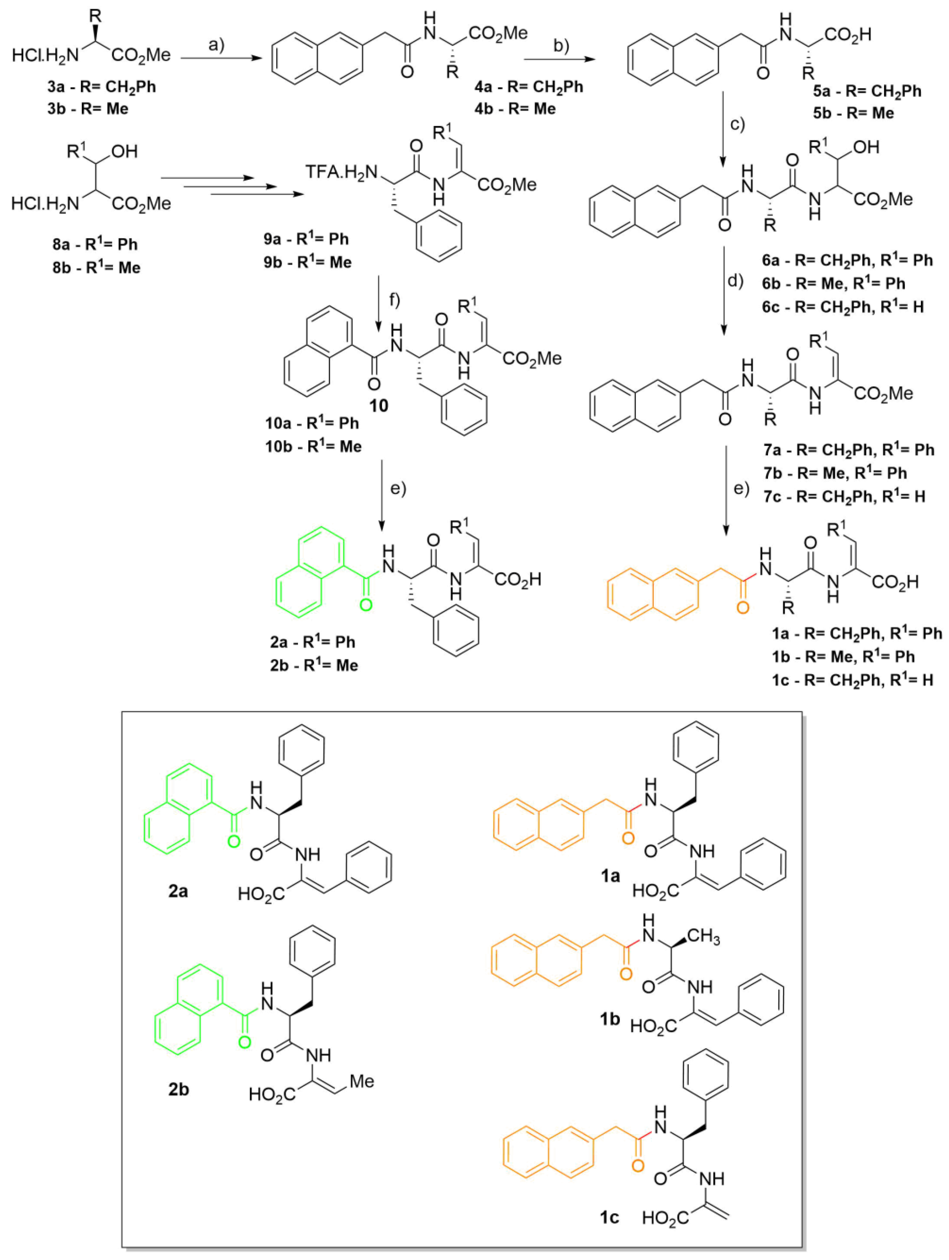
Synthesis of 2-naphthylacetylamino acids (4a,b): 2-Naphthylacetic acid was dissolved in acetonitrile (10 mL mmol-1) and put in an ice bath. HOBt (1.0 equiv.), DCC (1.0 equiv.), amino acid methyl ester (1.0 equiv.) and triethylamine (2.0 equiv.) were added, waiting about 2 min between each addition. The mixture was left stirring at rt overnight (~18 h). The urea was filtered and the solvent removed under reduced pressure. Acetone was added and the mixture was stored in the freezer for 2 h. The urea was filtered again. Evaporation at reduced pressure gave a residue that was partitioned between ethyl acetate (50 mL) and KHSO4 (30 mL, 1 M). The organic phase was thoroughly washed with KHSO4 (1 M), NaHCO3 (1 M) and brine (3 × 30 mL, each), and dried with MgSO4. Removal of the solvent afforded compounds 4a,b.
2-Naph-L-Phe-OMe (4a): 2-Naphthylacetic acid (0.37 g, 2.00 mmol) and H-L-Phe-OMe,HCl (3a) gave compound 4a as a white solid (0.64 g, 93%); mp: 85.0–87.0 °C; 1H NMR (400 MHz, CDCl3, δ): 3.01 (dq, J = 5.6 and 14.0 Hz, 2H, βCH2), 3.70 (s, 3H, OCH3), 3.72 (s, 2H, CH2), 4.86 (td, J = 5.6 and 7.6 Hz, 1H, αCH), 5.83 (d, J = 7.2 Hz, 1H, NH), 6.79 (d, J = 7.4 Hz, 2H, Ho Phe), 6.99 (t, J = 8.0 Hz, 2H, Hm Phe), 7.10 (tt, J = 2.0 and 7.4 Hz, 1H, Hp Phe), 7.30 (dd, J = 1.6 and 8.4 Hz, 1H, H-3 Naph), 7.49–7.54 (m, 2H, 2 × Ar H Naph), 7.66 (s, 1H, H-1 Naph), 7.79–7.87 (m, 3H, 3 × Ar H Naph); 13C NMR (100.6 MHz, CDCl3, δ): 37.52 (βCH2), 43.81 (CH2), 52.30 (OCH3), 52.89 (αCH), 126.05 (CH Naph), 126.37 (CH Naph), 126.97 (Cp Phe), 127.19 (CH-3 Naph), 127.70 (2 × CH Naph), 128.26 (CH-1 Naph), 128.40 (Cm Phe), 128.80 (CH Naph), 128.99 (Co Phe), 131.85 (C-2 Naph), 132.57 (C-4a Naph), 133.56 (C-8a Naph), 135.33 (Ci Phe), 170.42 (C=O Naph), 171.72 (C=O Phe); HRMS (ESI) m/z: [M+H]+ calcd for C22H22NO3+ 348.1594; found, 348.1592.
2-Naph-L-Ala-OMe (4b): 2-Naphthylacetic acid (0.55 g, 2.95 mmol) and H-L-Ala-OMe,HCl (3b) gave compound 4b as a white solid (0.62 g, 78%); mp: 93.5–96.0 °C; 1H NMR (400 MHz, CDCl3, δ): 1.33 (d, J = 7.2 Hz, 3H, βCH3), 3.71 (s, 3H, OCH3), 3.77 (s, 2H, CH2), 4.61 (quint, J = 7.2 Hz, 1H, αCH), 6.05 (d, J = 6.4 Hz, 1H, NH), 7.41 (dd, J = 2.0 and 8.4 Hz, 1H, H-3), 7.46–7.53 (m, 2H, Ar H), 7.75 (s, 1H, H-1), 7.82–7.87 (m, 3H, Ar H); 13C NMR (100.6 MHz, CDCl3, δ): 18.26 (βCH3), 43.72 (CH2), 48.10 (αCH), 52.40 (OCH3), 125.99 (CH), 126.35 (CH), 127.19 (CH-3), 127.66 (CH), 127.70 (CH), 128.23 (CH-1), 128.74 (CH), 131.96 (C-2), 132.54 (C-4a), 133.54 (C-8a), 170.41 (C=O Naph), 173.30 (C=O Ala); HRMS (ESI) m/z: [M+H]+ calcd for C16H18NO3+ 272.1281; found, 272.1280.
Synthesis of compounds 5a,b: The amino acid derivative was dissolved in methanol (until 10 mL) and NaOH (1 M) (1.5 equiv.). The reaction was followed by TLC (Thin Layer Chromatography) until no starting material was detected. The organic solvent was removed under reduced pressure, and the reaction mixture was acidified to pH 3 with KHSO4 (1 M). The solid formed was filtered, affording compounds 5a,b.
2-Naph-L-Phe-OH (5a): 2-Naph-L-Phe-OMe (4a) (0.55 g, 1.58 mmol) gave compound 5a as a white solid (0.48 g, 91%); mp: 145.0–147.0 °C; 1H NMR (400 MHz, DMSO-d6, δ): 2.88 (dd, J = 9.6 and 14.0 Hz, 1H, βCH), 3.07 (dd, J = 4.8 and 14.0 Hz, 1H, βCH), 3.58 (dd, J = 14.0 and 21.2 Hz, 2H, CH2), 4.41–4.48 (m, 1H, αCH), 7.19 (brs, 5H, Ar H Phe), 7.27 (dd, J = 1.6 and 8.4 Hz, 1H, Ar H), 7.43–7.50 (m, 2H, Ar H), 7.65 (s, 1H, Ar H), 7.76–7.80 (m, 2H, Ar H), 7.85 (d, J = 8.0 Hz, 1H, Ar H), 8.44 (d, J = 8.0 Hz, 1H, NH), 12.63 (brs, 1H, CO2H); 13C NMR (100.6 MHz, DMSO-d6, δ): 36.71 (βCH2), 42.09 (CH2), 53.49 (αCH), 125.45 (CH), 125.99 (CH), 126.36 (Cp Phe), 127.21 (CH), 127.32 (CH), 127.43 (CH), 127.45 (CH), 127.56 (CH), 128.10 (Cm Phe), 129.11 (Co Phe), 131.72 (C), 132.91 (C), 133.85 (C), 137.53 (Ci Phe), 169.96 (C=O), 172.99 (CO2H); HRMS (ESI) m/z: [M+H]+ calcd for C21H20NO3+ 334.1438; found, 334.1435.
2-Naph-L-Ala-OH (5b): 2-Naph-L-Ala-OMe (4b) (0.37 g, 1.36 mmol) gave compound 5b as a white solid (0.28 g, 80%); mp: decomposes above 157.0 °C; 1H NMR (400 MHz, DMSO-d6, δ): 1.27 (d, J = 7.2 Hz, 3H, βCH3), 3.62 (s, 2H, CH2), 4.21 (t, J = 7.2 Hz, 1H, αCH), 7.41–7.48 (m, 3H, Ar H), 7.76 (s, 1H, Ar H), 7.82–7.87 (m, 3H, Ar H), 8.45 (d, J = 7.2 Hz, 1H, NH), 12.49 (brs, 1H, CO2H); 13C NMR (100.6 MHz, DMSO-d6, δ): 17.21 (βCH3), 41.94 (CH2), 47.58 (αCH), 125.46 (CH), 126.02 (CH), 127.23 (CH), 127.34 (CH), 127.43 (CH), 127.48 (CH), 127.63 (CH), 131.73 (C), 132.94 (C), 133.94 (C), 169.83 (C=O), 174.13 (CO2H); HRMS (ESI) m/z: [M+H]+ calcd for C15H16NO3+ 258.1125; found, 258.1125.
Synthesis of dipeptide derivatives (6ac): The procedure is similar to the one described for amino acid derivatives 4a,b, but 2-naphthylacetic acid is substituted by the N-protected amino acid derivative (5a,b).
2-Naph-L-Phe-D,L-Phe(β-OH)-OMe (6a): 2-Naph-L-Phe-OH (5a) (0.24 g, 0.72 mmol) and H-D,L-Phe(β-OH)-OMe,HCl gave compound 6a as a light solid (0.27 g, 73%); mp: 130.0–133.0 °C; 1H NMR (400 MHz, CDCl3, δ): 2.56–2.61 (m, 1H, βCH), 2.72–2.79 (m, 2H, 2 × βCH), 2.86 (dd, J = 6.4 and 14.0 Hz, 1H, βCH), 3.55 (s, 4H, 2 × CH2), 3.61 (s, 6H, 2 × OCH3), 4.60–4.76 (m, 4H, 4 × αCH), 5.14 (d, J = 3.2 Hz, 1H, βCH), 5.21 (d, J = 3.2 Hz, 1H, βCH), 5.84–5.89 (m, 1H, NH), 5.94–5.99 (m, 1H, NH), 6.51–6.56 (m, 2H, Ar H), 6.82–6.89 (m, 4H, Ar H), 6.95–7.27 (m, 19H, 17 × Ar H, 2 × NH), 7.40–7.48 (m, 6H, Ar H), 7.66–7.69 (m, 3H, Ar H), 7.73–7.75 (m, 2H, Ar H); 13C NMR (100.6 MHz, CDCl3, δ): 37.25 (βCH2), 37.47 (βCH2), 43.58 (CH2), 43.63 (CH2), 52.61 (OCH3), 52. 67 (OCH3), 53.68 (αCH Phe), 53.97 (αCH Phe), 58.32 (αCH), 58.38 (αCH), 73.38 (βCH), 73.49 (βCH), 125.78 (CH), 125.84 (CH), 126.01 (CH), 126.09 (CH), 126.30 (CH), 126.38 (CH), 126.70 (CH), 126.76 (CH), 127.08 (2 × CH), 127.65 (CH), 127.68 (CH), 127.70 (CH), 127.95 (CH), 128.03 (CH), 128.08 (CH), 128.22 (CH), 128.25 (CH), 128.27 (CH), 128.30 (CH), 128.42 (CH), 128.45 (CH), 128.75 (CH), 128.81 (CH), 129.08 (CH), 129.15 (CH), 131.48 (C), 131.59 (C), 133.49 (2 × C), 133.52 (2 × C), 135.64 (Ci Phe), 135.90 (Ci Phe), 139.54 [Ci Phe(β-OH)], 139.68 [Ci Phe(β-OH)], 170.44 (C=O), 170.63 (C=O), 170.84 (C=O), 171.03 (C=O), 171.14 (C=O), 171.16 (C=O); HRMS (ESI) m/z: [M+H]+ calcd for C31H31N2O5+ 511.2228; found, 511.2237; [M+Na]+ calcd for C31H30N2NaO5+ 533.2047; found, 533.2061.
2-Naph-L-Ala-D,L-Phe(β-OH)-OMe (6b): 2-Naph-L-Ala-OH (5b) (0.44 g, 1.71 mmol) and H-D,L-Phe(β-OH)-OMe,HCl gave compound 6b as a light oil, that spontaneously crystallized (0.33 g, 45%); mp: 112.0–115.0 ◦C; 1H NMR (400 MHz, CDCl3, δ): 1.01 (d, J = 7.2 Hz, 3H, βCH3), 1.24 (d, J = 6.8 Hz, 3H, βCH3), 3.68 (s, 2H, CH2), 3.69 (s, 2H, CH2), 3.70 (s, 3H, OCH3), 3.71 (s, 3H, OCH3), 4.45–4.56 (m, 2H, αCH), 4.80 (dd, J = 3.6 and 8.8. Hz, 1H, αCH), 4.85 (dd, J = 3.2 and 8.8 Hz, 1H, αCH), 5.25 (d, J = 3.6 Hz, 1H, βCH), 5.32 (d, J = 3.2 Hz, 1H, βCH), 6.20 (d, J = 7.6 Hz, 1H, NH), 6.35 (d, J = 7.6 Hz, 1H, NH), 7.20–7.35 (m, 14H, 12 × Ar H and 2 × NH), 7.42–7.50 (m, 4H, Ar H), 7.66 (d, J = 8.0 Hz, 2H, Ar H), 7.76–7.82 (m, 6H, Ar H); 13C NMR (100.6 MHz, CDCl3, δ): 18.12 (βCH3), 18.53 (βCH3), 43.52 (CH2), 43.56 (CH2), 48.78 (αCH), 48.88 (αCH), 52.56 (OCH3), 52.61 (OCH3), 58.10 (αCH), 58.37 (αCH), 73.43 (βCH), 73.48 (βCH), 125.66 (2 × CH), 125.87 (2 × CH), 125.97 (CH), 126.02 (CH), 126.30 (CH), 126.36 (CH), 127.05 (2 × CH), 127.66 (2 × CH), 127.67 (2 × CH), 127.85 (CH), 127.93 (CH), 128.20 (2 × CH), 128.27 (2 × CH), 128.30 (2 × CH), 128.74 (CH), 128.76 (CH), 131.62 (C), 131.64 (C),132.51 (C), 132.52 (C), 133.49 (2 × C), 139.65 (2 × C), 170.59 (C=O), 170.62 (C=O), 171.13 (C=O Naph), 171.14 (C=O Naph), 172.08 (C=O Ala), 172.12 (C=O Ala); HRMS (ESI) m/z: [M+H]+ calcd for C25H27N2O5+ 435.1915; found, 435.1912.
2-Naph-L-Phe-D,L-Ser-OMe (6c): 2-Naph-L-Phe-OH (5a) (0.36 g, 1.08 mmol) and H-D,L-Ser-OMe,HCl gave compound 6c as a white solid (0.24 g, 51%), after a column chromatography (petroleum ether, ethyl acetate, mixtures of crescent polarity); mp: 166.0–168.0 °C; 1H NMR (300 MHz, CDCl3, δ): 2.92 (dd, J = 8.0 and 14.0 Hz, 1H, βCH Phe), 3.06 (dd, J = 5.9 and 14.0 Hz, 1H, βCH Phe), 3.58 (t, J = 6.5 Hz, 1H, OH), 3.65 (s, 2H, CH2 Naph), 3.73 (s, 3H, OCH3), 3.82–3.88 (m, 2H, βCH2 Ser), 4.54–4.59 (m, 1H, αCH Ser), 4.71–4.78 (m, 1H, αCH Phe), 6.37 (d, J = 7.8 Hz, 1H, NH Phe), 6.92–6.95 (m, 2H, 2 × Ho Phe), 7.00–7.05 (m, 2H, 2 × Hm Phe), 7.08–7.13 (m, 1H, Hp Phe), 7.21 (dd, J = 1.7 and 8.3 Hz, 1H, Ar H Naph), 7.25 (s, 1H, NH Ser), 7.46–7.52 (m, 12H, 2 × Ar H Naph), 7.58 (s, 1H, Ar H Naph), 7.73–7.76 (m, 2H, 2 × Ar H Naph), 7.80–7.83 (m, 1H, Ar H Naph); 13C NMR (75.5 MHz, CDCl3, δ): 37.84 (βCH2 Phe), 43.47 (CH2 Naph), 52.61 (OCH3), 54.48 (αCH Phe), 54.85 (αCH Ser), 62.51 (βCH2 Ser), 126.02 (CH Naph), 126.32 (CH Naph), 126.87 (CHp Phe), 127.08 (CH Naph), 127.64 (CH Naph), 127.69 (CH Naph), 128.22 (CH Naph), 128.42 (CHm Phe), 128.72 (CH Naph), 129.09 (CHo Phe), 131.60 (C Naph), 132.50 (C Naph), 133.46 (C Naph), 135.79 (Ci Phe), 170.53 (C=O Ser), 171.17 (C=O Phe), 171.70 (C=O Naph); Anal. calcd for C25H26N2O5: C 69.11, H 6.03, N 6.45; found: C 68.52, H 6.485, N 7.151; HRMS (ESI) m/z: [M+H]+ calcd for C25H27N2O5+ 435.1915; found, 435.1914.
Synthesis of dehydrodipeptide derivatives (7ac): DMAP (0.1 equiv.) was added to solutions of compounds 6ac in dry acetonitrile (1 M) followed by Boc2O (1.0 equiv.) under rapid stirring at room temperature. The reaction was monitored by 1H NMR until all the reactant had been consumed. Then TMG (2% in volume) was added, stirring was con-tinued and the reaction followed by 1H NMR. When all the reactant had been consumed, evaporation at reduced pressure gave a residue that was partitioned between ethyl acetate (50 mL) and KHSO4 (30 mL, 1 M). The organic phase was thoroughly washed with KHSO4 (1 M), NaHCO3 (1 M) and brine (3 × 30 mL, each), and dried with MgSO4. Removal of the solvent afforded compounds 7ac.
2-Naph-L-Phe-Z-ΔPhe-OMe (7a): 2-Naph-L-Phe-D,L-Phe(β-OH)-OMe (6a) (0.27 g, 0.53 mmol) gave compound 7a as a light solid (0.16 g, 62%); mp: 197.0–200.0 °C; 1H NMR (400 MHz, DMSO-d6, δ): 2.84 (dd, J = 10.4 and 13.6 Hz, 1H, βCH), 3.13 (dd, J = 4.0 and 13.6 Hz, 1H, βCH), 3.59 (q, J = 14.0 Hz, 2H, CH2), 3.69 (s, 3H, OCH3), 4.72 (ddd, J = 4.0, 8.4 and 10.4 Hz, 1H, αCH), 7.16–7.25 (m, 6H, 5 × Ar H and βCH), 7.29–7.31 (m, 4H, Ar H), 7.42–7.49 (m, 2H, Ar H), 7.60–7.62 (m, 3H, Ar H), 7.73–7.77 (m, 2H, Ar H), 7.83–7.85 (m, 1H, Ar H), 8.53 (d, J = 8.4 Hz, 1H, NH), 9.92 (s, 1H, NH); 13C NMR (100.6 MHz, DMSO-d6, δ): 37.11 (βCH2), 42.15 (CH2 Naph), 52.17 (OCH3), 54.09 (αCH Phe), 125.41 (CH), 125.86 (αC ΔPhe), 125.94 (CH), 126.30 (CH), 127.18 (CH), 127.31 (CH), 127.40 (CH), 127.56 (CH), 128.03 (CH), 128.93 (CH), 129.23 (CH), 128.40 (CH), 130.02 (CH), 131.69 (C), 132.01 (CH), 132.01 (βCH ΔPhe), 132.89 (C), 133.93 (C-2 Naph), 137.76 (Ci Phe), 138.18 (Ci ΔPhe), 165.33 (C=O ΔPhe), 170.05 (C=O Naph), 171.47 (C=O Phe); HRMS (ESI) m/z: [M+H]+ calcd for C31H29N2O4+ 493.2122; found, 493.2128; [M+Na]+ calcd for C31H28N2NaO4+ 515.1941; found, 515.1949.
2-Naph-L-Ala-Z-ΔPhe-OMe (7b): 2-Naph-L-Ala-D,L-Phe(β-OH)-OMe (6b) (0.26 g, 0.60 mmol) gave compound 7b as a light yellow solid (0.21 g, 84%); mp: 159.5–167.0 ◦C; 1H NMR (400 MHz, CDCl3, δ): 1.36 (d, J = 6.8 Hz, 3H, βCH3), 3.69 (s, 2H, CH2), 3.78 (s, 3H, OCH3), 4.70–4.77 (m, 1H, αCH), 6.24 (d, J = 7.2 Hz, 1H, NH), 7.28–7.33 (m, 4H, Ar H), 7.37 (s, 1H, βCH), 7.43–7.50 (m, 4H, Ar H), 7.67 (s, 1H, Ar H), 7.75–7.82 (m, 3H, Ar H), 8.04 (s, 1H, NH); 13C NMR (100.6 MHz, CDCl3, δ): 17.53 (βCH3), 43.51 (CH2), 49.10 (αCH), 52.59 (OCH3), 123.88 (αC), 126.01 (CH), 126.35 (CH), 127.03 (CH), 127.65 (CH), 127.67 (CH), 128.20 (CH), 128.53 (CH), 128.78 (CH), 129.54 (CH), 129.67 (CH), 131.64 (C), 132.52 (C), 133.03 (βCH), 133.41 (C), 133.49 (C), 165.25 (C=O ΔPhe), 170.83 (C=O Ala), 171.31 (C=O Naph); HRMS (ESI) m/z: [M+H]+ calcd for C25H25N2O4+ 417.1809; found, 417.1806; [M+Na]+ calcd for C25H24N2NaO4+ 439.1628; found, 439.1626.
2-Naph-L-Phe-ΔAla-OMe (7c): 2-Naph-L-Phe-D,L-Ser-OMe (6c) (0.30 g, 0.69 mmol) gave compound 7c as a white solid (0.21 g, 72%); mp: 65.0–67.0 °C; 1H NMR (400 MHz, CDCl3, δ): 2.99–3.01 (m, 2H, βCH2), 3.73 (s, 2H, CH2), 3.81 (s, 3H, OCH3), 4.71–4.76 (m, 1H, αCH), 5.89 (s, 1H, βCH), 5.99 (d, J = 7.6 Hz, 1H, NH), 6.55 (s, 1H, βCH), 6.89 (d, J = 7.2 Hz, 2H, Ho Phe), 7.02 (t, J = 7.4 Hz, 2H, Hm Phe), 7.11 (t, J = 7.4 Hz, 1H, Hp Phe), 7.23–7.29 (m, 1H, H-3 Naph), 7.50–7.54 (m, 2H, Ar H Naph), 7.63 (s, 1H, H-1 Naph), 7.78–7.81 (m, 2H, Ar H Naph), 7.84.7.87 (m, 1H, Ar H Naph), 8.24 (s, 1H, NH); 13C NMR (100.6 MHz, CDCl3, δ): 37.29 (βCH2), 43.62 (CH2 Naph), 52.91 (OCH3), 54.94 (αCH Phe), 109.37 (βCH2), 126.11 (CH Naph), 126.38 (CH Naph), 126.95 (Cp Phe), 127.09 (CH-3 Naph), 127.67 (CH Naph), 127.71 (CH Naph), 128.34 (CH-1 Naph), 128.56 (Cm Phe), 128.91 (CH Naph and Co Phe), 130.58 (αC), 131.45 (C-2 Naph), 132.57 (C-4a Naph), 133.53 (C-8a Naph), 135.55 (Ci Phe), 163.95 (C=O ΔAla), 169.41 (C=O Phe), 171.19 (C=O Naph); HRMS (ESI) m/z: [M+H]+ calcd for C25H25N2O4+ 417.18088; found, 417.18060; [M+Na]+ calcd for C25H24N2NaO4+ 439.16283; found, 439.16256.
Synthesis of 1-naphthoyl dehydrodipeptides (10a,b): The methyl ester of the peptide (1.1 equiv.) was dissolved in DCM (5 mL) and put in an ice bath. Triethylamine (2.2 equiv.) was added and, slowly, 1-naphthoyl chloride. The mixture was left stirring at rt overnight (~18 h). The mixture was filtered. Evaporation at reduced pressure gave a residue that was partitioned between ethyl acetate (50 mL) and KHSO4 (30 mL, 1 M). The organic phase was thoroughly washed with KHSO4 (1 M), NaHCO3 (1 M) and brine (3 × 30 mL, each), and dried with MgSO4. Removal of the solvent afforded compounds 10a,b.
1-Nap-L-Phe-Z-ΔPhe-OMe (10a): H-L-Phe-Z-ΔPhe-OMe.TFA (9a) (0.39 g, 0.90 mmol) and 1-naphthoyl chloride gave compound 10a as a white solid (0.38 g, 88%); mp: 197.0–198.0 °C; 1H NMR (400 MHz, DMSO-d6, δ): 2.95–3.02 (dd, J = 11.2 and 2.4 Hz, 1H, βCH2), 3.23–3.28 (dd, J = 3.6 and 10.4 Hz, 1H, βCH2 Phe), 3.74 (s, 3H, OCH3), 4.99–5.06 (m, 1H, αCH Phe), 7.25–7.29 (m, 1H, Ar H), 7.32–7.41 (m, 7H, Ar H and βCH ΔPhe), 7.42–7.46 (m, 3H, Ar H), 7.48–7.53 (m, 2H, Ar H), 7.74–7.77 (m, 2H, Ar H), 7.87 (d, J = 8.4 Hz, 1H, Ar H), 7.93 (d, J = 8.0 Hz, 1H, Ar H), 7.98 (d, J = 8.4 Hz, 1H, Ar H), 8.86 (d, J = 8.4 Hz, 1H, NH Phe), 10.02 (s, 1H, NH ΔPhe); 13C NMR (100.6 MHz, DMSO-d6, δ): 36.63 (βCH2 Phe), 52.25 (OCH3), 54.85 (αCH Phe), 124.82 (CH), 125.21 (CH), 125.54 (CH), 125.96 (αC), 126.12 (CH), 126.40 (CH), 126.42 (CH), 127.99 (CH), 128.15 (CH), 128.60 (CH), 129.31 (CH), 129.53 (CH), 129.68 (C), 129.77 (CH), 130.14 (CH), 132.10 (βCH ΔPhe), 132.99 (C), 133.31 (C), 134.38 (C),138.20 (C), 165.41 (C=O), 168.70 (C=O), 171.57 (C=O); Anal. calcd for C30H26N2O4: C 75.30, H 5.48, N 5.85; found: C 74.84, H 5.57, N 5.92.
1-Nap-L-Phe-Z-ΔAbu-OMe (10b): H-L-Phe-Z-ΔAbu-OMe.TFA (9b) (0.53 g, 1.4 mmol) and 1-naphthoyl chloride gave compound 10b as a white solid (0.47 g, 80%); mp: 164.0–165.0 °C; 1H NMR (400 MHz, CDCl3, δ): 1.70 (d, J = 7.2 Hz, 3H, γCH3 ΔAbu), 3.22–3.25 (m, 1H, βCH2), 3.36–3.40 (m, 1H, βCH2 Phe), 3.73 (s, 3H, OCH3), 5.22 (q, J = 7.6 Hz, 1H, αCH Phe), 6.74–6.83 (m, 2H, NH Phe and βCH ΔAbu), 7.28–7.41 (m, 6H, Ar H), 7.43–7.52 (m, 3H, Ar H), 7.84 (d, J = 8.0 Hz, 2H, Ar H), 7.89 (d, J = 8.0 Hz, 1H, Ar H), 8.07 (d, J = 8.4 Hz, 1H, NH ΔPhe); 13C NMR (100.6 MHz, CDCl3, δ): 14.42 (γCH3 ΔAbu), 37.78 (βCH2 Phe), 52.30 (OCH3), 54.87 (αCH Phe), 124.62 (CH), 125.18 (CH), 125.39 (CH), 125.92 (C), 126.41 (CH), 127.11 (CH), 127.19 (CH), 128.25 (CH), 128.77 (CH), 129.41 (CH), 129.99 (C), 131.04 (CH), 133.21 (C), 133.58 (C), 134.69 (βCH ΔAbu), 136.38 (C), 164.53 (C=O), 169.38 (C=O), 169.74 (C=O); HRMS (micrO-TOF) m/z: [M+H]+ calcd. for C25H25N2O4+ 417.1814; found, 417.1809.
Synthesis of the N-capped C-deprotected dehydrodipeptides (1ac, 2a,b): The N-protected dehydrodipeptide (7ac and 10a,b) was dissolved in 1,4-dioxane (10 mL) and NaOH (1 M) (1.5 equiv.). The reaction was followed by TLC until no starting material was detected. The organic solvent was removed under reduced pressure, and the reaction mixture was acidified to pH 3 with KHSO4 (1 M). The solid formed was filtered, affording compounds 1ac, 2a,b.
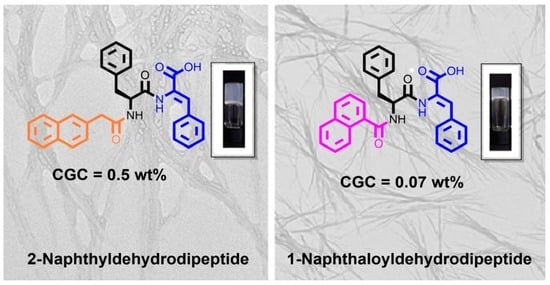
2-Naph-L-Phe-Z-ΔPhe-OH (1a): 2-Naph-L-Phe-Z-ΔPhe-OMe (7a) (0.34 g, 0.69 mmol) gave compound 1a as a white solid (0.31 g, 94%); mp: 165.0–167.0 °C; 1H NMR (400 MHz, DMSO-d6, δ): 2.78–2.84 (m, 1H, βCH), 3.11–3.16 (m, 1H, βCH), 3.51–3.59 (m, 2H, CH2), 4.68–4.74 (m, 1H, αCH), 7.15–7.24 (m, 5H, Ar H), 7.27–7.30 (m, 5H, Ar H and βCH), 7.42–7.48 (m, 2H, Ar H), 7.58–7.60 (m, 3H, Ar H), 7.72–7.76 (m, 2H, Ar H), 7.82–7.85 (m, 1H, Ar H), 8.47 (d, J = 8.4 Hz, 1H, NH), 9.73 (s, 1H, NH); 13C NMR (100.6 MHz, DMSO-d6, δ): 37.16 (βCH2), 42.18 (CH2), 54.14 (αCH), 125.43 (CH), 125.95 (CH), 126.27 (CH), 126.54 (αC), 127.19 (CH), 127.33 (CH), 127.41 (CH), 127.57 (CH), 128.02 (CH), 128.46 (CH), 129.18 (CH), 129.25 (CH), 129.93 (CH), 131.70 (C), 131.93 (βCH), 132.89 (C), 133.54 (C), 133.94 (C-2), 137.85 (Ci Phe), 166.19 (C=O), 170.06 (C=O), 171.16 (C=O); HRMS (ESI) m/z: [M+Na]+ calcd for C30H26N2NaO4+ 501.17848; found, 501.17831; [M+H]+ calcd for C30H27N2O4+ 479.1965; found, 479.1963.
2-Naph-L-Ala-Z-ΔPhe-OH (1b): 2-Naph-L-Ala-Z-ΔPhe-OMe (7b) (0.18 g, 0.42 mmol) gave compound 1b as a light solid (0.13 g, 76%); mp: 161.0–163.0 °C; 1H NMR (400 MHz, DMSO-d6, δ): 1.30 (d, J = 7.2 Hz, 3H, βCH3), 3.66 (s, 2H, CH2), 4.46 (quint, J = 7.2 Hz, 1H, αCH), 7.24–7.31 (m, 4H, 3 × Ar H and βCH), 7.43–7.49 (m, 3H, Ar H), 7.59–7.62 (m, 2H, Ar H), 7.76 (s, 1H, Ar H), 7.81–7.87 (m, 3H, Ar H), 8.40 (d, J = 7.2 Hz, 1H, NH Ala), 9.50 (s, 1H, NH ΔPhe), 12.68 (brs, 1H, CO2H); 13C NMR (100.6 MHz, DMSO-d6, δ): 17.84 (βCH3), 42.01 (CH2), 48.32 (αCH), 125.44 (CH), 125.98 (CH), 126.60 (C), 127.30 (CH), 127.35 (CH), 127.43 (CH), 127.49 (CH), 127.71 (CH), 128.37 (CH), 129.07 (CH), 129.93 (CH), 131.73 (βCH), 131.74 (C), 132.95 (C), 133.62 (C), 134.05 (C), 166.23 (C=O ΔPhe), 169.88 (C=O Naph), 171.93 (C=O Ala); HRMS (ESI) m/z: [M+H]+ calcd for C24H23N2O4+ 403.1652; found, 403.1659.
2-Naph-L-Phe-ΔAla-OH (1c): 2-Naph-L-Phe-ΔAla-OMe (7c) (0.21 g, 0.50 mmol) gave compound 1c as a white solid (0.13 g, 65%); mp: 159.0–160.0 °C; 1H NMR (400 MHz, DMSO-d6, δ): 2.82 (dd, J = 10.4 and 13.6 Hz, 1H, βCH), 3.08 (dd, J = 4.0 and 13.6 Hz, 1H, βCH), 3.52–3.61 (m, 2H, CH2), 4.72–4.76 (m, 1H, αCH), 5.73 (s, 1H, βCH), 6.31 (s, 1H, βCH), 7.16–7.23 (m, 4H, 2 × Hm Phe, Hp Phe, Ar H Naph), 7.27–7.29 (m, 2H, 2 × Ho Phe), 7.44–7.48 (m, 2H, 2 × Ar H Naph), 7.59 (s, 1H, H-1 Naph), 7.73 (d, J = 8.4 Hz, 1H, Ar H Naph), 7.77 (dd, J = 2.0 and 7.8 Hz, 1H, Ar H Naph), 8.56 (dd, J = 1.6 and 7.6 Hz, 1H, Ar H Naph), 8.56 (d, J = 8.0 Hz, 1H, NH Phe), 9.28 (s, 1H, NH ΔAla), 12.80 (brs, 1H, CO2H); 13C NMR (100.6 MHz, DMSO-d6, δ): 36.85 (βCH2), 42.07 (CH2 Naph), 54.67 (αCH Phe), 108.10 (βCH2), 125.46 (CH Naph), 125.98 (CH Naph), 126.27 (Cp Phe), 127.17 (CH-1 Naph), 127.32 (CH), 127.41 (CH), 127.48 (2 × CH), 127.99 (CH), 129.24 (Co Phe), 131.70 (C Naph), 132.80 (αC), 132.89 (C Naph), 133.69 (C Naph), 137.64 (Ci Phe), 164.79 (C=O ΔAla), 170.27 (C=O Naph), 170.92 (C=O Phe); HRMS (ESI) m/z: [M+H]+ calcd for C24H23N2O4+ 403.16578; found, 403.16522; [M+Na]+ calcd for C24H22N2NaO4+ 425.1477; found, 425.14716; [M+K]+ calcd for C24H22N2KO4+ 441.1217; found, 441.1210.
1-Nap-L-Phe-Z-ΔPhe-OH (2a): 1-Nap-L-Phe-Z-ΔPhe-OMe (10a) (0.32 g, 0.67 mmol) gave compound 2a as a white solid (0.29 g, 94%); mp: 181.0–182.0 °C; 1H NMR (400 MHz, DMSO-d6, δ): 2.92–2.98 (dd, J = 11.6 and 2.4 Hz, 1H, βCH2 Phe), 3.24–3.29 (dd, J = 3.2 and 10.4 Hz, 1H, βCH2 Phe), 4.97–5.03 (m, 1H, αCH Phe), 7.25–7.36 (m, 7H, Ar H and βCH ΔPhe), 7.38–7.51 (m, 6H, Ar H), 7.61 (d, J = 6.8 Hz, 2H, Ar H), 7.80 (d, J = 8.4 Hz, 1H, Ar H), 7.91 (d, J = 8.0 Hz, 1H, Ar H), 7.96 (d, J = 7.6 Hz, 1H, Ar H), 8.87 (d, J = 8.4 Hz, 1H, NH Phe), 9.74 (s, 1H, NH ΔPhe); 13C NMR (100.6 MHz, DMSO-d6, δ): 36.65 (βCH2 Phe), 55.09 (αCH Phe), 124.85 (CH), 125.17 (CH), 125.60 (CH), 126.12 (CH), 126.34 (CH), 126.42 (CH), 127.95 (CH), 128.14 (CH), 128.24 (CH), 128.44 (CH), 128.64 (C), 129.34 (CH), 129.65 (C), 129.71 (CH), 129.76 (CH), 132.98 (C), 134.53 (C), 134.67 (C), 138.37 (C), 166.14 (C=O), 170.93 (C=O), 173.33 (C=O); HRMS (micrOTOF) m/z: [M+Na]+ calcd. for C29H24N2NaO4+ 487.1634; found, 487.1629.
1-Nap-L-Phe-Z-ΔAbu-OH (2b): 1-Nap-L-Phe-Z-ΔAbu-OMe (10b) (0.33 g, 0.79 mmol) gave compound 2b as a white solid (0.31 g, 97%); mp: 200.0–201.0 °C; 1H NMR (400 MHz, DMSO-d6, δ): 1.69 (d, J = 7.2 Hz, 3H, γCH3 ΔAbu), 2.92–2.99 (dd, J = 11.2 and 2.4 Hz, 1H, βCH2), 3.20–3.25 (dd, J = 4.0 and 10.0 Hz, 1H, βCH2 Phe), 4.97–5.03 (m, 1H, αCH Phe), 6.62 (q, J = 7.2 Hz, 1H, γCH ΔAbu), 7.11 (dd, J = 2.8 and 6.0 Hz, 1H, Ar H), 7.18–7.31 (m, 6H, Ar H), 7.56 (s, 1H, Ar H), 7.23–7.27 (m, 1H, Ar H), 7.30–7.33 (m, 2H, Ar H), 7.39–7.43 (m, 4H, Ar H), 7.46–7.52 (m, 2H, Ar H), 7.78 (d, J = 8.4 Hz, 1H, Ar H), 7.91 (d, J = 8.0 Hz, 1H, Ar H), 7.96 (d, J = 8.0 Hz, 1H, Ar H), 8.76 (d, J = 8.4 Hz, 1H, NH Phe), 9.39 (s, 1H, NH ΔAbu), 12.55 (brs, 1H, CO2H); 13C NMR (100.6 MHz, DMSO-d6, δ): 13.79 (γCH3 ΔAbu), 37.32 (βCH2 Phe), 54.64 (αCH Phe), 124.83 (CH), 125.07 (CH), 125.54 (CH), 126.11 (CH), 126.32 (CH), 126.42 (CH), 127.95 (CH), 128.10 (CH), 128.20 (αC ΔAbu), 129.33 (CH), 129.64 (C), 129.67 (CH), 132.18 (βCH ΔAbu), 132.97 (C), 134.57 (C), 138.21 (C), 165.55 (C=O), 168.50 (C=O), 170.10 (C=O); HRMS (micrOTOF) m/z: [M+Na]+ calcd for C24H22N2NaO4+ 425.1477; found, 425.1488.
Self-assembly: All solutions were made up with ultra-filtered (18 MΩ) water from a Barnstead Nanopure system (Thermofisher, Waltham, MA, USA). Phosphate buffer was prepared from sodium dihydrogen phosphate monohydrate (NaH2PO4·H2O, Fluka BioChemika, Honeywell, Charlotte, NC, USA) and sodium phosphate dibasic dodecahydrate (Na2HPO4·12H2O, Fluka, Honeywell, Charlotte, NC, USA) with a final concentration of 0.1 M and pH 6.00, 7.19 or 8.06 (Mettler Toledo FiveEasy pH Meter, Merck, Darmstadt, Germany). D-glucono-δ-lactone (GdL, Sigma, St. Louis, MO, USA), aqueous NaOH 1 M and HCl 0.1 M were used.
Self-assembly with buffer: Compounds were weighed into sample vials. Buffer was added and the mixture was sonicated at room temperature, followed by heating to 80 °C under magnetic stirring. Solutions were left to cool to room temperature for 24 h.
Self-assembly with GdL: Compounds were weighed into sample vials. Water was added and the suspension was adjusted to pH circa 10 with aqueous NaOH 1 M. The mixture was briefly sonicated at room temperature and added into a vial with the weighed amount of GDL. The mixture was stirred (1000 rpm during 10 s) and left standing at room temperature overnight. The pH of the hydrogel was measured with pH paper.
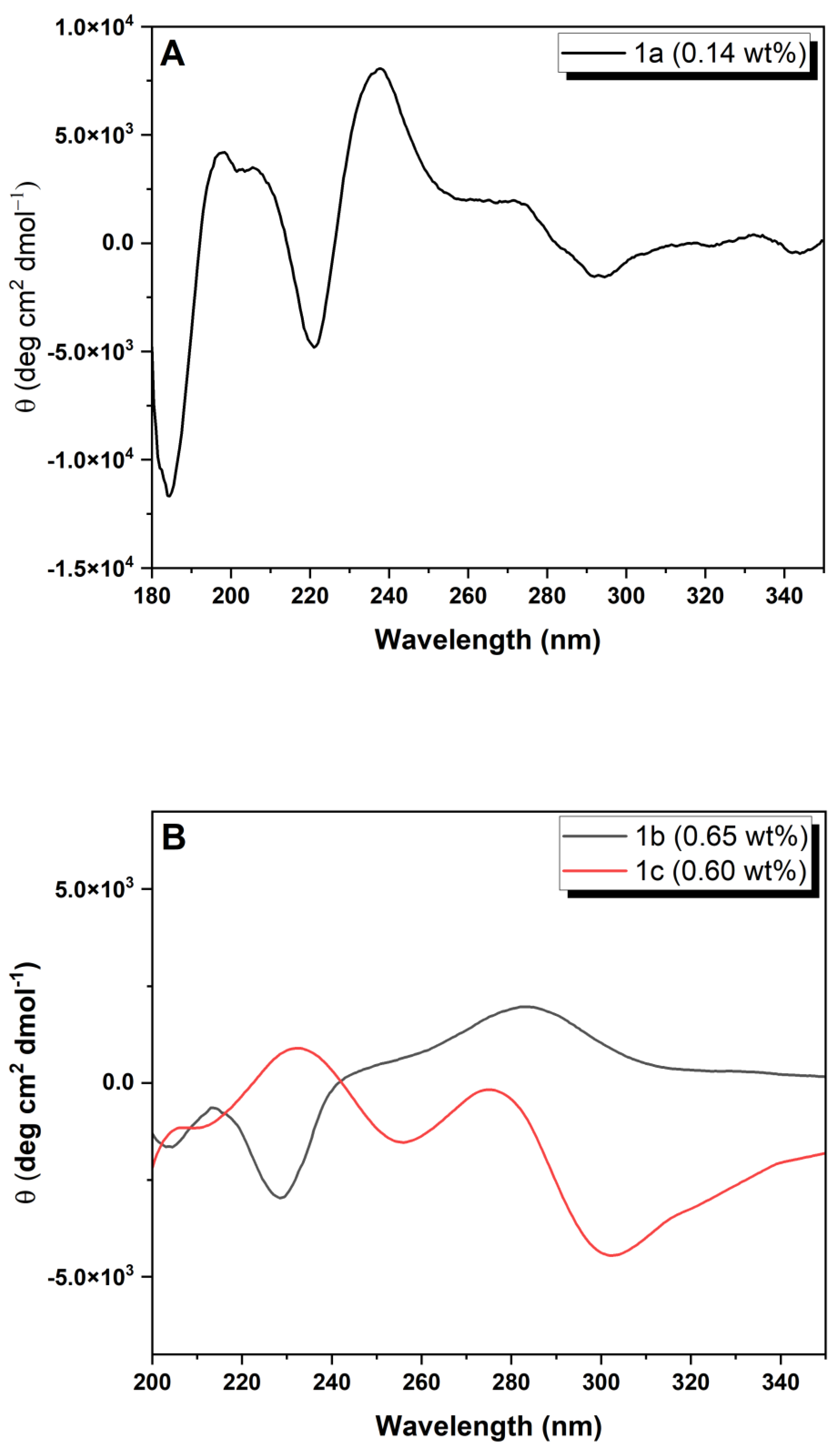
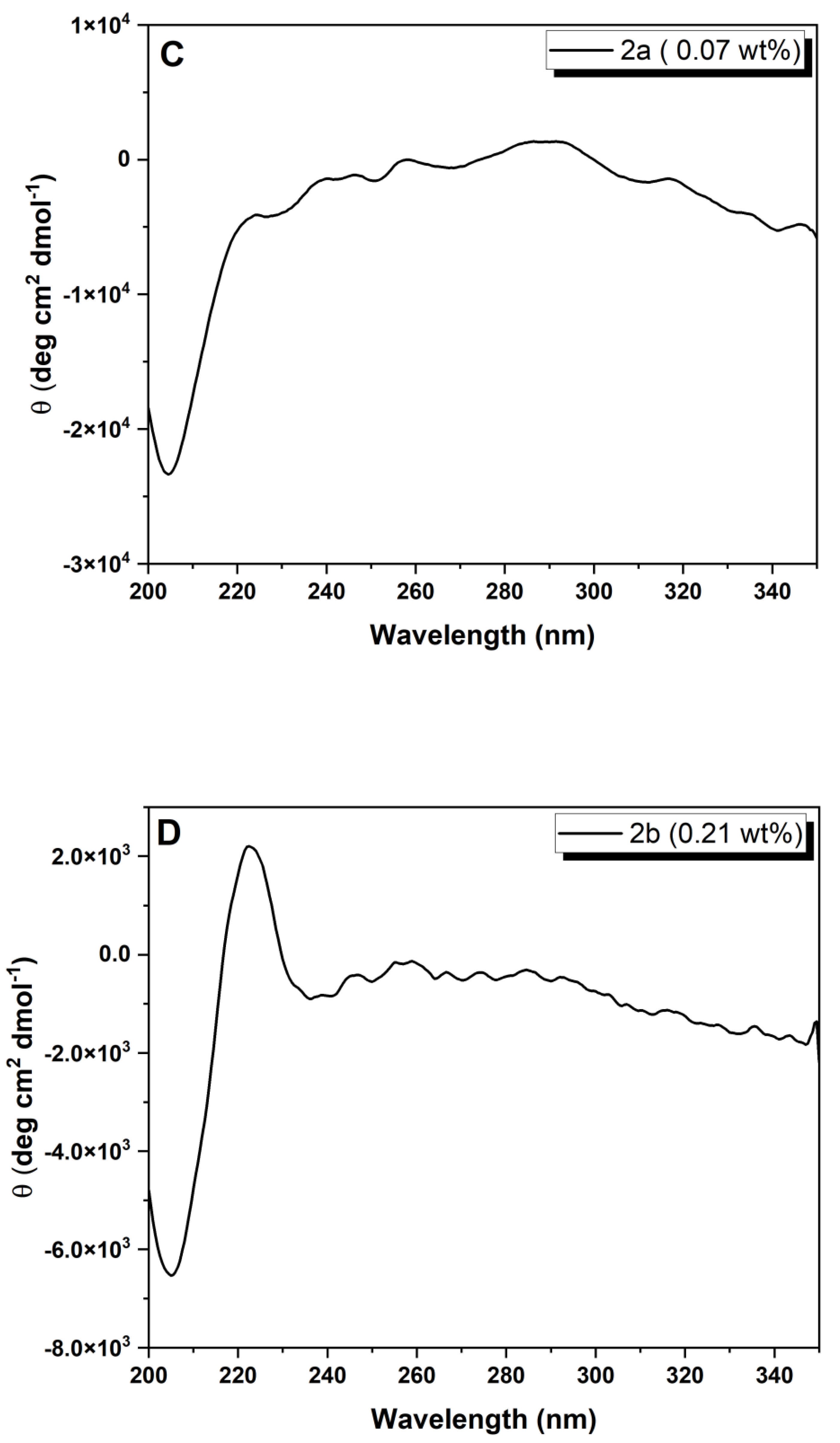
Circular dichroism: The CD spectra were recorded at 20 °C on a Chirascan spectropolarimeter (AppliedPhotophysics, Leatherhead, Surrey, UK). Peptide hydrogels were loaded into 0.1 mm quartz cells. Spectra display absorbance <2 at any measured point with 0.5 nm step, 1 nm bandwidth and 1 s collection time per step, taking three averages. The post-acquisition smoothing tool from Chirascan software (Version 4.13) was used to remove random noise elements from the averaged spectra. A residual plot was generated for each curve in order to check for spectral distortion effects during the smoothing process. CD data were normalized to molar mean residue ellipticity.
Scanning transmission electron microscopy: STEM experiments were performed using an ultra-high resolution field emission gun scanning electron microscopy (FEG-SEM), NOVA 200 Nano SEM, FEI Company, Hillsboro, Oregon, USA (SEMAT/UM), operated at 15 and 18.5 kV, using a STEM detector (PNDetector, Munich, Germany). Cu-C grids (S160-4 AGAR) were immersed in the peptide hydrogels. The grid was then allowed to dry at room temperature.
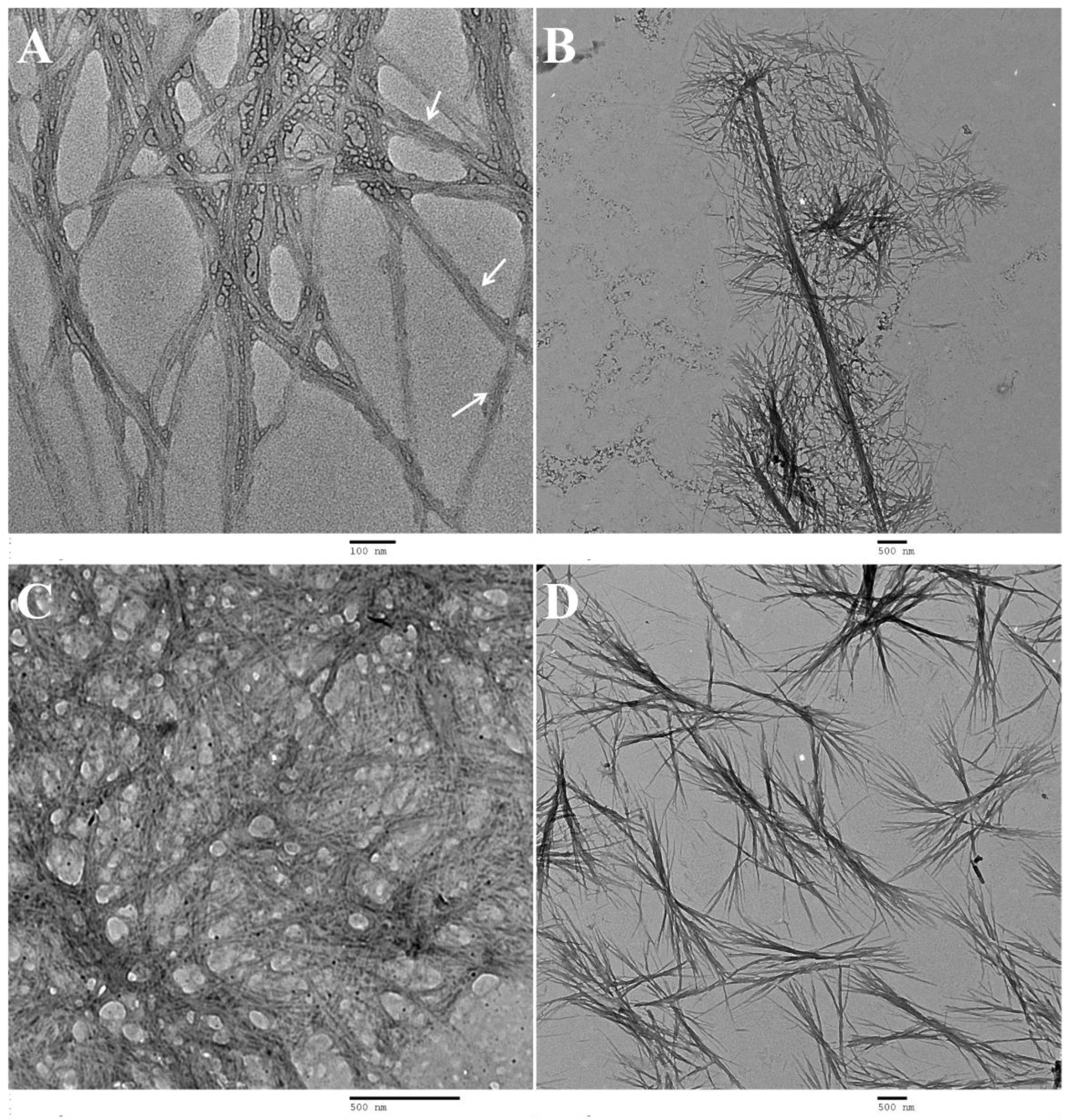
Transmission electron microscopy: TEM experiments were performed using a Philips CM20 (FELMI-ZFE, Graz, Austria) transmission electron microscope operating at 200 kV. Diluted solutions, 5 times more diluted than the measured cgc, were prepared in conditions similar to those used for preparation of the hydrogels. The shiny side of 300 mesh Cu grids coated with a carbon film (Agar Scientific, Stansted, Essex, UK) was placed over a drop of dehydrodipeptide solution (1a and 1c) during 1 min; the excess solution at the sides of the grid was carefully cleaned. One drop of the peptide solution (1b, 2a and 2b) was placed on the shiny side of 300 mesh Cu grids coated with a carbon film (Agar Scientific, Stansted, Essex, UK) for 1 min; the excess solution at the sides of the grid was carefully cleaned. In all cases, the shiny side of the grid was placed over a drop of aqueous uranyl acetate (1 wt%) (Agar Scientific, Stansted, Essex, UK) for 1 min. The excess at the sides of the grid was cleaned very carefully and the grid was then allowed to dry at room temperature.
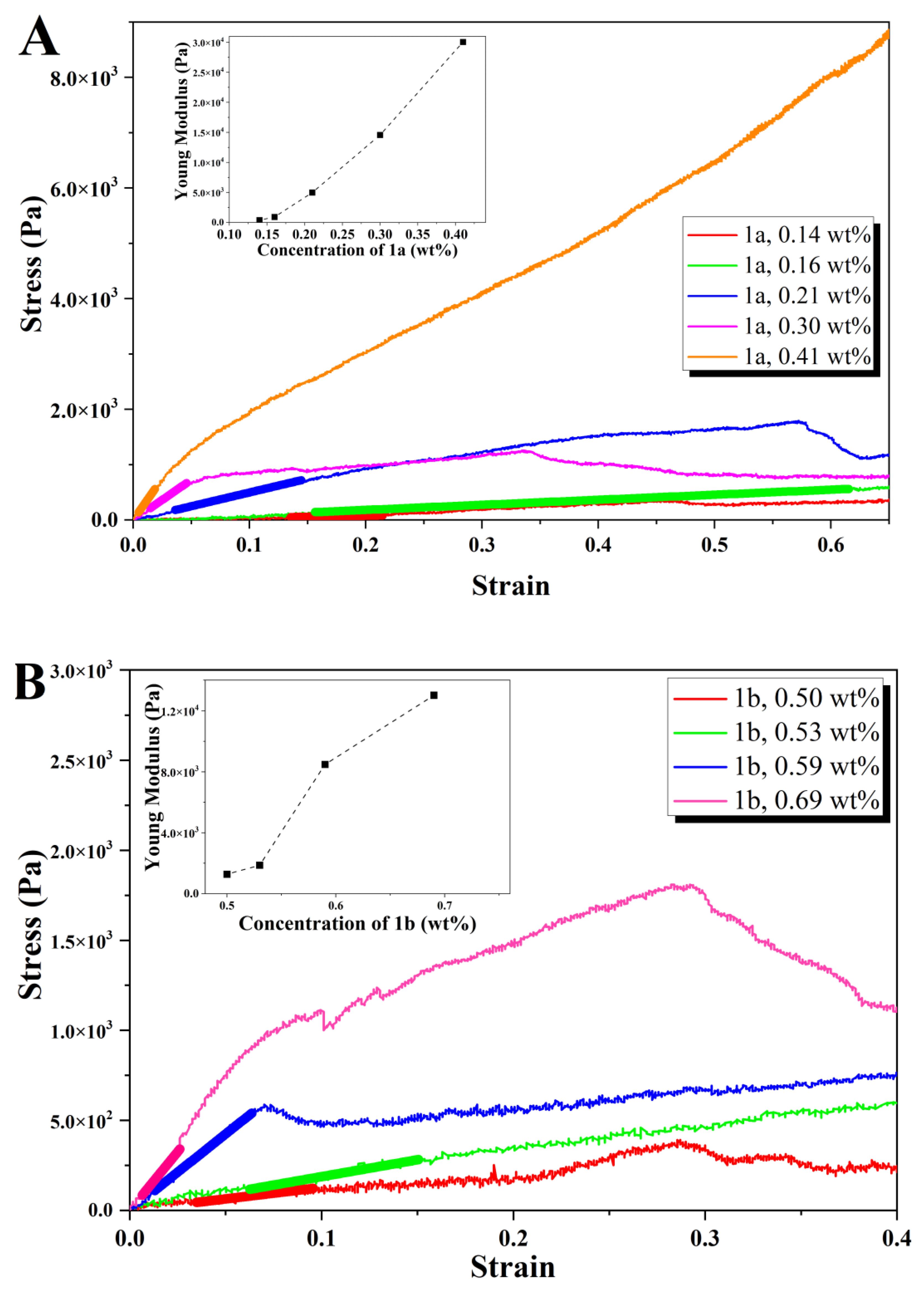
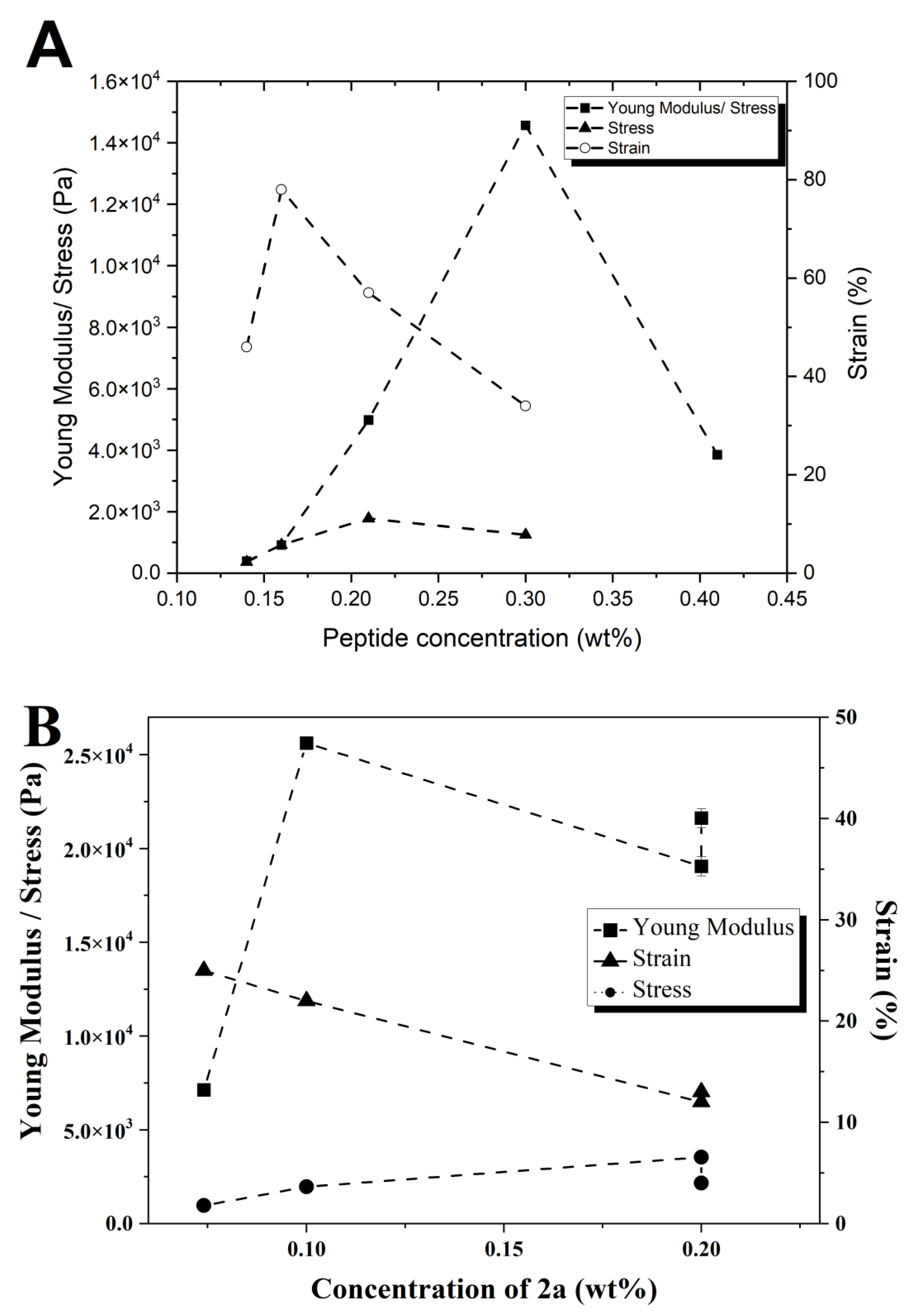
Rheology: Rheology experiments were carried out using a PaarPhysica MCR300 rheometer, Graz, Austria, equipped with a TEK 350-C plate with TC20/EDT/TEK temperature control and a cylindrical plunger with a diameter of 1 cm. Penetration tests on gelled samples were preferred to dynamic shear rheology performed on gelling samples in the rheometer. This is because very long (within 24 h) gel kinetics of samples would jeopardize the practicality of the study, which focuses on the viscoelastic comparison between gels and not on the gelation properties. The hydrogels were prepared in soda glass specimen tubes (Samco, Warwickshire, UK), which also served as the cups for the rheological measurements. The vials had diameters of 25 mm and the measurements were performed till a gap of 1.5 mm to the bottom of the vials was reached, avoiding significant end effects. The normal force was measured as a function of penetration distance in the gel. Young’s moduli were computed neglecting any plunger buoyancy effect, following the approach of Oakenfull et al. [40]. The slope and associated standard error of the linear regime of the stress–strain responses (Young’s moduli) and the power law exponents for the concentration regimes in 2b were determined using OriginPro 8 software. Strain and stress at break correspond to the location of the first maximum in the stress–strain curves followed by a deep decrease in stress.
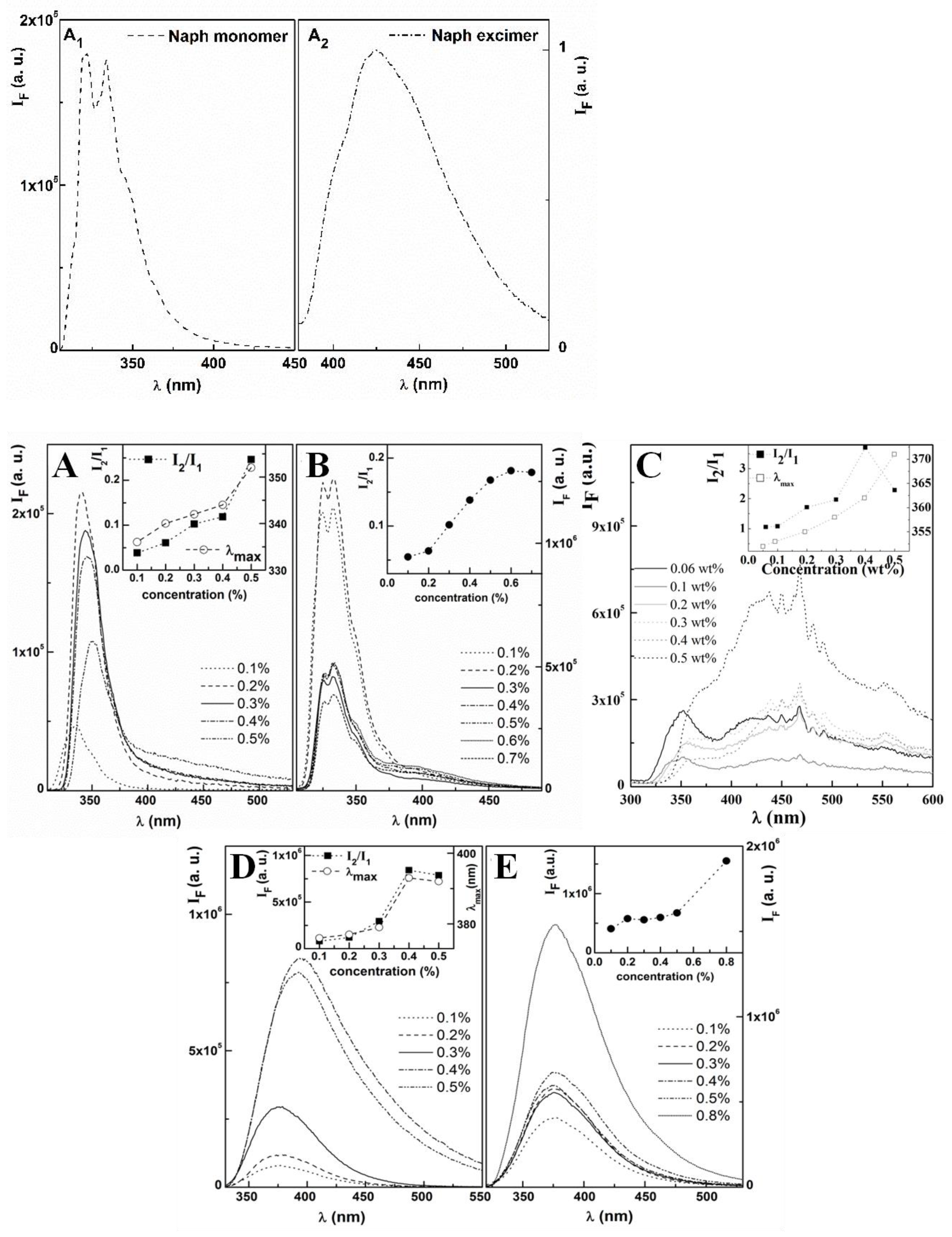
Spectroscopic measurements: Fluorescence measurements were performed using a Fluorolog 3 spectrofluorimeter (Kyoto, Japan), equipped with double monochromators in both excitation and emission, Glan-Thompson polarizers and a temperature-controlled cuvette holder. Fluorescence emission and excitation spectra were corrected for the instrumental response of the system.
Molecular dynamics (MD) simulations: The α,β-dehydroamino acids, ΔzPhe, ΔAla and ΔzAbu and Napthtalene were parameterized and validated in a previous work by some of the authors [29], from bonded and non-bonded parameters based on equivalent encoded amino acids of the GROMOS 54a7 force field [45]. The N-protected groups 2-naphthylacetyl (2-Naph) and 1-naphthoyl (1-Nyl) were parameterized based on the Naproxen group also used previously [29]. Topology files for 2-Naph, 1-Nyl, Npx, ΔzPhe, ΔAla and ΔzAbu are available upon request.
The initial molecular structure of the dehydrodipeptides was built with the program PyMOL [45]. MD simulations of dehydrodipeptides monomers were performed in octahedral boxes of SPC water with a hydration layer of at least 1.5 nm between the peptide and the periodic boundary conditions in all directions [46,47]. Boxes were made neutral with the addition of one Na+ ion. Each system was subjected to 12,000 steps of energy minimization with the steepest descent algorithm. Then, 100 ns of MD simulations were run, 40 ns of equilibration and 60 ns of production. MD simulations were run with the GROMACS 4.5.4 software package [48,49,50]. In all MD simulations, the system was maintained at constant temperature and pressure, 310 K and 1 atm, respectively, with the Berendsen thermostat and barostat methods, with ΔT = 0.10 ps and ΔP = 1.0 ps [51]. The SETTLE algorithm was used to constrain bond lengths and angles of water molecules, while the bond lengths and angles of peptides were constrained with the LINCS algorithm, which allowed the use of a 2 fs time step [52,53]. For the treatment of long-range interactions, we used the reaction field method, with a cutoff of 1.4 nm and a dielectric constant of 54 (corresponding to SPC water). The van der Waals interactions were truncated with a twin-range cutoff of 0.8 and 1.4 nm. The conformation population was computed with a clustering algorithm through a single-linkage method by first fitting the heavy atoms on the dehydrodipeptides and then analyzing the RMSD matrix vs. time to determine the most likely conformations for each peptide. The MD procedure used to simulate the dimers was similar to that used for the monomers. In this case, the dimers were solvated with SPC water in cubic boxes of 5 × 5 × 5 nm neutralized with two Na+ ions; the same energy minimization, equilibration and production run (40 + 60 ns) was also followed for the dimers. The aggregation analysis was done by monitoring the distance between the centers of mass of the peptides in the box vs. time for 60 ns and used to populate 0.1 nm bin histograms.
The most likely conformations for each molecule were also studied using quantum chemical DFT calculations. DFT calculations were performed with the program Gaussian 09 with the BLYP-D3 Hamiltonian that has been found to yield improved geometries in π–π stacked systems [54,55,56]. Calculations were performed with the SMD implicit water solvent with the def2-TVZP basis set.

Supplementary Materials

The following supporting information can be downloaded at: https://www.mdpi.com/article/10.3390/gels9060464/s1, Figure S1: Phase diagrams for hydrogelators obtained by addition of GDL to peptide solutions adjusted to pH circa 10 with NaOH 1M: (A) 1b; (B) 1c. *approximate pH of the gel (measured with indicator paper). Figure S2. Phase diagrams for dehydrodipeptide 2a, obtained by addition of GdL to alkaline solutions of this peptide (4 %(v/v) of NaOH 1 m); (A) Correlation between pH and peptide concentration; (B) Correlation between equivalents of GdL and peptide concentration. Figure S3. Phase diagrams for dehydrodipeptide 2b, obtained by addition of GdL to alkaline solutions of this peptide (4 %(v/v) of NaOH 1 m); (A) Correlation between pH and peptide concentration (arrows represent the drop in pH by addition of more GdL to the same solution); (B) Correlation between equivalents of GdL and peptide concentration. Figure S4. UV-VIS absorption spectra for hydrogelators 1ac and 2a,b in gel phase at the same concentration and pH as that deployed for the CD measurements: (A) gels 1ac; (B) gels 2a,b. UV-VIS absorption spectra for hydrogelators 1ac and 2a,b in solution, at concentrations well below the CGC: (C) gels 1ac; (D) gels 2a,b. Figure S5. STEM images: (A,B) 1a (gel at 0.5 wt% in phosphate buffer, pH 8), scale (A) 2 μm, (B) 500 nm; (C) 1b [gel at 0.77 wt%] scale 1 μm; (D) 1c [gel at 0.6 wt%], scale 1 μm. Figure S6. TEM images: (A,B,C) 1b (0.65 wt%), scale (A) 2 μm; (B,C) 500 nm; (D,E) 2a (0.014 wt%), scale 100 nm; (F) 2b (0.040 wt%), scale 500 nm. Figure S7. Stress-strain response curves for penetration tests for hydrogels at 25 μm s−1 plunger speed.: (A) gel 2a; (B) gel 2b; (C) Zoom of the linear regime of the stress-strain response curve of compounds 2b.

Author Contributions

Conceptualization, P.M.T.F. and J.A.M.; methodology P.M.T.F., J.A.M., I.H., L.H., E.M.S.C. and M.M.-F.; investigation, H.V., A.C., T.C., J.A.M., P.M.T.F., I.H., L.H., E.M.S.C. and M.M.-F.; writing P.M.T.F., J.A.M., I.H., L.H., E.M.S.C. and M.M.-F. All authors have read and agreed to the published version of the manuscript.

Funding

This work was funded by National Funds through FCT-Portuguese Foundation for Science and Technology under the Project PTDC/QUI-QOR/29015/2017, CQ/UM UID/QUI/00686/2013 and UID/QUI/0686/2016, UID/CTM/50025/2019 and CF-UM-UP (UIDB/04650/2020). The NMR spectrometers are part of the National NMR Network (PTNMR) and are partially supported by Infrastructure Project No 022161 (co-financed by FEDER through COMPETE 2020, POCI and PORL and FCT through PIDDAC).

Acknowledgments

For computing resources: Search-ON2: Revitalization of HPC infrastructure of UMinho, (NORTE-07-0162-FEDER-000086), co-funded by the North Portugal Regional Operational Programme (ON.2-O Novo Norte), under the National Strategic Reference Framework (NSRF), through the European Regional Development Fund (ERDF). Norte (ON.2) through the project Matepro—Optimizing Materials and Processes, with reference NORTE-07-0124-FEDER-000037 FEDER COMPETE is also acknowledged. T. Castro thanks the senior position funded by the European Union through the European Regional Development Fund (ERDF) under the Competitiveness Operational Program (BioCell-NanoART = Novel Bio-inspired Cellular Nano-architectures, POC-A1.1.4-E-2015 nr. 30/01.09.2016). M. Melle-Franco acknowledges support by the FCT through the programs Ciência 2008, the project IF/00894/2015 and within the Projects Scope: UID/CEC/00319/2013 and UIDB/50011/2020 and UIDP/5 0011/2020 (CICECO-Aveiro Institute of Materials). FCT Investigator Programme through grant IF/00606/2014 to L. Hilliou, is acknowledged. IWH was supported by EPSRC grant EP/L020599/1.

Conflicts of Interest

The authors declare no conflict of interest.

References

  1. Lowry, G.V.; Avellan, A.; Gilbertson, L.M. Opportunities and challenges for nanotechnology in the agritech revolution. Nat. Nanotechnol. 2019, 14, 517–522. [Google Scholar] [CrossRef] [PubMed]
  2. Ariga, K.; Shrestha, L.K. Supramolecular nanoarchitectonics for functional materials. APL Mater. 2019, 7, 120903. [Google Scholar] [CrossRef]
  3. Doshi, N.; Mitragotr, S. Designer Biomaterials for Nanomedicine. Adv. Funct. Mater. 2009, 19, 3843–3854. [Google Scholar] [CrossRef]
  4. d’Orlye, F.; Trapiella-Alfonso, L.; Lescot, C.; Pinvidic, M.; Doan, B.T.; Varenne, A. Synthesis, Characterization and Evaluation of Peptide Nanostructures for Biomedical Applications. Molecules 2021, 26, 4587. [Google Scholar] [CrossRef] [PubMed]
  5. Abbas, M.; Atiq, A.; Xing, R.R.; Yan, X.H. Silver-incorporating peptide and protein supramolecular nanomaterials for biomedical applications. J. Mater. Chem. B 2021, 9, 4444–4458. [Google Scholar] [CrossRef]
  6. Chibh, S.; Mishra, J.; Kour, A.; Chauhan, V.S.; Panda, J.J. Recent advances in the fabrication and bio-medical applications of self-assembled dipeptide nanostructures. Nanomedicine 2021, 16, 2. [Google Scholar] [CrossRef]
  7. Yang, J.; An, H.W.; Wang, H. Self-Assembled Peptide Drug Delivery Systems. ACS Appl. Bio Mater. 2021, 4, 24–46. [Google Scholar] [CrossRef]
  8. Talebian, S.; Mehrali, M.; Taebnia, N.; Pennisi, C.P.; Kadumudi, F.B.; Foroughi, J.; Hasany, M.; Nikkhah, M.; Akbari, M.; Orive, G.; et al. Self-Healing Hydrogels: The Next Paradigm Shift in Tissue Engineering? Adv. Sci. 2019, 6, 1801664. [Google Scholar] [CrossRef] [Green Version]
  9. Means, A.C.; Grunlan, M.A. Modern Strategies To Achieve Tissue-Mimetic, Mechanically Robust Hydrogels. ACS Macro Lett. 2019, 8, 705–713. [Google Scholar] [CrossRef] [Green Version]
  10. Sangeetha, N.M.; Maitra, U. Supramolecular gels: Functions and uses. Chem. Soc. Rev. 2005, 34, 821–836. [Google Scholar] [CrossRef] [Green Version]
  11. Zhao, F.; Ma, M.L.; Xu, B. Molecular hydrogels of therapeutic agents. Chem. Soc. Rev. 2009, 38, 883–891. [Google Scholar] [CrossRef]
  12. Knowles, T.P.J.; Mezzenga, R. Amyloid Fibrils as Building Blocks for Natural and Artificial Functional Materials. Adv. Mater. 2016, 28, 6546–6561. [Google Scholar] [CrossRef] [PubMed]
  13. Du, X.; Zhou, J.; Xu, B. Supramolecular hydrogels made of basic biological building blocks. Chem.-Asian J. 2014, 9, 1446–1472. [Google Scholar] [CrossRef] [PubMed] [Green Version]
  14. Amit, M.; Yuran, S.; Gazit, E.; Reches, M.; Ashkenasy, N. Tailor-Made Functional Peptide Self-Assembling Nanostructures. Adv. Mater. 2018, 30, 1707083. [Google Scholar] [CrossRef] [PubMed]
  15. Okesola, B.O.; Wu, Y.; Derkus, B.; Gani, S.; Wu, D.; Knani, D.; Smith, D.K.; Adams, D.J.; Alvaro Mata, A. Supramolecular Self-Assembly To Control Structural and Biological Properties of Multicomponent Hydrogels. Chem. Mater. 2019, 31, 7883–7897. [Google Scholar] [CrossRef] [Green Version]
  16. Li, J.; Xing, R.; Bai, S.; Yan, X. Recent advances of self-assembling peptide-based hydrogels for biomedical applications. Soft Matter 2019, 15, 1704. [Google Scholar] [CrossRef] [PubMed]
  17. Veloso, R.S.V.; Ferreira, P.M.T.; Martins, J.A.; Coutinho, P.J.G.; Castanheira, E.M.S. Magnetogels: Prospects and Main Challenges in Biomedical Applications. Pharmaceutics 2018, 10, 145. [Google Scholar] [CrossRef] [Green Version]
  18. Carvalho, A.; Gallo, J.; Valentão, P.; Andrade, P.B.; Pereira, D.M.; Hilliou, L.; Ferreira, P.M.T.; Bañobre-López, M.; Martins, J.A. Magnetic dehydrodipeptide-based self-assembled hydrogels for theragnostic applications. Nanomaterials 2019, 9, 541. [Google Scholar] [CrossRef] [Green Version]
  19. Veloso, S.R.S.; Martins, J.A.; Hilliou, L.; Amorim, C.O.; Amaral, V.S.; Almeida, B.G.; Jervis, P.J.; Moreira, R.; Pereira, D.M.; Coutinho, P.J.G.; et al. Dehydropeptide-based plasmonic magnetogels: A supramolecular composite nanosystem for multimodal cancer therapy. J. Mater. Chem. B 2020, 8, 45–64. [Google Scholar] [CrossRef]
  20. Veloso, S.R.S.; Magalhães, C.A.B.; Rodrigues, A.R.O.; Vilaça, H.; Queiroz, M.-J.R.P.; Martins, J.A.; Coutinho, P.J.G.; Ferreira, P.M.T.; Castanheira, E.M.S. Novel dehydropeptide-based magnetogels containing manganese ferrite nanoparticles as antitumor drug nanocarriers. Phys. Chem. Chem. Phys. 2019, 21, 10377–10390. [Google Scholar] [CrossRef]
  21. Marchesan, S.; Vargiu, A.V.; Styan, K.E. The Phe-Phe Motif for Peptide Self-Assembly in Nanomedicine. Molecules 2015, 20, 19775–19788. [Google Scholar] [CrossRef] [PubMed] [Green Version]
  22. Kopeček, J.; Yang, J. Peptide-directed self-assembly of hydrogels. Acta Biomater. 2009, 5, 805–816. [Google Scholar] [CrossRef] [PubMed] [Green Version]
  23. Bakota, E.L.; Sensoy, O.; Ozgur, B.; Sayar, M.; Hartgerink, J.D. Self-Assembling Multidomain Peptide Fibers with Aromatic Cores. Biomacromolecules 2013, 13, 1370–1378. [Google Scholar] [CrossRef] [PubMed]
  24. Bowerman, C.J.; Liyanage, W.; Federation, A.J.; Nilsson, B.L. Tuning β-Sheet Peptide Self-Assembly and Hydrogelation Behavior by Modification of Sequence Hydrophobicity and Aromaticity. Biomacromolecules 2011, 12, 2735–2745. [Google Scholar] [CrossRef]
  25. Chakraborty, P.; Tang, Y.; Yamamoto, T.; Yao, Y.; Guterman, T.; Zilberzwige-Tal, S.; Adadi, N.; Dvir, W.J.T.; Ramamoorthy, A.; Wei, G.; et al. Unusual Two-Step Assembly of a Minimalistic Dipeptide-Based Functional Hypergelator. Adv. Mater. 2020, 32, 1906043. [Google Scholar] [CrossRef]
  26. Adams, D.J.; Mullen, L.M.; Berta, M.; Chen, L.; Frith, W.J. Relationship between molecular structure, gelation behaviour and gel properties of Fmoc-dipeptides. Soft Matter 2010, 6, 1971–1980. [Google Scholar] [CrossRef]
  27. Vilaça, H.; Castro, T.; Costa, F.M.G.; Melle-Franco, M.; Hilliou, L.; Hamley, I.W.; Castanheira, E.M.S.; Martins, J.A.; Ferreira, P.M.T. Self-assembled RGD dehydropeptide hydrogels for drug delivery applications. J. Mater. Chem. B 2017, 5, 8607–8617. [Google Scholar] [CrossRef] [Green Version]
  28. Vilaça, H.; Hortelão, A.C.; Castanheira, E.M.S.; Queiroz, M.-J.R.P.; Hilliou, L.; Hamley, I.W.; Martins, J.A.; Ferreira, P.M.T. Dehydrodipeptide Hydrogelators Containing Naproxen N-Capped Tryptophan: Self-Assembly, Hydrogel Characterization, and Evaluation as Potential Drug Nanocarriers. Biomacromolecules 2015, 16, 3562–3573. [Google Scholar] [CrossRef]
  29. Vilaça, H.; Pereira, G.; Castro, T.G.; Hermenegildo, B.F.; Shi, J.; Faria, T.Q.; Micaêlo, N.; Brito, R.M.M.; Xu, B.; Castanheira, E.M.S.; et al. New self-assembled supramolecular hydrogels based on dehydropeptides. J. Mater. Chem. B 2015, 3, 6355–6367. [Google Scholar] [CrossRef]
  30. Moreira, R.; Jervis, P.J.; Carvalho, A.; Ferreira, P.M.T.; Martins, J.A.; Valentão, P.; Andrade, P.B.; Perreira, D.M. Biological Evalua-tion of Naproxen-Dehydrodipeptide Conjugates with Self-Hydrogelation Capacity as Dual LOX/COX Inhibitors. Pharmaceutics 2020, 12, 122. [Google Scholar] [CrossRef] [Green Version]
  31. Ferreira, P.M.T.; Monteiro, L.S.; Castanheira, E.M.S.; Pereira, G.; Lopes, C.; Vilaça, H. Electrochemical reduction of dehydroamino acids: Synthesis and photophysical properties of β,β-diarylalanines. Tetrahedron 2011, 67, 193. [Google Scholar] [CrossRef]
  32. Berlman, I.B. Handbook of Fluorescence Spectra of Aromatic Molecules; Academic Press: London, UK, 1971. [Google Scholar]
  33. Yang, Z.; Liang, G.; Ma, M.; Gao, Y.; Xu, B. Conjugates of naphthalene and dipeptides produce molecular hydrogelators with high efficiency of hydrogelation and superhelical nanofibers. J. Mater. Chem. 2007, 17, 850–854. [Google Scholar] [CrossRef]
  34. Chen, L.; Revel, S.; Morris, K.; Serpell, L.C.; Adams, D.J. Effect of molecular structure on the properties of naphthalene-dipeptide hydrogelators. Langmuir 2010, 26, 13466–13471. [Google Scholar] [CrossRef]
  35. Nakanishi, K.; Berova, N.; Woody, R.W. Circular Dichroism Principles and Applications; VCH Publishers, Inc.: New York, NY, USA, 1994. [Google Scholar]
  36. Yang, Z.; Liang, G.; Ma, M.; Abbah, A.S.; Luc, W.W.; Xu, B. D-Glucosamine-based supramolecular hydrogels to improve wound healing. Chem. Commun. 2007, 8, 843–845. [Google Scholar] [CrossRef] [PubMed]
  37. Kurouski, D.; Lu, X.; Popova, L.; Wan, W.; Shanmugasundaram, M.; Stubbs, G.; Dukor, R.K.; Lednev, I.K.; Nafie, L.A. Is Supramolecular Filament Chirality the Underlying Cause of Major Morphology Differences in Amyloid Fibrils? J. Am. Chem. Soc. 2014, 136, 2302–2312. [Google Scholar] [CrossRef] [PubMed]
  38. Liu, M.; Zhang, L.; Wang, T. Supramolecular Chirality in Self-Assembled Systems. Chem. Rev. 2015, 115, 7304–7397. [Google Scholar] [CrossRef]
  39. Xing, Q.; Zhang, J.; Xie, Y.; Wang, Y.; Qi, W.; Rao, H.; Su, R.; He, Z. Aromatic Motifs Dictate Nanohelix Handedness of Tripeptides. ACS Nano 2018, 12, 12305–12314. [Google Scholar] [CrossRef]
  40. Oakenfull, D.G.; Parker, N.S.; Tanner, R.I. Method for Determining Absolute Shear Modulus of Gels From Compression Tests. J. Texture Stud. 1988, 19, 407–417. [Google Scholar] [CrossRef]
  41. Guenet, J.-M. Structure versus rheological properties in fibrillar thermoreversible gels from polymers and biopolymers. J. Rheol. 2000, 44, 947–960. [Google Scholar] [CrossRef]
  42. Jones, J.L.; Marques, C.M. Rigid polymer network models. J. Phys. 1990, 51, 1113. [Google Scholar] [CrossRef] [Green Version]
  43. Dobrynin, A.V.; Carrillo, J.-M.Y. Universality in nonlinear elasticity of biological and polymeric networks and gels. Macromolecules 2011, 44, 140. [Google Scholar] [CrossRef]
  44. Vilaça, H. Synthesis of New Peptide Derivatives that Self-Assemble into Nanostructured Hydrogels for Biomedical Applications. Ph.D. Thesis, Universidade do Minho, Braga, Portugal, 2015. Available online: https://hdl.handle.net/1822/40563 (accessed on 10 March 2023).
  45. Huang, W.; Lin, Z.; Van Gunsteren, W.F. Validation of the GROMOS 54A7 force field with respect to β-peptide folding. J. Chem. Theory Comput. 2011, 7, 1237–1243. [Google Scholar] [CrossRef] [PubMed]
  46. Schrodinger, L. The PyMOL Molecular Graphics System. 2010. Available online: https://www.schrodinger.com/products/pymol (accessed on 7 May 2023).
  47. Smith, P.E.; Van Gunsteren, W.F. Consistent dielectric properties of the simple point charge and extended simple point charge water models at 277 and 300 K. J. Chem. Phys. 1994, 100, 3169–3174. [Google Scholar] [CrossRef]
  48. Van Der Spoel, D.; Van Maaren, P.J.; Berendsen, H.J.C. A systematic study of water models for molecular simulation: Derivation of water models optimized for use with a reaction field. J. Chem. Phys. 1998, 108, 10220–10230. [Google Scholar] [CrossRef] [Green Version]
  49. Bekker, H.; Berendsen, H.J.C.; Dijkstra, E.J.; Achterop, S.; Vondrumen, R.; Vanderspoel, D.; Sijbers, A.; Keegstra, H.; Renardus, M.K.R. GROMACS—A parallel computer for molecular-dynamics symulations. In Physics Computing’92; DeGroot, R.A., Nadrchal, J., Eds.; World Scientific Publishing: Singapore, 1993; pp. 252–256. [Google Scholar]
  50. Hess, B.; Kutzner, C.; van der Spoel, D.; Lindahl, E. GROMACS 4: Algorithms for Highly Efficient, Load-Balanced, and Scalable Molecular Simulation. J. Chem. Theory Comput. 2008, 4, 435–447. [Google Scholar] [CrossRef] [Green Version]
  51. Spoel, D.V.D.; Linddahl, E.; Hess, B.; Buuren, A.R.V.; Apol, E.; Meulenhoff, P.J.; Tieleman, D.P.; Sijbers, A.L.T.M.; Feenstra, K.A.; Drunen, R.V. Gromacs User Manual. 2010. Available online: https://ftp.gromacs.org/pub/manual/manual-4.5.4.pdf (accessed on 7 May 2023).
  52. Berendsen, H.J.C.; Postma, J.P.M.; Van Gunsteren, W.F.; Dinola, A.; Haak, J.R. Molecular dynamics with coupling to an external bath. J. Chem. Phys. 1984, 81, 3684–3690. [Google Scholar] [CrossRef] [Green Version]
  53. Hess, B. P-LINCS: A parallel linear constraint solver for molecular simulation. J. Chem. Theory Comput. 2008, 4, 116–122. [Google Scholar] [CrossRef]
  54. Hess, B.; Bekker, H.; Berendsen, H.J.C.; Fraaije, J.G.E.M. LINCS: A Linear Constraint Solver for molecular simulations. J. Comput. Chem. 1997, 18, 1463–1472. [Google Scholar] [CrossRef]
  55. Frisch, M.J.; Trucks, G.W.; Schlegel, H.B.; Scuseria, G.E.; Robb, M.A.; Cheeseman, J.R.; Scalmani, G.; Barone, V.; Petersson, G.A.; Nakatsuji, H. Gaussian 09, Revision A.02; Gaussian, Inc.: Wallingford, CT, USA, 2016. [Google Scholar]
  56. Strutyński, K.; Melle-Franco, M.; Gomes, J.A.N.F. New parameterization scheme of DFT-D for graphitic materials. J. Phys. Chem. A 2013, 117, 2844–2853. [Google Scholar] [CrossRef]
Figure 1. Synthetic pathway for dehydrodipeptides N-protected with 2-naphthylacetyl (2-Naph; 1ac) and 1-naphthaloyl (1-Nap; 2a,b) groups: (a) 2-(naphthalene-2-yl)acetic acid, DCC/HOBt, NEt3, ACN, rt; (b) i. NaOH (1 M), methanol, rt, ii. HCl (1 M); (c) phenylserine methyl ester hydrochloride (R1 = Ph), threonine methyl ester hydrochloride (R1 = Me), serine methyl ester hydrochloride (R1 = H), DCC/HOBt, NEt3, ACN, rt; (d) i. Boc2O, DMAP, dry ACN, rt, ii. TMG; (e) i. NaOH (1 M), 1,4-dioxane, rt, ii. KHSO4 (1 M). Synthesis of 1-naphthaloyldehydrodipeptides 2a and 2b: (f) NEt3, 1-naphthaloylchloride, DCM, rt.
Gels 09 00464 g001
Figure 2. Optical images of hydrogels of dehydrodipeptides: (A) 1a (0.14 wt%, phosphate buffer 0.1 M, pH 8.0); (B) 1b (0.5 wt%, GDL, pH 6.0); (C) 1c (0.6 wt%, GDL, pH 4.0); (D) 2a (0.07 wt%, GDL, pH 5.0); (E) 2b (0.2 wt%, GDL, pH 5.0).
Gels 09 00464 g002
Figure 3. Fluorescence spectra (λexc = 280 nm) of naphthalene in ethanol: (A1) monomeric, non-associated form, at 10−5 M; (A2) associated form, excimer, at 10−1 M. (AE): Concentration dependence of the fluorescence emission (λexc = 280 nm) of hydrogelators/hydrogels: (A) 1a (phosphate buffer, 0.1 M, pH 8); (B) 1b (NaOH/GDL, pH 6); (C) 1c (NaOH/GDL, pH 4); (D) 2a (NaOH/GDL, pH 6); (E) 2b (NaOH/GDL, pH 6). Insets: concentration dependence of the wavelength of maximum emission for the non-aggregated form, λmax, and intensity ratio I2/I1. For hydrogelators/hydrogels 1c (C) and 2b (E), the wavelength of maximum emission does not show concentration dependence.
Gels 09 00464 g003
Figure 4. CD spectra of peptides: (A) 1a (0.14 wt%, phosphate buffer 0.1 M, pH 8.0); (B) 1b (0.65 wt%, pH 6) and 1c (0.60 wt%, pH 4); (C) 2a (0.07 wt%, pH 5); (D) 2b (0.21 wt%, pH 5).
Gels 09 00464 g004aGels 09 00464 g004b
Figure 5. TEM images: (A) 1a (0.028 wt%, pH 8), scale 100 nm (arrows indicate the twisting seen in the fibres); (B) 1b (0.65 wt%, pH 6), scale 500 nm; (C) 2a (0.014 wt%, pH 6), scale 500 nm; (D) 2b (0.04 wt%, pH 6), scale 500 nm.
Gels 09 00464 g005
Figure 6. Stress-strain response curves for penetration tests for hydrogels at 25 μm·s−1 plunger speed: (A) hydrogel 1a; inset: concentration dependence of Young’s modulus. (B) hydrogel 1b; inset: concentration dependence of Young’s modulus. Young’s modulus was determined from the slope of the linear regime of the stress–strain response curves (indicated by bold lines).
Gels 09 00464 g006
Figure 7. Concentration dependence of Young’s modulus and stress and strain at break for: (A) gel 1a; (B) gel 2a. (C,D) Concentration dependence of the elasticity for gel 2b. The red and blue lines represent the linear fitting [blue line: ln(E) = 11.66 ± 0.51 + ln(conc) 3.56 ± 0.44, r2 = 0.96; red line: ln(E) = 9.94 ± 0.16 + ln(conc) 1.44 ± 0.26, r2 = 0.94].
Gels 09 00464 g007aGels 09 00464 g007b
Figure 8. Most populated conformations obtained for hydrogelators 1ac and 2a and 2b in water from MD simulations refined at the BLYP-D3/DEF2-TZVP level. Only polar hydrogen atoms are shown. C atoms are represented in green, H in gray, O in red and N in blue. The yellow lines and the associated figures represent the dihedral angle describing the orientation of the naphthalene ring in relation to the amide plane.
Gels 09 00464 g008
Figure 9. Radial distribution function of the centers of mass for two peptide molecules, together with illustrative snapshots of the molecular structure of bound dimers. Only polar hydrogen atoms are shown. C atoms are represented in green, H in gray, O in red and N in blue. Hydrogen bonds are represented by yellow dashed lines and the associated numbers represent the length of the hydrogen bonds.
Gels 09 00464 g009
Table 1. Critical gelation concentration (CGC), gel-sol phase-transition pH (pHgs) and hydrophobicity for hydrogelators 1ac and 2a,b.
HydrogelatorCGCmiLogP 2
wt%mMNaOH 1 M:GDL (%v/v; wt%) 1pH
2-Naph-L-Phe-ΔPhe-OH, 1a0.14 32.93Phosphate buffer85.01
0.5 410.51.25:0.36-
2-Naph-L-Ala-ΔPhe-OH, 1b0.5 412.44.0:0.5963.55
2-Naph-L-Phe-ΔAla-OH, 1c0.6 414.91.9:1.543.30
1-Nap-L-Phe-ΔPhe-OH, 2a0.07 41.514.0:0.7554.89
-0.4 44.314.0:0.766-
1-Nap-L-Phe-ΔAbu-OH, 2b0.2 45.124.0:0.8453.44
-0.4 410.34.0:0.656-
1 % v/v of added NaOH 1 M and % (m/v) of added GDL. 2 miLogP is the logP value (octanol-water partition coefficient), a molecular descriptor of hydrophobicity/lipophilicity, calculated by the online resource molinspiration properties calculator (https://www.molinspiration.com/services, accessed on 4 May 2023). 3 cooling on-standing to rt a hot (80 °C) solution of hydrogelator in phosphate buffer (0.1 M, pH 8.0). 4 pH dropping with added GDL.
Table 2. Rheological properties of the hydrogels of peptides 1a,b and 2a,b.
CompoundStrain 1
[%]
Stress 1
[Pa]
Young Modulus 2
[Pa]
Shear Modulus 3
[kPa]
wt%mM
1a0.306.2734124914,567 ± 53.84.90 ± 0.02
1b0.6917.1529180013,013.8 ± 105.34.34 ± 0.03
2a0.102.1522196925,625.3 ± 98.58.54 ± 0.03
2b0.8120.132715,07114,358.7 ± 106.14.79 ± 0.03
1 Values taken at the breaking point. 2 Determined from the slope of the linear regime of the stress–strain response curve showing up right after the non-linear strain regime for probe accommodation to the gel’s surface. 3 Determined from Young’s modulus (G = E/3).
Disclaimer/Publisher’s Note: The statements, opinions and data contained in all publications are solely those of the individual author(s) and contributor(s) and not of MDPI and/or the editor(s). MDPI and/or the editor(s) disclaim responsibility for any injury to people or property resulting from any ideas, methods, instructions or products referred to in the content.

