
A Nanoclay-Enhanced Hydrogel for Self-Adhesive Wearable Electrophysiology Electrodes with High Sensitivity and Stability
 , and
, and 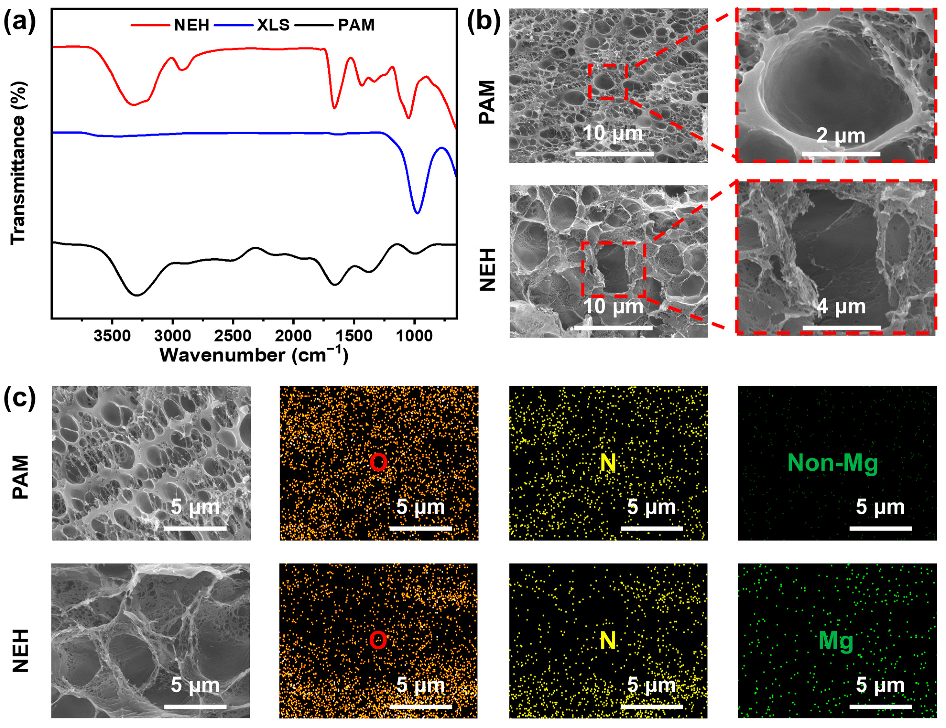
Abstract
:1. Introduction
2. Results and Discussion
2.1. Basic Characterization of NEH
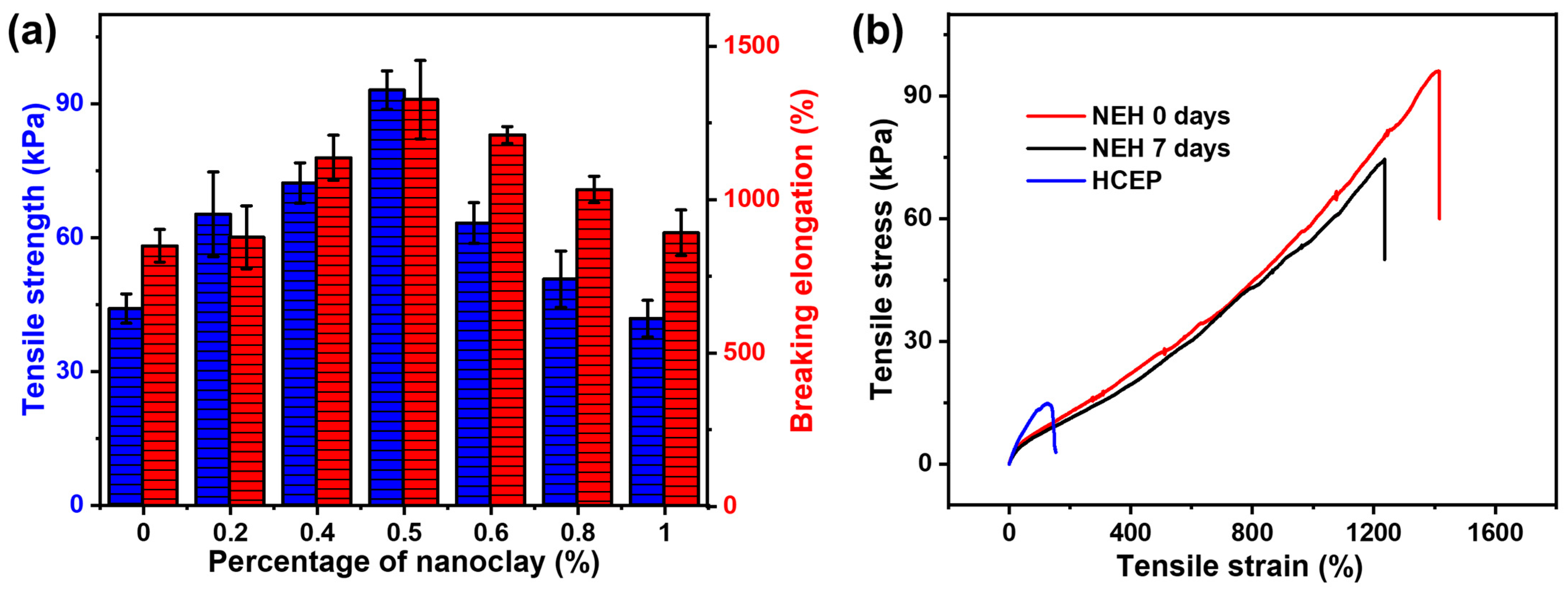
2.2. Mechanical Properties of NEH
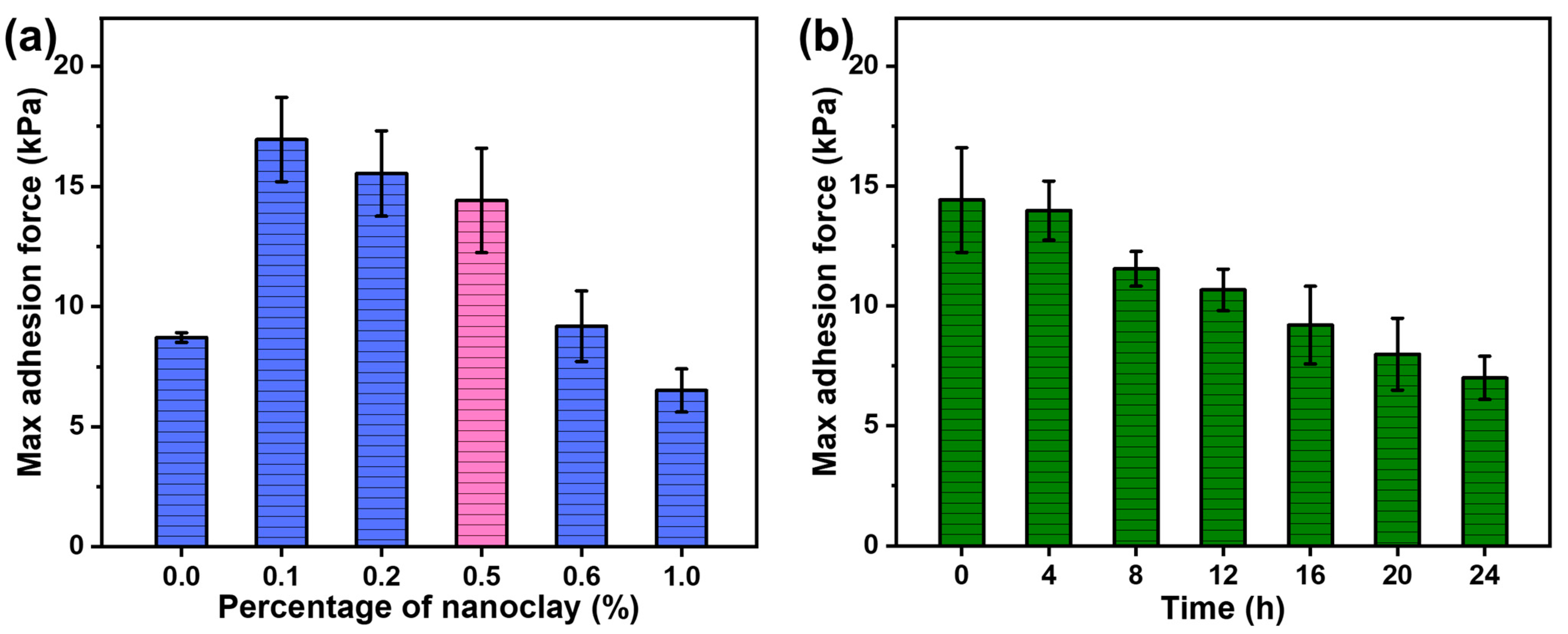
2.3. Adhesive Performance of NEH
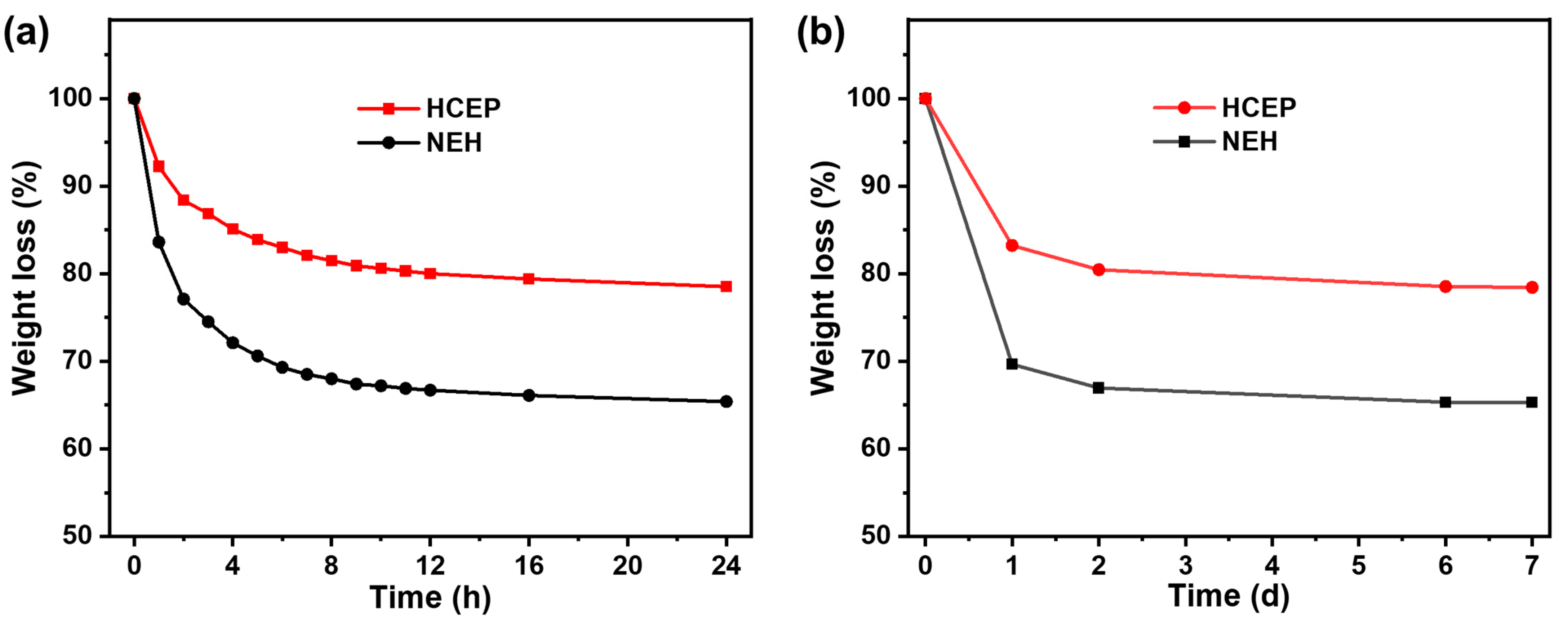
2.4. Water Retention Performance of NEH
2.5. Conductivity of NEH
2.6. Measurement of Impedance
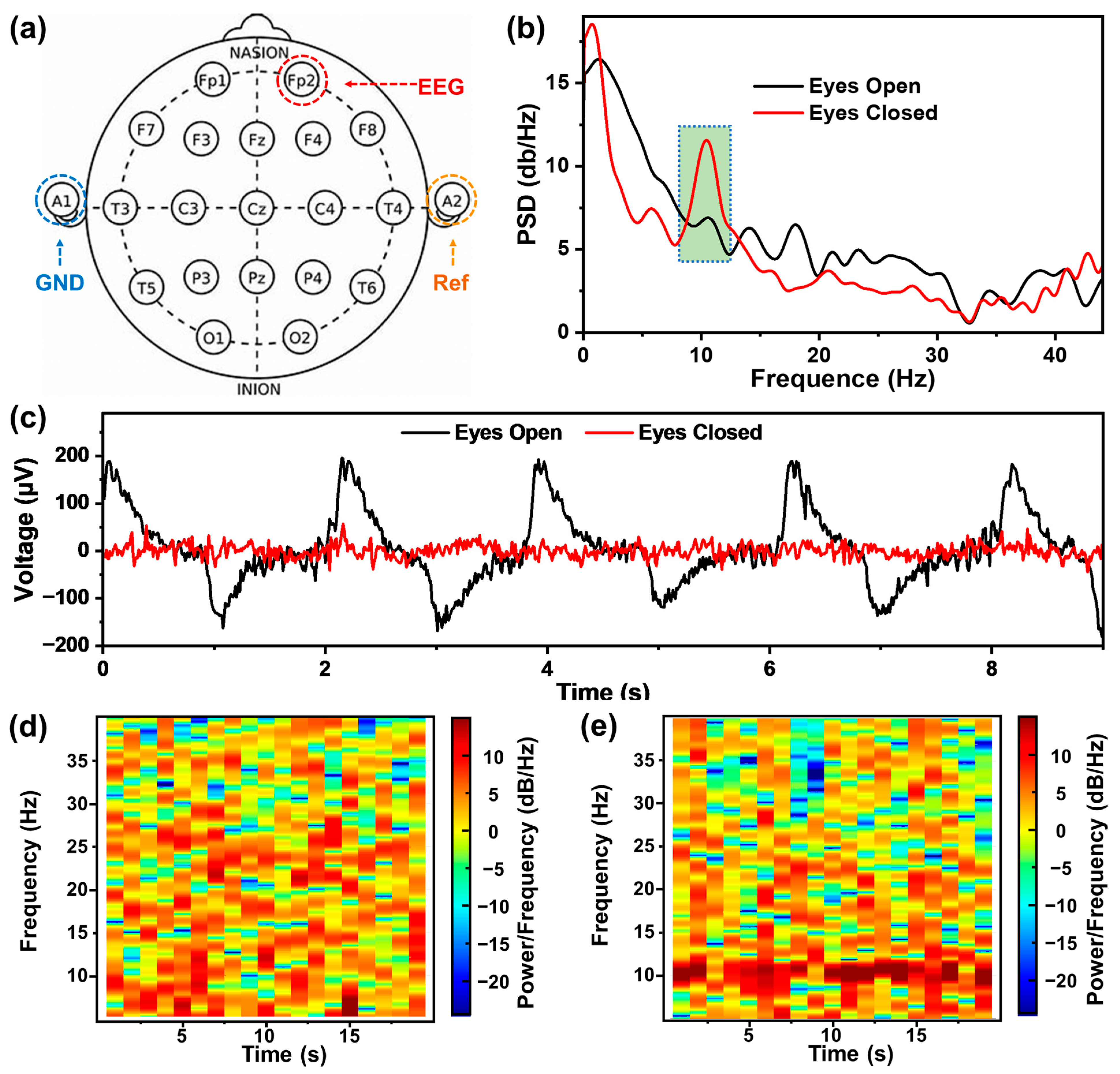
2.7. EEG Recording by the NEH Self-Adhesive Electrodes
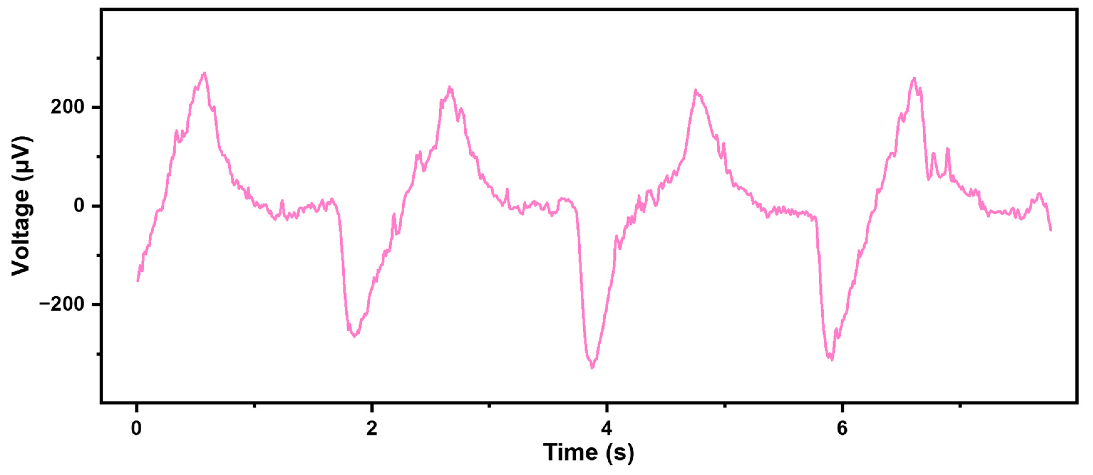
2.8. Signal Stability of Time and Motion by the NEH Self-Adhesive Electrode
3. Conclusions
4. Materials and Methods
4.1. Materials
4.2. Characterization
4.3. Experimental Methods
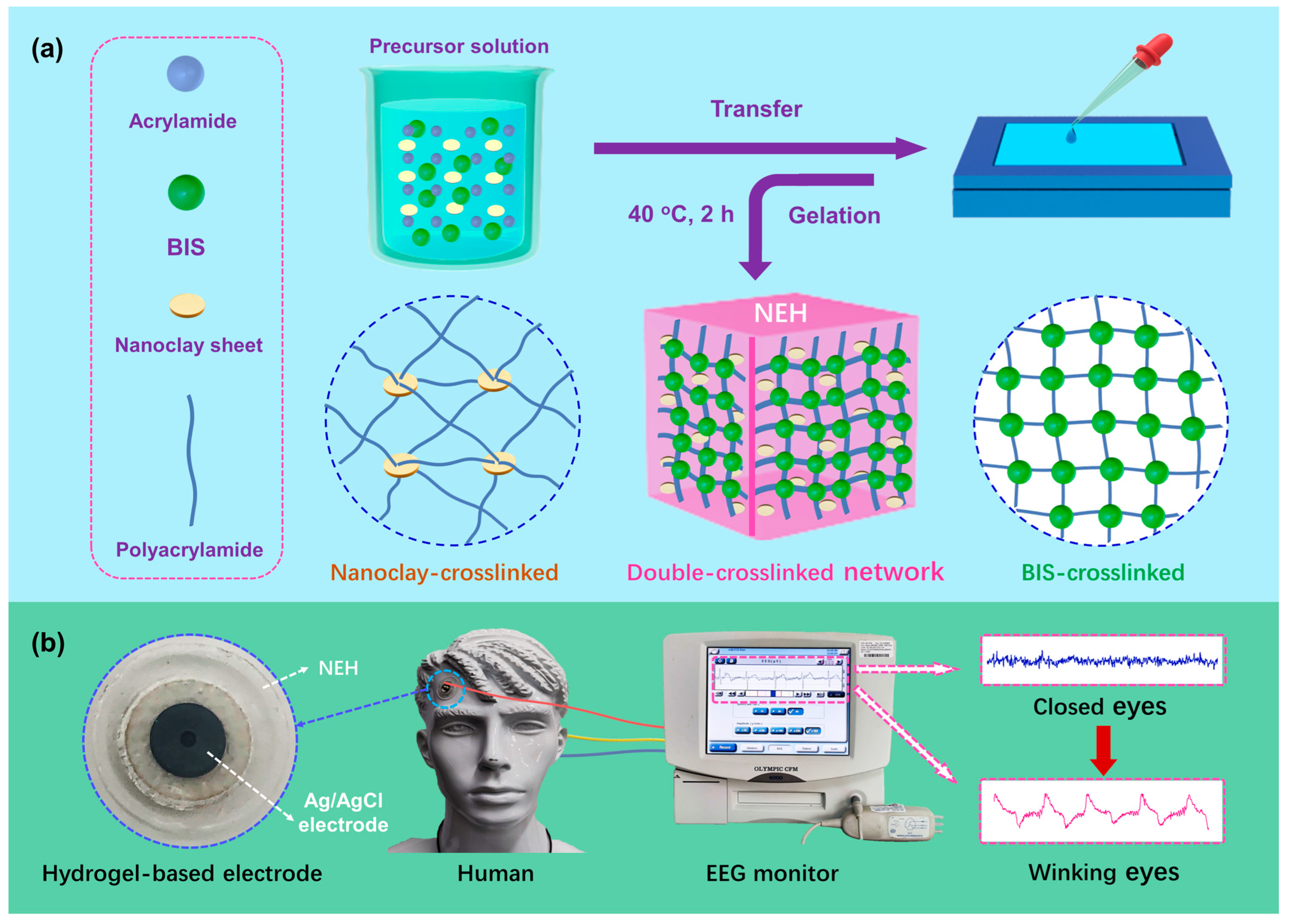
4.3.1. Preparation of the NEH
4.3.2. Mechanical Performance
4.3.3. Light Transmittance Property
4.3.4. Adhesive Performance
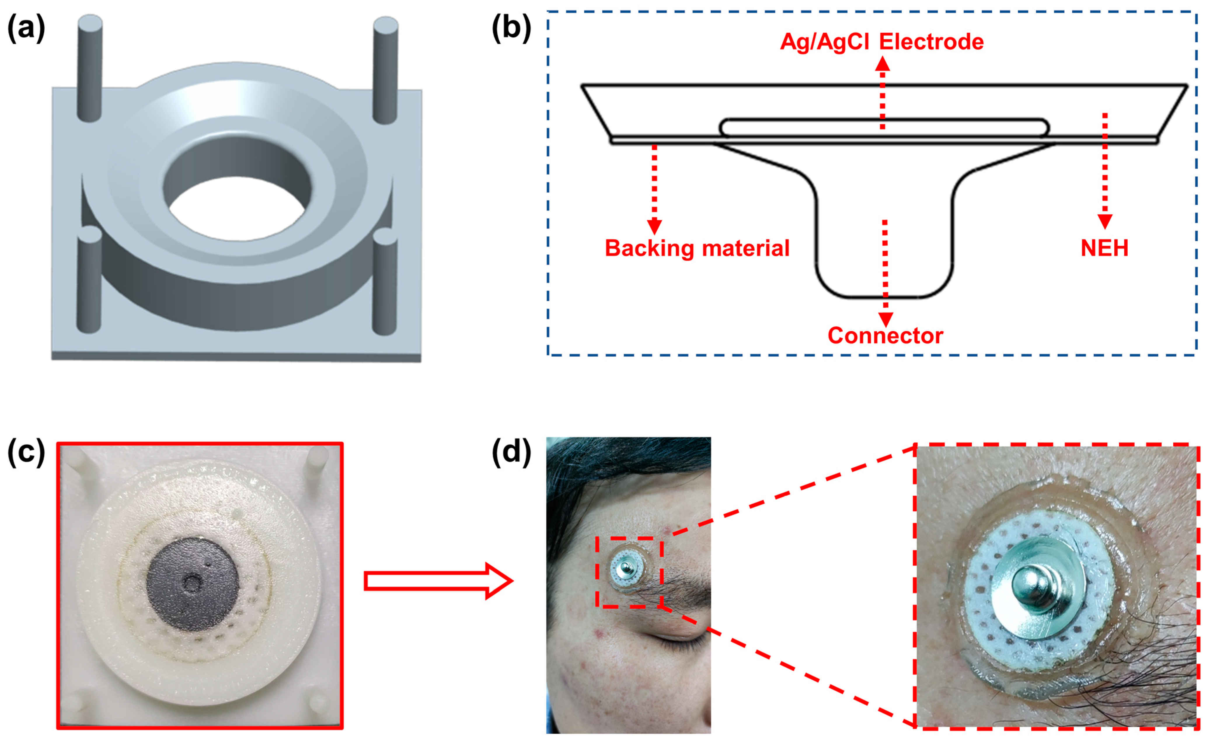
4.3.5. Preparation of the NEH Self-Adhesive Electrode
4.3.6. Measurement for Conductivity
4.3.7. Measurement for Impedance
Supplementary Materials
Author Contributions
Funding
Institutional Review Board Statement
Informed Consent Statement
Data Availability Statement
Conflicts of Interest
References
- Thymi, M.; Lobbezoo, F.; Aarab, G.; Ahlberg, J.; Baba, K.; Carra, M.C.; Gallo, L.M.; De Laat, A.; Manfredini, D.; Lavigne, G.; et al. Signal acquisition and analysis of ambulatory electromyographic recordings for the assessment of sleep bruxism: A scoping review. J. Oral Rehabil. 2021, 48, 846–871. [Google Scholar] [CrossRef]
- Ikeda, T. Current Use and Future Needs of Noninvasive Ambulatory Electrocardiogram Monitoring. Intern. Med. 2021, 60, 9–14. [Google Scholar] [CrossRef]
- Gonzalez Otarula, K.A.; Balaguera, P.; Schuele, S. Ambulatory EEG to Classify the Epilepsy Syndrome. J. Clin. Neurophysiol. 2021, 38, 87–91. [Google Scholar] [CrossRef]
- McDevitt, W.M.; Gul, T.; Jones, T.J.; Scholefield, B.R.; Seri, S.; Drury, N.E. Perioperative electroencephalography in cardiac surgery with hypothermic circulatory arrest: A narrative review. Interact. Cardiovasc. Thorac. Surg. 2022, 35, ivac198. [Google Scholar] [CrossRef] [PubMed]
- Liu, Q.; Yang, L.; Zhang, Z.; Yang, H.; Zhang, Y.; Wu, J. The Feature, Performance, and Prospect of Advanced Electrodes for Electroencephalogram. Biosensors 2023, 13, 101. [Google Scholar] [CrossRef] [PubMed]
- Yuan, H.; Li, Y.; Yang, J.; Li, H.; Yang, Q.; Guo, C.; Zhu, S.; Shu, X. State of the Art of Non-Invasive Electrode Materials for Brain-Computer Interface. Micromachines 2021, 12, 1521. [Google Scholar] [CrossRef]
- Li, G.L.; Wu, J.T.; Xia, Y.H.; He, Q.G.; Jin, H.G. Review of semi-dry electrodes for EEG recording. J. Neural Eng. 2020, 17, 26. [Google Scholar] [CrossRef] [PubMed]
- Teplan, M. Fundamentals of EEG measurement. Meas. Sci. Rev. 2002, 2, 1–11. [Google Scholar]
- Searle, A.; Kirkup, L. A direct comparison of wet, dry and insulating bioelectric recording electrodes. Physiol. Meas. 2000, 21, 271–283. [Google Scholar]
- Yang, L.; Liu, Q.; Zhang, Z.; Gan, L.; Zhang, Y.; Wu, J. Materials for Dry Electrodes for the Electroencephalography: Advances, Challenges, Perspectives. Adv. Mater. Technol. 2021, 7, 2100612. [Google Scholar] [CrossRef]
- Wang, Y.; Pei, W.; Guo, K.; Gui, Q.; Li, X.; Chen, H.; Yang, J. Dry electrode for the measurement of biopotential signals. Sci. China Inf. Sci. 2011, 54, 2435–2442. [Google Scholar] [CrossRef] [Green Version]
- Yang, L.; Gan, L.; Zhang, Z.; Zhang, Z.; Yang, H.; Zhang, Y.; Wu, J. Insight into the Contact Impedance between the Electrode and the Skin Surface for Electrophysical Recordings. ACS Omega 2022, 7, 13906–13912. [Google Scholar] [CrossRef]
- Dai, R.R.; Zhou, H.; Huang, W.; Li, C.Y.; Qin, C.; Liu, X.M.; Pan, Z.F. Conductive Hydrogel-Based Electronics for Intelligent Sensing and Smart Controlling. J. Nanoelectron. Optoelectron. 2021, 16, 689–698. [Google Scholar] [CrossRef]
- Ma, P.Q.; Chen, Y.; Lai, X.Y.; Zheng, J.; Ye, E.Y.; Loh, X.J.; Zhao, Y.; Parikh, B.H.; Su, X.Y.; You, M.L.; et al. The Translational Application of Hydrogel for Organoid Technology: Challenges and Future Perspectives. Macromol. Biosci. 2021, 21, 2100191. [Google Scholar] [CrossRef]
- Wang, H.N.; Xu, Z.J.; Zhao, M.; Liu, G.T.; Wu, J. Advances of hydrogel dressings in diabetic wounds. Biomater. Sci. 2021, 9, 1530–1546. [Google Scholar] [CrossRef]
- Lan, L.; Li, F.; Li, W.; Chen, R.; Xiong, Z.; He, Y.; Ouedraogo, N.A.N.; Ai, B.; Tao, L.; Sun, K.; et al. Highly Skin-Compliant Polymeric Electrodes with Synergistically Boosted Conductivity toward Wearable Health Monitoring. ACS Appl. Mater. Interfaces 2022, 14, 20113–20121. [Google Scholar] [CrossRef] [PubMed]
- Wang, C.; Wang, H.; Wang, B.; Miyata, H.; Wang, Y.; Nayeem, M.O.G.; Kim, J.J.; Lee, S.; Yokota, T.; Onodera, H.; et al. On-skin paintable biogel for long-term high-fidelity electroencephalogram recording. Sci. Adv. 2022, 8, eabo1396. [Google Scholar] [CrossRef] [PubMed]
- Aguzin, A.; Luque, G.C.; Ronco, L.I.; Del Agua, I.; Guzman-Gonzalez, G.; Marchiori, B.; Gugliotta, A.; Tome, L.C.; Gugliotta, L.M.; Mecerreyes, D.; et al. Gelatin and Tannic Acid Based Iongels for Muscle Activity Recording and Stimulation Electrodes. ACS Biomater. Sci. Eng. 2022, 8, 2598–2609. [Google Scholar] [CrossRef]
- Hsieh, J.C.; Li, Y.; Wang, H.; Perz, M.; Tang, Q.; Tang, K.W.K.; Pyatnitskiy, I.; Reyes, R.; Ding, H.; Wang, H. Design of hydrogel-based wearable EEG electrodes for medical applications. J. Mater. Chem. B 2022, 10, 7260–7280. [Google Scholar] [CrossRef]
- Hsieh, J.C.; Alawieh, H.; Li, Y.; Iwane, F.; Zhao, L.; Anderson, R.; Abdullah, S.I.; Kevin Tang, K.W.; Wang, W.; Pyatnitskiy, I.; et al. A highly stable electrode with low electrode-skin impedance for wearable brain-computer interface. Biosens. Bioelectron. 2022, 218, 114756. [Google Scholar] [CrossRef]
- Sun, C.; Luo, J.; Jia, T.; Hou, C.; Li, Y.; Zhang, Q.; Wang, H. Water-resistant and underwater adhesive ion-conducting gel for motion-robust bioelectric monitoring. Chem. Eng. J. 2022, 431, 134012. [Google Scholar] [CrossRef]
- Luo, J.; Sun, C.; Chang, B.; Jing, Y.; Li, K.; Li, Y.; Zhang, Q.; Wang, H.; Hou, C. MXene-Enabled Self-Adaptive Hydrogel Interface for Active Electroencephalogram Interactions. ACS Nano 2022, 16, 19373–19384. [Google Scholar] [CrossRef]
- Shen, G.; Gao, K.; Zhao, N.; Yi, Z.; Jiang, C.; Yang, B.; Liu, J. A novel flexible hydrogel electrode with a strong moisturizing ability for long-term EEG recording. J. Neural Eng. 2021, 18, 066047. [Google Scholar] [CrossRef] [PubMed]
- Yi yan, L.; Wen bo, C. Analysis and Discus sion on S kin Alle rgy Caused by Dis posable Electroca rdiogram Electrode. Chin. J. Pharmacovigil. 2014, 11, 687–692. [Google Scholar]
- Qiao, F.; Li, Q.; Qi, Z.; Wang, F. Preparation, characterization and properties of polyamide/nanoclay-composites. Polym. Bull. 1997, 1997, 7–15. [Google Scholar]
- Haraguchi, K.; Takehisa, T. Nanocomposite hydrogels: A unique organic-inorganic network structure with extraordinary mechanical, optical, and swelling/de-swelling properties. Adv. Mater. 2002, 14, 1120–1124. [Google Scholar] [CrossRef]
- Xiong, L.J.; Hu, X.B.; Liu, X.X.; Zhen, T. Polymer-Laponite nanocomposite hydrogels with super-elongation. Prog. Chem. 2008, 20, 464–468. [Google Scholar]
- Xu, L.; Gao, S.; Guo, Q.; Wang, C.; Qiao, Y.; Qiu, D. A Solvent-Exchange Strategy to Regulate Noncovalent Interactions for Strong and Antiswelling Hydrogels. Adv. Mater. 2020, 32, e2004579. [Google Scholar] [CrossRef]
- Nie, J.J.; Du, B.Y.; Oppermann, W. Swelling, elasticity, and spatial inhomogeneity of poly(N-isopropylacrylamide)/clay nanocomposite hydrogels. Macromolecules 2005, 38, 5729–5736. [Google Scholar] [CrossRef]
- Haraguchi, K.; Takehisa, T.; Fan, S. Effects of clay content on the properties of nanocomposite hydrogels composed of poly(N-isopropylacrylamide) and clay. Macromolecules 2002, 35, 10162–10171. [Google Scholar] [CrossRef]
- Hammadi, A. Electrical conductance, density, and viscosity in mixtures of alkali-metal halides and glycerol. Int. J. Thermophys. 2004, 25, 89–111. [Google Scholar] [CrossRef]
- Lee, Y.; Yim, S.G.; Lee, G.W.; Kim, S.; Kim, H.S.; Hwang, D.Y.; An, B.S.; Lee, J.H.; Seo, S.; Yang, S.Y. Self-Adherent Biodegradable Gelatin-Based Hydrogel Electrodes for Electrocardiography Monitoring. Sensors 2020, 20, 5737. [Google Scholar] [CrossRef]
- Li, Y.; Yang, D.; Wu, Z.; Gao, F.-L.; Gao, X.-Z.; Zhao, H.-Y.; Li, X.; Yu, Z.-Z. Self-adhesive, self-healing, biocompatible and conductive polyacrylamide nanocomposite hydrogels for reliable strain and pressure sensors. Nano Energy 2023, 109, 108324. [Google Scholar] [CrossRef]
- Zhu, M.; Wang, H.; Li, S.; Liang, X.; Zhang, M.; Dai, X.; Zhang, Y. Flexible Electrodes for In Vivo and In Vitro Electrophysiological Signal Recording. Adv. Healthc. Mater. 2021, 10, e2100646. [Google Scholar] [CrossRef]
- Oostenveld, R.; Praamstra, P. The five percent electrode system for high-resolution EEG and ERP measurements. Clin. Neurophysiol. 2001, 112, 713–719. [Google Scholar]
- Rains, J.C.; Penzien, D.B. Sleep and chronic pain: Challenges to the alpha-EEG sleep pattern as a pain specific sleep anomaly. J. Psychosom. Res. 2003, 54, 77–83. [Google Scholar]
- Li, G.L.; Wu, J.T.; Xia, Y.H.; Wu, Y.Y.; Tian, Y.L.; Liu, J.; Chen, D.C.; He, Q.G. Towards emerging EEG applications: A novel printable flexible Ag/AgCl dry electrode array for robust recording of EEG signals at forehead sites. J. Neural Eng. 2020, 17, 026001. [Google Scholar] [CrossRef]
- Perez-Riera, A.R.; Barbosa-Barros, R.; Daminello-Raimundo, R.; de Abreu, L.C. Main artifacts in electrocardiography. Ann. Noninvasive Electrocardiol. 2018, 23, e12494. [Google Scholar] [CrossRef] [PubMed] [Green Version]
- Littmann, L. Electrocardiographic artifact. J. Electrocardiol. 2021, 64, 23–29. [Google Scholar] [CrossRef]


Disclaimer/Publisher’s Note: The statements, opinions and data contained in all publications are solely those of the individual author(s) and contributor(s) and not of MDPI and/or the editor(s). MDPI and/or the editor(s) disclaim responsibility for any injury to people or property resulting from any ideas, methods, instructions or products referred to in the content. |
© 2023 by the authors. Licensee MDPI, Basel, Switzerland. This article is an open access article distributed under the terms and conditions of the Creative Commons Attribution (CC BY) license (https://creativecommons.org/licenses/by/4.0/).
Share and Cite
Wang, F.; Yang, L.; Sun, Y.; Cai, Y.; Xu, X.; Liu, Z.; Liu, Q.; Zhao, H.; Ma, C.; Liu, J. A Nanoclay-Enhanced Hydrogel for Self-Adhesive Wearable Electrophysiology Electrodes with High Sensitivity and Stability. Gels 2023, 9, 323. https://doi.org/10.3390/gels9040323
Wang F, Yang L, Sun Y, Cai Y, Xu X, Liu Z, Liu Q, Zhao H, Ma C, Liu J. A Nanoclay-Enhanced Hydrogel for Self-Adhesive Wearable Electrophysiology Electrodes with High Sensitivity and Stability. Gels. 2023; 9(4):323. https://doi.org/10.3390/gels9040323
Chicago/Turabian StyleWang, Fushuai, Lang Yang, Ye Sun, Yiming Cai, Xin Xu, Zhenzhong Liu, Qijie Liu, Hongliang Zhao, Chunxin Ma, and Jun Liu. 2023. "A Nanoclay-Enhanced Hydrogel for Self-Adhesive Wearable Electrophysiology Electrodes with High Sensitivity and Stability" Gels 9, no. 4: 323. https://doi.org/10.3390/gels9040323