
Anti-Inflammatory Effect and Toxicological Profile of Pulp Residue from the Caryocar Brasiliense, a Sustainable Raw Material
 , , , ,
Abstract
:1. Introduction
2. Results and Discussion
2.1. Flavonoid Content of EPPR
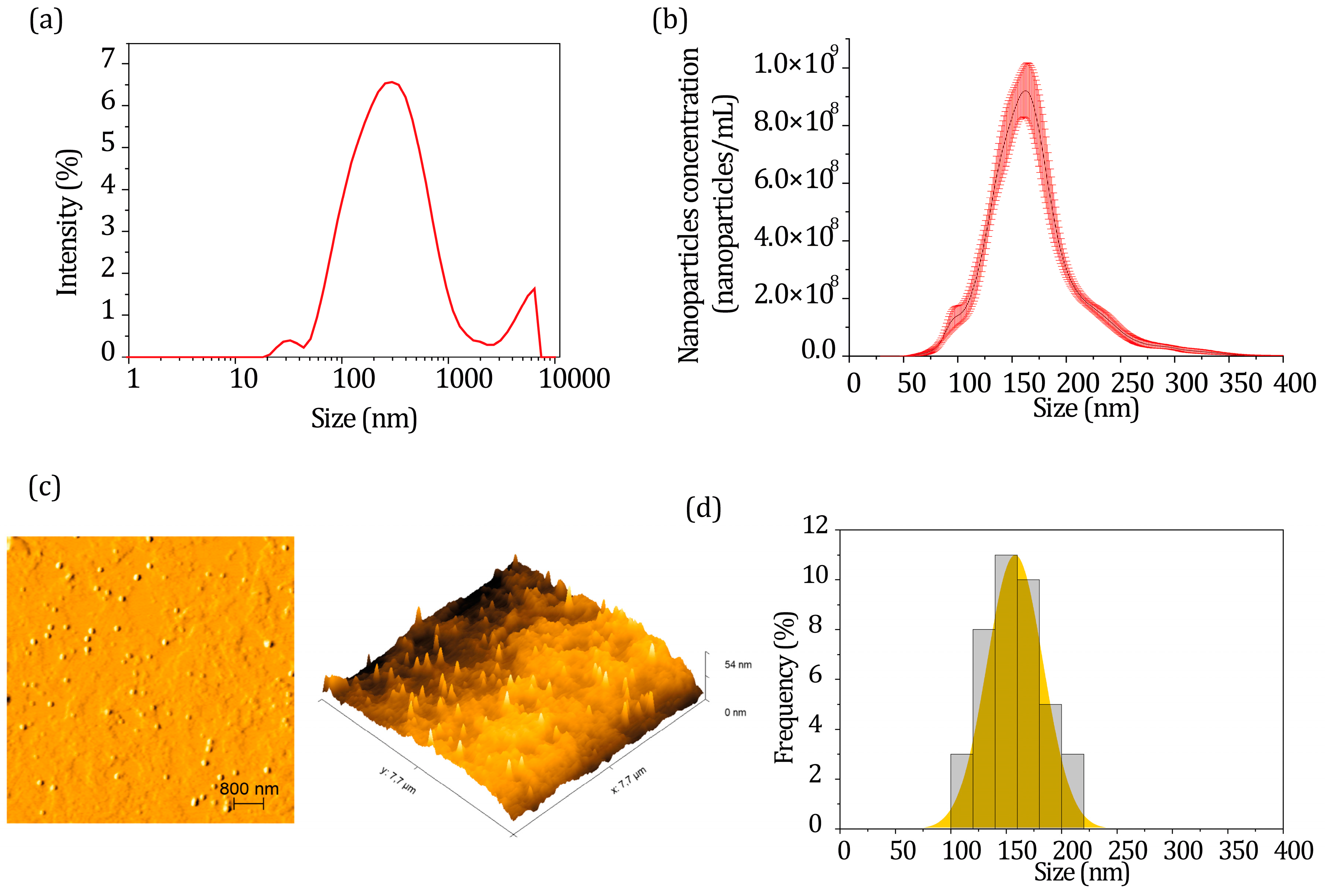
2.2. Characterization of CTS Nanoparticles Containing EPPR
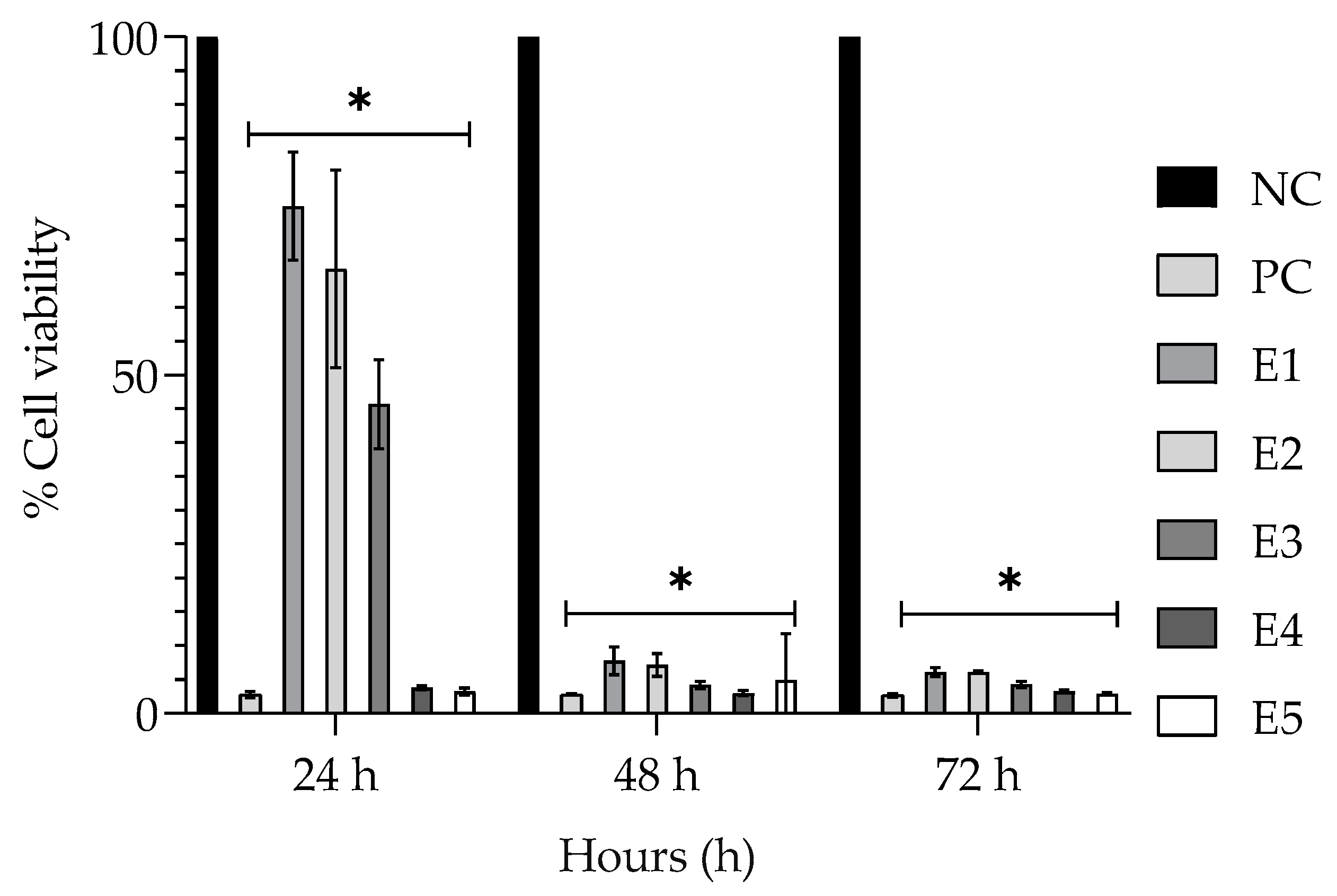
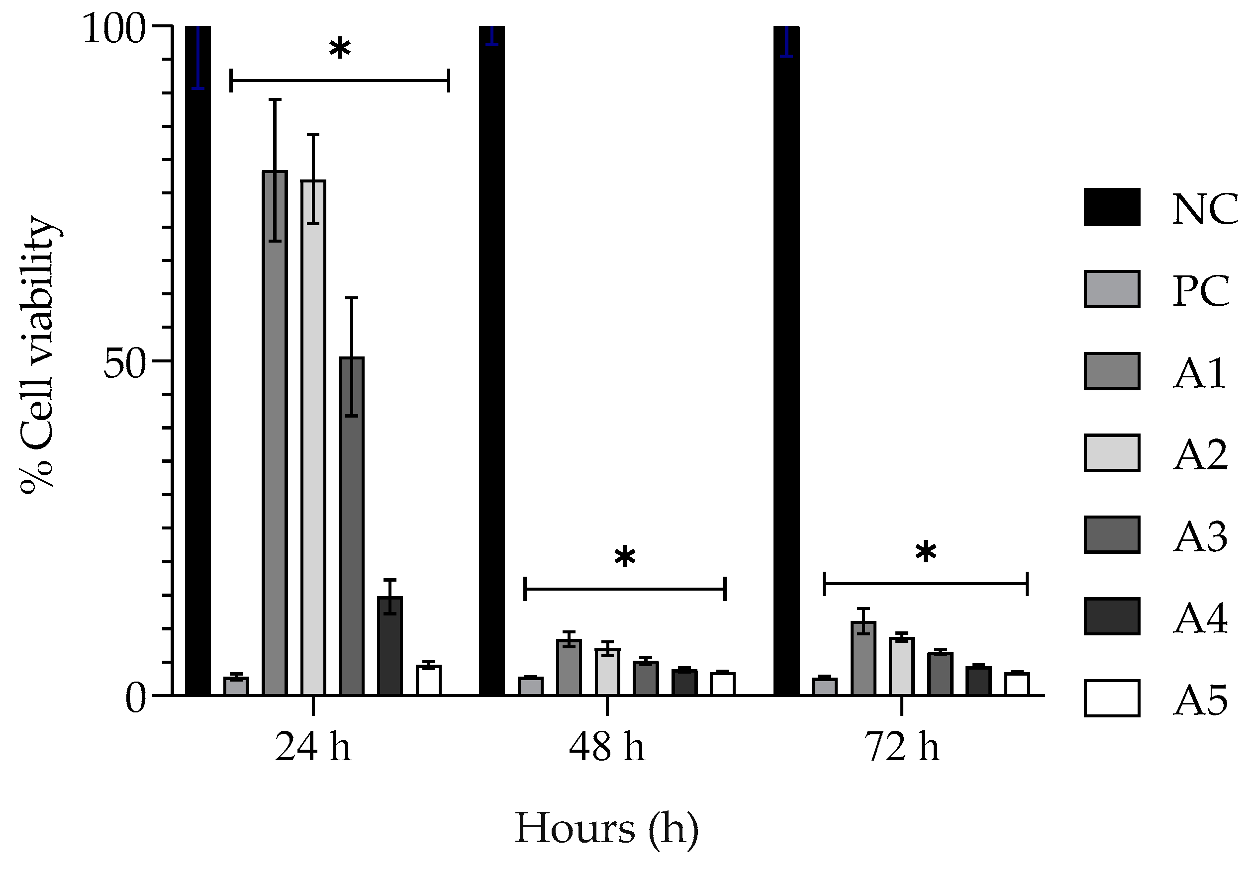
2.3. Determination of Toxicity of the Encapsulated EPPR by the 3-(4,5-dimethylthiazol-2-yl)-2,5-diphenyltetrazolium Bromide (MTT) Assay
2.4. Determination of In Vitro Anti-Inflammatory Activity of Non-Encapsulated Extract (EPPR)
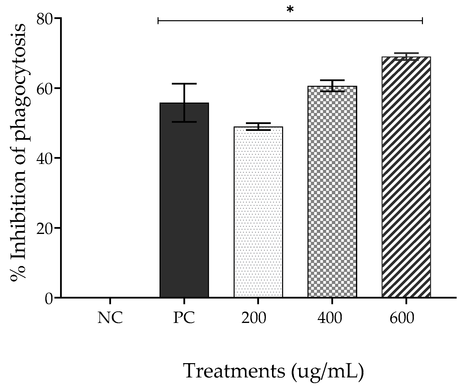
2.4.1. Phagocytosis
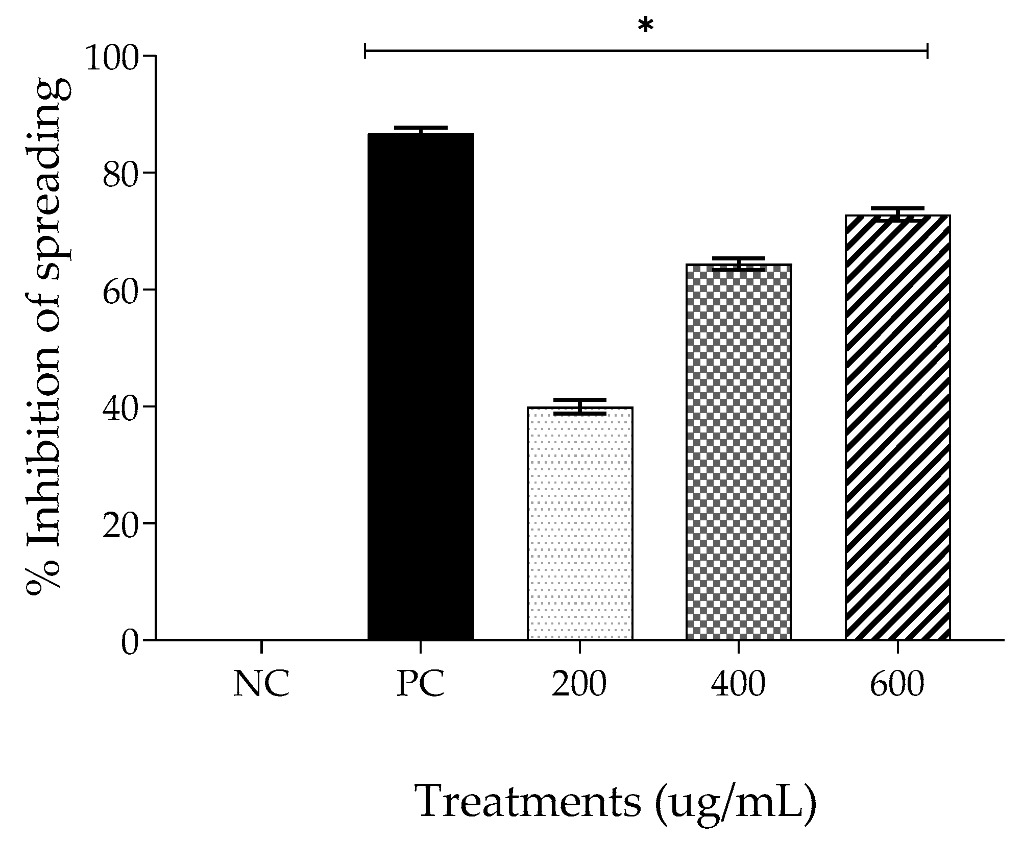
2.4.2. Spreading
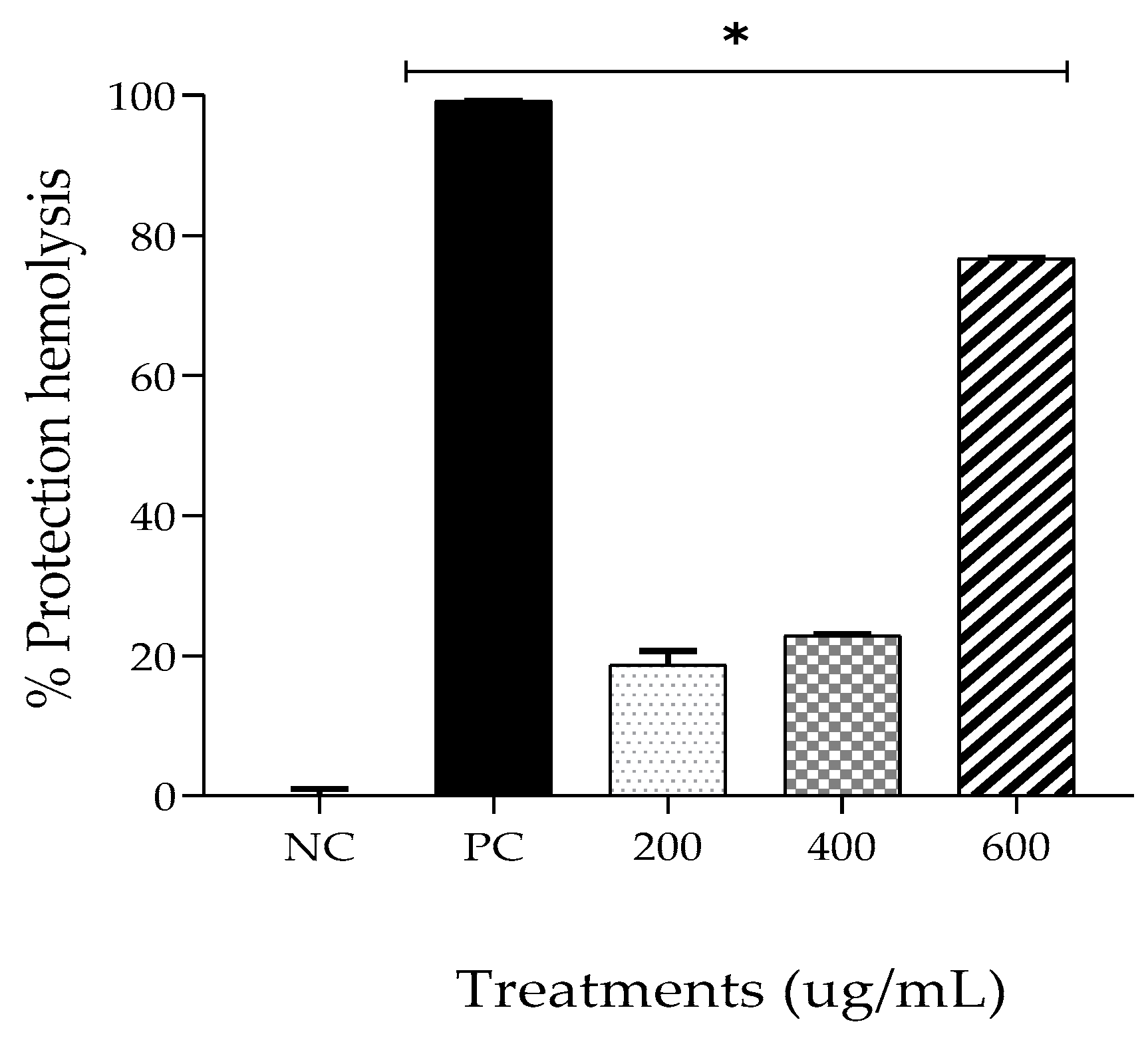
2.4.3. Membrane Stabilization
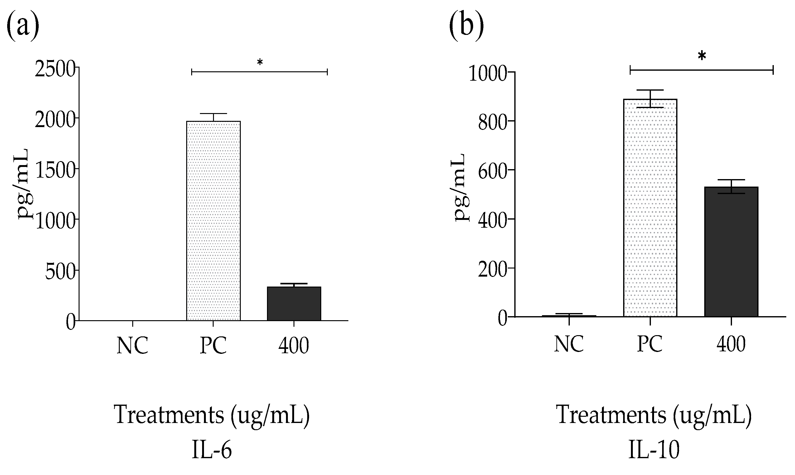
2.5. Quantification of the Levels of Cytokines IL-6 and IL-10 Induced by the Non-Encapsulated Extract (EPPR)
2.6. Acute Toxicity In Vivo of the Non-Encapsulated Extract (EPPR)
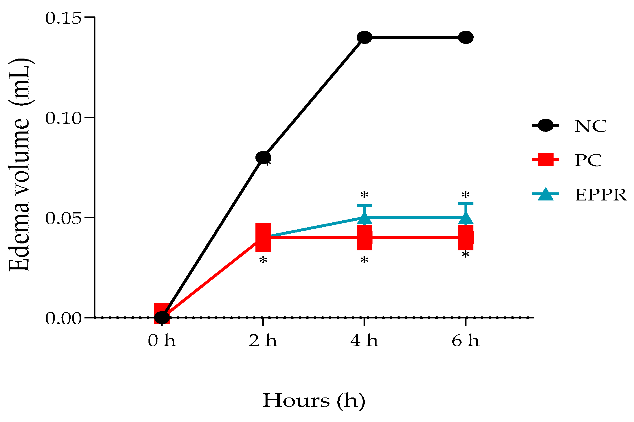
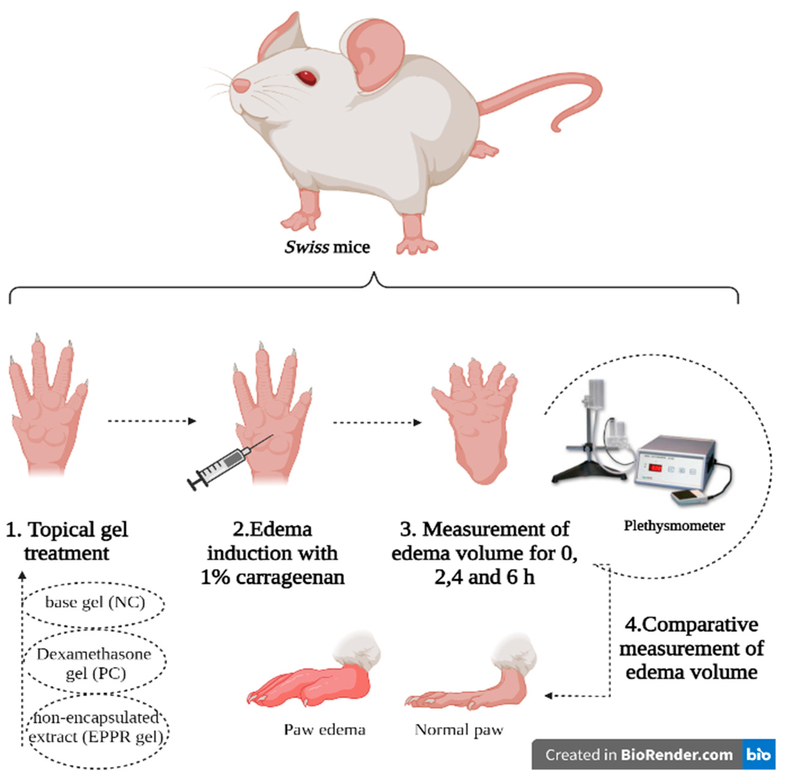
2.7. Determination of the In Vivo Anti-Inflammatory Activity of the Gel Containing the Non-Encapsulated Extract (EPPR gel) on Carrageenan-Induced Paw Edema
2.8. In Vitro Ocular Irritability Test of the Gel Containing the Non-Encapsulated Extract (EPPR gel) in the Chorioallantoic Membrane of Chicken Eggs
2.9. Preliminary Stability Evaluation of the Gel Containing the Non-Encapsulated Extract (EPPR Gel)
3. Conclusions
4. Materials and Methods
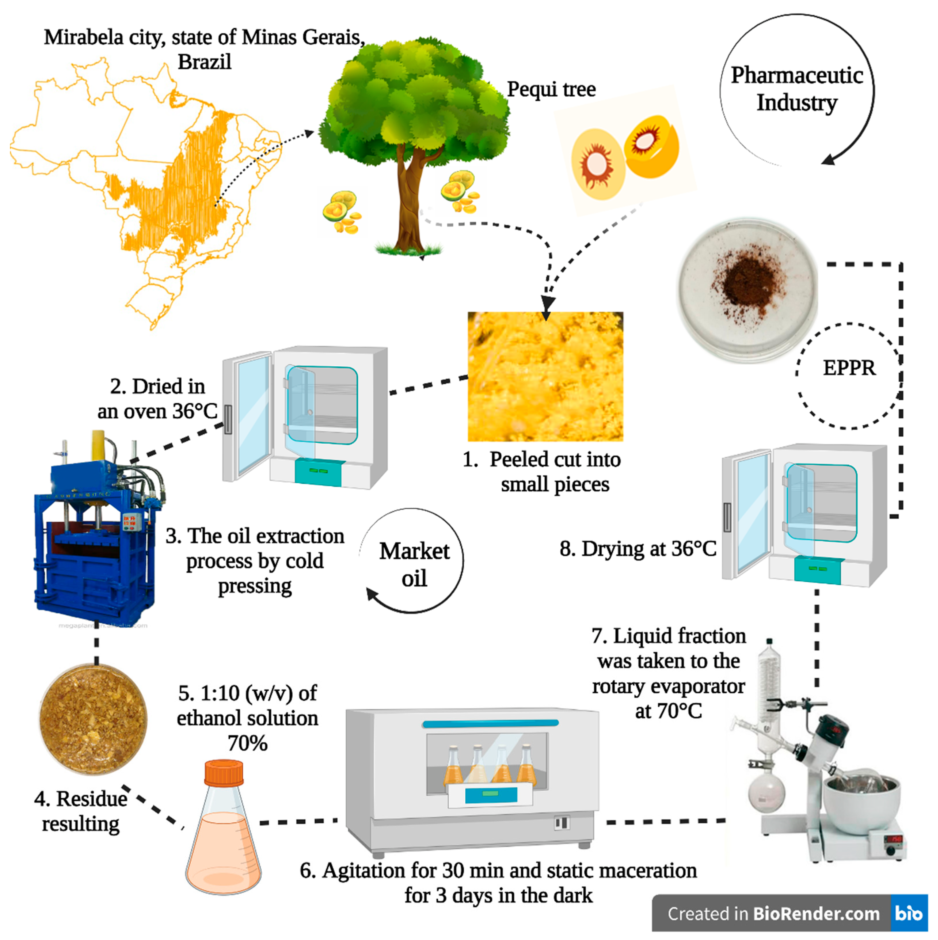
4.1. Material of Vegetable Origin
4.2. Preparation of the Hydroethanolic EPPR
4.3. Flavonoid Content of EPPR
4.4. Encapsulation of EPPR in CTS via Ionic Gelatinization
4.5. Characterization of CTS Nanoparticles Containing EPPR
4.5.1. DLS
4.5.2. Surface Charge
4.5.3. NTA
4.5.4. AFM
4.6. Determination of Toxicity of the Encapsulated EPPR by the MTT Assay
4.7. Determination of In Vitro Anti-Inflammatory Activity of the Non-Encapsulated Extract (EPPR)
4.7.1. Treatments
4.7.2. Cell Culture
4.7.3. Selection of Macrophages
4.7.4. Phagocytosis
4.7.5. Macrophage Spreading
4.7.6. Membrane Stabilization
4.8. Quantification of the Levels of the Cytokines IL-6 and IL-10 Induced by the Non-Encapsulated Extract (EPPR)
4.9. Animals
4.10. Determination of In Vivo Toxicity of the Non-Encapsulated Extract (EPPR)
4.11. Preparation of the Gel Formulation Containing the Non-Encapsulated Extract (EPPR Gel)
4.12. Determination of the In Vivo Anti-Inflammatory Effect of the Gel Containing the Non-Encapsulated Extract (EPPR Gel) on Carrageenan-Induced Paw Edema
4.13. Ex Vivo Ocular Irritability Test of the Gel Containing the Non-Encapsulated Extract (EPPR Gel) in the Chorioallantoic Membrane of Chicken Eggs (MCA)
4.14. Preliminary Stability Evaluation of the Gel Containing the Non-Encapsulated Extract (EPPR Gel)
4.14.1. Accelerated Stability Test or Centrifugation Test
4.14.2. Thermal Stress Test
4.14.3. pH evaluation
4.15. Statistical Analysis
5. Patents
Author Contributions
Funding
Institutional Review Board Statement
Informed Consent Statement
Data Availability Statement
Conflicts of Interest
References
- Sabat, R.; Wolk, K.; Loyal, L.; Döcke, W.-D.; Ghoreschi, K. T Cell Pathology in Skin Inflammation. Semin. Immunopathol. 2019, 41, 359–377. [Google Scholar] [CrossRef] [PubMed] [Green Version]
- Santos, T.F.B. Bioprospecting of endophytic filamentous fungi of medicinal species with potential to produce molecules of biotechnological interest. Master’s Dissertation, Federal University of São Carlos, Sorocaba, Brazil, 2018. [Google Scholar]
- Tanaka, T.; Narazaki, M.; Kishimoto, T. IL-6 in Inflammation, Immunity, and Disease. Cold Spring Harb. Perspect. Biol. 2014, 6, a016295. [Google Scholar] [CrossRef] [PubMed]
- Santos, F.S.; Santos, R.F.; Dias, P.P.; Zanão, L.A.Z., Jr.; Tomassoni, F. The culture of pequi (Caryocar brasiliense Camb.). Acta Iguazu 2013, 2, 46–57. [Google Scholar] [CrossRef]
- Amaral, L.F.B.; Moriel, P.; Foglio, M.A.; Mazzola, P.G. Evaluation of the Cytotoxicity and Phototoxicity of Caryocar Brasiliense Supercritical Carbon Dioxide Extract. BMC Complement. Altern. Med. 2014, 14, 450. [Google Scholar] [CrossRef] [Green Version]
- Bezerra, R.G. Pre-clinical evaluation of the pequi oil (Caryocar coriaceum Wittm.) and emulsion based on it for the treatment of dermatitis: Formulation, characterization and antimicrobial and anti-inflammatory effects. Master’s Dissertation, Ceará’s Federal University, Faculty of Pharmacy Dentistry and Nursing, Fortaleza, Brazil, 2021. [Google Scholar]
- Viroli, S.L.M.; Rodrigues, F.M.; Viroli, S.G.; Carvalho, N.P.; Alves, T.T.; Lança, A.C.; Vivan, J.V.; Ramos, M.L.; Feitosa, K.P.; Veloso, C.; et al. Extraction and Characterization of Oil from the Pulp of Pequi (Caryocar Brasiliense) Produced Manually in the Allotment Piauzinho Municipality of Pium-TO. Res. Soc. Dev. 2022, 11, e49911427711. [Google Scholar] [CrossRef]
- Araújo, A.C.M.A.; Menezes, E.G.T.; Terra, A.W.C.; Dias, B.O.; de Oliveira, É.R.; Queiroz, F. Bioactive Compounds and Chemical Composition of Brazilian Cerrado Fruits’ Wastes: Pequi Almonds, Murici, and Sweet Passionfruit Seeds. Food Sci. Technol. 2018, 38, 203–214. [Google Scholar] [CrossRef] [Green Version]
- Roll, M.M.; Miranda-Vilela, A.L.; Longo, J.P.F.; da Silveira Agostini-Costa, T.; Grisolia, C.K. The Pequi Pulp Oil (Caryocar brasiliense Camb.) Provides Protection against Aging-Related Anemia, Inflammation and Oxidative Stress in Swiss Mice, Especially in Females. Genet. Mol. Biol. 2018, 41, 858–869. [Google Scholar] [CrossRef] [Green Version]
- Cunha, L.M.S.; Pires, R.F.; dos Santos, K.G.; Dantas, S.C. Comparison of yield by different methods of oil extraction from pequi pulp. Res. Soc. Dev. 2020, 9, e342973876. [Google Scholar] [CrossRef]
- Pegorin Brasil, G.S.; Borges, F.A.; de Machado, A.A.; Mayer, C.R.M.; Udulutsch, R.G.; Herculano, R.D.; Funari, C.S.; dos Santos, A.G.; Santos, L. A Sustainable Raw Material for Phytocosmetics: The Pulp Residue from the Caryocar brasiliense Oil Extraction. Rev. Bras. Farmacogn. 2022, 32, 827–833. [Google Scholar] [CrossRef]
- Bonifácio, B.V.; da Silva, P.B.; Ramos, M.A.D.S.; Negri, K.M.S.; Bauab, T.M.; Chorilli, M. Nanotechnology-Based Drug Delivery Systems and Herbal Medicines: A Review. Int. J. Nanomed. 2014, 9, 1–15. [Google Scholar] [CrossRef] [Green Version]
- Lal, H.M.; Uthaman, A.; Thomas, S. Silver Nanoparticle as an Effective Antiviral Agent. In Polymer Nanocomposites Based on Silver Nanoparticles: Synthesis, Characterization and Applications; Lal, H.M., Thomas, S., Li, T., Maria, H.J., Eds.; Engineering Materials; Springer International Publishing: Cham, Switzerland, 2021; pp. 247–265. [Google Scholar]
- Uthaman, A.; Lal, H.M.; Thomas, S. Fundamentals of Silver Nanoparticles and Their Toxicological Aspects. In Polymer Nanocomposites Based on Silver Nanoparticles: Synthesis, Characterization and Applications; Lal, H.M., Thomas, S., Li, T., Maria, H.J., Eds.; Engineering Materials; Springer International Publishing: Cham, Switzerland, 2021; pp. 1–24. ISBN 978-3-030-44259-0. [Google Scholar]
- Barreto, G.P.M.; Benassi, M.T.; Mercadante, A.Z. Bioactive Compounds from Several Tropical Fruits and Correlation by Multivariate Analysis to Free Radical Scavenger Activity. J. Braz. Chem. Soc. 2009, 20, 1856–1861. [Google Scholar] [CrossRef]
- Tungmunnithum, D.; Thongboonyou, A.; Pholboon, A.; Yangsabai, A. Flavonoids and Other Phenolic Compounds from Medicinal Plants for Pharmaceutical and Medical Aspects: An Overview. Medicines 2018, 5, 93. [Google Scholar] [CrossRef]
- Frasao, B.; Costa, M.; Silva, F.; Rodrigues, B.; Baltar, J.; Araujo, J.; Moreira, D.; Torrezan, R.; Conte-Junior, C. Effect of Pequi (Caryocar brasiliense) and Juçara (Euterpe Edulis) Waste Extract on Oxidation Process Stability in Broiler Meat Treated by UV-C. PLoS ONE 2018, 13, e0208306. [Google Scholar] [CrossRef] [Green Version]
- Roesler, R.; Malta, L.G.; Carrasco, L.C.; Holanda, R.B.; Sousa, C.A.S.; Pastore, G.M. Antioxidant activity of cerrado fruits. Food Sci. Technol. 2007, 27, 53–60. [Google Scholar] [CrossRef] [Green Version]
- Magalhães, F.S.; de Souza Martins Sá, M.; Luiz Cardoso, V.; Hespanhol Miranda Reis, M. Recovery of Phenolic Compounds from Pequi (Caryocar brasiliense Camb.) Fruit Extract by Membrane Filtrations: Comparison of Direct and Sequential Processes. J. Food Eng. 2019, 257, 26–33. [Google Scholar] [CrossRef]
- Nascimento-Silva, N.R.R.; Mendes, N.S.R.; Silva, F.A. Nutritional composition and total phenolic compounds content of pequi pulp (Caryocar brasiliense Cambess.). J. Bioenergy Food Sci. 2020, 7, 2812019. [Google Scholar] [CrossRef]
- Ribeiro, D.M.; Fernandes, D.C.; Alves, A.M.; Naves, M.M.V. Carotenoids Are Related to the Colour and Lipid Content of the Pequi (Caryocar Brasiliense Camb.) Pulp from the Brazilian Savanna. Food Sci. Technol. 2014, 34, 507–512. [Google Scholar] [CrossRef] [Green Version]
- Malacrida, C.R.; Moraes, I.C.F.; de Rosso, V.V.; da Costa Rodrigues, C.E.; de Souza, A.C. Effect of the Application of an Enzymatic Pretreatment on Bioactive Compounds of Caryocar brasiliense Camb Pulp Oil. J. Food Process. Preserv. 2018, 42, e13828. [Google Scholar] [CrossRef]
- Bakshi, P.S.; Selvakumar, D.; Kadirvelu, K.; Kumar, N.S. Chitosan as an Environment Friendly Biomaterial—A Review on Recent Modifications and Applications. Int. J. Biol. Macromol. 2020, 150, 1072–1083. [Google Scholar] [CrossRef]
- Coutinho, A.J.; Costa Lima, S.A.; Afonso, C.M.M.; Reis, S. Mucoadhesive and PH Responsive Fucoidan-Chitosan Nanoparticles for the Oral Delivery of Methotrexate. Int. J. Biol. Macromol. 2020, 158, 180–188. [Google Scholar] [CrossRef]
- De Oliveira, T.D.; Riani, L.R.; Costa, M.P.; Fabri, R.L.; Nascimento, J.W.L.; Silva, F.P.; da Silva Filho, A.A.; Tavares, G.D. Baccharis Dracunculifolia Extract-Loaded Chitosan Nanoparticles: Development, Physicochemical Characterization and Cytotoxicity Evaluation. Braz. J. Dev. 2021, 7, 72010–72022. [Google Scholar] [CrossRef]
- Mahmoudi, R.; Tajali Ardakani, M.; Hajipour Verdom, B.; Bagheri, A.; Mohammad-Beigi, H.; Aliakbari, F.; Salehpour, Z.; Alipour, M.; Afrouz, S.; Bardania, H. Chitosan Nanoparticles Containing Physalis Alkekengi-L Extract: Preparation, Optimization and Their Antioxidant Activity. Bull. Mater. Sci. 2019, 42, 131. [Google Scholar] [CrossRef] [Green Version]
- Mondéjar-López, M.; Rubio-Moraga, A.; López-Jimenez, A.J.; García Martínez, J.C.; Ahrazem, O.; Gómez-Gómez, L.; Niza, E. Chitosan Nanoparticles Loaded with Garlic Essential Oil: A New Alternative to Tebuconazole as Seed Dressing Agent. Carbohydr. Polym. 2022, 277, 118815. [Google Scholar] [CrossRef] [PubMed]
- Abosabaa, S.A.; ElMeshad, A.N.; Arafa, M.G. Chitosan Nanocarrier Entrapping Hydrophilic Drugs as Advanced Polymeric System for Dual Pharmaceutical and Cosmeceutical Application: A Comprehensive Analysis Using Box-Behnken Design. Polymers 2021, 13, 677. [Google Scholar] [CrossRef] [PubMed]
- Kaiser, M.; Pereira, S.; Pohl, L.; Ketelhut, S.; Kemper, B.; Gorzelanny, C.; Galla, H.-J.; Moerschbacher, B.M.; Goycoolea, F.M. Chitosan Encapsulation Modulates the Effect of Capsaicin on the Tight Junctions of MDCK Cells. Sci. Rep. 2015, 5, 10048. [Google Scholar] [CrossRef] [PubMed] [Green Version]
- Doughty, D. A Rational Approach to the Use of Topical Antiseptics. J. Wound Ostomy Cont. Nurs. 1994, 21, 224–231. [Google Scholar] [CrossRef]
- Eming, S.A.; Wynn, T.A.; Martin, P. Inflammation and Metabolism in Tissue Repair and Regeneration. Science 2017, 356, 1026–1030. [Google Scholar] [CrossRef] [Green Version]
- Wynn, T.A.; Vannella, K.M. Macrophages in Tissue Repair, Regeneration, and Fibrosis. Immunity 2016, 44, 450–462. [Google Scholar] [CrossRef] [Green Version]
- Barman, P.K.; Koh, T.J. Macrophage Dysregulation and Impaired Skin Wound Healing in Diabetes. Front. Cell Dev. Biol. 2020, 8, 528. [Google Scholar] [CrossRef]
- Athira, K.; Keerthi, T.R. Analyses of Methanol Extracts of Two Marine Sponges, Spongia Officinalis Var. Ceylonensis and Sigmadocia Carnosa from Southwest Coast of India for Their Bioactivities. Int. J. Curr. Microbiol. Appl. Sci. 2016, 5, 722–734. [Google Scholar] [CrossRef]
- Kumar, V.; Bhat, Z.A.; Kumar, D.; Khan, N.; Chashoo, I. Evaluation of Anti-Inflammatory Potential of Leaf Extracts of Skimmia Anquetilia. Asian Pac. J. Trop. Biomed. 2012, 2, 627–630. [Google Scholar] [CrossRef] [Green Version]
- Dias, G.T. Evaluation of In Silico Toxicity and In Vitro Hemolytic, Antioxidant and Antibacterial Activities of the Essential Oil and Microencapsulated Essential Oil forms of Lippia Pedunculosa; Paraíba’s Federal University: João Pessoa, Brazil, 2018. [Google Scholar]
- De Sousa, A.P.; Cordeiro, L.V.; da Silva Souza, H.D.; de Souza, M.D.F.V.; da Silveira, R.D.C.; de Oliveira Filho, A.A. Evaluation of in vitro cytotoxicity and ex-vivo genotoxicity in compounds from Pavonia Glazioviana Gürke (Malvaceae). Rev. Ciências Médicas Biológicas 2022, 21, 53–59. [Google Scholar] [CrossRef]
- De Reis, L.D.O.; Nassif, P.A.N.; Tabushi, F.I.; Milléo, F.Q.; Favero, G.M.; Ariede, B.L.; Reis, C.F.D.D.; Dalabona, B.F. Preliminary analysis of interleukin-6 changes in pre- and postoperative in diabetic patients with bmi < 35 submitted to partial duodenal switch. Arq. Bras. Cir. Dig. 2016, 29, 252–256. [Google Scholar] [CrossRef] [Green Version]
- Grazia Roncarolo, M.; Gregori, S.; Battaglia, M.; Bacchetta, R.; Fleischhauer, K.; Levings, M.K. Interleukin-10-Secreting Type 1 Regulatory T Cells in Rodents and Humans. Immunol. Rev. 2006, 212, 28–50. [Google Scholar] [CrossRef]
- Iyer, S.S.; Cheng, G. Role of Interleukin 10 Transcriptional Regulation in Inflammation and Autoimmune Disease. Crit. Rev. Immunol. 2012, 32, 23–63. [Google Scholar] [CrossRef] [Green Version]
- De Torres, O.L.R.; de Santana, F.C.; Torres-Leal, F.L.; de Melo, I.L.P.; Yoshime, L.T.; Matos-Neto, E.M.; Seelaender, M.C.L.; Araújo, C.M.M.; Cogliati, B.; Mancini-Filho, J. Pequi (Caryocar brasiliense Camb.) Almond Oil Attenuates Carbon Tetrachloride-Induced Acute Hepatic Injury in Rats: Antioxidant and Anti-Inflammatory Effects. Food Chem. Toxicol. 2016, 97, 205–216. [Google Scholar] [CrossRef]
- Guarnier, L.P.; Romão, P.V.M.; Palozi, R.A.C.; Silva, A.O.; Lorençone, B.R.; Marques, A.A.M.; dos Santos, A.C.; Souza, R.I.C.; Souza, K.D.; Lourenço, E.L.B.; et al. Development of a Predictive Model to Induce Atherogenesis and Hepato-Renal Impairment in Female Rats. Biomolecules 2019, 9, 664. [Google Scholar] [CrossRef] [Green Version]
- Di Santo, M.C.; D’ Antoni, C.L.; Domínguez Rubio, A.P.; Alaimo, A.; Pérez, O.E. Chitosan-Tripolyphosphate Nanoparticles Designed to Encapsulate Polyphenolic Compounds for Biomedical and Pharmaceutical Applications—A Review. Biomed. Pharmacother. 2021, 142, 111970. [Google Scholar] [CrossRef]
- Darif, D.; Hammi, I.; Kihel, A.; El Idrissi Saik, I.; Guessous, F.; Akarid, K. The Pro-Inflammatory Cytokines in COVID-19 Pathogenesis: What Goes Wrong? Microb. Pathog. 2021, 153, 104799. [Google Scholar] [CrossRef]
- Diniz, D.M. Anti-Inflammatory Activity of Microemulsion Containing Pequi Oil (Caryocar coriaceum W.); Paraíba’s State University: Campina Grande, Brazil, 2015. [Google Scholar]
- Santos, E.; de Araújo, S.P.; Moraes, C.L.M.; Araújo, S.d.S.; Raesel, G.; Justi, P.N.; Argandonac, E.J.S.; Kassuyac, C. Antiedematogenic activity of Pequi Caryocar brasiliense oil. Exp. Clin. Perspect. Biomed. Innov. Health Educ. (PECIBES) 2015, 1, 1. [Google Scholar]
- Mansur, M.C.P.P.R.; Leitão, S.G.; Cerqueira-Coutinho, C.; Vermelho, A.B.; Silva, R.S.; Presgrave, O.A.F.; Leitão, Á.A.C.; Leitão, G.G.; Ricci-Júnior, E.; Santos, E.P. In Vitro and In Vivo Evaluation of Efficacy and Safety of Photoprotective Formulations Containing Antioxidant Extracts. Rev. Bras. De Farmacogn. 2016, 26, 251–258. [Google Scholar] [CrossRef] [Green Version]
- Santos, L.; Pegorin, S.G.; Machado, A.A.; Dos Santos, A.G.; Funari, C.S.; Ribeiro-Paes, J.T. Method of Obtaining Phytocosmetic in Antioxidant and Photoprotective Gel Cream and Formulation of Phytocosmetic in Antioxidant and Photoprotective Gel Cream Containing Extract of Caryocar brasiliense Camb.-pequi; Depositor: São Paulo State University, Julio Mesquita Filho. Proxy: Renan Padron Almeida. BR 102020023090. Magazine: 12 dez. 2020; São Paulo State University: São Paulo, Brazil, 2021. [Google Scholar]
- Serdar, G.; Sökmen, M.; Demir, E.; Sökmen, A.; Bektaş, E. Extraction of Antioxidative Principles of Achillea Biserrata M. Bieb. and Chromatographic Analyses. Int. J. Second. Metab. 2015, 2, 3–15. [Google Scholar] [CrossRef]
- Calvo, P.; Remuñan-López, C.; Vila-Jato, J.L.; Alonso, M.J. Chitosan and Chitosan/Ethylene Oxide-Propylene Oxide Block Copolymer Nanoparticles as Novel Carriers for Proteins and Vaccines. Pharm. Res. 1997, 14, 1431–1436. [Google Scholar] [CrossRef] [PubMed]
- Tsuboy, M.S.; Marcarini, J.C.; Luiz, R.C.; Barros, I.B.; Ferreira, D.T.; Ribeiro, L.R.; Mantovani, M.S. In Vitro Evaluation of the Genotoxic Activity and Apoptosis Induction of the Extracts of Roots and Leaves from the Medicinal Plant Coccoloba Mollis (Polygonaceae). J. Med. Food 2010, 13, 503–508. [Google Scholar] [CrossRef] [Green Version]
- Azedo, M.R.; Blagitz, M.G.; Souza, F.N.; Benesi, F.J.; Della Libera, A.M.M.P. Functional Evaluation of Monocytes in Cattle Naturally Infected with the Bovine Leucosis Virus. Arq. Bras. Med. Veterinária Zootec. 2011, 63, 1131–1140. [Google Scholar] [CrossRef] [Green Version]
- Bastos, C.R.; Blagitz, M.G.; Souza, F.N.; Batista, C.F.; Stricagnolo, C.R.; Azedo, M.R.; Della Libera, A.M.M.P. Cell viability, phagocytosis and spreading by mononuclear phagocytes and hydrogen peroxide release by leukocytes from healthy and infected bovine mammary glands. Pesqui. Vet. Bras. 2012, 32, 850–854. [Google Scholar] [CrossRef] [Green Version]
- Ananthi, T.; Chitra, M. Screening of in vitro anti-inflammatory activity of Michelia champaca linn. Asian J. Pharm. Clin. Res. 2013, 6, 71–72. [Google Scholar]
- Ethics Committee on the Use of Animals. OECD/OCDE 423 OECD Guideline for Testing of Chemicals Acute Oral Toxicity-Acute Toxic Class Method Introduction; Ethics Committee on the Use of Animals: São José do Rio Preto, Brazil, 2001. [Google Scholar]
- Winter, C.A.; Risley, E.A.; Nuss, G.W. Carrageenin-Induced Edema in Hind Paw of the Rat as an Assay for Antiiflammatory Drugs. Proc. Soc. Exp. Biol. Med. 1962, 111, 544–547. [Google Scholar] [CrossRef]
- Journal Officiel de la Republique Française. Decree of December 27, 1996 Relating to the Methods of Analysis Necessary to Control the Composition of Cosmetic Products. Appendix IV: Official Method for Evaluating the Potential Irritant by Application to the Chorioallantoic Membrane of the Hen’s Egg; Journal Officiel de la Republique Française: Paris, France, 1996; pp. 19137–19138. [Google Scholar]


| Sample | mg QE/g |
|---|---|
| EPPR | 5.88 ± 0.15 |
| General Aspects | Evaluation Time after Administration | ||||
|---|---|---|---|---|---|
| 30 min | 1 h | 2 h | 3 h | 4 h | |
| CNS Hyperexcitability | 0.00 | 0.11 | 0.32 | 0.20 | 0.00 |
| Hypnosis | 0.20 | 0.15 | 0.50 | 0.77 | 0.40 |
| Parameter | Control | EPPR |
|---|---|---|
| Starting weight | 45 ± 10.00 | 39 ± 15.31 |
| Final weight | 51 ± 14.00 | 57 ± 16.44 |
| Treatments (Gels) | % Anti-Inflammatory Activity | |||
|---|---|---|---|---|
| 0 h | 2 h | 4 h | 6 h | |
| NC | 0% | 0% | 0% | 0% |
| PC | 0% | 53.33% * | 70.27% * | 70.27% * |
| EPPR | 0% | 49.95% * | 67.79% * | 67.79% * |
| Treatments | Grade Average | Final Rating |
|---|---|---|
| SS | 0 | Non-Irritant |
| NC | 0 | Non-Irritant |
| PC | 21 | Severe Irritant |
| EPPR | 1 | Mild Irritant |
| Phenomenon | 30 s | 30 and 60 s | 60 and 300 s |
|---|---|---|---|
| Hyperemia | 5 | 3 | 1 |
| Bleeding | 7 | 5 | 3 |
| Coagulation | 9 | 7 | 5 |
Disclaimer/Publisher’s Note: The statements, opinions and data contained in all publications are solely those of the individual author(s) and contributor(s) and not of MDPI and/or the editor(s). MDPI and/or the editor(s) disclaim responsibility for any injury to people or property resulting from any ideas, methods, instructions or products referred to in the content. |
© 2023 by the authors. Licensee MDPI, Basel, Switzerland. This article is an open access article distributed under the terms and conditions of the Creative Commons Attribution (CC BY) license (https://creativecommons.org/licenses/by/4.0/).
Share and Cite
Fracasso, J.A.R.; Ibe, M.B.; da Costa, L.T.S.; Guarnier, L.P.; Viel, A.M.; Brito, G.R.d.; Parron, M.C.; Pereira, A.E.d.S.; Pegorin Brasil, G.S.; Farias Ximenes, V.; et al. Anti-Inflammatory Effect and Toxicological Profile of Pulp Residue from the Caryocar Brasiliense, a Sustainable Raw Material. Gels 2023, 9, 234. https://doi.org/10.3390/gels9030234
Fracasso JAR, Ibe MB, da Costa LTS, Guarnier LP, Viel AM, Brito GRd, Parron MC, Pereira AEdS, Pegorin Brasil GS, Farias Ximenes V, et al. Anti-Inflammatory Effect and Toxicological Profile of Pulp Residue from the Caryocar Brasiliense, a Sustainable Raw Material. Gels. 2023; 9(3):234. https://doi.org/10.3390/gels9030234
Chicago/Turabian StyleFracasso, Julia Amanda Rodrigues, Mariana Bittencourt Ibe, Luísa Taynara Silvério da Costa, Lucas Pires Guarnier, Amanda Martins Viel, Gustavo Reis de Brito, Mariana Conti Parron, Anderson Espírito do Santo Pereira, Giovana Sant’Ana Pegorin Brasil, Valdecir Farias Ximenes, and et al. 2023. "Anti-Inflammatory Effect and Toxicological Profile of Pulp Residue from the Caryocar Brasiliense, a Sustainable Raw Material" Gels 9, no. 3: 234. https://doi.org/10.3390/gels9030234