Article

Diversity and Antimicrobial Activity of Intestinal Fungi from Three Species of Coral Reef Fish

1
University Joint Laboratory of Guangdong Province, Hong Kong and Macao Region on Marine Bioresource Conservation and Exploitation, College of Marine Sciences, South China Agricultural University, Guangzhou 510642, China
2
Guangdong Provincial Key Laboratory of Aquatic Product Processing and Safety, College of Food Science and Technology, Guangdong Ocean University, Zhanjiang 524088, China
3
Technical Center of Xiamen Customs, Xiamen 361026, China
*
Authors to whom correspondence should be addressed.
These authors contributed equally to this work.
J. Fungi 2023, 9(6), 613; https://doi.org/10.3390/jof9060613
Submission received: 30 March 2023 / Revised: 18 May 2023 / Accepted: 25 May 2023 / Published: 26 May 2023

Abstract

:
Although intestinal microbiota play crucial roles in fish digestion and health, little is known about intestinal fungi in fish. This study investigated the intestinal fungal diversity of three coral reef fish (Lates calcarifer, Trachinotus blochii, and Lutjanus argentimaculatus) from the South China Sea using a culturable method. A total of 387 isolates were recovered and identified by sequencing their internal transcribed spacer sequences, belonging to 29 known fungal species. The similarity of fungal communities in the intestines of the three fish verified that the fungal colonization might be influenced by their surrounding environments. Furthermore, the fungal communities in different intestines of some fish were significantly different, and the number of yeasts in the hindgut was less than that in fore- and mid-intestines, suggesting that the distribution of fungi in fishes’ intestines may be related to the physiological functions of various intestinal segments. In addition, 51.4% of tested fungal isolates exhibited antimicrobial activity against at least one marine pathogenic microorganism. Notably, isolate Aureobasidium pullulans SCAU243 exhibited strong antifungal activity against Aspergillus versicolor, and isolate Schizophyllum commune SCAU255 displayed extensive antimicrobial activity against four marine pathogenic microorganisms. This study contributed to our understanding of intestinal fungi in coral reef fish and further increased the library of fungi available for natural bioactive product screening.

1. Introduction

Coral reef ecosystems are famous for their great biodiversity and ecological and economic value [1]. Although representing only 0.2% of the world’s ocean area proportionally, they provide habitats for at least 25% of known marine species [2,3]. In recent decades, a series of natural and anthropogenic disturbances have led to severe degradation of coral reefs [4]. As one of the most important communities in coral reef ecosystems, coral reef fish significantly contribute to ecosystem functioning and reef resilience [5,6]. At the same time, the microbiome, the collection of microorganisms associated with animals, is essential for the optimal growth and survival of the host fish [7,8]. Specifically, it plays a crucial role in host digestion, energy, and health [9,10]. Therefore, the microbial flora residing in the fish intestine has been studied in many species of coral reef fish, which greatly enriches our understanding of the complex interactions that occur between microorganisms residing in the fish intestine and their hosts [11].
Recently, the intestinal bacteria of coral reef fish have been researched intensively, which has revealed variable core flora among the different fish species. For example, in Golden Pompano (Trachinotus ovatus), Proteobacteria, Tenericutes, Spirochaetes, and Firmicutes were the dominant phyla [12]. In Barramundi (Lates calcarifer), Firmicutes was the most abundant and diverse phylum, while at the genus level, Acinetobacter, Citrobacter, Exiguobacterium, and Pseudomonas were the predominant genera [13]. In Mangrove Red Snapper (Lutjanus argentimaculatus), Proteobacteria and Firmicutes were the predominant phyla, and Vibrio, Morganella, Schewanella, Photobacterium, Pantoea, and Bacillus were the major genera [14]. Although the bacterial species in the intestines of different fish are not identical, the phylum Proteus, Firmicutes, and Bacteroides have been found to account for 90% of the studied intestinal microbial communities of fish, indicating that these bacteria may have an important influence and role in the intestinal function of fish. Further, microorganisms in the fish intestine can be roughly divided into two parts: autochthonous (being able to colonize in the gut) and allochthonous (considered to be free-living) [15]. From the host’s point of view, autochthonous bacteria are considered to be more important than allochthonous, as they promote the digestion and absorption of nutrients, stimulate the maturation and function of the immune system, and resist the invasion and infection of pathogens in three aspects [16]. These studies have contributed to our understanding of the species and composition of bacteria in the intestines of different coral reef fish. A recent study reported that the environmental microbiota that live in complex and dynamic seawater might affect the microorganisms in the intestine of fish [17]. Although there is still no definitive explanation for how microbial component taxa aggregate from their regional species pools, a host of possible drivers have been hypothesized, involving physiological host effect habitat filters, stochastic processes of colonization and migration, and different microbial competition processes [18].
As a diversified and widespread group of eukaryotes, fungi are major decomposers of microorganisms [19]. They should be regarded as essential parts of the intestinal microbiota due to their ability to maintain the normal functioning of physiological processes and tissue health and promote the proper progression of the immune system [20,21]. A recent study revealed that the fungal communities were not only related to fish species specificity but also to the respective physiological functions of different intestinal segments [19]. Among fungi, yeasts have been recognized as part of the normal microbiota of fish. Genus Rhodotorula was relatively frequent in both freshwater and marine fish, and the genus Debaryomyces were dominant in rainbow trout [22]. Caruffo et al. reported that many yeasts isolated from the gut of fish could be potential probiotics, reducing the mortality associated with the Vibrio anguillarum challenge [23]. However, the role of yeasts in fish health and nutrition is still not very clear [24]. Although many new methods of molecular biology, such as DNA microarray technology [25], fluorescence in situ hybridization [26], and high-throughput sequencing [27], can comprehensively analyze the species and an abundance of information of intestinal fungi, acquiring pure cultures of fungi is a critical step in the study of microbial function.
Barramundi (L. calcarifer), Golden Permit (T. blochii), and Mangrove Red Snapper (L. argentimaculatus) are three common and carnivorous coral reef fish in the South China Sea. These coral reef fish generally have a complex life history. Spawning and larval settlement occurred in the marine areas of river mouths, and then juvenile fish migrated upstream to fresh water, where they grew and became mature as males [28]. They exhibited a relatively high trophic status in the food chain and played a crucial role in maintaining the ecological balance of coral reef systems. They fed on fish and crustaceans but consumed a variety of algae as well [2].
In this research, we surveyed and compared the diversity and antimicrobial activity of culturable fungi associated with different intestinal segments of the three coral reef fish and their living seawater. Based on the internal transcribed spacer (ITS) sequences of culturable fungi that are associated with the fish intestine and seawater, we analyzed the phylogenetic diversity of these fungi. Furthermore, we also calculated the species and quantity in different intestinal segments separately and compared the dissimilarity of the fungal communities in the fish intestines and their living seawater. The information on fungal diversity and distribution in different intestinal segments of coral reef fish is very scarce and important, which provides the baseline data for gut microbiota in coral reef fish from the South China Sea and a reference for the study on gut microbiota in other marine organisms. In addition, 37 representative fungal isolates were selected for screening their antimicrobial activity against six marine pathogenic microorganisms, which can check the potential of these fungi from different intestinal segments in the production of bioactive molecules against pathogenic microorganisms and provide a good resource for our subsequent screening of marine microbial active substances.

2. Materials and Methods

2.1. Sample Collection and Preparation

Three species of coral reef fish and their surrounding environments (seawater) were collected from Daya Bay (114°32″59 E, 22°40″37 N), Shenzhen, South China, in August 2020. Located in the northeast of the South China Sea, the bay is a closed bay of 650 square kilometers with an annual average temperature of 22 degrees Celsius. The maximum sea surface temperature (SST) is 30 °C, and the minimum temperature is 15 °C [29].
All fish were mature, and males (with a length of 20~30 cm), and obtained by sea fishing and identified by Dr. Xiao Chen (South China Agricultural University, Guangzhou, China) as belonging to L. argentimaculatus (Samples LA1~LA3), T. blochii (Samples TB1~TB3), and L. calcarifer (Samples LA1~LA3), and were categorized as coral reef fish according to their habitat in reference to descriptions from Fish Base (https://www.fishbase.in/search.php (accessed on 18 March 2019), Copenhagen, Denmark) [2]. Three replicates of each fish species were sampled, and a total of nine samples from the three fish species was collected (Samples LA1~LA3, TB1~TB3, and LA1~LA3). Moreover, the phylogenetic tree was constructed to show the evolutionary relationships between the three species of fish based on the sequences of the cytochrome c oxidase subunit I (COI) genes downloaded from the National Center for Biotechnology Information (NCBI, Bethesda, MD, USA). The results demonstrated that L. argentimaculatus is less closely related to T. blochii and L. calcarifer (Figure 1). Three replicate seawater samples around the three fish were collected below the water surface using sterile seawater samplers (wiped with 75% alcohol before use). The depth and temperature of the three sets of seawater sampling sites were 5–10 m and 26 ± 1 °C, respectively [30].
Obtained fish were immediately put into sterile Ziploc plastic bags onboard, placed in a cool box during transportation to the laboratory, and kept at 4 °C before isolation (time from collection to isolation <12 h). Fish intestinal samples and seawater samples were collected and transported to the laboratory as soon as possible.

2.2. Fungal Isolation of Fish Intestines and Seawater

The obtained fish were placed in a dissecting tray, and the body surface of the fish was wiped with 75% alcohol. After each fish was cut in an arc along the anus in an upward direction with dissecting scissors, its intestine was separated and divided into fore-, mid- and hind-intestinal segments with dissecting scissors [31]. Then the mucosa (including intestinal contents) of each intestinal segment was individually collected with sterile dissecting scissors, and finally, 0.5 g of the mucosa was gently scraped into a centrifuge tube [32].
Then, 0.5 mL of each of the three seawater samples was diluted into 4.5 mL of sterile seawater. For each of the 27 fish intestinal samples, 1 mL of sterile water and 0.1 g of intestinal sample were added to a mortar, then an appropriate amount of sterilized quartz sand was added and ground well with a grinding rod (the sand serves to make the sample fully ground), and 0.1 mL of the mixture was taken into the sterile seawater (0.9 mL) and mixed well [2].
Treated seawater samples and fish intestinal samples were diluted with sterile seawater at the ratio of 1:10 and 1:100. Then 100 µL aliquots were plated on six different isolation media (including GPA (1% Glucose, 0.1% peptone, 0.1% K2HPO4, 0.025% MgSO4, 2% agar), GYMA (0.4% Glucose, 0.4% yeast extract, 0.5% malt extract, 2% agar), GPSA (1% Glucose, 0.1% peptone, 1% starch, 0.1% K2HPO4, 0.1% MgSO4, 2% agar), GYPA (0.5% Glucose, 0.1% yeast extract, 0.5% peptone, 2% agar), PDA (20% Potato, 2% glucose, 2% agar), and SYA (1% Starch, 0.5% yeast extract, 2% agar)) with a sterile (sterilized by autoclaving at 121 °C for 30 min) glass spreading rod, and cultured in a constant temperature incubator at 26 °C until the morphology of the isolates could be observed clearly (The approximate days took to see the morphology of isolates was 5–7 days) [30]. Censored isolates were transferred to new PDA (200 g of diced potato, 20 g of glucose, 20 g of agar, and 1 L of seawater) medium individually, and the appearance and quantity of the isolated fungi were recorded and purified gradually until single colonies were obtained.

2.3. Extraction of Fungal Genomic DNA

The extraction of fungal genomic DNA was performed according to a published procedure [33]. Briefly, about 0.5–1.0 g of fresh mycelium of each selected fungal isolate and liquid nitrogen were taken into a pre-cooled mortar and ground thoroughly. The ground mycelium was transferred to a centrifuge tube, and 600 μL of sodium dodecyl sulfate (SDS) buffer was added to it, and the mixture was put in a water bath at 65 °C for 45 min and then centrifuged at 12,000 r/min (revolutions per minute) for 5 min. The supernatant was pipetted and transferred to a new centrifuge tube, and an equal volume of saturated phenol.,: chloroform: isoamyl alcohol with a volume ratio of 25:24:1 was added to the centrifuge tube. The mixture was slowly shaken and centrifuged at 12,000 r/min for 5 min. Then the supernatant was taken to a new centrifuge tube, and an equal volume of isopropanol was added to the centrifuge tube. The centrifuge tube was precipitated at −20 °C for 30 min and centrifuged at 12,000 r/min for 20 min. After that, the supernatant was removed; the DNA pellet was rinsed with 70% alcohol and inverted in the ultra-clean table to make the alcohol evaporate. Then the 20 μL TE buffer (10 mmol/L Tris-HCl, 1 mmol/L EDTA, pH 8.0) was added to the tube to dissolve the DNA, and the integrity of the remaining DNA was detected by electrophoresis and stored at −20 °C.

2.4. PCR Amplification of Target DNA and Sequencing of ITS Fragments

PCR amplification was performed using primers ITS1(5’-TCCGTAGGTGAACCTGCGG-3’) and ITS4 (5’-TCCTCCGCTTATTGATATGC-3’) for the extracted fungal genome [34]. ITS-r DNA amplification was amplified as a 25 μL PCR reaction system: 0.25 (10 μM) μL each for ITS1 and ITS4, 1 μL template DNA, 0.75 μL DMSO, 12.5 μL Taq premix (TakaRa, Beijing, China) and 10.25 μL H2O. Reaction conditions: after pre-denaturation at 95 °C for 5 min, amplification was performed with 30 cycles, denaturation at 94 °C for 45 s, annealing at 50 °C for 45 s, extension at 72 °C for 45 s, and extension at 72 °C for 10 min. PCR amplification products were detected by 1.2% agarose gel electrophoresis. The PCR amplification products were sequenced (Sanger sequencing method) on an ABI 3130 Genetic Analyzer (Applied Biosystems, Foster City, CA, USA) using the ITS1-ITS4 primers and BigDye Terminator V3.1 cycle sequencing kit (Applied Biosystems) [35].

2.5. Sequence Alignment and Phylogenetic Analyses

All sequencing results were analyzed for shearing using Mega 6.0 software, and the sequences were matched with those in the GenBank database using BLAST (a sequence alignment engine) at NCBI. The species name was assigned to the selected isolate when the top three matching BLAST hits were from the same species and were ≥95% similar to the query sequence. All ITS sequences were aligned using Clustal W in MEGA 6.0. Phylogenetic analysis of ITS sequences was carried out using the neighbor-joining method with 1000 bootstrap iterations of MEGA 6.0 software [36].

2.6. Bioassay of Antimicrobial Activity of Fungal Isolates

Six species of marine pathogenic bacteria and fungi were selected to test the antimicrobial activity of fungal isolates obtained from the intestines of three coral fish and their surrounding environments. Three species of marine pathogenic bacteria included Vibrio alginolyticus (UST981130-062, VA), Pseudoaltermonas piscida (UST010723-006, PP), and Micrococcus luteus (UST950701-006, ML) [37]; and three species of marine pathogenic fungi included Aspergillus versicolor (SCSGAF0096, AV), A. sydowii (SCSGAF0035, AS), and Penicillium citrinum (SCSGAF0192, PC) [38].
Selected and tested fungal isolates were grown on PDA plates and incubated for seven days at 26 °C. Agar blocks containing the strains were cut into discs and placed on analysis plates lined with pathogenic microorganisms. Pathogenic bacteria were cultured on LBA (0.5% peptone, 0.3% yeast extract, 2% agar) plates for 18 h at 30 °C, and pathogenic fungi were cultured on PDA (20% potato, 2% glucose, 2% agar) plates for 72 h at 26 °C. The antimicrobial activity of the experimental strains was represented by measuring the diameter of the growth inhibition zone (mm), and each antimicrobial assay was conducted in triplicate.

2.7. Data Analysis

In order to analyze the differences in fungal communities from different intestinal segments of multiple fish species and their surrounding environments, Bray–Curtis dissimilarity was chosen in this study. The Bray–Curtis analysis can show a linear response to the transfer of abundance from a given species in one plot to the same species in another plot in which the species is less abundant. In addition, five other coefficients can show a rather gradual, although nonlinear, change along with the transfer of abundances [39]. Recently, this method has been widely applied in the study of the analysis of differences in microbial communities [31,40]. The species Bray–Curtis coefficient was calculated from the presence (represented by 1)/absence (represented by 0) matrix of the fungi separated from the three fish intestines and the corresponding seawater, using SPSS software for Windows (Version 11.5) [31,40].

2.8. Nucleotide Sequence Accession Number

The ITS sequences of the 29 representative fungal isolates obtained in this experiment were registered in GenBank. Accession numbers are OK275106-OK275134.

3. Results

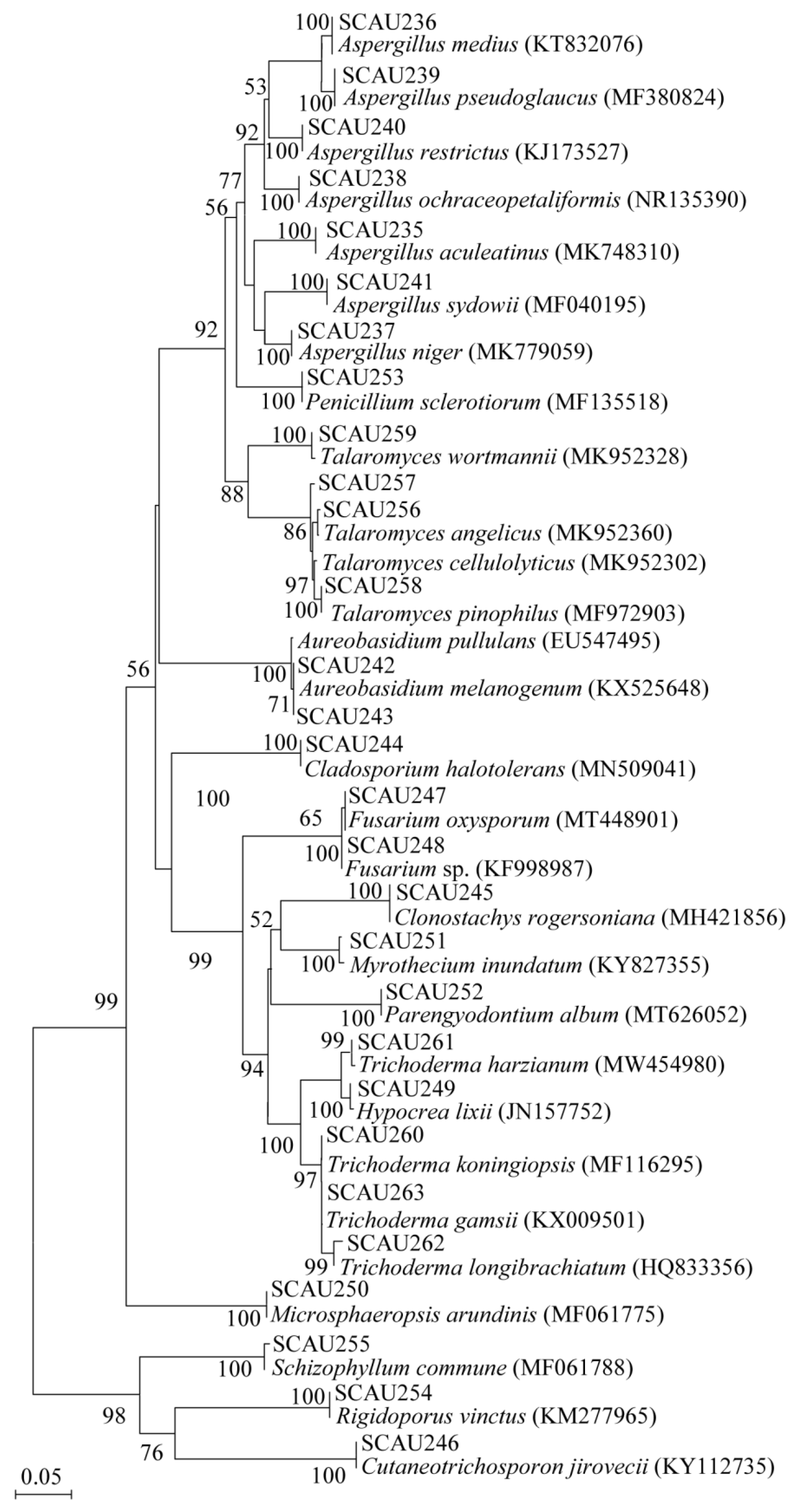
3.1. Isolation and Phylogenetic Diversity of Intestinal Fungi from the Three Coral Reef Fish

A total of 387 fungal isolates were recovered from the three fish intestines (240 isolates) and the seawater (147 isolates) in which the fish lived, based on the size, color, and other morphological observations of the fungus. After being sequenced (ITS sequences) and BLAST searched in GenBank, only one isolate shared 90.40% (low) similarity with its closest NCBI relative (Absidi psychrophilia strain SYM0202, its accession number is JN942684), which was excluded from our further analysis. The remaining 386 isolates shared 98–100% similarity to their closest NCBI relatives (Table 1).
Based on the fungal ITS-rDNA sequences, a phylogenetic tree of 29 different fungal species in 15 known genera is shown in (Figure 2), which was constructed using the neighbor-joining method. The 15 known genera recovered in this study mainly included Aspergillus, Penicillium, Talaromyces, Aureobasidium, Cladosporium, Fusarium, Clonostachys, Myrothecium, Parengyodontium, Trichoderma, Hypocrea, Microsphaeropsi, Schizophyllum, Rigidoporus, and Cutaneotrichosporon.

3.2. Dissimilarity of Fungal Communities in the Intestines of Three Coral Reef Fish and Seawater

The Bray-Curtis analysis showed distinct dissimilarity of the intestinal fungal communities between the three fish, which was from 41.2% to 71.4% (Table 2), indicating that the difference in the intestinal fungal communities between the three fish was obvious. After further analysis of intestinal fungi from the three fish species, it was found that many fungal species, such as Aspergillus niger and Aspergillus pseudoglaucus, were commonly recovered in all three fish species (Table 1). However, there were some distinctive fungal species distributed in different fish. For example, Aspergillus medius, Aspergillus sydowii, Aureobasidium melanogenum, Aureobasidium pullulans, and Cladosporium halotolerans were only isolated from L. calcarifer, while Aspergillus ochraceopetaliformis, Microsphaeropsis arundinis, Myrothecium inundatum, and Rigidoporus vinctus were unique to L. argentimaculatus. Aspergillus restrictus, Cutaneotrichosporon jirovecii, and Penicillium sclerotiorum were recovered only from T. blochii (Table 1).
The Bray–Curtis analysis showed a relatively high dissimilarity of fungal communities between fish intestinal fungi and surrounding seawater fungi, ranging from 66.7% to 81.8% (Table 2). This result indicated that the fungal community in three fish intestines differed from that in the seawater in which fish live.

3.3. Comparison of the Fungal Community in Fore-, Mid- and Hind-Intestinal Segments

Bray–Curtis analysis showed that the dissimilarities of the fungal communities in different segments of the same fish were from 27.3% to 100.0% (Table 3). The fungal isolates from different intestinal segments in the same fish showed distinct dissimilarity in different fish. The dissimilarities between the fore- and mid-intestinal segments of L. calcarifer, L. argentimaculatus, and T. blochii were 27.3%, 60.0%, and 100.0%, respectively. The dissimilarity between the mid- and hind-intestinal segments in L. argentimaculatus was 55.6%, yet the dissimilarity was 100.0% in L. calcarifer and T. blochii. The dissimilarity between fore- and hind-intestinal segments in L. calcarifer, L. argentimaculatus, and T. blochii was 71.4%, 33.3%, and 75.0%, respectively.

3.4. Analysis of the Antimicrobial Activity of the Tested Fungi

Thirty-seven fungal representative isolates (29 species) were tested against the three marine pathogenic fungi and three marine pathogenic bacteria. Nineteen isolates (51.4%) exhibited distinct antimicrobial activity against at least one pathogenic bacterium or fungus (Table 4). It is worth mentioning that isolate A. pullulans SCAU243 exhibited very strong antifungal activity against A. versicolor, while isolate Schizophyllum commune SCAU255 displayed moderate or strong antibacterial activity against four species of marine pathogenic microbial species (Table 4). In addition, A. pseudoglaucus SCAU239-1, F. oxysporum SCAU247-3, and Hypocrea lixii SCAU249 displayed moderate antimicrobial activity against three pathogenic microorganisms; and eight fungi showed antimicrobial activity against two pathogenic microorganisms.

4. Discussion

In this research, a total of 387 fungal isolates were recovered, and only 386 isolates were identified as 29 known fungal species, indicating the presence of relatively diverse and abundant fungal floras in the three coral reef fish intestines and their surrounding environments (seawater). One isolate showed 90.40% similarity with its closest relative, indicating that it may be a new species in the genus Absidi. The identification of the novel candidate still needs further investigation through morphological observation and multigene analysis. The 386 identified isolates belonged to Ascomycota and Basidiomycota (phylum level), while Ascomycota fungi were common both in the three coral reef fish intestines and in seawater (Table 1). These results are consistent with the observations in Tilapia and Bighead Carp intestines, deep oceans, deep-sea sediments, and seawater [19,41,42,43,44].

4.1. Comparison of Fungal Community in Environments and Intestines from the Three Fish

In general, Bray–Curtis analysis showed a distinct dissimilarity in the fungal species in the fish intestines of the three reef fish species (Table 2). Previous studies generally believe that the similarity of intestinal microbial colonization in fish is partly due to the fluctuation in the environmental habitat [18]. Among the environmental habitats, the microbial compositions of the water and diet are the decisive factors [45,46], suggesting that environmental factors were able to influence the composition of the intestinal microbiota of diseased fish, and their experimental results showed that the two most important environmental factors affecting the intestinal microbiota of “red operculum” diseased fish were ammonia concentration and water temperature. Coincidentally, all three fish species are carnivorous and live in the same environment in this study. Therefore, it was hypothesized that the same living environment might cause the three fish to have similar intestinal fungal communities. In addition, the reasons for the similarity of the intestinal fungi of the three fish should include the fact that only 1% of the total microbial community was culturable [47]. On the other hand, although they all live in the same environment in this experiment, and the effects of salinity, temperature, and microbial composition of seawater can be excluded, there is still a certain degree of dissimilarity of environmental conditions between them. Ref. [18] reported that the host could select different fungal species from the environment to colonize in the intestines. Besides surrounding environments, the intestinal fungal communities of the three coral reef fish species in the South China Sea may also be affected by potential factors such as the sex, size, life stages, feeding strategies, and potential prey of these fish species. For example, Li et al. revealed that the difference had occurred in the intestinal microbial communities between male and female wild largemouth bronze gudgeon [48]. Furthermore, [49] found that the intestinal microflora of Atlantic cod Gadus morhua differed depending on whether the fish were fed fish meal, fermented soy protein, or standard soy protein.

4.2. Comparison of Fungal Community in Fore-, Mid- and Hind-Intestinal Segments

The dissimilarity of the fungal isolates in different segments of the same fish was from 27.3% to 100.0% (Table 3), suggesting that the diversity and distribution of the fungal community varied with different intestinal segments in the fish. Different intestinal segments of vertebrates, including fish, show various features, such as digestion and absorption, which lead to different microbial compositions in different intestinal segments [50,51]. For example, microbial communities in the intestines of adult honeybees and wild-caught adult Penaeus monodon differed significantly from the crop to the rectum, and the similarity between communities was significantly reduced, demonstrating a niche partitioning and compartment specificity [52,53]. However, in the present study, significant differences were observed only between the fore- and mid-intestines of L. calcarifer and between the three different intestinal segments of T. blochii. While no remarkable differences were observed for fungal communities in other segments of the intestine, which was comparable to the findings for bighead carp, where there were no remarkable differences in fungi between intestinal segments [19]. Among the intestinal fungi, we found a dramatic decrease in the number of yeasts in the hind-intestine in comparison to the fore- and mid-intestines, in addition to differences in the distribution of Trichoderma spp. and Talaromyces spp. In a survey of tilapia and bighead carp, it was also found that the hind-intestines showed a significant reduction in pathogenicity and saprotrophicity compared with the fore-intestine [19]. These results indicated that the distribution of the fungi in the fish intestine was associated with the physiological functions of various intestinal segments except for the specificity of the host species.

4.3. Potential Antimicrobial Properties of Fungi from Fish Intestine and Seawater

Thirty-seven fungal representatives from the intestine of three coral reef fish and seawater were tested for antimicrobial activity against six pathogenic bacteria and fungi. After initial screening, more than half of the fungal isolates (51.4%) showed significant antimicrobial activities in the experiment, indicating that fungi from the fish intestines or seawater were an excellent source for the extraction of natural products with great bioactivity [54,55]. A. versicolor is a pathogen [38] that can be inhibited by a variety of fungi from fish intestines and seawater in this investigation (Table 4). It is widely distributed in humid indoor environments and causes not only remarkable lethal activity to brine shrimp but also respiratory and pulmonary diseases in humans; moreover, it accounts for 5.8% of the fungi that cause gray nails in recent statistics [56,57]. In this assay, five isolates of experimental fungi demonstrated high inhibitory activity against A. versicolor (Table 4), and the inhibitory activity of A. pullulans SCAU243 far exceeded that of other fungi.
Among the fungal isolates with broad antimicrobial activity, S. commune SCAU255 exhibited extensive and high-intensity antibacterial activity [58]. As a widespread edible fungus with distribution on almost every continent except Antarctica, S. commune produces the neutral extracellular polysaccharide lytic polysaccharide, consisting of a 1,6-β-d-glucosyl side group and a 1,3-β-d-linked glucose residue backbone, with remarkable anticancer and antitumor activity and good immunomodulatory activity, yet it is less frequently found in seawater [59,60]. In previous studies, isolates of S. commune found in marine sediments showed biological activity inconsistent with continental isolates, and it was hypothesized that isolates from marine sources might have unique biological characteristics that assist them in adapting to extreme seafloor environments [61].
For the remaining isolates with antimicrobial activities, except for some species of Talaromyces and Aspergillus that exhibited relatively high antibacterial and antifungal activities, Microsphaeropsis arundinis SCAU250 and Penicillium sclerotiorum SCAU253 only had high antibacterial activities. Interestingly, three fungal isolates of Fusarium oxysporum and two isolates of Aspergillus pseudoglaucus demonstrated different antibacterial and antifungal activities. These findings illustrate that different isolates of F. oxysporum and A. pseudoglaucus may have relatively high variability and differ in morphology, biological activity, and pathogenicity. The reason for this may be attributed to diverse karyotypes among isolates in the same environment [62]. As a fish pathogenic fungus, F. oxysporum has been found in many fish in recent years, and it poses a great danger to fish health [63,64,65]. Different countermeasures, such as different biological activity screening modes, and different application directions, etc., must be taken regarding different isolates.

5. Conclusions

In conclusion, fungal diversity in the intestine of three species of coral reef fish and seawater was investigated using culturable methods and ITS sequences. Both fish intestine and seawater fungi showed high diversity. There were similarities in the intestinal fungal communities between the three fish and seawater, which verified the influence of seawater on the intestinal microbial colonization of fish. In the comparison of the variability of different segments of fish intestinal fungi, significant differences were found only between the fore- and mid-intestines of Lates calcarifer and between the three different intestinal segments of Trachinotus blochii. However, the number of culturable microbes is quite different from the actual number of microbes in the environment and fish intestine. Relying on culture technology alone cannot fully reflect the true situation of marine microbial diversity and may lead to the omission of microbial resources with potential application value, which is a shortcoming of our study. The combination of culturable and high-throughput sequencing will be the direction of future research on fungal diversity.

Author Contributions

Experiment materials were collected by X.L. and J.Y.; data collection and analysis were performed by Z.Z., J.W. and Q.Y. The first draft of the manuscript was written by X.L., J.W. and D.X.; the manuscript was revised by X.Z. and S.Z.; and all authors commented on previous versions of the manuscript. All authors have read and agreed to the published version of the manuscript.

Funding

This work was supported by the Guangdong Natural Science Foundation of China (2023A1515011859) and the Foundation of Guangdong Provincial Key Laboratory of Aquatic Product Processing and Safety, College of Food Science and Technology, Guangdong Ocean University (GDPKLAPPS2202).

Institutional Review Board Statement

This article does not contain any studies with human participants or animals performed by any of the authors.

Informed Consent Statement

Not applicable.

Data Availability Statement

The ITS sequences of the 29 representative fungal isolates obtained in this experiment were registered in GenBank. Accession numbers are OK275106-OK275134.

Acknowledgments

The authors are grateful to Muhammad Amin (College of Pharmacy, Oregon State University, Corvallis, OR, USA) for his kindness in English editing and polishing of the revised manuscript.

Conflicts of Interest

The authors have declared that they have no conflict of interest.

References

  1. Fisher, R.; O’Leary, R.A.; Low-Choy, S.; Mengersen, K.; Knowlton, N.; Brainard, R.E.; Caley, M.J. Species richness on coral reefs and the pursuit of convergent global estimates. Curr. Biol. 2015, 25, 500–505. [Google Scholar] [CrossRef] [PubMed]
  2. Gao, Y.M.; Zou, K.S.; Zhou, L.; Huang, X.D.; Li, Y.Y.; Gao, X.Y.; Chen, X.; Zhang, X.Y. Deep insights into gut microbiota in four carnivorous coral reef fishes from the south china sea. Microorganisms 2020, 8, 426. [Google Scholar] [CrossRef] [PubMed]
  3. Graham, N.A.J.; Nash, K.L. The importance of structural complexity in coral reef ecosystems. Coral Reefs 2013, 32, 315–326. [Google Scholar] [CrossRef]
  4. Pandolfi, J.M.; Connolly, S.R.; Marshall, D.J.; Cohen, A.L. Projecting coral reef futures under global warming and ocean acidification. Science 2011, 333, 418–422. [Google Scholar] [CrossRef]
  5. Brandl, S.J.; Casey, J.M.; Meyer, C.P. Dietary and habitat niche partitioning in congeneric cryptobenthic reef fish species. Coral Reefs 2020, 39, 305–317. [Google Scholar] [CrossRef]
  6. Kane, C.N.; Tissot, B.N. Trophic designation and live coral cover predict changes in reef-fish community structure along a shallow to mesophotic gradient in hawaii. Coral Reefs 2017, 36, 891–901. [Google Scholar] [CrossRef]
  7. Ray, A.K.; Ghosh, K.; Ringø, E. Enzyme-producing bacteria isolated from fish gut: A review. Aquac. Nutr. 2012, 18, 465–492. [Google Scholar] [CrossRef]
  8. Reinoso, S.; Gutierrez, M.S.; Dominguez-Borbor, C.; Arguello-Guevara, W.; Bohorquez-Cruz, M.; Sonnenholzner, S.; Nova-Baza, D.; Mardones, C.; Navarrete, P. Selection of autochthonous yeasts isolated from the intestinal tracts of cobia fish (Rachycentron canadum) with probiotic potential. J. Fungi 2023, 9, 274. [Google Scholar] [CrossRef]
  9. Lynch, S.V.; Pedersen, O. The human intestinal microbiome in health and disease. New Engl. J. Med. 2016, 375, 2369–2379. [Google Scholar] [CrossRef]
  10. Tran, N.T.; Zhang, J.; Xiong, F.; Wang, G.T.; Li, W.X.; Wu, S.G. Altered gut microbiota associated with intestinal disease in grass carp (Ctenopharyngodon idellus). World J. Microbiol. Biotechnol. 2018, 34, 71. [Google Scholar] [CrossRef]
  11. Talwar, C.; Nagar, S.; Lal, R.; Negi, R.K. Fish gut microbiome: Current approaches and future perspectives. Indian J. Microbiol. 2018, 58, 397–414. [Google Scholar] [CrossRef] [PubMed]
  12. You, C.; Chen, B.; Wang, M.; Wang, S.; Zhang, M.; Sun, Z.; Juventus, A.J.; Ma, H.; Li, Y. Effects of dietary lipid sources on the intestinal microbiome and health of golden pompano (Trachinotus ovatus). Fish Shellfish. Immunol. 2019, 89, 187–197. [Google Scholar] [CrossRef] [PubMed]
  13. Zheng, X.; Yang, R.; Hu, J.; Lin, S.; Gu, Z.; Ma, Z. The gut microbiota community and antioxidant enzymes activity of barramundi reared at seawater and freshwater. Fish Shellfish Immunol. 2019, 89, 127–131. [Google Scholar] [CrossRef] [PubMed]
  14. Reshma, K.J.; Sumithra, T.G.; Nair, A.V.; Stefi Raju, V.; Kishor, T.G.; Sreenath, K.R.; Sanil, N.K. An insight into the gut microbiology of wild-caught mangrove red snapper, Lutjanus argentimaculatus (forsskal, 1775). Aquaculture 2018, 497, 320–330. [Google Scholar] [CrossRef]
  15. Banerjee, G.; Ray, A.K. Bacterial symbiosis in the fish gut and its role in health and metabolism. Symbiosis 2017, 72, 1–11. [Google Scholar] [CrossRef]
  16. Nie, L.; Zhou, Q.J.; Qiao, Y.; Chen, J. Interplay between the gut microbiota and immune responses of ayu (plecoglossus altivelis) during vibrio anguillarum infection. Fish Shellfish. Immunol. 2017, 68, 479–487. [Google Scholar] [CrossRef]
  17. Perez, T.; Balcazar, J.L.; Ruiz-Zarzuela, I.; Halaihel, N.; Vendrell, D.; de Blas, I.; Muzquiz, J.L. Host-microbiota interactions within the fish intestinal ecosystem. Mucosal Immunol. 2010, 3, 355–360. [Google Scholar] [CrossRef]
  18. Schmidt, V.T.; Smith, K.F.; Melvin, D.W.; Amaral-Zettler, L.A. Community assembly of a euryhaline fish microbiome during salinity acclimation. Mol. Ecol. 2015, 24, 2537–2550. [Google Scholar] [CrossRef]
  19. Zhou, L.; Han, Y.; Wang, D.; Li, Y.; Huang, X.; He, A. Comparison of fungal community composition within different intestinal segments of tilapia and bighead carp. J. Oceanol. Limnol. 2021, 39, 1961–1971. [Google Scholar] [CrossRef]
  20. Maas, E.; Penders, J.; Venema, K. Fungal-bacterial interactions in the human gut of healthy individuals. J. Fungi 2023, 9, 139. [Google Scholar] [CrossRef]
  21. Satala, D.; Bras, G.; Kozik, A.; Rapala-Kozik, M.; Karkowska-Kuleta, J. More than just protein degradation: The regulatory roles and moonlighting functions of extracellular proteases produced by fungi pathogenic for humans. J. Fungi 2023, 9, 121. [Google Scholar] [CrossRef] [PubMed]
  22. Gatesoupe, F.J. Live yeasts in the gut: Natural occurrence, dietary introduction, and their effects on fish health and development. Aquaculture 2007, 267, 20–30. [Google Scholar] [CrossRef]
  23. Caruffo, M.; Navarrete, N.; Salgado, O.; Díaz, A.; López, P.; García, K.; Feijóo, C.G.; Navarrete, P. Potential probiotic yeasts isolated from the fish gut protect zebrafish (Danio rerio) from a vibrio anguillarum challenge. Front. Microbiol. 2015, 6, 1093. [Google Scholar] [CrossRef] [PubMed]
  24. Reyes-Becerril, M.; Tovar-Ramírez, D.; Ascencio-Valle, F.; Civera-Cerecedo, R.; Gracia-López, V.; Barbosa-Solomieu, V. Effects of dietary live yeast debaryomyces hansenii on the immune and antioxidant system in juvenile leopard grouper mycteroperca rosacea exposed to stress. Aquaculture 2008, 280, 39–44. [Google Scholar] [CrossRef]
  25. Rajilić-Stojanović, M.; Heilig, H.G.H.J.; Molenaar, D.; Kajander, K.; Surakka, A.; Smidt, H.; de Vos, W.M. Development and application of the human intestinal tract chip, a phylogenetic microarray: Analysis of universally conserved phylotypes in the abundant microbiota of young and elderly adults. Environ. Microbiol. 2009, 11, 1736–1751. [Google Scholar] [CrossRef]
  26. Huber, I.; Spanggaard, B.; Appel, K.F.; Rossen, L.; Nielsen, T.; Gram, L. Phylogenetic analysis and in situ identification of the intestinal microbial community of rainbow trout (Oncorhynchus mykiss, walbaum). J. Appl. Microbiol. 2004, 96, 117–132. [Google Scholar] [CrossRef]
  27. Shang, S.; Ren, J.; Wang, J.; Xin, S.; Xia, J.; Tang, X. High-throughput sequencing reveals significant diversity in the gut microbiomes of humpback (Chanodichthys dabryi) and crucian carp (Carassius carassius). Biologia 2021, 76, 655–662. [Google Scholar] [CrossRef]
  28. McCulloch, M.; Cappo, M.; Aumend, J.; Müller, W. Tracing the life history of individual barramundi using laser ablation mc-icp-ms sr-isotopic and sr/ba ratios in otoliths. Mar. Freshw. Res. 2005, 56, 637–644. [Google Scholar] [CrossRef]
  29. Shi, R.; Xu, S.; Qi, Z.; Zhu, Q.; Huang, H.; Weber, F. Influence of suspended mariculture on vertical distribution profiles of bacteria in sediment from daya bay, southern china. Mar. Pollut. Bull. 2019, 146, 816–826. [Google Scholar] [CrossRef]
  30. Zhang, X.Y.; Hao, H.L.; Lau, S.; Wang, H.Y.; Han, Y.; Dong, L.M.; Huang, R.M. Biodiversity and antifouling activity of fungi associated with two soft corals from the south china sea. Arch. Microbiol. 2019, 201, 757–767. [Google Scholar] [CrossRef]
  31. Wu, F.; Chen, B.; Liu, S.; Xia, X.; Gao, L.; Zhang, X.; Pan, Q. Effects of woody forages on biodiversity and bioactivity of aerobic culturable gut bacteria of tilapia (Oreochromis niloticus). PLoS ONE 2020, 15, e235560. [Google Scholar] [CrossRef] [PubMed]
  32. Zhang, X.Y.; Zeng, X.N.; Liu, S.; Wu, F.; Li, Y.Y.; Pan, Q. Effects of dietary four different woody forages on gut microbiota of nile tilapia (Oreochromis niloticus). Aquac. Res. 2021, 52, 1733–1742. [Google Scholar] [CrossRef]
  33. Tejesvi, M.V.; Kajula, M.; Mattila, S.; Pirttilä, A.M. Bioactivity and genetic diversity of endophytic fungi in rhododendron tomentosum harmaja. Fungal Divers. 2011, 47, 97–107. [Google Scholar] [CrossRef]
  34. Lai, X.; Cao, L.; Tan, H.; Fang, S.; Huang, Y.; Zhou, S. Fungal communities from methane hydrate-bearing deep-sea marine sediments in south china sea. ISME J. 2007, 1, 756–762. [Google Scholar] [CrossRef]
  35. Cale, J.A.; Scott, N.; Pec, G.J.; Landhausser, S.M.; Karst, J. Choices on sampling, sequencing, and analyzing dna influence the estimation of community composition of plant fungal symbionts. Appl. Plant Sci. 2021, 9, e11449. [Google Scholar] [CrossRef]
  36. Saitou, N.; Nei, M. The neighbor-joining method: A new method for reconstructing phylogenetic trees. Mol. Biol. Evol. 1987, 4, 406–425. [Google Scholar] [CrossRef] [PubMed]
  37. Lau, S.C.K.; Mak, K.K.W.; Chen, F.; Qian, P.Y. Bioactivity of bacterial strains isolated from marine biofilms in Hong Kong waters for the induction of larval settlement in the marine polychaete Hydroides elegans. Mar. Ecol. Prog. Ser. 2002, 226, 301–310. [Google Scholar] [CrossRef]
  38. Phongpaichit, S.; Preedanan, S.; Rungjindama, N.; Sakayaroj, J.; Benzies, C.; Chuaypat, J.; Plathong, S. Aspergillosis of the gorgonian sea fan Annella sp. After the 2004 tsunami at Mu Ko Similan National Park, Andaman Sea, Thailand. Coral Reefs 2006, 25, 296. [Google Scholar] [CrossRef]
  39. Ricotta, C.; Podani, J. On some properties of the bray-curtis dissimilarity and their ecological meaning. Ecol. Complex. 2017, 31, 201–205. [Google Scholar] [CrossRef]
  40. Zhang, X.Y.; Fu, W.; Chen, X.; Yan, M.T.; Huang, X.D.; Bao, J. Phylogenetic analysis and antifouling potentials of culturable fungi in mangrove sediments from techeng isle, china. World J. Microbiol. Biotechnol. 2018, 34, 90. [Google Scholar] [CrossRef]
  41. Bass, D.; Howe, A.; Brown, N.; Barton, H.; Demidova, M.; Michelle, H.; Li, L.; Sanders, H.; Watkinson, S.C.; Willcock, S.; et al. Yeast forms dominate fungal diversity in the deep oceans. Proc. R. Soc. B-Biol. Sci. 2007, 274, 3069–3077. [Google Scholar] [CrossRef] [PubMed]
  42. Jebaraj, C.S.; Raghukumar, C.; Behnke, A.; Stoeck, T. Fungal diversity in oxygen-depleted regions of the arabian sea revealed by targeted environmental sequencing combined with cultivation. FEMS Microbiol. Ecol. 2010, 71, 399–412. [Google Scholar] [CrossRef]
  43. Edgcomb, V.P.; Beaudoin, D.; Gast, R.; Biddle, J.F.; Teske, A. Marine subsurface eukaryotes: The fungal majority. Environ. Microbiol. 2011, 13, 172–183. [Google Scholar] [CrossRef] [PubMed]
  44. Goncalves, V.N.; Vitoreli, G.A.; de Menezes, G.; Mendes, C.; Secchi, E.R.; Rosa, C.A.; Rosa, L.H. Taxonomy, phylogeny and ecology of cultivable fungi present in seawater gradients across the northern antarctica peninsula. Extremophiles 2017, 21, 1005–1015. [Google Scholar] [CrossRef] [PubMed]
  45. Li, T.; Li, H.; Gatesoupe, F.J.; She, R.; Lin, Q.; Yan, X.; Li, J.; Li, X. Bacterial signatures of “red-operculum” disease in the gut of crucian carp (Carassius auratus). Microb. Ecol. 2017, 74, 510–521. [Google Scholar] [CrossRef]
  46. Wang, A.R.; Ran, C.; Ringø, E.; Zhou, Z.G. Progress in fish gastrointestinal microbiota research. Rev. Aquac. 2018, 10, 626–640. [Google Scholar] [CrossRef]
  47. Romero, J.; Navarrete, P. 16s rdna-based analysis of dominant bacterial populations associated with early life stages of coho salmon (Oncorhynchus kisutch). Microb. Ecol. 2006, 51, 422–430. [Google Scholar] [CrossRef]
  48. Li, X.; Yan, Q.; Ringø, E.; Wu, X.; He, Y.; Yang, D. The influence of weight and gender on intestinal bacterial community of wild largemouth bronze gudgeon (Coreius guichenoti, 1874). BMC Microbiol. 2016, 16, 191. [Google Scholar] [CrossRef]
  49. Ringø, E.; Sperstad, S.; Myklebust, R.; Refstie, S.; Krogdahl, Å. Characterisation of the microbiota associated with intestine of atlantic cod (Gadus morhua L.): The effect of fish meal, standard soybean meal and a bioprocessed soybean meal. Aquaculture 2006, 261, 829–841. [Google Scholar] [CrossRef]
  50. Le, H.T.M.D.; Lie, K.K.; Giroud-Argoud, J.; Ronnestad, I.; Saele, O. Effects of cholecystokinin (cck) on gut motility in the stomachless fish ballan wrasse (Labrus bergylta). Front. Neurosci. 2019, 13, 553. [Google Scholar] [CrossRef]
  51. Lin, K.; Wang, W.; Ruan, H.; Dai, J.; Sun, J.; Liu, L.; Huang, X. Transcriptome analysis of differentially expressed genes in the fore- and hind-intestine of ovate pompano Trachinotus ovatus. Aquaculture 2019, 508, 76–82. [Google Scholar] [CrossRef]
  52. Mongkol, P.; Bunphimpapha, P.; Rungrassamee, W.; Arayamethakorn, S.; Klinbunga, S.; Menasveta, P.; Chaiyapechara, S. Bacterial community composition and distribution in different segments of the gastrointestinal tract of wild-caught adultpenaeus monodon. Aquac. Res. 2018, 49, 378–392. [Google Scholar] [CrossRef]
  53. Callegari, M.; Crotti, E.; Fusi, M.; Marasco, R.; Gonella, E.; De Noni, I.; Romano, D.; Borin, S.; Tsiamis, G.; Cherif, A.; et al. Compartmentalization of bacterial and fungal microbiomes in the gut of adult honeybees. Npj Biofilms Microbiomes 2021, 7, 42. [Google Scholar] [CrossRef] [PubMed]
  54. Mohamed, O.G.; Khalil, Z.G.; Capon, R.J. N-amino-l-proline methyl ester from an australian fish gut-derived fungus: Challenging the distinction between natural product and artifact. Mar. Drugs 2021, 19, 151. [Google Scholar] [CrossRef]
  55. Qi, J.; Chen, C.; He, Y.; Wang, Y. Genomic analysis and antimicrobial components of m7, an aspergillus terreus strain derived from the south china sea. J. Fungi 2022, 8, 1051. [Google Scholar] [CrossRef]
  56. Gao, H.; Zhou, L.; Cai, S.; Zhang, G.; Zhu, T.; Gu, Q.; Li, D. Diorcinols b-e, new prenylated diphenyl ethers from the marine-derived fungus aspergillus versicolor zln-60. J. Antibiot. 2013, 66, 539–542. [Google Scholar] [CrossRef]
  57. Torres-Rodriguez, J.M.; Madrenys-Brunet, N.; Siddat, M.; Lopez-Jodra, O.; Jimenez, T. Aspergillus versicolor as cause of onychomycosis: Report of 12 cases and susceptibility testing to antifungal drugs. J. Eur. Acad. Dermatol. Venereol. 1998, 11, 25–31. [Google Scholar] [CrossRef]
  58. Miskovic, J.; Karaman, M.; Raseta, M.; Krsmanovic, N.; Berezni, S.; Jakovljevic, D.; Piattoni, F.; Zambonelli, A.; Gargano, M.L.; Venturella, G. Comparison of two schizophyllum commune strains in production of acetylcholinesterase inhibitors and antioxidants from submerged cultivation. J. Fungi 2021, 7, 115. [Google Scholar] [CrossRef]
  59. Klaus, A.; Kozarski, M.; Niksic, M.; Jakovljevic, D.; Todorovic, N.; Van Griensven, L.J.L.D. Antioxidative activities and chemical characterization of polysaccharides extracted from the basidiomycete schizophyllum commune. Lwt—Food Sci. Technol. 2011, 44, 2005–2011. [Google Scholar] [CrossRef]
  60. Toyotome, T.; Takino, M.; Takaya, M.; Yahiro, M.; Kamei, K. Identification of volatile sulfur compounds produced by schizophyllum commune. J. Fungi 2021, 7, 465. [Google Scholar] [CrossRef]
  61. Alam, N.; Cha, Y.J.; Shim, M.J.; Lee, T.S.; Lee, U.Y. Cultural conditions for mycelial growth and molecular phylogenetic relationship in different wild strains of schizophyllum commune. Mycobiology 2010, 38, 17–25. [Google Scholar] [CrossRef] [PubMed]
  62. Ayukawa, Y.; Komatsu, K.; Taga, M.; Arie, T. Cytological karyotyping of fusarium oxysporum by the germ tube burst method (GTBM). J. Gen. Plant Pathol. 2018, 84, 254–261. [Google Scholar] [CrossRef]
  63. Structure, function and diversity of the healthy human microbiome. Nature 2012, 486, 207–214. [CrossRef]
  64. Kulatunga, D.C.; Dananjaya, S.H.; Park, B.K.; Kim, C.H.; Lee, J.; De Zoysa, M. First report of fusarium oxysporum species complex infection in zebrafish culturing system. J. Fish Dis. 2017, 40, 485–494. [Google Scholar] [CrossRef] [PubMed]
  65. Mallik, S.K.; Shahi, N.; Joshi, N.; Pant, K.; Kala, K.; Chandra, S.; Sarma, D. The emergence of zoonotic fusarium oxysporum infection in captive-reared fingerlings of golden mahseer, tor putitora (hamilton, 1822) from the central himalayan region of india. Transbound. Emerg. Dis. 2020, 67, 555–563. [Google Scholar] [CrossRef] [PubMed]
Figure 1. Neighbor-Joining phylogenetic tree of the cytochrome c oxidase subunit I (COI) gene of the three coral reef fish (Lates calcarifer, Trachinotus blochii, and Lutjanus argentimaculatus) with the maximum composite likelihood and bootstrap method (Q = 1000) was rooted by Thunnus thynnus, Thalassoma bifasciatum, and constructed with Mega 6.0. The clone sequences of the fish were downloaded from the National Center for Biotechnology Information (NCBI, Bethesda, MD, USA), and the corresponding locus tags were KT352986.1, HM379862.1, GU673901.1, GU673843.1, and JX983354.1.
Jof 09 00613 g001
Figure 2. Neighbor-joining phylogenetic tree from analysis of the internal transcribed spacer (ITS) sequences of culturable fungi isolated from three coral reef fish intestines and seawater. The number on the node represents the percentage of bootstrapping support based on a neighbor-joining analysis of 1000 resampled datasets. Only values of >50% are shown. Scale bar: 0.05 substitutions per nucleotide position.
Jof 09 00613 g002
Table 1. Fungi isolated from three fish intestines and environmental seawater as identified by internal transcribed spacer (ITS) sequences.
Fungal SpeciesRepresentative Isolates
(Accession Number in GenBank)
Similarity of ITS SequenceNumber of Fungal Isolates
LCLATBSW
Aspergillus aculeatinusSCAU235 (OK275106)99.6%
A. mediusSCAU236 (OK275107)99.4%4 ± 1
A. nigerSCAU237 (OK275108)100.0%1 ± 19 ± 25 ± 27 ± 1
A. ochraceopetaliformisSCAU238 (OK275109)99.8% 3 ± 2
A. pseudoglaucusSCAU239 (OK275110)99.0%2 ± 12 ± 02 ± 1
A. restrictusSCAU240 (OK275111)99.1% 1 ± 1
A. sydowiiSCAU241 (OK275112)99.3%10 ± 3 4 ± 1
Aureobasidium melanogenumSCAU242 (OK275113)99.6%1 ± 0 3 ± 1
A. pullulansSCAU243 (OK275114)99.8%2 ± 1 4 ± 15 ± 2
Cladosporium halotoleransSCAU244 (OK275115)100.0%3 ± 1 4 ± 32 ± 1
Clonostachys rogersonianaSCAU245 (OK275116)99.6% 3 ± 1
Cutaneotrichosporon jiroveciiSCAU246 (OK275117)99.4% 1 ± 0
Fusarium oxysporumSCAU247 (OK275118)99.8% 2 ± 0
Fusarium sp.SCAU248 (OK275119)99.8% 2 ± 1
Hypocrea lixiiSCAU249 (OK275120)99.3% 3 ± 2
Microsphaeropsis arundinisSCAU250 (OK275121)99.0% 2 ± 1
Myrothecium inundatumSCAU251 (OK275122)99.67% 2 ± 1
Parengyodontium albumSCAU252 (OK275123)99.6% 2 ± 12 ± 1
Penicillium sclerotiorumSCAU253 (OK275124)99.5% 2 ± 0
Rigidoporus vinctusSCAU254 (OK275125)99.5% 1 ± 0 2 ± 1
Schizophyllum communeSCAU255 (OK275126)99.7% 2 ± 1
Talaromyces angelicusSCAU256 (OK275127)98.4% 1 ± 0
T. cellulolyticusSCAU257 (OK275128)98.9%2 ± 1
T. pinophilusSCAU258 (OK275129)99. 5% 3 ± 1
T. wortmanniiSCAU259 (OK275130)99.5% 3 ± 1
Trichoderma gamsiiSCAU260 (OK275131)99.7% 3 ± 1
T. harzianumSCAU261 (OK275132)100.0% 1 ± 0
T. koningiopsisSCAU262 (OK275133)99.1% 2 ± 1
T. longibrachiatumSCAU263 (OK275134)99.7% 6 ± 22 ± 13 ± 1
LC Lates calcarifer, LA Lutjanus argentimaculatus, and TB Trachinotus blochii. “SW” refers to the surrounding seawater.
Table 2. Dissimilarity of fungal communities in guts of three species of coral reef fish and environmental seawater.
LCLATBSW
LC/71.4%41.2%66.7%
LA71.4%/60.0%81.8%
TB41.2%60.0%/76.0%
SW66.7%81.8%76.0%/
LC Lates calcarifer, LA Lutjanus argentimaculatus, and TB Trachinotus blochii. “SW” refers to the surrounding seawater.
Table 3. The dissimilarity of fungal communities in fore-, mid-, and hind-intestinal segments of the same fish.
LC-FLC-MLC-HLA-FLA-MLA-HTB-FTB-MTB-H
LC-F/27.3%71.4%100.0%60.0%100.0%75.0%66.7%100.0%
LC-M27.3%/100.0%81.8%45.5%80.0%77.8%71.4%81.8%
LC-H71.4%100.0%/100.0%100.0%100.0%60.0%100.0%100.0%
LA-F100.0%81.8%100.0%/60.0%33.3%75.0%100.0%20.0%
LA-M60.0%45.5%100.0%60.0%/55.6%75.0%66.7%60.0%
LA-H100.0%80.0%100.0%33.3%55.6%/71.4%100.0%33.3%
TB-F75.0%77.8%60.0%75.0%75.0%71.4%/100.0%75.0%
TB-M66.7%71.4%100.0%100.0%66.7%100.0%100.0%/100.0%
TB-H100.0%81.8%100.0%20.0%60.0%33.3%75.0%100.0%/
LC Lates calcarifer, LA Lutjanus argentimaculatus, and TB Trachinotus blochii. F fore-intestinal segments, M mid-intestinal segments, and H hind-intestinal segments.
Table 4. Antimicrobial activities of fungi in three fish intestines and environmental seawater.
Fungal SpeciesRepresentative Diameter of the Growth Inhibition Zone (mm)
IsolatesPPVAMLAVASPC
AspergillusochraceopetaliformisSCAU238/11.3 ± 5.3////
A. pseudoglaucusSCAU239-1/10.8 ± 0.3//19.0 ± 2.017.0 ± 1.0
A. pseudoglaucusSCAU239-212 ± 1.0//20.0 ± 2.3//
A. sydowiiSCAU241///20.5 ± 0.5//
Aureobasidium melanogenumSCAU24213.5 ± 0.513.5 ± 1.5////
A. pullulansSCAU243///36.0 ± 1.0//
Cladosporium halotolerans halo toleransSCAU24411.5 ± 0.5///17.5 ± 0.5/
Fusarium oxysporumSCAU247-1/////17.0 ± 1.0
F. oxysporumSCAU247-214.5 ± 2.5/////
F. oxysporumSCAU247-314.5 ± 0.515.5 ± 3.5///20.5 ± 0.5
Hypocrea lixiiSCAU24912.0 ± 0.313.0 ± 1.018.0 ± 4.0///
Microsphaeropsis arundinisSCAU250///22.5 ± 0.5/17.5 ± 1.5
Parengyodontium albumSCAU25213.5 ± 1.513.5 ± 2.5////
Penicillium sclerotiorumSCAU253////15.5 ± 0.519.5 ± 2.5
Schizophyllum communeSCAU25512.5 ± 0.513.3 ± 2.713.0 ± 1.0//26.5 ± 3.5
Talaromyces cellulolyticusSCAU257///17.8 ± 2.8/12.5 ± 0.5
T. wortmanniiSCAU259///26.0 ± 4.0/18.5 ± 1.5
T. gamsiiSCAU260/8.3 ± 3.9////
T. harzianumSCAU261//20.5 ± 5.5///
PP Pseudoaltermonas piscida, VA Vibrio alginolyticus, ML Micrococcus luteus, AV Aspergillus versicolor, AS A. sydowii, and PC Penicillium citrinum. The experiment in the inhibition circle size differences manifest the degree of inhibition activity (Each trial was conducted three times): Zone of inhibition circle less than or equal to 10 mm for weak activity; ranged between 10 and 20 mm for moderate activity; greater than 20 mm for strong activity; “/”: no traces or no antagonistic effects were detected.
Disclaimer/Publisher’s Note: The statements, opinions and data contained in all publications are solely those of the individual author(s) and contributor(s) and not of MDPI and/or the editor(s). MDPI and/or the editor(s) disclaim responsibility for any injury to people or property resulting from any ideas, methods, instructions or products referred to in the content.

