Article

Sesquiterpenes from the Fungus Antrodiella albocinnamomea with Cytotoxicity and Antibacterial Activity

School of Pharmaceutical Sciences, South-Central Minzu University, Wuhan 430074, China
*
Authors to whom correspondence should be addressed.
These authors contributed equally to this work.
J. Fungi 2023, 9(5), 521; https://doi.org/10.3390/jof9050521
Submission received: 10 April 2023 / Revised: 20 April 2023 / Accepted: 25 April 2023 / Published: 28 April 2023

Abstract

:
Eight new sesquiterpenes, namely, albocinnamins A−H (18), along with two known ones (9 and 10), have been isolated from the fungus Antrodiella albocinnamomea. Compound 1 possesses a new backbone that might be derived from cadinane-type sesquiterpene. Structures of the new compounds were elucidated by detailed spectroscopic data analysis, single-crystal X-ray diffraction, and ECD calculations. Compounds 1a and 1b showed cytotoxicity against SW480 and MCF-7 cells, with IC50 values ranging from 19.3 to 33.3 μM, while compound 2 displayed cytotoxicity against the HL-60 cell with an IC50 value of 12.3 μM. In addition, compounds 5 and 6 exhibited antibacterial activity against Staphylococcus aureus with MIC values of 64 and 64 µg/mL, respectively.

1. Introduction

Antrodiella albocinnamomea is a white-rot basidiomycetous fungus belonging to Basidiomycota, which is widely distributed in temperate to subtropical areas of China [1]. Previous studies show that A. albocinnamomea is highly productive for bioactive sesquiterpenes, including cadinane, triquinane, chamigrane, humulane, and gymonmitrane classes [2,3,4,5,6,7,8]. Representative sesquiterpenes are antroalbocin A, antroxazole A, antrodillin, and antroalbol H. Antroalbocin A is a novel bridged tricyclic sesquiterpene with antibacterial activity against Staphylococcus aureus [9]. Antroxazole A is an interesting chamigrane dimer containing an oxazole moiety, showing selective inhibition on LPS-induced B lymphocyte cell proliferation [10]. Antrodillin is a triquinane sesquiterpene derivative that also showed immunosuppressive activity [11]. Antroalbol H is a chamigrane sesquiterpene that has potential anti-diabetic activity [12]. Such rich sesquiterpene resources prompted us to carry out further research on this fungus. As part of our long-term research into the chemical composition of fungi, we conducted the secondary metabolites on the cultural broth of the fungus A. albocinnamomea in rice medium. Herein, the isolation, structural elucidation, and antibacterial activity of these isolates (Figure 1) are reported.

2. Materials and Methods

2.1. General Expriment Procedures

Melting points were measured on an X-4 micro melting point apparatus. Optical rotations were acquired using a Rudolph Autopol IV polarimeter. UV and CD spectra were recorded on a UH5300 UV-VIS Double Beam Spectrophotometer and an Applied Photophysics Chirascan-Plus spectrometer. IR spectra were conducted on a Shimadzu Fourier transform infrared spectrometer with KBr pellets. 1D and 2D NMR spectra were recorded on a Bruker Advance III 600 spectrometer using TMS as a ternal standard. Chemical shifts (δ) are reported in parts per million (ppm). HRESIMS data were obtained on a Thermo Scientific Q Exactive Orbitrap MS system. X-ray crystallographic analysis was conducted on the BRUKER D8 QUEST. Colum chromatography (CC) was carried out using silica gel (200–300 and 500–800 mesh, Qingdao Marine Chemical Ltd., Qingdao, China), RP-18 gel (20–45 µ, Fuji Silysia Chemical Ltd., Aichi, Japan), and Sephadex LH-20 (Pharmacia Fine Chemical Co., Ltd., Uppsala, Sweden). Medium Pressure Liquid Chromatography (MPLC) was performed on Biotage SP1 equipment, and columns were packed with RP-18 gel. High performance liquid chromatography (HPLC) was performed on Agilent 1260 system, equipped with DAD detector, Agilent ZORBAX SB-C18 column (5 µm, 4.6 × 150 mm) and Agilent XDB-C18 column (5 µm, 9.4 × 150 mm or 21.2 × 150 mm). The results were monitored by thin-layer chromatography (TLC). All solvents used were of analytical grade.

2.2. Fungal Material

The fungus A. albocinnamomea was collected from rotting poplar trees in Changbai Mountain Nature Reserve, Jilin Province of China, on 20 October 2009. It was identified by Professor Yu-Cheng Dai (Beijing Forestry University). The fungal specimen (CGBWSHF00182-4) has been deposited at the School of Pharmaceutical Sciences, South-Central Minzu University, China. The strain was cultured on plates of potato dextrose agar (PDA) medium at 25 °C for 6 days. After that, several pieces of mycelium were inoculated into rice culture medium (100 g of rice, 100 mL of water, in each 500 mL culture bottle). A total of 200 bottles were incubated fixedly at 25 °C for 40 days in a dark place.

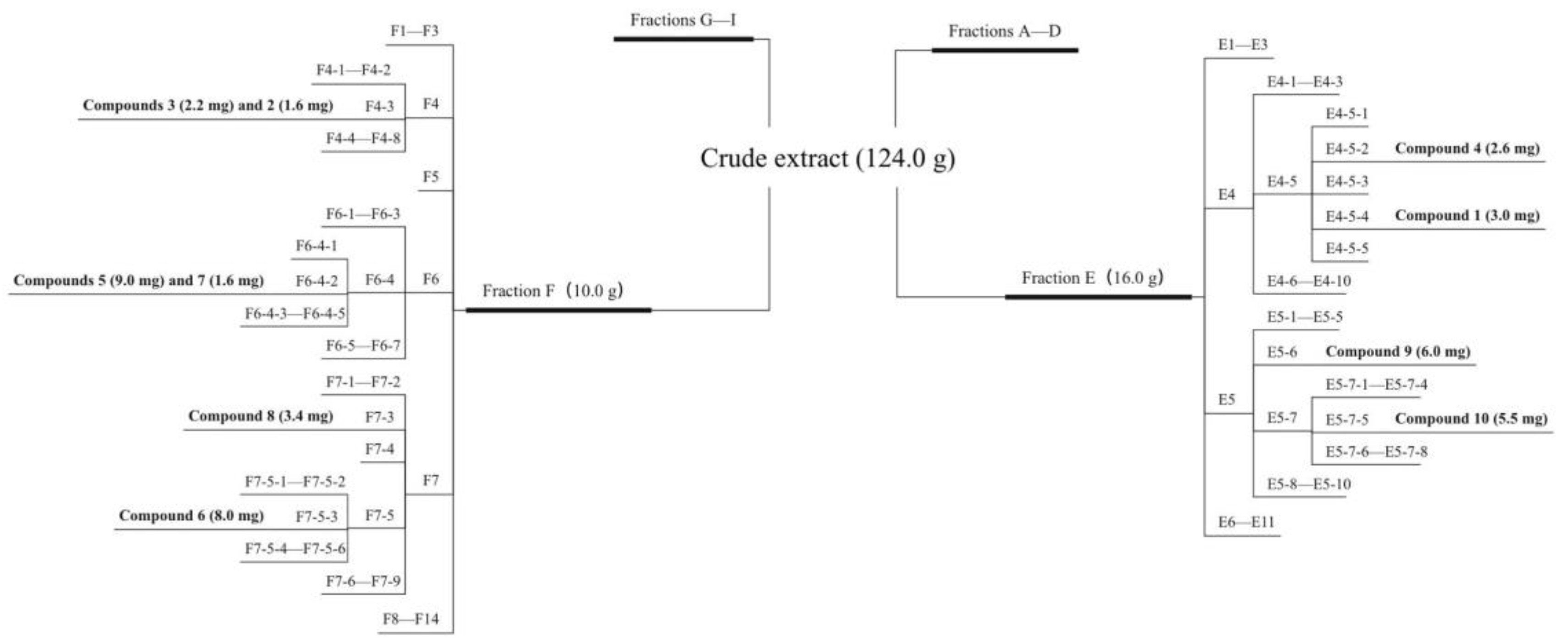
2.3. Extraction and Isoation

The fermented material was extracted five times with absolute methanol. The extract was dissolved in water and EtOAc, and extracted four times with EtOAc to yield 124.0 g. As shown in Scheme 1, the crude extract was separated into 9 fractions (A−I) using a CC over silica gel column (80–100 mesh) with a solvent system of CH2Cl2−MeOH (from 100:0 to 0:100). Fr. E (16.0 g) was separated by MPLC over RP-18 silica gel eluted with MeOH/H2O (from 10:90 to 100:0) to obtain 11 fractions (E1−E11). Fr. E4 and Fr. E5 were both isolated with a silica gel column with a step gradient of CH2Cl2−MeOH (from 100:1 to 0:100) to afford 10 parts (E4-1−E4-10 and E5-1−E5-10). Fr. E4-5 was fractionated by Sephadex LH-20 CC eluted with acetone into five subfractions (E4-5-1−E4-5-5). Fr. 4-5-2 was purified by prep-HPLC to yield compound 4 (2.6 mg). Fr. E4-5-4 was subjected to Sephadex LH-20 CC eluted with MeOH and further purified by prep-HPLC to yield compound 1 (3.0 mg). Fr. E5-6 was prepared with HPLC to yield compound 9 (6.0 mg). Fr. E5-7 was separated into 8 parts (Fr. E5-7-1−5-7-8) using RP-18 silica gel eluted with MeOH/H2O (from 20:80 to 0:100). Fr. E5-7-5 was purified by prep-HPLC to yield compound 10 (5.5 mg). Fr. F (10.0 g) was first fractionated by MPLC over RP-18 eluted with MeOH/H2O (from 20:80 to 0:100) to give 14 subfractions (F1−F14). Fr F4 was isolated with Sephadex LH-20 CC eluted with CH2Cl2/MeOH (1:1) to afford 8 parts (F4-1−F4-8). Further purification of Fr. 4-3 with prep-HPLC gave compounds 3 (2.2 mg) and 2 (1.6 mg). Fr. F6 was separated into 7 subfractions (F6-1−F6-7) by a silica gel column of CH2Cl2/MeOH (from 50:1 to 0:100). Fr. F6-4 was purified by Sephadex LH-20 CC eluted with MeOH to obtain 5 parts (F6-4-1−F6-4-5). Fr. F6-4-2 was then further purified via prep-HPLC to give compounds 5 (9.0 mg) and 7 (1.6 mg). Fr. F7 was separated into 9 subfractions (F7-1−F7-9) by a silica gel column of petroleum ether/acetone (from 30:1 to 0:1). Fr. F7-3 was prepared with HPLC to yield compound 8 (3.4 mg). Fr. F7-5 was isolated with Sephadex LH-20 CC eluted with acetone to afford 6 parts (F7-5-1−F7-5-6). Fr. 7-5-3 was purified by prep-HPLC to yield compound 6 (8.0 mg).
Albocinnamin A (1a/1b): colorless crystals (H2O); mp 182.5–186.8 °C; 1a: [α]25D + 46.9 (c 0.50, MeOH), 1b: [α]25D − 22.5 (c 0.50, MeOH); UV (H2O) λmax (log ε) 215 (3.78), 230 (3.68), 290 (3.22) nm; 1H (methanol-d4, 600 MHz) and 13C NMR (methanol-d4, 150 MHz) data, see Table 1; HRESIMS m/z 263.12769 [M + H] + (calcd for C15H20O3, 263.12779).
Albocinnamin B (2): colorless oil; [α]23.6D − 23.6 (c 0.5, MeOH); UV (MeOH) λmax (log ε) 235 (3.49) nm; 1H (600 MHz) and 13C NMR (150 MHz) data (methanol-d4), see Table 1; HRESIMS m/z 235.16927 [M + H] + (calcd. for C15H23O3, 235.16926).
Albocinnamin C (3): yellow powder; [α]23.6D + 9.6 (c 0.5, MeOH); UV (MeOH) λmax (log ε) 235 (3.59) nm; 1H (600 MHz) and 13C NMR (150 MHz) data (methanol-d4), see Table 1; HRESIMS m/z 287.16153 [M + Na] + (calcd. for C16H24NaO3, 287.16177).
Albocinnamin D (4): colorless oil; [α]23.6D + 13.6 (c 0.5, MeOH); UV (MeOH) λmax (log ε) 235 (3.86) nm; 1H (600 MHz) and 13C NMR (150 MHz) data (methanol-d4), see Table 1; HRESIMS m/z 251.16422 [M + H] + (calcd. for C15H23O3, 251.16417).
Albocinnamin E (5): colorless crystals (H2O); mp 234–236 °C; [α]27D − 108.7 (c 0.5 MeOH); UV (MeOH) λmax (log ε) 235 (3.98) nm; IR (KBr) νmax 3329, 2960, 1697, 1649, 1379, 1022 cm−1; 1H (600 MHz) and 13C NMR (150 MHz) data (methanol-d4), see Table 2; HRESIMS m/z 251.16414 [M + H] + (calcd. for C15H23O3, 251.16417).
Albocinnamin F (6): colorless oil (H2O); [α]27D − 53.2 (c 0.5 MeOH); UV (MeOH) λmax (log ε) 245 (3.83) nm; 1H (600 MHz) and 13C NMR (150 MHz) data (DMSO-d6), see Table 2; HRESIMS m/z 267.15892 [M + H] + (calcd. for C15H23O4, 267.15909).
Albocinnamin G (7): colorless oil (H2O); [α]27D + 166.7 (c 0.5, MeOH); UV (MeOH) λmax (log ε) 270 (3.20) nm; 1H (600 MHz) and 13C NMR (150 MHz) data (methanol-d4), see Table 2; HRESIMS m/z 249.14854 [M + H] + (calcd. for C15H21O3, 249.14852).
Albocinnamin H (8): colorless oil; [α]27D + 44.4 (c 0.5, MeOH); UV (MeOH) λmax (log ε) 230 (3.47) nm; 1H (600 MHz) and 13C NMR (150 MHz) data (methanol-d4), see Table 2; HRESIMS m/z 305.13573 [M + Na] + (calcd. for C15H22NaO5, 305.13594).

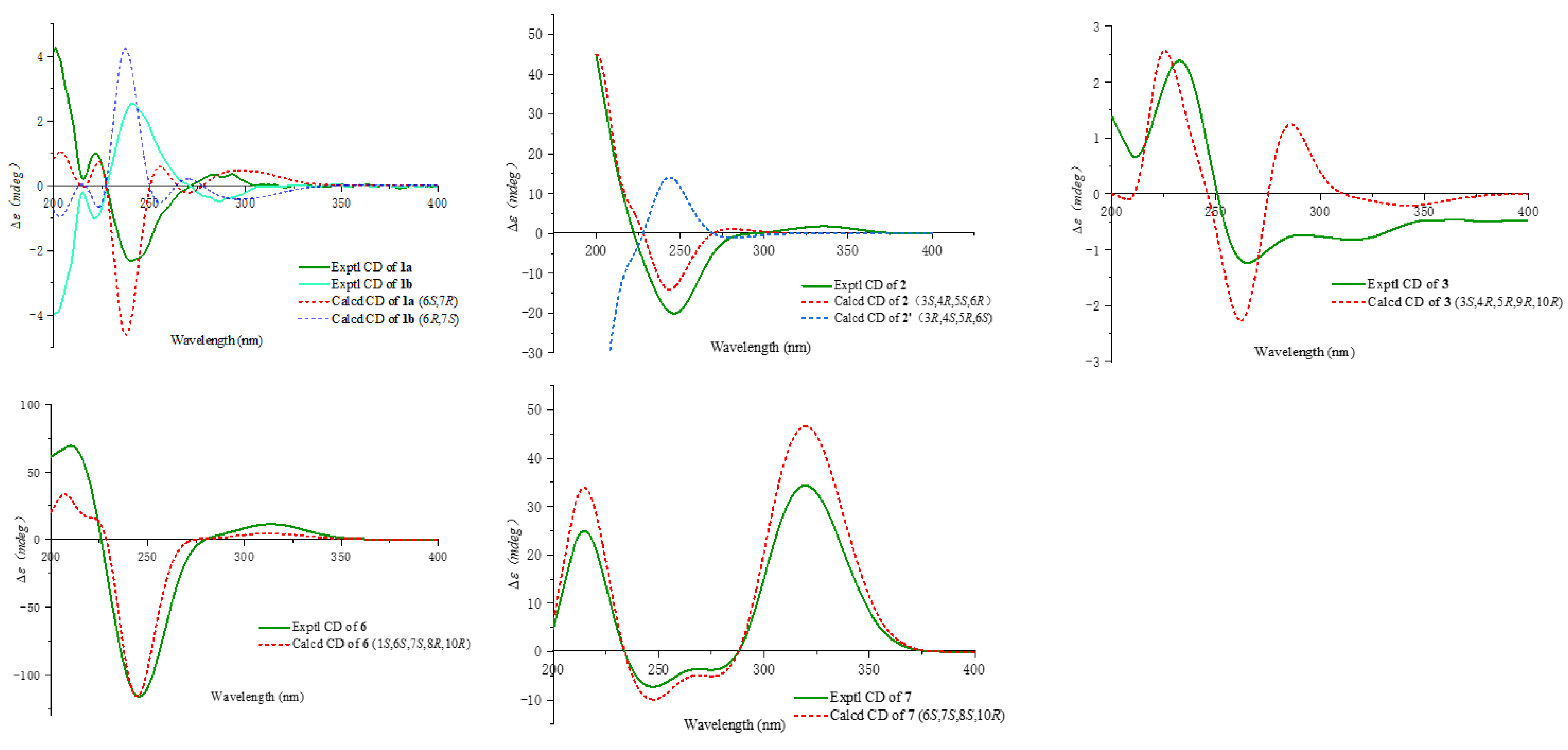
2.4. ECD Calculations

The Gaussian 16 program package was used for the calculations of the ECD spectra of 13, 67. The stable conformers subjected to ECD calculation were optimized using the time-dependent density functional theory (TDDFT) method at the B3LYP/6-311G (d, p) level of theory [13,14]. The ECD curves were extracted by SpecDis 1.60 and weighted by Boltzmann distribution after UV correction [15]. For details, see the Supporting Information below.

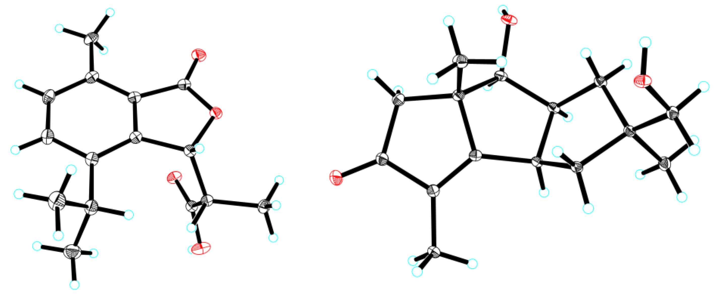
2.5. X-Ray Crystallographic Analysis

Single crystals of compounds 1(1a/1b) and 5 were obtained from MeOH and H2O, and all single crystals were collected by a Bruker D8 QUEST diffractometer, which was equipped with Cu-Kα radiation (λ 1.54178 Å). The structure was solved with ShelXT, using direct methods and refined with ShelXT using least square minimization. Crystallographic data for compounds 1 and 5 have been deposited at the Cambridge Crystallographic Data Centre (CCDC number for 1: 2253029, and 5: 2253030).
X-ray crystallographic data for 1 (1a and 1b): C15H18O4, (M = 262.29 g/mol): monoclinic, space group Pbca, a = 11.1153(5) Å, b = 13.4703(6) Å, c = 18.6151(8) Å, α = 90°, β = 90°, γ = 90°, V = 2787.2(2) Å3, Z = 8, T = 100(2) K, μ(Cu Kα) = 0.739 mm−1, Dcalc = 1.250 mg/m3, 22,765 reflections measured, 2749 independent reflections (Rint = 0.0608). The final R1 was 0.0347 (I > 2σ(I)), and wR(F2) was 0.0923 (I > 2σ(I)). The final R1 was 0.0456 (all data), and wR(F2) was 0.0962 (all data).
X-ray crystallographic data for 5: C15H22O3, (M = 250.32 g/mol): monoclinic, space group P212121, a = 7.7864(4) Å, b = 9.0991(4) Å, c = 19.0099(9) Å, α = 90°, β = 90°, γ = 90°, V = 1346.84(11) Å3, Z = 4, T = 100(2) K, μ(Cu Kα) = 0.676 mm−1, Dcalc = 1.235 mg/m3, 12,794 reflections measured, 2639 independent reflections (Rint = 0.0419). The final R1 was 0.0309 (I > 2σ(I)), and wR(F2) was 0.0806 (I > 2σ(I)). The final R1 was 0.0312 (all data), and wR(F2) was 0.0808 (all data). Flack parameter = 0.01(5).

2.6. Antibacterial Assay

All compounds were subjected to minimal inhibitory concentration (MIC) tests against two species of bacteria (S. aureus and Mycobacterium tuberculosis). Both bacteria were purchased from China General Microbiological Culture Collection Center (CGMCC). All strains were cultured in Mueller Hinton broth (MHB) (Guangdong Huankai Microbial Sci. &Tech. Co., Ltd., Guangzhou, China) at 37 °C. A sample of each culture was then diluted 40-fold in fresh MHB broth and incubated at 37 °C with shaking (200 rpm) for 2.5 h [16]. The resultant mid-log phase cultures were diluted to a concentration of 5 × 10⁵ CFU/mL, and then 50 mL was added to each well of the compound-containing plates, giving a final compound concentration range of 128 or 50 mg/mL. The plates were observed after 24 h incubation at 37 °C [17]. Inhibition rates were determined using photometry at OD625 nm. Rifampicin was used as the positive control (MIC < 2.5 µg/mL).

2.7. Cytotoxicity Assay

All compounds were assessed for their cytotoxicity toward the human promyelocytic leukemia (HL-60), colon cancer (SW480), and breast cancer (MCF-7) cell lines. All the cells were seeded into 96-well plates containing DMEM or RPMI1640 medium with 10% FBS under a 5% CO2 atmosphere at 37 °C. The assays were performed by the MTS method according to the manufacturer’s instructions [18]. Briefly, the isolated compounds dissolved in dimethyl sulfoxide (DMSO) and were then diluted with culture media to produce difference concentrations (40, 20, 10, 5, 2.5, 1.25, 0.625 µM). After incubation for 24 h, various levels of compounds were added to each well and incubated for 48 h. A total of 100 µL of culture media and 20 µL of MTS solution were added, which incubated for 3 h at 37 °C [19]. The absorbance of each well was measured at 490 nm using the Multi-Mode microplate reader. Paclitaxel was used as a positive control, and the concentrations for paclitaxel were 0.5, 0.25, 0.125, 0.0625, 0.03125, 0.015625, and 0.0078125 µM (IC50 < 0.08 µM).

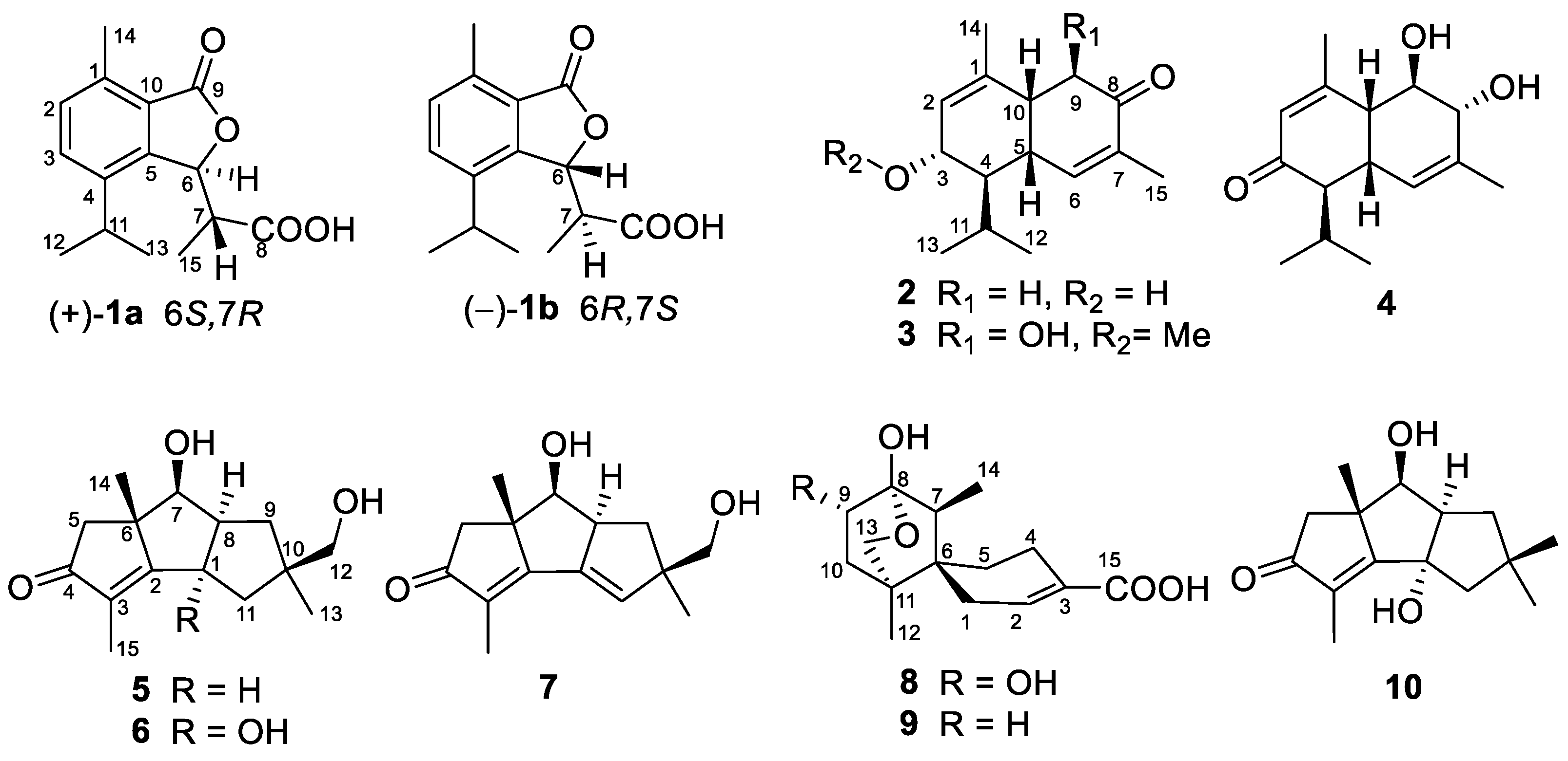
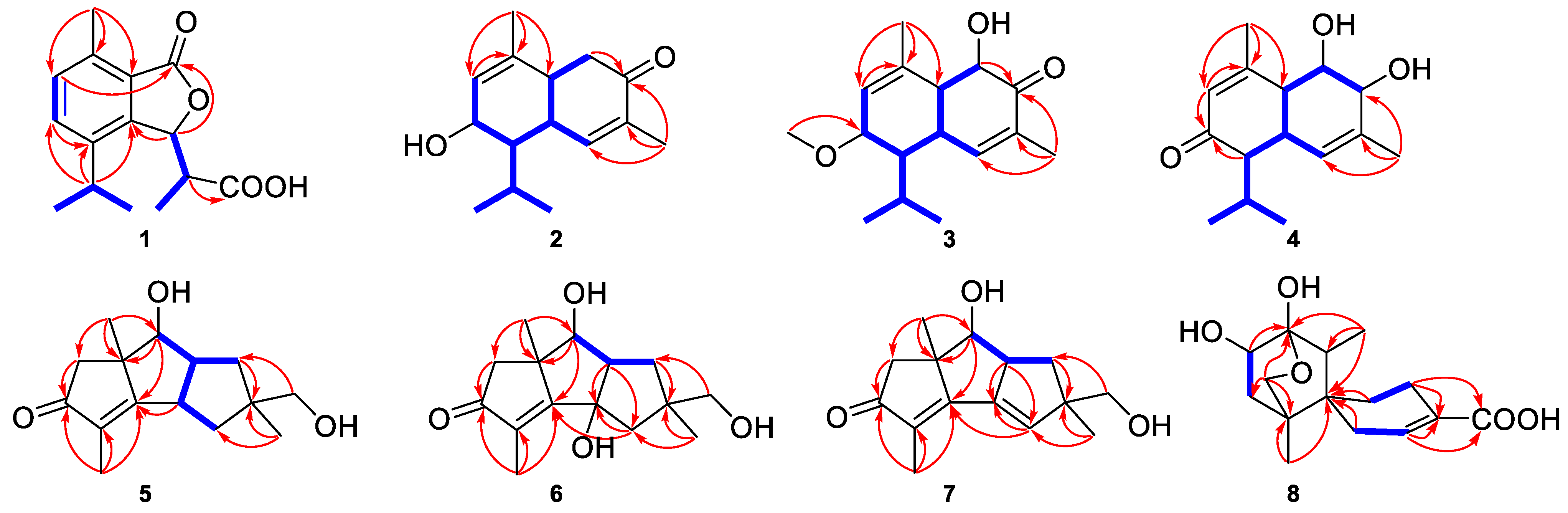
3. Results and Discussion

Compound 1 was isolated as colorless crystals. Its molecular formula was determined as C15H19O3 by HRESIMS (measured at m/z 263.12769 [M + H]+; calcd for C15H20O3, 263.12779), which accounted for seven double-bond equivalents. The 1H NMR and 13C NMR spectrum (Table 1) showed 15 carbon signals, including four CH3, five CH, and six non-protonated carbons. Primary analysis of these data indicated that 1 had a benzene group and two carbonyl carbons. The 1H-1H COSY data revealed three fragments, as shown in Figure 2. Based on this, the HMBC data revealed the planar structure of 1 (Figure 2). At first, the HMBC correlations from H3-14 to C-1 and from H-11 to C-3, C-4, C-5 indicated one methyl group placed at C-1 and an isopropyl group placed at C-4 of the benzene group, respectively. In addition, the HMBC correlations from H-6 to C-9 (δC 173.3) suggested a γ-lactone fused with the benzene group. Finally, the HMBC correlations from H3-15, H-6 and H-7 to C-8 (δC 176.9) suggested a carboxyl group of C-8, in connection with C-7. Hence, the planar structure of 1 was established as an aromatic sesquiterpene with a novel backbone. A single crystal X-ray diffraction experiment was performed (Figure 3), and the result confirmed the planar structure as given above. In addition, the data revealed the relative configuration of 1 and suggested that 1 should be a racemate. Therefore, compound 1 was separated into two pure enantiomers by chiral-phase HPLC (Figure S22), and the absolute configurations were determined by comparing the calculated and experimental ECD spectra (1a/1b, Figure 4). This experiment enabled 1a and 1b to be determined as (+)-(6S,7R) and (−)-(6R,7S), respectively. Consequently, the structure of 1 was characterized and trivially named as (±)-albocinnamin A.
Compound 2 was isolated as a colorless oil. Its molecular formula was determined as C15H22O2 by HRESIMS (measured at m/z 235.16927 [M + H]+; calcd for C15H23O2 235.16926), which accounted for five degrees of unsaturation. The 1H and 13C NMR data (Table 1) revealed four CH3, one CH2, seven CH, and three non-protonated carbons. Of them, data at δC 152.1 (d, C-6), 135.1 (s, C-7) and 201.0 (s, C-8) indicated the presence of an α,β-unsaturated keto moiety, while one signal at δC 69.3 (d, C-3) suggested one OH group. The 1H-1H COSY correlations disclosed a long link, as shown in Figure 2, which established the OH position at C-3 and revealed the α,β-unsaturated keto moiety to be 6,7-en-8-one. Based on this, the HMBC correlations from H-14 to C-1, C-2, and C-10 constructed a six-membered ring. In addition, the HMBC correlations from H-9 to C-8 and C-7 established another six-membered ring. Therefore, compound 2 was assigned as a bicyclic cadinane sesquiterpene [20]. In the ROESY spectrum (Figure 5), observed cross peaks of H-3/H-5, H-12 and H-5/H-10 indicated that H-3, H-5 and H-10 were in the same orientation, and H-4 was in the other orientation. There, correlations revealed the relative configuration of 2 (3S,4R,5S,6R or 3R,4S,5R,6S). Finally, the absolute configurations were determined by comparing the calculated and experimental ECD spectra (Figure 4). Hence, the structure of 2 was identified and trivially named as albocinnamin B.
Compound 3 was isolated as a yellow solid. Its molecular formula was determined as C16H24O3 by HRESIMS (measured at m/z 287.16153 [M + Na]+; calcd for C16H24NaO3 287.16177). The 1H and 13C NMR data (Table 1) showed similarities to those of 2, except for the substitutions at C-3 and C-9. There is a hydroxyl group at C-3 of 2, while C-3 of 3 is a methoxy group. Finally, the signal at δC 76.0 (d, C-9) indicated a OH group placed at C-9. These changes were substantiated by the HMBC correlation from H-16 to C-3, and the 1H-1H COSY correlation from H-9 to H-10. In the ROESY spectrum (Figure 5), observed cross peaks of H-3/H-5, H-12, H-5/H-10 and H-4/H-9 indicated that H-3, H-5 and H-10 were in the same orientation, and that H-4 and H-9 were in the same orientation. Finally, the absolute configuration of 3 was determined by ECD calculations (Figure 4). Consequently, the structure of 3 was identified and trivially named as albocinnamin C.
Compound 4 was isolated as a colorless oil. Its molecular formula was determined as C15H22O3 by HRESIMS (measured at m/z 251.16422 [M + H]+; calcd for C15H23O3 251.16417). Primary analysis of 1D and 2D data (Table 1) was similar to those of 3. The difference was that one carbonyl carbon (δC 204.5) placed at C-3 and one OH group placed at C-8. This conclusion was supported by the HMBC correlations from H-4, H-2 to C-3, and from H-15 to C-6, C-7, and C-8. In the ROESY spectrum, cross peaks of H-5/H-10, H-8, and H-4/H-9 were observed (Figure 2). Considering the homology of biological sources, the absolute configuration of 4 should be the same as 3. Hence, the structure of 4 was identified and trivially named as albocinnamin D.
Compound 5 was isolated as colorless crystals. Its molecular formula was determined as C15H22O3 by HRESIMS (measured at m/z 251.16414 [M + H]+; calcd for C15H23O3 251.16417), which accounted for five double-bond equivalents. The 1H NMR and 13C NMR spectrum (Table 2) of compound 5 showed signals for 15 carbons, including three CH3, four CH2, three CH, and five non-protonated carbons. Primary analysis of 1D and 2D data showed that 5 was similar to cocumin F [21]. The major difference between 5 and cocumin F is that the oxygenated methylene in 5 replaced the methyl of C-12 in cocumin F, which was confirmed by the HMBC correlations from H-12 to C-9, C-10 and C-11 (Figure 2). In the ROESY spectrum (Figure 5), the cross peaks of H-1/H-8, H-1/H-13, H-7/H-8, H-8/H-13 were observed, and not cross peaks of H3-14/H-1, H-7 and H-8. The planar structure of 5 was indicated. Finally, a single-crystal X-ray diffraction not only confirmed the planar structure, but also established the absolute configuration of 5 (Figure 3), named as albocinnamin E.
Compound 6 was isolated as a colorless oil. Its molecular formula was determined as C15H22O4 by HRESIMS (measured at m/z 267.15892 [M + H]+; calcd for C15H23O4 267.15909). The 1D and 2D NMR data (Table 2) showed that it was extremely similar with those of 5, except for the presence of an additional OH group, which was located at C-1, as confirmed by the HMBC correlations from H-8 and H-11 to C-1. In the ROESY spectrum (Figure 5), observed cross peaks of 1-OH/H-7, H-8, and without cross peaks of H3-14/H-7, H-8, indicating that 1-OH, H-7, and H-8 were in the same orientation and H3-14 was in the other orientation. Finally, the absolute configuration of 6 was confirmed by ECD calculations (Figure 4). Hence, the structure of 6 was identified and trivially named as albocinnamin F.
Compound 7 was isolated as a colorless oil. Its molecular formula was determined as C15H20O3 by HRESIMS (measured at m/z 249.14854 [M + H]+; calcd for C15H21O3 249.14852), accounting for six degrees of unsaturation. The 1D data (Table 2) displayed high similarity to those of 5, except for the signals at δC 143.6 and δC 135.7, which suggested one double bond between C-1 and C-11 of 7. This assumption was supported by the HMBC correlations from H-8 to C-1, C-11 (Figure 2). Detailed analysis of 2D NMR data suggested that other parts of 7 were identical to those of 5. The absolute configurations were determined by comparing the calculated and experimental ECD spectra (Figure 4). Therefore, the structure of 7 was established and trivially named as albocinnamin G.
Compound 8 was isolated as a colorless oil. Its molecular formula was determined as C15H22O5 by HRESIMS (measured at m/z 305.13573 [M + Na]+; calcd for C15H22NaO5 305.13594), which accounted for five degrees of unsaturation. Primary analysis of the 1H and 13C NMR (Table 2) spectra of 8 showed its close resemblance to ochracine F,15 and the difference of 8 was the presence of one OH group placed at C-9, as supported by the 1H-1H COSY correlation from H-9 to H-10 and its molecular weight. In the ROESY spectrum, observed cross peaks of H-9/H-14 indicated H-9 and H-14 in the same orientation (Figure 5). Considering the homology of biological sources, the absolute configuration of 8 was the same as that of ochracine F. Consequently, the structure of 8 was identified and trivially named as albocinnamin H.
In addition, two known compounds were identified as ochracine F (9) [22] and cerrenin C (10) [23] by comparison of their spectroscopic data to the reported data in the literature. Structurally, compound 1 possessed a new backbone, and it may be derived from a precursor of the cadinane sesquiterpene 2 via a key Baeyer−Villiger oxidation. After further hydrolysis, oxidation, dehydration, and aromatization, a novel product 1 was finally built (Scheme 2).
Previous studies have proved that sesquiterpenes in A. albocinnamomea have cytotoxicity and antibacterial activity [9]. Therefore, all compounds were tested for their cytotoxicity activity against three human cancer cell lines (HL-60, SW480 and MCF-7) and for their antibacterial activity against S. aureus and M. tuberculosis. As a result, compounds 1a and 1b showed moderate cytotoxicity against SW480 and MCF-7 cells with IC50 values ranging from 19.3 to 33.3 μM, while compound 2 showed cytotoxicity against HL-60 cells with an IC50 value of 12.3 μM (Table 3). In addition, compounds 5 and 6 exhibited antibacterial activity against S. aureus with MIC values of 64 and 64 µg/mL, respectively.

4. Conclusions

A total of ten sesquiterpenes, including eight new ones, have been characterized from the fungus A. albocinnamomea. The new structures with absolute configurations were established by means of spectroscopic methods, single-crystal X-ray diffraction, and ECD calculations. Compounds 1a and 1b showed cytotoxicity against SW480 and MCF-7 cells, while compound 2 displayed cytotoxicity against HL-60 cell. In addition, compounds 5 and 6 exhibited antibacterial activity against S. aureus. This indicates that A. albocinnamomea is rich in sesquiterpenes, which have potential cytotoxicity and antibacterial application prospects.

Supplementary Materials

The following supporting information can be downloaded at: https://www.mdpi.com/article/10.3390/jof9050521/s1. Spectra of 1D, 2D-NMR and HRESIMS for compounds 18, X-ray crystallographic data of 1, 5 and ECD calculations for compounds 13 and 67 (PDF).

Author Contributions

Conceptualization, J.H. and T.F.; methodology, J.N., F.W., J.L., J.H. and T.F.; validation, J.H. and T.F.; investigation, J.N. and F.W.; resources, J.L.; data curation, J.N. and F.W.; writing—original draft preparation, J.N. and F.W.; writing—review and editing, T.F.; supervision, J.H.; project administration, J.H. and T.F.; funding acquisition, J.H. and T.F. All authors have read and agreed to the published version of the manuscript.

Funding

This research was funded by the National Natural Science Foundation of China (22177139, 22277147) and the Fundamental Research Funds for the Central Universities, South-Central Minzu University (CZP21001).

Institutional Review Board Statement

Not applicable.

Informed Consent Statement

Not applicable.

Data Availability Statement

The data of this study are available from the corresponding author (T.F.).

Acknowledgments

The authors thank Analytical & Measuring Center, School of Pharmaceutical Sciences, South-Central Minzu University for MS and NMR spectra test.

Conflicts of Interest

The authors declare no conflict of interest.

References

  1. Yuan, H.S.; Dai, Y.C.; Steffen, K. Screening and evaluation of white rot fungi to decolourise synthetic dyes, with particular reference to Antrodiella albocinnamomea. Mycology 2012, 3, 100–108. [Google Scholar]
  2. Chen, Z.M.; Fan, Q.Y.; Yin, X.; Yang, X.Y.; Li, Z.H.; Feng, T.; Liu, J.K. Three new humulane sesquiterpenes from cultures of the fungus Antrodiella albocinnamomea. Nat. Prod. Bioprospect. 2014, 4, 207–211. [Google Scholar] [CrossRef] [PubMed]
  3. Chen, Z.M.; Yang, X.Y.; Fan, Q.Y.; Li, Z.H.; Wei, K.; Chen, H.P.; Feng, T.; Liu, J.K. Three novel degraded steroids from cultures of the Basidiomycete Antrodiella albocinnamomea. Steroids 2014, 87, 21–25. [Google Scholar] [CrossRef] [PubMed]
  4. Chen, Z.M.; Chen, H.P.; Wang, F.; Li, Z.H.; Feng, T.; Liu, J.K. New triquinane and gymnomitrane sesquiterpenes from fermentation of the basidiomycete Antrodiella albocinnamomea. Fitoterapia 2015, 102, 61–66. [Google Scholar] [CrossRef]
  5. Yang, Y.L.; Yu, W.W.; Li, Z.H.; Liu, J.K.; Feng, T. Antrodiellins A–C, triquinane sesquiterpenoids from fungus Antrodiella albocinnamomea with their antibacterial activity. Phytochem. Lett. 2022, 47, 24–27. [Google Scholar] [CrossRef]
  6. Yu, W.W.; Ma, J.T.; He, J.; Li, Z.H.; Liu, J.K.; Feng, T. Cadinane sesquiterpenoids from the fungus Antrodiella albocinnamomea and their inhibitory activity against nitric oxide production. Phytochemistry 2022, 196, 113081. [Google Scholar] [CrossRef]
  7. Dai, Q.; Zhang, F.L.; Feng, T. Sesquiterpenoids specially produced by fungi: Structures, biological activities, chemical and biosynthesis (2015–2020). J. Fungi 2021, 7, 1026. [Google Scholar] [CrossRef]
  8. Luo, C.; Lou, D.J.; Li, Y.F.; Yang, L.; Shao, L.J.; Liu, J.K.; Yang, X.Y. A new chamigrane sesquiterpene from the basidiomycete Antrodiella albocinnamomea. J. Asian Nat. Prod. Res. 2023, 25, 191–196. [Google Scholar] [CrossRef]
  9. Li, W.; He, J.; Feng, T.; Yang, H.X.; Ai, H.L.; Li, Z.H.; Liu, J.K. Antroalbocin A, an antibacterial sesquiterpenoid from higher fungus Antrodiella albocinnamomea. Org. Lett. 2018, 20, 8019–8021. [Google Scholar] [CrossRef]
  10. He, J.; Yu, W.W.; Isaka, M.; Cox, R.J.; Liu, J.K.; Feng, T. Antroxazole A, an oxazole-containing chamigrane dimer from the fungus Antrodiella albocinnamomea with immunosuppressive activity. Org. Biomol. Chem. 2022, 20, 7278–7283. [Google Scholar] [CrossRef]
  11. Liang, D.D.; Yi, X.W.; Wu, H.; Li, Z.H.; Wang, G.K.; Cheng, G.G.; Feng, T. Antrodillin, an immunosuppressive sesquiterpenoid from higher fungus Antrodiella albocinnamomea. RSC Adv. 2021, 11, 1124–1127. [Google Scholar] [CrossRef] [PubMed]
  12. Wang, F.; Yang, X.; Lu, Y.; Li, Z.; Xu, Y.; Hu, J.; Liu, J.K.; Xiong, W. The natural product antroalbol H promotes phosphorylation of liver kinase B1 (LKB1) at threonine 189 and thereby enhances cellular glucose uptake. J. Biol. Chem. 2019, 294, 10415–10427. [Google Scholar] [CrossRef] [PubMed]
  13. Vainio, M.J.; Johnson, M.S. Generating conformer ensembles using a multiobjective genetic algorithm. J. Chem. Inf. Model. 2007, 47, 2462–2474. [Google Scholar] [CrossRef]
  14. Frisch, M.J.; Trucks, G.W.; Schlegel, H.B.; Scuseria, G.E.; Robb, M.A.; Cheeseman, J.R.; Scalmani, G.; Barone, V.; Petersson, G.A.; Nakatsuji, H.; et al. Gaussian 16; Revision B.01; Gaussian, Inc.: Wallingford, CT, USA, 2016. [Google Scholar]
  15. Bruhn, T.; Schaumloeffel, A.; Hemberger, Y.; Bringmann, G. SpecDis: Quantifying the comparison of calculated and experimental electronic circular dichroism spectra. Chirality 2013, 25, 243–249. [Google Scholar] [CrossRef]
  16. Moghadamtousi, S.Z.; Abdul Kadir, H.; Hassandarvish, P.; Tajik, H.; Abubakar, S.; Zandi, K. A review on antibacterial, antiviral, and antifungal activity of curcumin. BioMed Res. Int. 2014, 2014, 186864. [Google Scholar] [CrossRef]
  17. Gutierrez Nicolas, F.; Reyes, G.; Audisio, M.C.; Uriburu, M.L.; Leiva Gonzalez, S.; Barboza, G.E.; Nicotra, V.E. Withanolides with Antibacterial Activity from Nicandra john-tyleriana. J. Nat. Prod. 2015, 78, 250–257. [Google Scholar] [CrossRef]
  18. Samadi, A.K.; Tong, X.; Mukerji, R.; Zhang, H.; Timmermann, B.N.; Cohen, M.S. Withaferin A, a cytotoxic steroid from Vassobia breviflora, induces apoptosis in human head and neck squamous cell carcinoma. J. Nat. Prod. 2010, 73, 1476–1481. [Google Scholar] [CrossRef] [PubMed]
  19. Maeda, G.; Gilissen, P.J.; Rudenko, A.; van der Wal, J.; Bourgard, C.; Gupta, A.K.; Sunnerhagen, P.; Munissi, J.J.E.; Nyandoro, S.S.; Erdélyi, M. Oxygenated cyclohexene derivatives from the stem and root barks of Uvaria pandensis. J. Nat. Prod. 2021, 84, 3080–3089. [Google Scholar] [CrossRef]
  20. Chen, D.L.; Chen, M.Y.; Hou, Y.; Wang, C.H.; Sun, Z.C.; Yang, Y.; Liang, H.Q.; Ma, G.X.; Xu, X.D.; Wei, J.H. Cadinane-type sesquiterpenoids with cytotoxic activity from the infected stems of the semi-mangrove Hibiscus tiliaceus. J. Nat. Prod. 2022, 85, 127–135. [Google Scholar] [CrossRef]
  21. Hellwig, V.; Dasenbrock, J.; Schumann, S.; Steglich, W.; Leonhardt, K.; Anke, T. New triquinane-type sesquiterpenoids from Macrocystidia cucumis (Basidiomycetes). Eur. J. Org. Chem. 1998, 1998, 73–79. [Google Scholar] [CrossRef]
  22. Zhao, Z.Z.; Zhao, Q.L.; Feng, W.S.; He, H.R.; Li, M.; Xue, G.M.; Chen, H.P.; Liu, J.K. Structure and absolute configuration assignments of ochracines F–L, chamigrane and cadinane sesquiterpenes from the basidiomycete Steccherinum ochraceum HFG119. RSC Adv. 2021, 11, 18693–18701. [Google Scholar] [CrossRef] [PubMed]
  23. Liu, H.X.; Tan, H.B.; Chen, K.; Chen, Y.C.; Li, S.N.; Li, H.H.; Zhang, W.M. Cerrenins A–C, cerapicane and isohirsutane sesquiterpenoids from the endophytic fungus Cerrena sp. Fitoterapia 2018, 129, 173–178. [Google Scholar] [CrossRef] [PubMed]
Figure 1. The structure of compounds 110.
Jof 09 00521 g001
Scheme 1. Separation Process for the Compounds 110.
Jof 09 00521 sch001
Figure 2. Key HMBC (arrows) and 1H-1H COSY (bold blue lines) correlations for compounds 18.
Jof 09 00521 g002
Figure 3. X-ray ORTEP drawings of compounds 1 (left) and 5 (right).
Jof 09 00521 g003
Figure 4. Experimental and calculated ECD spectra for compounds 13 and 67.
Jof 09 00521 g004
Figure 5. Key ROESY correlations for compounds 28.
Jof 09 00521 g005
Scheme 2. Plausible Biosynthesis Pathway for 1.
Jof 09 00521 sch002
Table 1. NMR Spectroscopic Data for Compounds 14 (600 MHz for 1H NMR and 150 MHz for 13C NMR).
1 a2 a3 a4 a
PositionδC, TypeδH, (J in Hz)δC, TypeδH, (J in Hz)δC, TypeδH, (J in Hz)δC, typeδH, (J in Hz)
1125.1, C 138.3, C 141.4, C 163.9, C
2132.5, CH7.24 d (7.8)127.9, CH5.40 m124.0, CH5.68 dd (3.5, 1.6)127.3, CH5.72 m
3131.9, CH7.47 d (7.8)69.3, CH4.03 dd (6.3, 2.6)77.9, CH3.76 m204.5, C
4142.8, C 48.7, CH1.71 m46.7, CH1.90 m59.4, CH2.00 m
5147.2, C 38.1, CH2.53 m38.3, CH2.66 m35.9, CH2.94 m
683.2 CH5.69 d (2.7)152.1, CH6.97 m151.6, CH6.89 m127.6, CH5.33 m
745.2, CH3.15 m135.1, C 133.2, C 135.2, C
8176.9, C 201.0, C 201.2, C 74.6, CH3.77 d (4.1)
9173.3, C 40.3, CH22.57 m
2.45 dd (17.6, 13.2)
76.0, CH4.32 d (10.8)73.2, CH4.17 dd (6.3, 4.1)
10137.5, C 41.0, CH2.56 m47.9, CH2.50 dd (10.8, 4.8)43.6, CH2.65 dd (6.3, 5.4)
1130.5, CH3.04 m28.3, CH2.08 m29.0, CH1.92 m29.2, CH1.92 dd (13.8, 6.8)
1223.8, CH31.30 d (6.8)20.0, CH31.07 d (7.1)19.6, CH30.97 d (6.9)21.8, CH31.06 d (6.7)
1323.4, CH31.21 d (6.8)20.8, CH31.01 d (7.1)20.8, CH31.03 d (6.9)20.5, CH30.85 d (6.7)
1417.1, CH32.55 s20.9, CH31.68 t (1.5)24.5, CH31.86 t (1.6)24.5, CH32.08 t (1.1)
1515.3, CH31.35 d (7.1)16.0, CH31.74 t (1.4)15.9, CH31.77 t (1.4)20.7, CH31.72 t (1.6)
16 55.2, CH33.30 s
a Measured in methanol-d4.
Table 2. NMR Spectroscopic Data for Compounds 58 (600 MHz for 1H NMR and 150 MHz for 13C NMR).
Position5 a6 b7 a8 a
δC, TypeδH, (J in Hz)δC, TypeδH, (J in Hz)δC, TypeδH, (J in Hz)δc, typeδH, (J in Hz)
143.1, CH3.45 m86.5, C 143.6, C 29.6, CH22.17 d (3.3)
2188.1, C 186.9, C 175.8, C 140.9, CH6.96 t (3.3)
3133.6, C 131.0, C 129.3, C 132.9, C
4212.9, C 209.2, C 212.9, C 22.7, CH22.49 m
2.24 m
554.0, CH22.23 d (17.4)
2.29 d (17.4)
53.3, CH22.20 d (17.1)
2.05 d (17.1)
51.7, CH22.41 d (17.4)
2.31 d (17.4)
31.1, CH21.83 m
1.43 m
653.1, C 51.8, C 55.8, C 39.2, C
778.7, CH3.92 d (9.2)75.1, CH3.96 d (9.4)75.5, CH4.14 d (10.2)46.9, CH1.80 q (7.2)
850.7, CH3.15 m59.8, CH2.76 m54.9, CH3.58 m99.1, C
937.4, CH22.17 dd (13.2, 10.4)
1.37 m
36.9, CH22.12 dd (13.5, 10.5)
1.26 m
40.0, CH22.13 dd (11.8, 6.9)
1.57 dd (11.8, 7.3)
66.7, CH3.84 dd (9.8, 4.0)
1049.5, C 47.4, C 57.6, C 41.6, CH22.32 m
1.29 dd (14.3, 3.9)
1141.2, CH21.76 m47.7, CH21.58 dd (13.5, 2.2)
2.00 m
135.7, CH5.89 d (2.9)36.4, C
1270.4, CH23.42 s68.4, CH23.16 s71.0, CH23.52 d (11.0)
3.42 d (11.0)
17.6, CH30.69 s
1322.8, CH31.02 s24.0, CH31.05 s20.8, CH31.16 s72.6, CH24.04 dd (9.0, 3.3)
3.56 d (9.0)
1423.0, CH31.28 s22.3, CH31.09 s23.6, CH31.02 s13.3, CH30.87 d (7.5)
158.2, CH31.65 s8.5, CH31.65 s8.8, CH31.69 s172.3, C2.17 d (3.3)
1-OH 5.03 s
7-OH 4.86 s
12-OH 4.64 s
a Measured in methanol-d4; b Measured in DMSO-d6.
Table 3. Cytotoxicity Data for the Isolates a.
CompoundsIC50 (μM)
HL-60SW480MCF-7
1a>4026.3 ± 1.6733.3 ± 1.19
1b>4029.6 ± 2.1419.3 ± 1.02
212.3 ± 1.87>40>40
Paclitaxel b<0.008<0.008<0.008
a Compounds with no cytotoxicity activities at the concentration of 40 μM to all cancer cell lines were not listed; b Positive control.
Disclaimer/Publisher’s Note: The statements, opinions and data contained in all publications are solely those of the individual author(s) and contributor(s) and not of MDPI and/or the editor(s). MDPI and/or the editor(s) disclaim responsibility for any injury to people or property resulting from any ideas, methods, instructions or products referred to in the content.

