Article

A Novel Effector FlSp1 Inhibits the Colonization of Endophytic Fusarium lateritium and Increases the Resistance to Ralstonia solanacearum in Tobacco

1
Department of Plant Pathology, College of Agriculture, Guizhou University, Guiyang 550000, China
2
Southwest Biomedical Resources of the Ministry of Education, Guizhou University, Guiyang 550000, China
*
Authors to whom correspondence should be addressed.
These authors contributed equally to this work.
J. Fungi 2023, 9(5), 519; https://doi.org/10.3390/jof9050519
Submission received: 7 March 2023 / Revised: 21 April 2023 / Accepted: 23 April 2023 / Published: 27 April 2023
(This article belongs to the Special Issue Plant-Associated Fungi)

Abstract

:
Effectors are crucial for the interaction between endophytes and their host plants. However, limited attention has been paid to endophyte effectors, with only a few reports published. This work focuses on an effector of Fusarium lateritium, namely FlSp1 (Fusarium-lateritium-Secreted-Protein), a typical unknown secreted protein. The transcription of FlSp1 was up-regulated after 48 h following fungal inoculation in the host plant, i.e., tobacco. The inactivation of FlSp1 with the inhibition rate decreasing by 18% (p < 0.01) resulted in a remarkable increase in the tolerance of F. lateritium to oxidative stress. The transient expression of FlSp1 stimulated the accumulation of reactive oxygen species (ROS) without causing plant necrosis. In comparison with the wild type of F. lateritium (WT), the FlSp1 mutant of the F. lateritium plant (ΔFlSp1) reduced the ROS accumulation and weakened the plant immune response, which resulted in significantly higher colonization in the host plants. Meanwhile, the resistance of the ΔFlSp1 plant to the pathogenic Ralstonia solanacearum, which causes bacterial wilt, was increased. These results suggest that the novel secreted protein FlSp1 might act as an immune-triggering effector to limit fungal proliferation by stimulating the plant immune system through ROS accumulation and thus balance the interaction between the endophytic fungi and their host plants.

1. Introduction

During growth, plants are confronted with a complex microenvironment in which pathogenic microbes and endophytes are present. In order to be protected from pathogens, plants have evolved two layers of immune systems: pattern-triggered immunity (PTI) and effector-triggered immunity (ETI). In order to avoid the recognition by their host plant, pathogens interfere or disrupt the PTI response by secreting effectors to suppress the plant’s immune system, while plants have evolved corresponding R proteins to specifically recognize effectors, triggering a higher-intensity defense response-ETI [1]. The effectors and plant defense systems evolve simultaneously in a dynamic balance and compete with each other. However, the function of endophytic effectors remains unclear. Therefore, exploring the mechanism of effectors in endophyte–plant interactions plays an important role in revealing the function of endophytes or the symbiotic relationship between endophytes and plants.
ROS burst plays an important role in plant immune systems [2]. For example, via the expression of defense-related genes through the overexpression of the transcription factors DEAXW and matrix metalloproteinases (MMP), both arabidopsis and tomato promote the accumulation of ROS and enhance their resistance to Botrytis cinerea [3,4]. In order to counter the defense strategy of ROS burst in the host plant, pathogens have developed new virulence mechanisms to escape plants’ immune systems. The Glomerella leaf spot of apple disease secretes an effector, namely Sntf2, to inhibit a plant’s immune system by reducing the accumulation of callose and H2O2 in the plant and enhancing its own virulence [5]. Additionally, Puccinia striiformis f. sp. tritic reduced host immunity with the increase in H2O2 accumulation in wheat and inhibited ROS-mediated cell death by silencing the effector pstGSRE1 [6]. Pathogenic effectors can also achieve ROS inhibition by activating or mimicking a plant’s ROS scavenging system. The fungal effector AvrPiz-t structurally mimicked ROD1 and activated the plant’s ROS scavenging system to suppress the plant immune system and promote fungi infestation [7]. These results suggest that the mechanism of ROS burst is the “Battlefield” of the competition between effectors and plant immunity. The accumulation of ROS enhances the resistance of plants. Fungi can inhibit the accumulation of ROS in plants, weaken the immune system of plants, and promote colonization and infection. In addition to pathogens, endophytic fungi can also affect ROS in plants [8], but the mechanism is still unclear. Based on the above, it can be concluded that the mechanisms of plant ROS burst play an important role in the plant immune response [9].
In previous studies, F. lateritium was usually considered to be a pathogen [10]. In our research, a strain of F. lateritium was isolated from Nothapodytes pittosporoides, which acted as an endophyte to promote plant growth and mediate disease resistance in the plants of Solanaceae [11,12,13]. However, the potential mechanisms by which F. lateritium regulates plant ROS burst and manipulates the plant immune system are still poorly understood. In this study, we investigate the mechanism of F. lateritium–plant interactions. An effector, namely FlSp1, was screened via bioinformatics analysis, revealing it to be an uncharacterized protein without a functional, structural domain. During the further examination of the function of the effector, we found that FlSp1 reduced the colonization of fungi and weakened tobacco resistance to Ralstonia solanacearum through the regulation of the plant immune system. The results of this study may provide a preliminary theoretical basis for F. lateritium–plant interactions and the mechanism by which F. lateritium helps plants to resist disease.

2. Materials and Methods

2.1. Plant Growing Conditions

Tobacco was cultured on solid Murashige and Skoog (MS) medium (0.443% MS Basal Medium with Vitamins; 0.7% agar powder; 3% sucrose; pH = 5.8). The tobacco seeds were washed 5 times with sterile water, then the seeds were soaked in 75% alcohol for 1 min followed by 2% NaOCl for 10 min, and finally, the seeds were washed 5 times with sterile water. The cleaned seeds were placed onto MS medium at 22 °C in a 16 h photoperiod/8 h dark cycle.
To cultivate the tobacco in soil, tobacco seeds were sown in soil and cultured at 28 °C in a 16 h photoperiod/8 h dark cycle and 40–60% humidity. Then, the seeds were transplanted to the pots after germination and cultured for 4 weeks until root irrigation or injection.

2.2. Strain Growth Conditions

The wild type and mutants of F. lateritium were incubated on potato dextrose agar (PDA) medium (0.5% potato extract; 2.0% glucose; 1.5% agar powder; 0.1 mg/mL chloramphenicol) at 25 °C in the dark for 8 d. The colonies were inoculated into 1/4 Sabouraud dextrose broth (SDB) liquid medium (0.5% yeast extract; 0.25% peptone; 1% glucose) for 5 d at 28 °C in the dark at 160 rpm. The mycelium was separated using triple filter paper and centrifuged for 5 min to obtain the spore precipitate. Finally, the concentration of the precipitate was adjusted to 1 × 10⁶ spores/mL for co-culture or root irrigation.
R. solanacearum was inoculated in BGT medium (0.5% yeast extract; 0.1% bacteriological peptone; 0.1% casamino acid; 0.5% glucose; 1.5% agar powder; 0.5% 2,3,5-triphenyltetrazolium chloride) and incubated at 28 °C for 48 h under dark conditions. Additionally, single colonies were placed into B medium (1% bacteriological pepton; 0.1% yeast extract; 0.1% casamino acid) at 28 °C and 180 rmp under dark conditions until OD600 = 1.0 for the next experiment.

2.3. Bioinformatic Prediction of Effectors in F. lateritium

Referring to the method by [14,15], we performed signal peptide prediction. All protein sequences of F. lateritium were analyzed using signalP4.0, a software that predicts the presence of potential signal peptide sequences in amino acid sequences and their cleavage sites. The results obtained by the software consisted of three main values: C, S, and Y. The highest value of C is the signal peptide cut site, while each amino acid corresponds to an S value, with higher S values in the signal peptide region, and the Y value is a parameter derived from the combined analysis of C and S values.
Secreted proteins prediction was performed as follows: transmembrane proteins were predicted using the software TMHMM for proteins containing signal peptides, and proteins containing transmembrane helices were transmembrane proteins. Among all the proteins containing signal peptides, those containing transmembrane structural domains were removed, and those remaining were secreted proteins.
Effector prediction involved the analysis of secreted proteins using the software EffectorP to predict whether they were effector proteins, and a prediction score was given.
For subcellular localization prediction, the subcellular localization of proteins was predicted using Protcomp9.0, and results were scored.

2.4. Plasmid Construction

The knockout of the gene was achieved using a homologous recombination strategy. FlSp1 was amplified from the F. lateritium genome and primers were designed using double-joint PCR to amplify upstream and downstream fragments of 800–1200 bp as homologous arms. At the same time, the hygromycin (hyg) sequence (1026 bp) was amplified, as well as its promoter GPDA (752 bp). The amplification primers corresponding to each fragment were designed by homologous recombination using the software Premier6. The upstream and downstream sequences of FlSp1 were homologously recombined with the GPDA + hyg fragment to replace the sequence after 50 bp of FlSp1. The recombined fragment was transformed into the pK2Gus vector and stored for backup.

2.5. Genetic Transformation

The agrobacterium tumefacien (AGL1)-mediated genetic transformation of F. lateritium with reference to Beauveria bassiana [16] was slightly improved. The recombinant plasmid was transformed into AGL1 and inoculated into agrobacterium rhizogene solid medium (YEB) (1% tryptone; 0.1% yeast extract; 0.05% MgSO4·7H2O; 0.5% sucrose; 1.5% agar powder; pH = 7.0; 50 μg/mL Kana, and 50 μg/mL Car) at 28 °C for 2 d. A single colony was placed into YEB liquid medium and incubated at 28 °C for 20 h at 180 rmp in the dark. Induction medium (IM) (0.3‰ NaCl; 0.3‰ MgSO4·7H2O; 0.3‰ K2HPO4; 0.78% MES; pH = 5.3; 10 mmol/mL glucose; 400μm/mL Acetosyringone (AS)) was added to resuspend the bacteria at OD660 = 0.15. The bacteria were incubated at 28 °C for about 6 h at 180 rmp in the dark. At the same time, the spore concentration of fungi was adjusted to 1 × 10⁴ spores/mL with IM and set aside. The spore suspension was mixed with an equal volume of AGL1, and the mixture of 100 μL was placed on microporous membrane on IM medium (200 μmol/L AS and 5 mmo/L glucose) and incubated for a total of 48 h at 22 °C. The microporous membrane was transferred to a Czapek medium plate (CZM) (3% sucrose; 0.2% NaNO3; 0.05% MgSO4·7H2O; 0.1% K2HPO4; 0.0001% FeSO4·7H2O; 0.05% KCl; pH = 7.0; 1.5% agar powder; containing 200 mg/mL Cephalosporins(Cef); 25 μg/mL hyg) with thaumatin resistance for the next step of transformer verification.

2.6. Transient Expression of FlSp1

Referring to the method of [17], the mCherry::FlSp1 transient expression vector was transformed into GV3101. Additionally, the transformed strain was incubated in YEB liquid medium at 180 rpm for 18 h. The precipitate was obtained by centrifugation for 5 min and resuspended in MES buffer (2.132 g MES, MgCl2·6H2O, pH = 5.6), and OD600 was adjusted to 0.6 and incubated at 180 rpm for 1 h. The Agrobacterium was injected into 5–6-week-old Nicotiana benthamiana with a 1 mm needle-free syringe. After 1 day, the leaves were stained by DAB to detect the ROS burst degree of the leaves. After 3–5 days, the incidence and the cell death area of the leaves were measured. INF1, which is a necrosis-inducing effector of a pathogen, was used as a positive control.

2.7. Phenotypic Analysis

The spore concentration of the WT or ΔFlSp1 was adjusted to 5 × 10⁵ spores/mL using Tween80, and 1 μL of spores suspension was inoculated onto CZM medium containing different stress factors (5.76 mM H2O2, pH = 10.4, pH = 4, 25 μg/mL Congo Red), respectively. CZM was used as base medium, and after 7–8 d of incubation at 25 °C under dark conditions, the diameter of each colony was measured using digital calipers and fungal growth inhibition rate statistics were performed. The inhibition rate of mycelial growth was calculated using the following equation: inhibition rate of mycelial growth (MGI) (%) = [(RGR − rgr)/RGR] × 100%. RGR is the radial growth rate of the control group, and rgr is the radial growth of fungi.

2.8. F. lateritium Co-Cultured with Tobacco

The spore concentration of fungi was adjusted to 5 × 10⁵ spores/mL. The sterile 4–6 week-old tobacco was placed in the spore suspension and co-cultured in an incubator at 25 °C with 16 h of light per day, or spore suspension was used to irrigate tobacco roots.

2.9. R. solanacearum Inoculation Treatment of Tobacco

R. solanacearum was activated according to the method in Section 2.2, the precipitate was collected by centrifugation at 6000 rmp/min for 10 min, and the concentration was adjusted to 1 × 10⁸ spores/mL with sterile water for inoculation. A needleless syringe was used to inject 200 μL of bacterial solution into the abaxial surface of the leaves. The injected parts of the leaves were cut off after 2 d, and the expression of defense-related genes was analyzed by RT-qPCR. After 14 days post-injection, the disease was observed according to disease-grading criteria. Six replicates were set up for each group of treatments. There are five disease severity ratings of the typical symptoms of bacterial wilt, ranging from 0 to 4 [18]: 0 = no symptoms; 1 = 1/4 inoculated leaves wilted; 2 = 1/4–1/2 inoculated leaves wilted; 3 = 1/2–3/4 inoculated leaves wilted; and 4 = whole plant wilted. The disease index was calculated according to the formula D = Σ(Mi × Si)/N, where D is the condition index, i is the number of disease stages, Mi is the number of strains with disease i, Si is the value of stage with disease i, and N is the total number of strains.

2.10. Histological Staining of Tobacco

Referring to the slightly improved method of Jing [19], the co-cultured tobacco roots were placed in 0.5 mM NBT solution (0.5 g NBT dissolved in 50 mm/L, pH = 7.8 sodium phosphate buffer, fixed to 500 mL), incubated in the dark for 1 h, washed three times with sterile water, and photographed under a fluorescent microscope.
Referring to the slightly improved method of L. Zhang [20], 0.1 g of DAB was dissolved in 100 mL sterile water, the pH was adjusted to 3.8 using HCl, and the mixture was kept away from light. The leaves were placed in DAB solution and stained overnight in the dark. Then, the leaves were placed in a decolorizing solution (anhydrous ethanol:propanetriol:lactic acid = 3:1:1), boiled for a few minutes until the green color of the leaves had completely faded, and then observed and photographed.
The method of F. lateritium mycelium staining was taken from [21]: tobacco roots were washed three times with sterile water after co-culturing, stained in solution (10 μg/mL WGA488, 15 μg/mL PI) for 40 min in the dark, and washed three times with 1 × PBS buffer. Finally, samples were analyzed microscopically on glass slides in 20% glycerol [22].

2.11. RNA Extraction and RT-qPCR

The collected samples were washed with sterile water and then snap-frozen. Total plant RNA was extracted using CWbio’s RNApure plant kit; the RNA was reverse-transcribed using the StarScript Ⅱ First-strand cDNA Synthesis Kit, and the RNA was extracted using SYBR® Green qPCR Mix (Monad) on a gradient fluorescent quantitative PCR system (RT-qPCR was performed on a Bio-Rad system using SYBR® Green qPCR Mix (Monad)).

2.12. Data Processing

The results of this study are expressed as mean values with standard deviation. The significance of the difference was determined via a one-way ANOVA test with Tukey’s multiple comparison method or t-test with SPSS [23]. Image processing was performed using GraphPad prism version 8.0.

3. Results

3.1. Prediction of F. lateritium Effectors

In order to investigate the mechanism of F. lateritium–plant interactions, we used bioinformatics strategies to predict the effectors of F. lateritium and to screen candidate proteins for further study. Firstly, the protein sequences were analyzed by signalP4.0. Additionally, it was found that among 14,756 protein sequences, there were 1502 amino acid sequences containing signal peptides, accounting for 10.17% of the total gene sequences. Further analysis of the proteins containing signal peptides using TMHMM and EffectorP revealed 1139 total secreted proteins and predicted 213 effectors, representing 7.7% and 1.4% of the total gene sequences, respectively (Table S1). The BLAST comparison revealed that 69 effectors were characterized as proteins, accounting for 32.4% of the total number of effectors (Table S2). As the number of the amino acid residues of the effectors mostly ranged from 50 to 300 aa, we excluded proteins with amino acid residue numbers greater than 300 aa. Finally, based on the predicted values returned by the software for each protein, we listed the 16 effectors with the highest predicted values (Table 1).
We used the gene with the highest predicted value, EV0011051.1, as the study object. A protein was found to contain 107 amino acid residues and an exocytotic signal structure at the N-terminal end with the signal peptide positioned at 1–17 aa, which had the highest effector likelihood score—0.979—for a typical unknown secreted protein, which was named as FlSp1 (Fusarium-lateritium-Secreted-Protein). Additionally, the subcellular localization of FlSp1 was predicted using Protcomp 9.0. Its secretion to the extracellular compartment had the highest predicted value of 2.4, which indicated that FlSp1 was most likely to be secreted to the extracellular compartment.

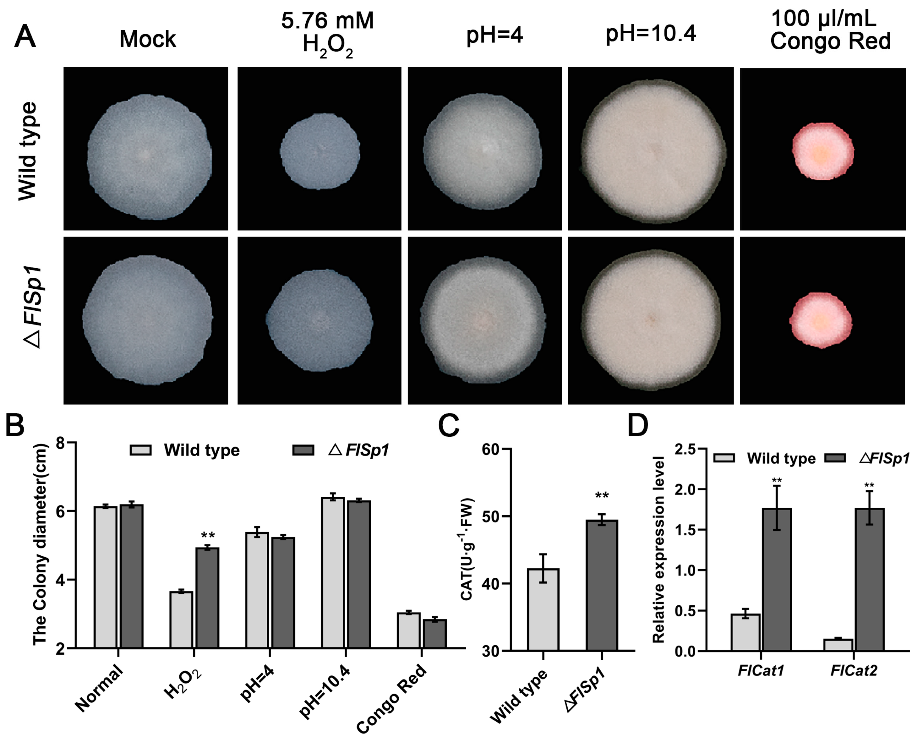
3.2. FlSp1 Negatively Mediates the Oxidative Stress Response in F. lateritium

To investigate the biological function of FlSp1, we obtained a gene mutant of FlSp1 using a homologous recombination strategy (Figure S1). Phenotypic analysis revealed no significant difference in the growth of the wild type (WT) and the FlSp1 mutant (ΔFlSp1) in normal basal medium (Figure 1A). Additionally, the knockout of Flsp1 did not affect the growth of the strain under acid-base conditions (pH = 4 and pH = 10.4) and cytostatic (Congo red) conditions. However, under oxidative stress (H2O2) conditions, the knockout of Flsp1 resulted in a significant increase in fungal tolerance to oxidative (Figure 1A,B) with relative inhibition rates of 18% (p < 0.01). Further analysis revealed that the knockout of Flsp1 resulted in a significant elevation (~1.25 fold) of peroxidase (CAT) activity in the strain in comparison with WT (p < 0.01) (Figure 1C). The analysis of transcriptional patterns revealed that the disruption of Flsp1 caused the significant up-regulation (~3.6- and 9.1-fold) of the expression of CAT synthesis-related genes—Flcat1 and Flcat2 (p < 0.01) (Figure 1D). This suggests that Flsp1 negatively mediates the oxidative stress responses in F. lateritium.

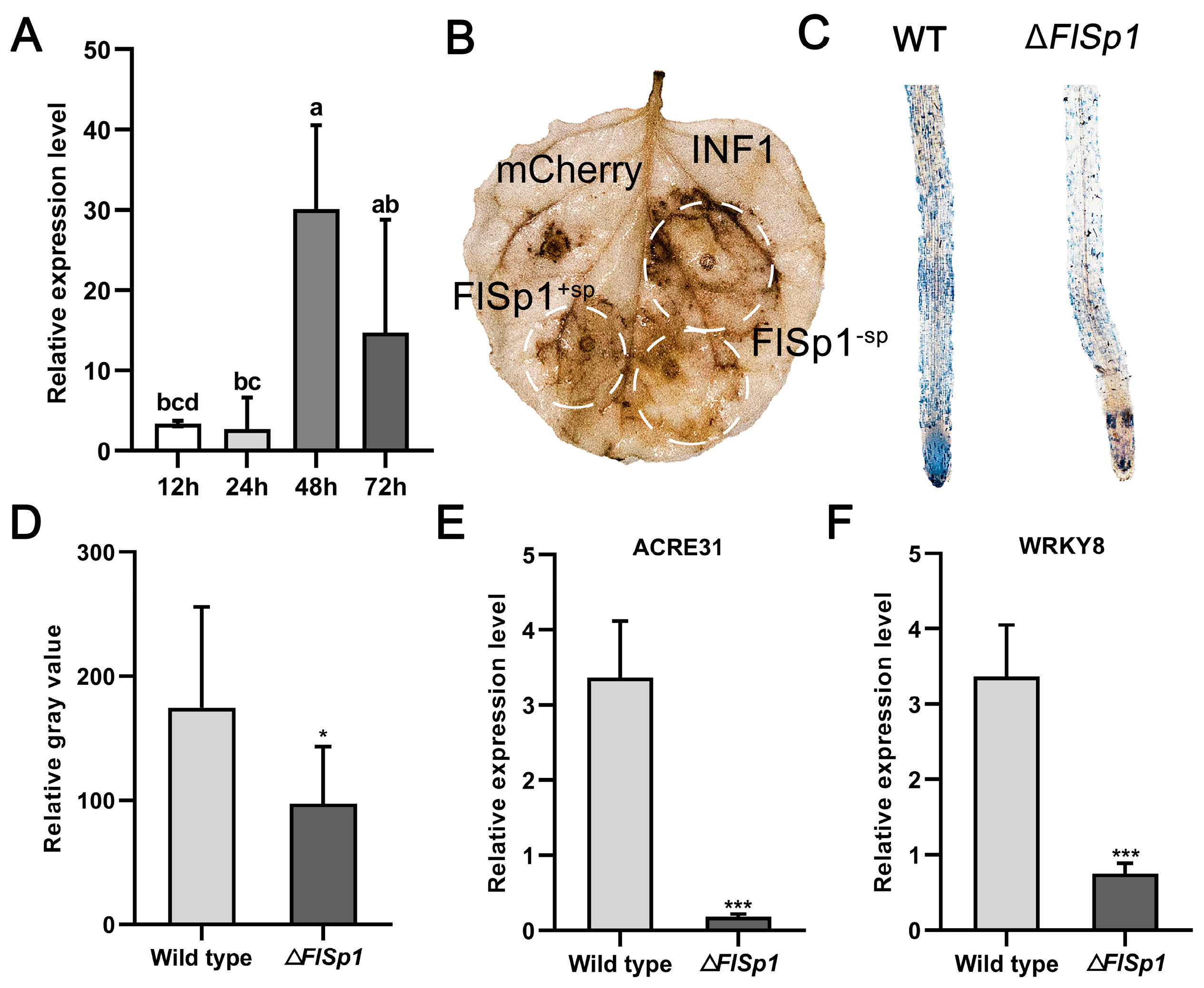
3.3. FlSp1 Stimulates Plant Immune Defense Response and Accumulates ROS

To further investigate the biological function of FlSp1 during fungal–plant interaction, the expression pattern of FlSp1 was analyzed during the interaction. The results showed that FlSp1 maintained low expression at 12 and 24 h of the interaction, reached peak expression at 48 h, and started to decrease at 72 h (Figure 2A), indicating that FlSp1 acts during the early stage of the interaction. Thus, we speculate that FlSp1 might be involved in the regulation of the plant immune systems during infestation with fungi. To test this theory, we examined the ability of FlSp1 to cause cell death or ROS accumulation via transient expression. The results showed that both the experimental group with a signal peptide (FlSp1+sp) and that without a signal peptide (FlSp1−sp) were able to cause the accumulation of ROS in comparison with the negative control (Figure 2B). In addition, we found that FlSp1 was unable to cause cell death in plant leaves, regardless of whether the signal peptide was removed (Figure S2). This result indicates that FlSp1, although it is able to stimulate plant immunity and ROS accumulation, does not cause a hypersensitive response to plant damage.
Therefore, we presume that FlSp1 may play a similar role in the interaction between F. lateritium and the host plant. We detected the early defense marker—O2—of the ROS through NBT staining. The results showed that the ΔFlSp1-treated roots produced significantly less blue insoluble material compared to the wild type (Figure 2C), which was examined for grayscale values, and it was found that the degree of ROS burst was reduced by approximately 50% (Figure 2D). Additionally, the significantly lower expression (~18.5 fold) of ACRE31, a marker gene for the plant immune system (p < 0.001), was detected in comparison with the wild type (Figure 2E,F). The expression of WRKY8, a transcription factor that positively regulates plant immune defense, was significantly down-regulated (~4.47 fold) (p < 0.001) (Figure 2F). These results suggest that FlSp1 triggers the accumulation of ROS in plants and mediates the immune system response of plants via the up-regulation of defense-related genes.

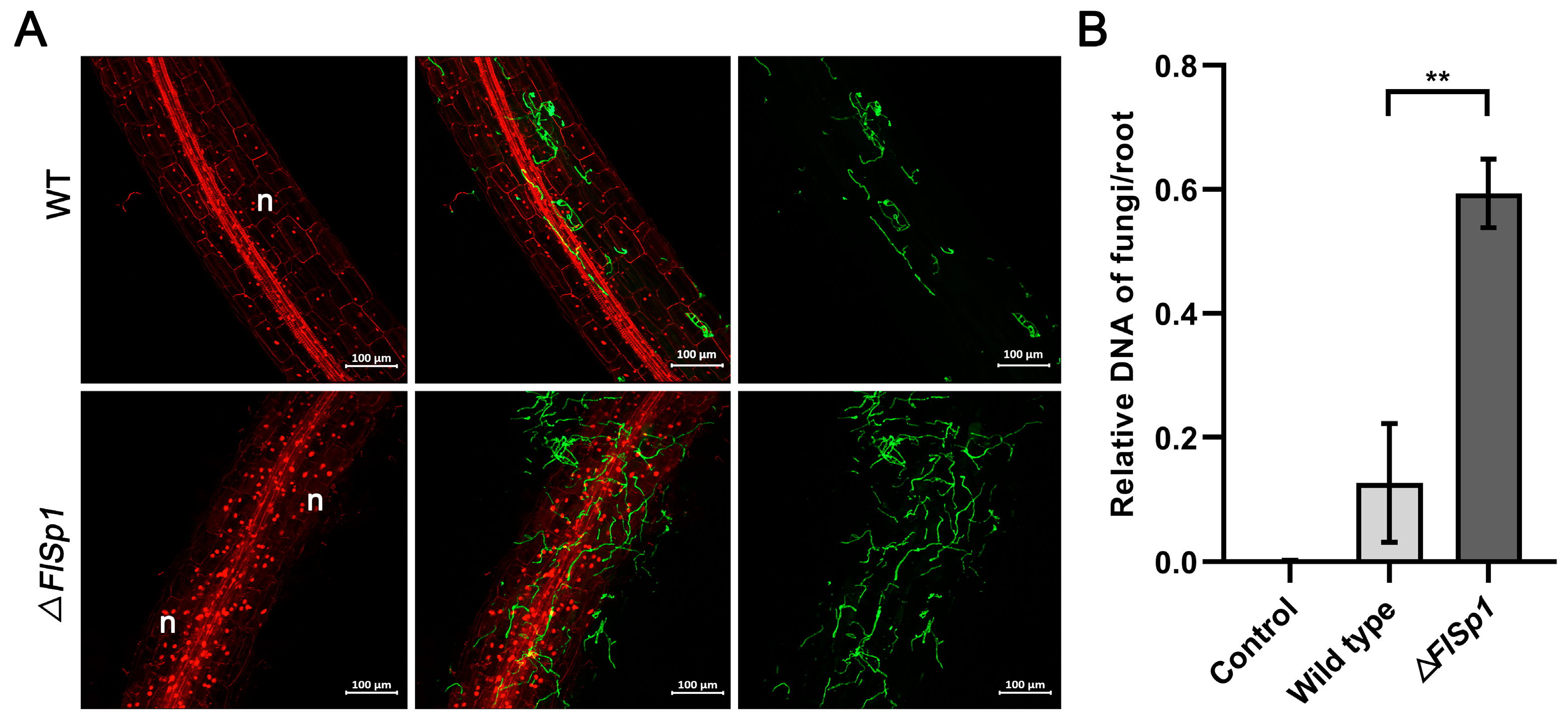
3.4. Disruption of FlSp1 Enhances the Colonization Rate of F. lateritium in Plants

The level of endophytic fungi colonization in plants is related to their function. To investigate the biological function of FlSp1 during interactions, we examined the effect of FlSp1 on fungal colonization in plant roots. The results revealed that the knockout of FlSp1 resulted in a significant increase in the fungal colonization of plants (Figure 3A), and the colonization of ΔFlSp1 in the root system was 3.3-fold higher than that of WT. (p < 0.01) (Figure 3B). It was shown that FlSp1 negatively regulated fungal colonization in plants.

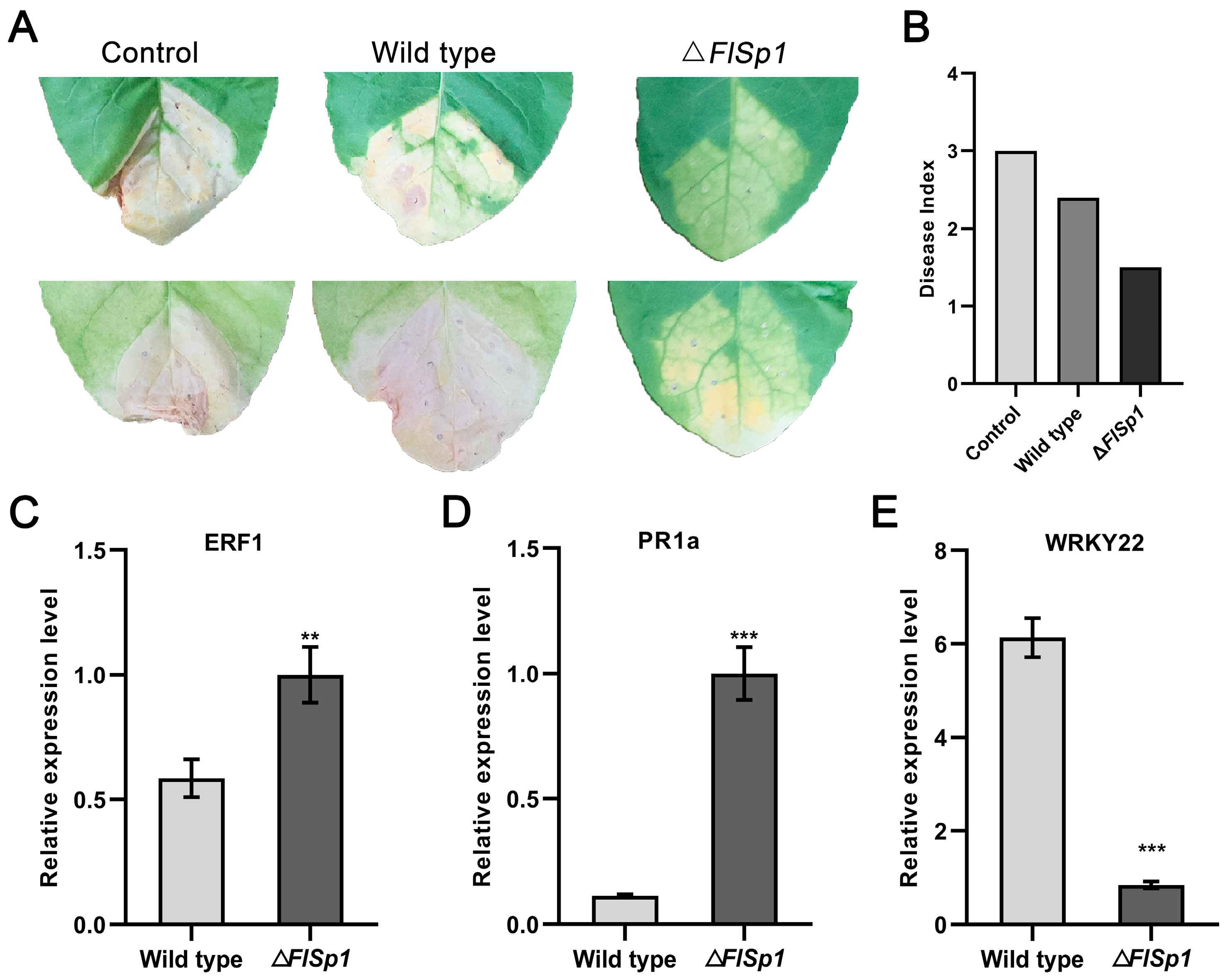
3.5. FlSp1 Negatively Mediates the Resistance of Tobacco to R. solanacearum

To investigate the effect of FlSp1 on the F. lateritium-mediated promotion of growth and disease resistance in the host plant, we first investigated the effect of FlSp1 on the growth-promoting effect of F. lateritium. Tobacco roots were irrigated with wild type and ΔFlSp1 spore suspension, and water was used as a control (Figure S4A). After 14 d, we counted the fresh weight, plant height, and chlorophyll content of tobacco leaves. The results showed that there was no significant effect on plant growth through FlSp1 knockdown (Figure S3B–E). To further investigate the effect of FlSp1 on plant resistance to disease, we used the spore suspensions of wild type and ΔFlSp1 to irrigate the tobacco roots. After 3 days, the plants were injected with R. solanacearum, and the plant disease index was created after 14 d. The results showed that ΔFlSp1 reduced the disease incidence of the plants compared with the wild type (Figure 4A). The disease indexes for the control, wild-type, and mutant groups were 3.0, 2.4, and 1.5, respectively. The results show that the ΔFlSp1 group had a significantly lower index of disease (Figure 4B). Meanwhile, ERF1, a key factor of the ethylene signaling pathway, and PR1a, a pathogenesis-related protein, were significantly up-regulated (~1.7- and 8.7-fold), while WRKY22, a transcription factor that negatively regulates plant immune defense, was significantly down-regulated (~7.2 fold) 2 d post-R. solanacearum injection (Figure 4C–E). The above results suggest that FlSp1 regulates plant resistance to R. solanacearum by mediating the expression of defense-related genes.

4. Discussion

In previous studies, F. lateritium has often been reported as a pathogenic fungus [10]. In our research group, a strain of F. lateritium was isolated from Nothapodytes pittosporoides, which acted as an endophyte to promote plant growth and mediate disease resistance in the plants of Solanaceae [11,12,13]. However, how the fungus mediates disease resistance in plants has not yet been reported. In this study, we focused on effectors, regarding them as an entry point, and predicted the effectors of F. lateritium through bioinformatics based on the F. lateritium genome. A secreted protein, FlSp1, which had the highest effector prediction score, was used as the study object. Subsequently, we analyzed the function of FlSp1 in fungal–plant interactions. Our results suggest that FlSp1 may act as an elicitor of plant defense responses, which stimulates plant immune responses (including ROS bursts) to limit the fungal colonization in plants. The reduction in fungal colonization helps to maintain balance with plants, so that F. lateritium does not damage plant tissue as much as the pathogenic fungus-causing disease.
During the process of fungal infection, ROS bursts caused by plant immune responses can inhibit fungal infection [25]. If fungi can better adapt to oxidative stress, it means that the colonization will be greatly increased [7]. Our data show that FlSp1 is highly expressed in the early stages of fungus–plant interaction, and the destruction of FlSp1 leads to a significant increase in the strain’s tolerance to oxidative stress. Therefore, we speculated that the FlSp1 mutant might be more adaptable to ROS bursts in the early interaction stages. The experimental results are consistent with our prediction that the colonization of ΔFlSp1 on tobacco significantly increased. However, it is interesting that the increase in the colonization of F. lateritium does not cause plant disease but rather enhances plant disease resistance. To clarify the reason for this, we initiated the transient expression of FlSp1. The results showed that FlSp1 could cause ROS bursts in plants but not hypersensitive responses. Further experimental data showed that the destruction of FlSp1 resulted in a significant decrease in the expression of defense-related genes. Those results indicate that FlSp1 may act as an elicitor of the plant defense response. FlSp1 is different from the elicitors of the pathogens that have been studied. For example, FoEG1 from Fusarium oxysporum can cause the cell death of N. benthamiana leaves, induce the accumulation of ROS, and enhance the resistance of tobacco to Botrytis cinerea [20]. The effector PvRxLR16 from Plasmopara viticola can cause cell death and the accumulation of ROS in N. benthamiana leaves and enhance the resistance to Phytophthora capsici [17]. It has a common characteristic that can trigger a strong immune response in N. benthamiana, leading to leaf necrosis and the accumulation of ROS, while FlSp1 does not cause necrosis. Therefore, we speculate that FlSp1, in addition to acting as an elicitor, may also have an important role in maintaining the endophytic relationship and the balance of the plant immune response, thus explaining the increase in ΔFlSp1 colonization. The growth of endophytes in the root system must be restricted; otherwise, there is the risk and possibility of causing disease [26]. However, why does an increase in the colonization of F. lateritium not cause disease? The reason may be that the level of colonization has not yet reached a critical value or that there are other factors that restrict F. lateritium colonization, keeping it within a certain colonization range. Our experimental data also showed that resistance to the pathogen was significantly higher in the ΔFlSp1-treated group after inoculation with R. solanacearum. Additionally, in this process, the expression of PR1a and ERF1, the marker genes of salicylic acid (SA)- and jasmonic acid (JA)-mediated signaling pathways, was significantly up-regulated. The results indicate that FlSp1 may indirectly influence the SA and JA signaling pathways in mediating resistance to R. solanacearum. However, this seems to contradict the result that ΔFlSp1 down-regulates plant defense genes. However, it is clear that the low immunity of the endophytes interacting with the plant does not affect the elevated immune defense response of the plant [27,28,29]. Therefore, we speculate that, within a certain range of colonization, there is a positive correlation between the enhancement of the plant immune response by endophytic F. lateritium.

Supplementary Materials

The following supporting information can be downloaded at: https://www.mdpi.com/article/10.3390/jof9050519/s1, Figure S1: Knockout of FlSp1 by homologous recombination strategy; Figure S2: FlSp1 causes plant immune response but not leaf death; Figure S3: FlSp1 does not affect tobacco growth; Table S1: Prediction and proportion of effectors; Table S2: According to BLAST comparison of F. lateritium, 69 effectors are characterized proteins.

Author Contributions

J.H., Z.H. and J.K. conceived and designed the experiment; J.H., G.L. and J.W. contributed to the data analysis; J.H., X.Z., Q.X. and Y.L. prepared the plants and fungi used in the experiment. All authors have read and agreed to the published version of the manuscript.

Funding

This work was supported by The National Natural Science Foundation of China (No. 32160667, 31901947, and 32170019). Qian Kehe-ZK [2021]-145/the Basic Project of Guizhou Provincial Natural Science Foundation. Gui Da Ren Ji He Zi (2019) 71/Guizhou University PhD Fund Project. Guizhou University Incubation [2019] 19/Guizhou University Incubation Program.

Institutional Review Board Statement

Not applicable.

Informed Consent Statement

Not applicable.

Data Availability Statement

Not applicable.

Acknowledgments

The author thanks their country and their two tutors who supported them continuously. Additionally, they sincerely thank Zhibo Zhao for support and resources (pCAMBIA1300).

Conflicts of Interest

The authors declare no conflict of interest.

References

  1. Martel, A.; Ruiz-Bedoya, T.; Breit-McNally, C.; Laflamme, B.; Desveaux, D.; Guttman, D.S. The ETS-ETI Cycle: Evolutionary Processes and Metapopulation Dynamics Driving the Diversification of Pathogen Effectors and Host Immune Factors. Curr. Opin. Plant Biol. 2021, 62, 102011. [Google Scholar] [CrossRef]
  2. Mittler, R.; Zandalinas, S.I.; Fichman, Y.; Van Breusegem, F. Reactive Oxygen Species Signalling in Plant Stress Responses. Nat. Rev. Mol. Cell Biol. 2022, 23, 663–679. [Google Scholar] [CrossRef]
  3. Ju, S.; Go, Y.S.; Choi, H.J.; Park, J.M.; Suh, M.C. DEWAX Transcription Factor Is Involved in Resistance to Botrytis Cinerea in Arabidopsis Thaliana and Camelina Sativa. Front. Plant Sci. 2017, 8, 1210. [Google Scholar] [CrossRef] [PubMed]
  4. Zhao, P.; Zhang, F.; Liu, D.; Imani, J.; Langen, G.; Kogel, K.-H. Matrix Metalloproteinases Operate Redundantly in Arabidopsis Immunity against Necrotrophic and Biotrophic Fungal Pathogens. PLoS ONE 2017, 12, e0183577. [Google Scholar] [CrossRef]
  5. Wang, M.; Ji, Z.; Yan, H.; Xu, J.; Zhao, X.; Zhou, Z. Effector Sntf2 Interacted with Chloroplast-Related Protein Mdycf39 Promoting the Colonization of Colletotrichum Gloeosporioides in Apple Leaf. Int. J. Mol. Sci. 2022, 23, 6379. [Google Scholar] [CrossRef] [PubMed]
  6. Qi, T.; Guo, J.; Liu, P.; He, F.; Wan, C.; Islam, M.A.; Tyler, B.M.; Kang, Z.; Guo, J. Stripe Rust Effector PstGSRE1 Disrupts Nuclear Localization of ROS-Promoting Transcription Factor TaLOL2 to Defeat ROS-Induced Defense in Wheat. Mol. Plant 2019, 12, 1624–1638. [Google Scholar] [CrossRef] [PubMed]
  7. Gao, M.; He, Y.; Yin, X.; Zhong, X.; Yan, B.; Wu, Y.; Chen, J.; Li, X.; Zhai, K.; Huang, Y.; et al. Ca2+ Sensor-Mediated ROS Scavenging Suppresses Rice Immunity and Is Exploited by a Fungal Effector. Cell 2021, 184, 5391–5404.e17. [Google Scholar] [CrossRef]
  8. Zhu, L.; Li, T.; Wang, C.; Zhang, X.; Xu, L.; Xu, R.; Zhao, Z. The Effects of Dark Septate Endophyte (DSE) Inoculation on Tomato Seedlings under Zn and Cd Stress. Environ. Sci. Pollut. Res. 2018, 25, 35232–35241. [Google Scholar] [CrossRef]
  9. Mori, M.P.; Penjweini, R.; Knutson, J.R.; Wang, P.; Hwang, P.M. Mitochondria and Oxygen Homeostasis. FEBS J. 2022, 289, 6959–6968. [Google Scholar] [CrossRef]
  10. Vitale, S.; Santori, A.; Wajnberg, E.; Castagnone-Sereno, P.; Luongo, L.; Belisario, A. Morphological and Molecular Analysis of Fusarium Lateritium, the Cause of Gray Necrosis of Hazelnut Fruit in Italy. Phytopathology 2011, 101, 679–686. [Google Scholar] [CrossRef]
  11. Xiao, Q. Effects of an Endophytic Fusarium Lateritium on Growth and Disease Resistance of Tomato. J. Huazhong Agric. Univ. 2022, 41, 173–180. [Google Scholar]
  12. Wang, J. Endophyte Fusarium Lateritium Showing Potato Growth Promotion and Disease Resistanceand the Construction of Its Genetic Transformation System. Mycosystema 2021, 40, 2008–2023. [Google Scholar]
  13. Zha, X. A Strain of Endophytic Fusarium Lateritium Promotes Growth and Resistance to Bacteriawilt of Tobacco. Mycosystema 2022, 41, 1658–1671. [Google Scholar]
  14. Sperschneider, J.; Dodds, P.N. EffectorP 3.0: Prediction of Apoplastic and Cytoplasmic Effectors in Fungi and Oomycetes. Mol. Plant-Microbe Interact. 2022, 35, 146–156. [Google Scholar] [CrossRef]
  15. Jones, D.A.; Bertazzoni, S.; Turo, C.J.; Syme, R.A.; Hane, J.K. Bioinformatic Prediction of Plant–Pathogenicity Effector Proteins of Fungi. Curr. Opin. Microbiol. 2018, 46, 43–49. [Google Scholar] [CrossRef]
  16. Ma, J.-C.; Zhou, Q.; Zhou, Y.-H.; Liao, X.-G.; Zhang, Y.-J.; Jin, D.; Pei, Y. The Size and Ratio of Homologous Sequence to Non-Homologous Sequence in Gene Disruption Cassette Influences the Gene Targeting Efficiency in Beauveria Bassiana. Appl. Microbiol. Biotechnol. 2009, 82, 891–898. [Google Scholar] [CrossRef] [PubMed]
  17. Xiang, J.; Li, X.; Yin, L.; Liu, Y.; Zhang, Y.; Qu, J.; Lu, J. A Candidate RxLR Effector from Plasmopara Viticola Can Elicit Immune Responses in Nicotiana Benthamiana. BMC Plant Biol. 2017, 17, 75. [Google Scholar] [CrossRef]
  18. Zhang, C.; Chen, H.; Cai, T.; Deng, Y.; Zhuang, R.; Zhang, N.; Zeng, Y.; Zheng, Y.; Tang, R.; Pan, R.; et al. Overexpression of a Novel Peanut NBS-LRR Gene AhRRS5 Enhances Disease Resistance to Ralstonia Solanacearum in Tobacco. Plant Biotechnol. J. 2017, 15, 39–55. [Google Scholar] [CrossRef]
  19. Jing, Y.; Shen, N.; Zheng, X.; Fu, A.; Zhao, F.; Lan, W.; Luan, S. Danger-Associated Peptide Regulates Root Immune Responses and Root Growth by Affecting ROS Formation in Arabidopsis. Int. J. Mol. Sci. 2020, 21, 4590. [Google Scholar] [CrossRef]
  20. Zhang, L.; Yan, J.; Fu, Z.; Shi, W.; Ninkuu, V.; Li, G.; Yang, X.; Zeng, H. FoEG1, a Secreted Glycoside Hydrolase Family 12 Protein from Fusarium Oxysporum, Triggers Cell Death and Modulates Plant Immunity. Mol. Plant Pathol. 2021, 22, 522–538. [Google Scholar] [CrossRef]
  21. Palmieri, D.; Vitale, S.; Lima, G.; Di Pietro, A.; Turrà, D. A Bacterial Endophyte Exploits Chemotropism of a Fungal Pathogen for Plant Colonization. Nat. Commun. 2020, 11, 5264. [Google Scholar] [CrossRef]
  22. Zuccaro, A.; Lahrmann, U.; Güldener, U.; Langen, G.; Pfiffi, S.; Biedenkopf, D.; Wong, P.; Samans, B.; Grimm, C.; Basiewicz, M.; et al. Endophytic Life Strategies Decoded by Genome and Transcriptome Analyses of the Mutualistic Root Symbiont Piriformospora Indica. PLoS Pathog. 2011, 7, e1002290. [Google Scholar] [CrossRef]
  23. Park, E.; Cho, M.; Ki, C.-S. Correct Use of Repeated Measures Analysis of Variance. Ann. Lab. Med. 2009, 29, 1–9. [Google Scholar] [CrossRef] [PubMed]
  24. Sperschneider, J.; Gardiner, D.M.; Dodds, P.N.; Tini, F.; Covarelli, L.; Singh, K.B.; Manners, J.M.; Taylor, J.M. E ffector P: Predicting Fungal Effector Proteins from Secretomes Using Machine Learning. New Phytol. 2016, 210, 743–761. [Google Scholar] [CrossRef] [PubMed]
  25. Kawano, Y.; Akamatsu, A.; Hayashi, K.; Housen, Y.; Okuda, J.; Yao, A.; Nakashima, A.; Takahashi, H.; Yoshida, H.; Wong, H.L.; et al. Activation of a Rac GTPase by the NLR Family Disease Resistance Protein Pit Plays a Critical Role in Rice Innate Immunity. Cell Host Microbe 2010, 7, 362–375. [Google Scholar] [CrossRef]
  26. Liberman, L.M. A Friend in Need (of Nutrients) Is A…. Cell 2016, 165, 269–271. [Google Scholar] [CrossRef] [PubMed]
  27. Guo, R.; Ji, S.; Wang, Z.; Zhang, H.; Wang, Y.; Liu, Z. Trichoderma Asperellum Xylanases Promote Growth and Induce Resistance in Poplar. Microbiol. Res. 2021, 248, 126767. [Google Scholar] [CrossRef]
  28. Pieterse, C.M.J.; Zamioudis, C.; Berendsen, R.L.; Weller, D.M.; Van Wees, S.C.M.; Bakker, P.A.H.M. Induced Systemic Resistance by Beneficial Microbes. Annu. Rev. Phytopathol. 2014, 52, 347–375. [Google Scholar] [CrossRef]
  29. Contreras-Cornejo, H.A.; Macías-Rodríguez, L.; Beltrán-Peña, E.; Herrera-Estrella, A.; López-Bucio, J. Trichoderma -Induced Plant Immunity Likely Involves Both Hormonal- and Camalexin-Dependent Mechanisms in Arabidopsis Thaliana and Confers Resistance against Necrotrophic Fungi Botrytis Cinerea. Plant Signal. Behav. 2011, 6, 1554–1563. [Google Scholar] [CrossRef]
Figure 1. FlSp1 negatively mediates the oxidative stress response in F. lateritium. (A) Effect of FlSp1 on the growth of F. lateritium under various stresses. (B) The diameter of the colonies was measured. Three replicates per treatment. (C) CAT enzyme biopsy assay of mycelium. The same mass was chosen from both wild type and ΔFlSp1 mycelium and tested for CAT enzyme activity under oxidative stress. (D) Analysis of the transcriptional patterns of Flcat1 and Flcat2. Three replicates were set up for each experiment; ** indicates highly significant difference (p < 0.01).
Jof 09 00519 g001
Figure 2. FlSp1 stimulates the plant immune system and accumulates ROS. (A) Expression pattern analysis of FlSp1. FlSp1 is highly expressed in the early stages of interaction. Separate one-way ANOVAs were used for each group, followed by pairwise comparisons at different time points using Tukey’s HSD method with different lowercase letters indicating significant differences in pairwise comparisons (p < 0.05). (B) FlSp1sp and FlSp1sp cause ROS accumulation in plant leaves. INF1 was used as a positive control. (C) ΔFlSp1 caused lower ROS burst in plant roots than in wild type. (D) Roots were stained for NBT and then examined for grey values using ImageJ. (E,F) Wild type and ΔFlSp1 were detected for the relative expression of defense-related genes after 1 d of co-culturing with N. benthamiana. Each experiment was repeated three times; * indicates significant difference (p < 0.05) and *** indicates extremely significant difference (p < 0.001).
Jof 09 00519 g002
Figure 3. Knockout of FlSp1 increases fungal colonization. (A) The mycelium was stained after 2 d of co-culturing and was photographed at 488 mM. n indicates plant cell nuclei. (B) After 2 d of co-culturing, the levels of wild types and mutant DNA were examined in the root system of the plant. Each experiment was repeated three times; ** indicates highly significant differences (p < 0.01).
Jof 09 00519 g003
Figure 4. FlSp1 negatively mediates the resistance of tobacco to R. solanacearum. (A) The ΔFlSp1 group had a significantly lower index of disease. (B) Disease index for each treatment group. (CE) After root irrigation treatment, detection of expression of defense-related genes 2 d after the R. solanacearum injection. Each experiment was repeated three times; ** indicates significant difference (p < 0.01) and *** indicates a highly significant difference (p < 0.001).
Jof 09 00519 g004
Table 1. Candidate proteins with the highest likelihood scores for 16 effectors. EffectorP predicts fungal effectors based on machine learning. Features that discriminate fungal effectors from secreted non-effectors are predominantly sequence length, molecular weight, and protein net charge, as well as cysteine, serine, and tryptophan content [24].
GeneIDEffector
Probability
(aa) Position of
Signal Peptide
(aa) Size of
Peptide
Subcellular LocalizationIntegral Prediction of the Protein Location
EVM0011051.10.9791–17108Extracellular2.4
EVM0014202.10.9711–23127Extracellular2.4
EVM0003861.10.9661–1694Extracellular2.3
EVM0007002.10.9651–2197Extracellular2.4
EVM0005253.10.9591–18121Extracellular3.2
EVM0002480.10.9521–1796Extracellular2.5
EVM0001443.10.9521–1897Extracellular2.4
EVM0000104.10.9521–1889Extracellular2.4
EVM0005809.10.9491–18124Extracellular2.6
EVM0004957.10.9481–1798Extracellular2.6
EVM0001142.10.9431–1988Extracellular2.7
EVM0005907.10.9281–17105Extracellular2.7
EVM0013376.10.9121–23131Extracellular2.4
EVM0007040.10.911–1765Extracellular2.4
EVM0011395.10.9091–16148Extracellular2.4
EVM0001445.10.9091–16140Extracellular2.8
Disclaimer/Publisher’s Note: The statements, opinions and data contained in all publications are solely those of the individual author(s) and contributor(s) and not of MDPI and/or the editor(s). MDPI and/or the editor(s) disclaim responsibility for any injury to people or property resulting from any ideas, methods, instructions or products referred to in the content.

