Article

Biodiversity of Basidiomycetous Yeasts Associated with Cladonia rei Lichen in Japan, with a Description of Microsporomyces cladoniophilus sp. nov

1
Department of Biological Science and Technology, Tokyo University of Science, Niijuku 6-3-1, Katsushika, Tokyo 125-8585, Japan
2
College of Science and Engineering, Kanto Gakuin University, Mutsuura-higashi 1-50-1, Kanazawa-ku, Yokohama 236-8501, Kanagawa, Japan
3
Medical Mycology Research Center, Chiba University, Inohana 1-8-1, Chuo-ku, Chiba 260-8673, Chiba, Japan
*
Author to whom correspondence should be addressed.
J. Fungi 2023, 9(4), 473; https://doi.org/10.3390/jof9040473
Submission received: 28 December 2022 / Revised: 8 April 2023 / Accepted: 10 April 2023 / Published: 14 April 2023

Abstract

:
For more than a century, lichens have been used as an example of dual-partner symbiosis. Recently, this has been challenged by the discovery of various basidiomycetous yeasts that coexist in multiple lichen species, among which Cladonia lichens from Europe and the United States were discovered to be highly specifically associated with the basidiomycetous yeast of the family Microsporomycetaceae. To verify this highly specific relationship, we investigated the diversity of basidiomycetous yeasts associated with Cladonia rei, a widely distributed lichen in Japan, by applying two approaches: yeast isolation from the lichen thalli and meta-barcoding analysis. We obtained 42 cultures of Cystobasidiomycetous yeast which were grouped into six lineages within the family Microsporomycetaceae. Unexpectedly, although the cystobasidiomycetes-specific primer was used, not only the cystobasidiomycetous yeasts but species from other classes were also detected via the meta-barcoding dataset; in particular, pucciniomycetous yeasts were found at a high frequency in some samples. Further, Halobasidium xiangyangense, which was detected in every sample with high abundance, is highly likely a generalist epiphytic fungus that has the ability to associate with C. rei. In the pucciniomycetous group, most of the detected species belong to the scale insect-associated yeast Septobasidium genus. In conclusion, even though Microsporomyces species are not the only yeast group associated with Cladonia lichen, our study demonstrated that the thalli of Cladonia rei lichen could be a suitable habit for them.

1. Introduction

Symbioses are widespread and essential to the diversity of our ecosystems. Lichen represents a successful model of the iconic symbiosis of stable dual mutualism of a single fungus and a photosynthesizing symbiont. With their long macro-evolutionary history, lichens can inhabit any environment or habitat and currently dominate nearly 10% of the Earth’s terrestrial ecosystem [1,2]. Among the lichen-forming fungi, Lecanoromycetes, as one of the largest clades, includes more than 80% of the lichen-forming species [3]. Belonging to this class, Cladonia is one of the most species-rich and morphologically distinctive genera of lichen-forming fungi, with a worldwide distribution [4].
Due to recent discoveries, lichens have been considered not only relevant to dual-partner symbiosis but also part of the complex ecosystems that are inhabited by other microscopic organisms, such as prokaryotes and fungi [5]. As lichens appeared 600 million years ago [6], the existence of secondary fungal species in Early Devonian (~415 million years ago) lichen fossils [7] indicates that this association might have existed throughout the evolutionary history of lichen. These coexistences have been detected in various lichen species [8,9,10]. As they have been mostly considered parasites or lichenicolous fungi [11], and a few as endolichenic fungi [12], and due to their rare isolation, the functions, influence, and roles on communities of lichens have been neglected or underestimated. The recent discovery by Spribille et al. [13] shed new light on this issue by proving the presence of basidiomycetous yeasts in the lichen cortex and their contribution to different phenotypes of two genetically inseparable Bryoria lichens. In the same study, the yeasts of the class Cystobasidiomycetes were detected in the cortex of various macro-lichen taxa, suggesting that the yeasts may represent the third essential constituent of lichen symbiosis. Multiple studies conducted to infer the diversity of lichen-associated basidiomycetous yeasts have delivered conflicting results. Although some supported the association between lichens and basidiomycetous yeasts [13,14,15], others argued against this association [16,17]. Spribille et al. [13], upon screening the lichen-associated yeasts from a wide range of lichen species, detected a specific group of basidiomycetous yeasts, Cyphobasidiales (Pucciniomycotina, Basidiomycota), associated with multiple species, but the yeasts detected from Cladonia lichens were grouped into one lineage within the Microsporomycetaceae family (Cystobasidiomycetes, Pucciniomycotina, Basidiomycota). Later, these unknown yeasts were discovered in a widespread species of this genus, despite their reproductive strategies [14,18]. This showed the high specificity of association with basidiomycetous yeasts of Cladonia lichen, while the reproductive and dispersal strategies were shown to be the key factors shaping photobiont diversity in this lichen group [18]. Surveys of these Cladonia-associated yeasts have been lacking in most of Asia, where several species from the same family have been discovered recently [19].
Most studies that have attempted to discover the biodiversity of lichen-associated basidiomycetous yeasts have utilized one or two approaches that might lead to bias. In some studies, Sanger sequencing, which provides high confidence for detected taxa from sequences, was utilized to determine the lichen-associated yeasts [16], but this approach is not sensitive enough for low abundance symbionts, and the probability of detection of certain species significantly depends on the biodiversity of the community within the lichens [20]. Although isolation from the thallus fragments method seems to have provided decent evidence of the existence of lichen-associated yeasts, it is highly limited by such factors as the type of culture media used for isolation and the skills of technicians [21,22]. Further, the contamination rate of cultures from Cladonia lichens due to their natural habitat [22] increases the difficulty of revealing the true diversity of the yeast community within the thalli of these lichens. Given advances in sequencing technologies, the meta-barcoding approach with new emerging high-throughput sequencing techniques seems to be a powerful tool for detecting the true diversity of the basidiomycetous yeast community within lichen thalli [23,24,25,26]. However, the meta-barcoding method also suffers numerous biases, so it requires affirmation from other methods [27,28,29].
Here, we focused on the biodiversity of basidiomycetous yeasts associated with Cladonia rei, which is widely distributed in Japan, using multiple approaches to infer the existence of lichen-associated yeast species and determine whether there is a high specificity in the third symbionts of Cladonia lichen in the region. The yeasts were isolated from multiple lichen specimens collected from various locations. The basidiomycetous yeasts were further screened via meta-barcoding analysis of conserved genomic sequences, amplified with the basidiomycete-specific primers designed by Spribille et al. [13].

2. Materials and Methods

2.1. Sampling

Fifteen specimens of Cladonia rei thallus were collected from two locations in Chiba prefecture, Japan, from March 2017 to November 2018 (Table 1). The species was confirmed via morphological and molecular methods. The collected thallus was frozen and stored at −20 °C.

2.2. Isolation, Culturing, and Characterization of the Yeast Strains

Strains were isolated from the thallus using the method described by Yamamoto et al. [22]. The thalli were inspected for external signs of discoloration and parasitism. For each isolation, around 10 podetia were selected and washed with running water for at least 1 h and then with 7 mL 1% sodium hypochlorite. The washed podetia were rinsed with sterilized distilled water thoroughly several times and then ground into small pieces using a mortar and pestle. Finally, the thallus fragments with sizes between 150 and 500 µm were selected using two nylon sieve meshes and then placed onto Malt/Yeast extract (YM) medium [30].
Yeasts grown from the thallus fragments were isolated into axenic cultures and were then maintained on YM medium at 25 °C. Morphological, physiological, and biochemical characteristics were examined following the methods of Kurtzman et al. [31]. We tested for ballistoconidia using the inverted plate method [32]. The glass slide containing the discharged spores was removed for examination under the microscope after 3–10 days. The potential sexual cycles were investigated using the Dalmau plate method on corn meal agar (CMA), YM agar (YMA), and potato–dextrose agar (PDA), according to Kurtzman et al. [31]. A loopful of cells of each strain was streaked on an agar plate and incubated at 17 °C for 1 month, and the cultures were examined microscopically every 2 weeks.

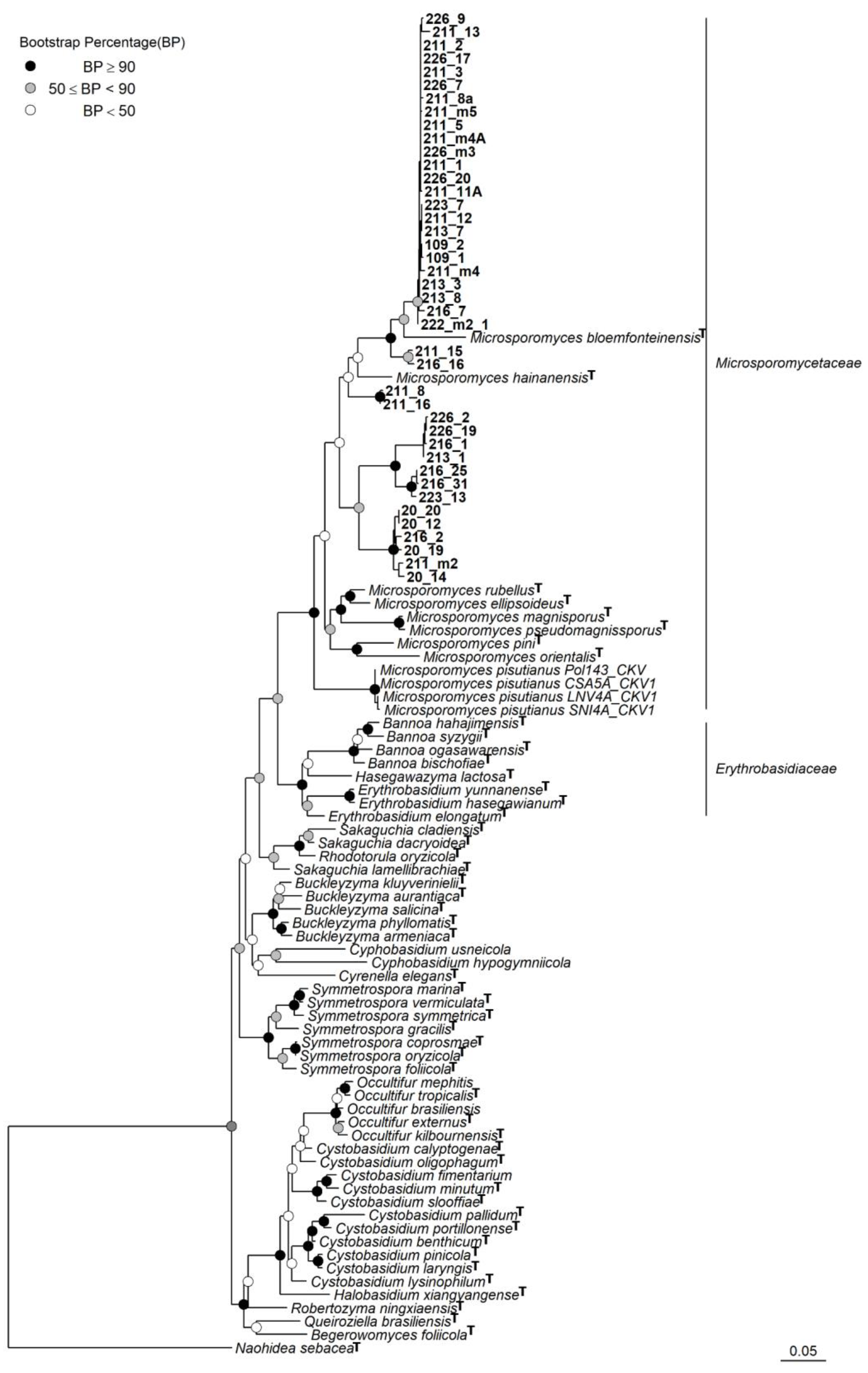
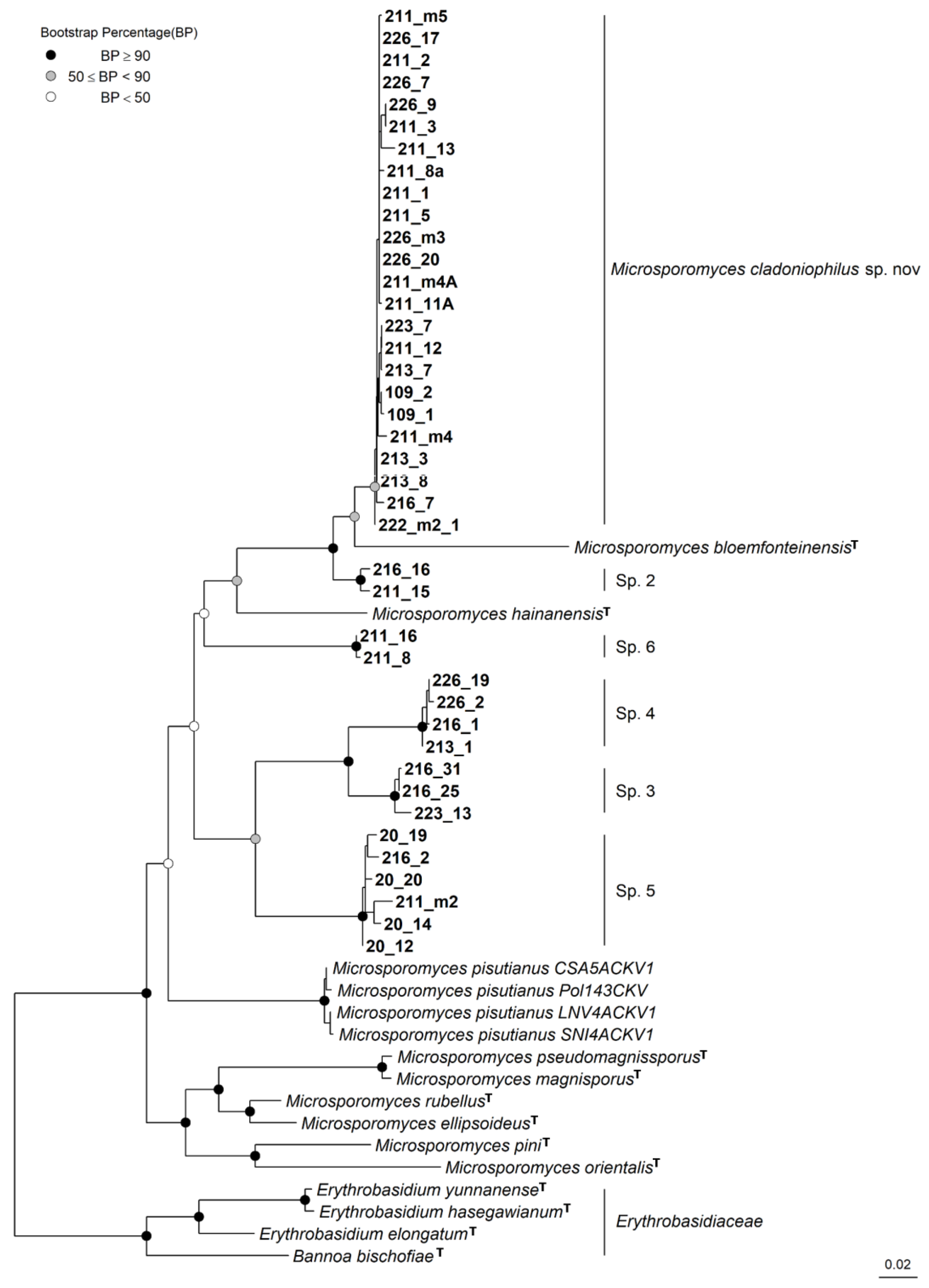
2.3. Sequencing and Phylogenetic Analyses

The total DNA of C. rei thallus and the yeast cultures was extracted by the method of Nakada et al. [33]. The ITS rDNA sequences, which were amplified with ITS4 and ITS5 primer, were used to confirm the species of lichen thallus [34]. For the isolated yeasts, the ITS region and partial LSU rDNA were amplified with ITS4 and ITS5; LR0 and LR5 primers were used, respectively [34,35]. PCR amplification for ITS began with denaturation at 95 °C for 5 min, followed by 35 cycles of denaturation at 95 °C for 30 s, annealing at 55 °C for 1 min, and elongation at 72 °C for 1 min, finished with extension at 72 °C for 5 min. PCR amplification was carried out for LSU denaturation at 95 °C for 5 min, which was followed by 35 cycles of denaturation at 95 °C for 30 s, annealing at 55 °C for 1 min, and elongation at 68 °C for 2 min, and finished with extension at 72 °C for 5 min. The PCR products were purified using a FastGeneTM Gel/PCR extract kit (Nippon Genetics, Tokyo, Japan). The forward and reverse strands of the purified PCR products were amplified using the same primers with a BigDyeTM Terminator v3.1 Cycle Sequencing Kit (Applied Biosystems, Waltham, MA, USA), following purification with Sephadex G-50. Sanger sequencing was conducted with a 3130xl Genetic Analyzer and a 3500 Genetic Analyzer (Applied Biosystems). The final sequences were assembled using Genetyx v. 13 (GENETYX, Tokyo, Japan).
To infer the phylogenetic position of our strains within the class Cystobasidiomycetes, we conducted a phylogenetic analysis on the basis of the phylogenetic results from Li et al. [19] and Černajová and Škaloud [14]. Sequences of the ITS region and LSU rDNA of species in the Cystobasidiomycetes class retrieved from GenBank, together with the sequences of the obtained cultures (Table S1), were aligned using MAFFT using the Q-INS-I method; each region was aligned separately. Gblocks v. 0.91b was used to remove the ambiguously aligned region [36]. Substitution models were estimated with Akaike Information Criterion (AIC) using JModelTest v.2.1.4 [37]. The model GTR+ I + G was selected for both LSU and ITS. Our cultures all clustered within the family Microsporomycetaceae (see Results). Thus, we reconstructed the phylogeny of this family with the currently accepted species [19] and our cultures. Bannoa bischofiae and three Erythobasidium species (Erythrobasidiaceae, Cystobasidiomycetes, Pucciniomycotina, Basidiomycota) were selected as outgroups. The sequences were processed as described above. We used RAxML-HPC v.8 to construct the maximum likelihood (ML) phylogeny [38] and perform 1000 rapid bootstrap replicates. The output trees were visualized using FigTree v.1.4.4 [39]. The final presentation was undertaken in free RStudio software [40,41] with the packages ggtree [42] and treeio [43].

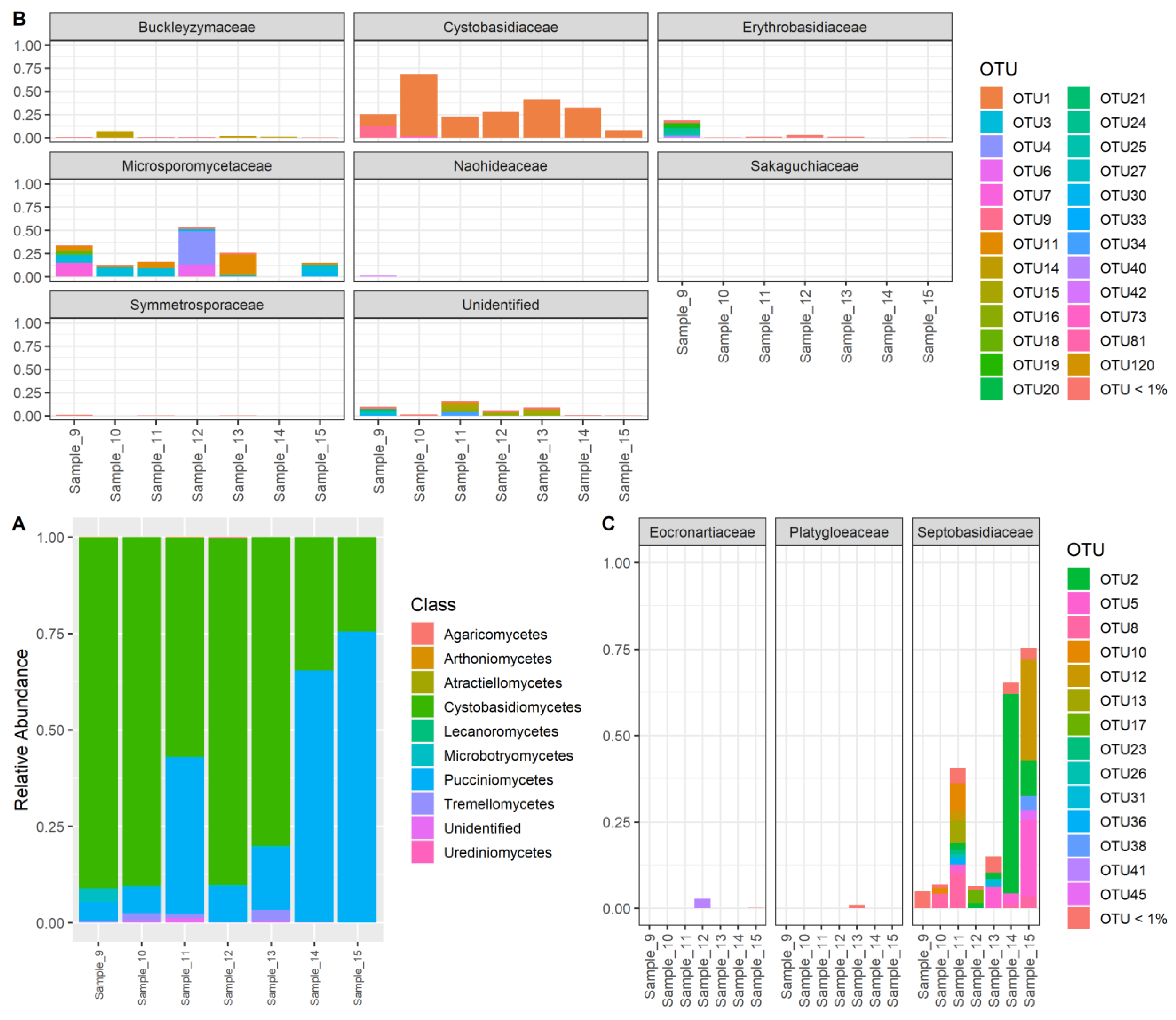
2.4. Meta-Barcoding Sequencing and OTU Identity Analysis

Seven specimens were selected for meta-barcoding (Table 1). The yeast ITS rDNA from the DNA of C. rei was amplified using the Cystobasidiomycetes-specific primer ITS_symrho_2F and LR0_symrho_R designed by Spribille et al. [13]. PCR products were checked on 2% agarose gel and quantified using Qubit 2.0. The libraries were generated with NEBNext®Ultra™ DNA Library Prep Kit for Illumina® (San Diego, CA, USA). Sequencing was completed with the paired-end protocol 2 × 300 bp on a PE300 MiSeq instrument at Novogene, Tokyo, Japan. All output reads are available in the Short Read Archive under accession PRJNA765160. Raw paired-end reads were merged using FLASH v.1.2.7 [44]. The adapters and barcodes were trimmed from sequences using fastx_trimmer, implemented in FASTX-Toolkit [45]. The operational taxonomic units (OTUs) were identified by clustering the trimmed sequences based on the sequence similarity threshold (97%) using USEARCH v.11 [46]. Each OTU’s taxonomy assignment was performed by blasting their reference sequences [47] against the full “UNITE + INSD” dataset for fungi version 2021-05-10 [48] with a 10 × 10−50 e-value threshold. The OTUs that were identified as cystobasidiomycetous yeasts and showed a relative abundance in any sample higher than 1% were further included in the phylogenetic analysis, with ITS region data to determine approximately their position within the class using the same method as above. To visualize the yeast community composition, we used the free RStudio software [40,41] with the ggplot2 package [49]. To avoid an over-detailed graph, the OTUs with a relative abundance of less than 1% were grouped.

3. Results

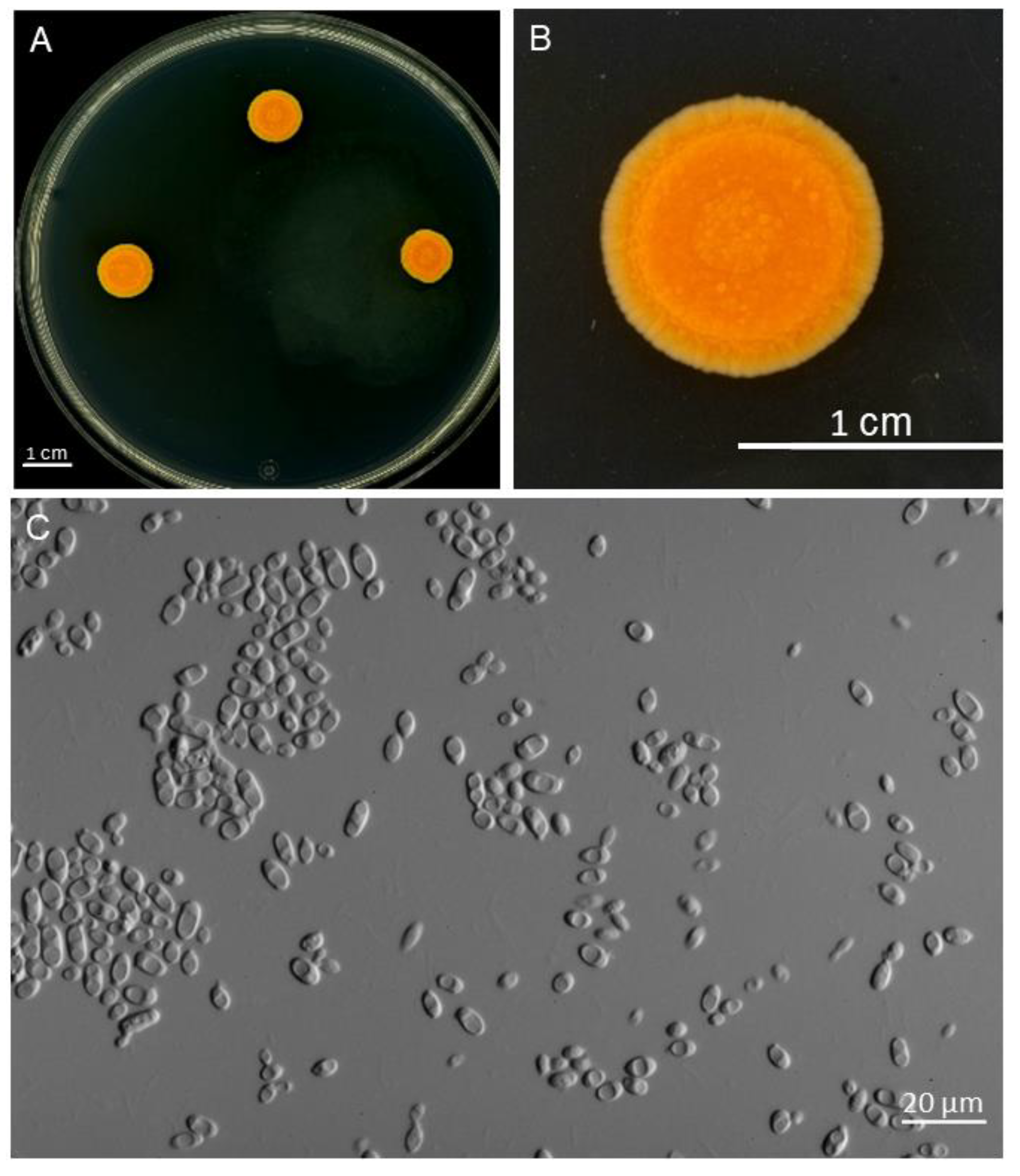
3.1. Isolated Cultures

Forty-two strains were cultured successfully from eight C. rei specimens and identified by sequencing the ITS and LSU rDNA (Table 2). Our phylogenetic analyses showed a consistent topology and were generally congruent with most of the major groups from [19] (Figure 1). Furthermore, the phylogenetic analyses also grouped Lichenozyma species with Microsporomyces species and our cultures. Hence, we agree with Li et al. (2020) that the Lichenozyma genus should be considered synonymous with Microsporomyces. Although the phylogenetic analysis of the Cystobasidiomycetes class was slightly incongruent in topology with the analysis on the family Microsporomycetaceae, both indicated that all of the isolated cultures belonged to Microsporomycetaceae, with high statistical support. Furthermore, the phylogenetic analysis of Microsporomycetaceae showed that our cultures could be grouped into six distinct lineages (Figure 2). The first lineage (Microsporomyces cladoniophilus) comprised most of the strains (24 strains) that appeared to be a sister of Microsporomyces bloemfonteinensis and the second lineage (Sp.2), with two isolated cultures 211_15 and 216_16 as their sister, with high support. The rest of the cultures were divided into two sister pairs, in which one was a sister of Microsporomyces hainanensis, but their relationships were not well supported. Furthermore, our phylogenetic results were congruent with Li et al. [19] in terms of the placement of the newly described Lichenozyma genus [14] (Microsporomyces pisutianus in Figure 1 and Figure 2) inside the Microsporomyces genus, with high statistical support, so we proposed Lichenozyma as a synonym of Microsporomyces.

3.2. Meta-Barcoding Results

A total of 3,809,740 reads were generated and clustered into 348 OTUs that mostly belonged to the Cystobasidiomycetes and Pucciniomycetes classes (Figure 3A). Within the Cystobasidiomycetes class, most of the OTUs with high relative abundance belonged to the two families Cystobasidiaceae and Microsporomycetaceae (Figure 3B). Based on the relative abundance of meta-barcoding reads, the OTU1, which occurred most abundantly in every sample, belonged to the Cystobasidiaceae family, along with two other OTUs (OTU9 and OTU81) (Figure 3B). We detected a high diversity of Microsporomycetaceae from our samples (six out of seven samples) since various OTUs were classified as belonging to the Microsporomycetaceae family. There were only three families from the Pucciniomycetes class detected from our data, and most of the OTUs were classified as Septobasidiaceae (Figure 3C). To infer the detailed phylogenetic positions of these OTUs, we conducted phylogenetic analyses on the OTUs that were identified as Cystobasidiomycetes and Pucciniomycetes fungi using the same method as described above. From the phylogenetic tree (Figure S1), OTU 14, 40, 9, and 1 were identified as Buckleyzyma aurantiaca, Erythrobasidium hasegawianum, Occultifur mephitis, and Halobasidium xiangyangense. This was consistent with the results from blasting against the “UNITE + INSD” dataset. Further, thirteen OTUs were grouped with the Microsporomyces species or our cultures, which suggested they belonged to these genera, while the remaining eight OTUs (19, 20, 21, 24, 25, 27, 33, 42) were unable to be classified. For the OTUs that belonged to the Pucciniomycetes class, most were identified as Septobasidum species, which are known for their association with scale insects (Figure S2), except OTU 41, which was classified as Eocronartium species (Figure 3C).
Figure 1. Phylogenetic tree of Cystobasidiomycetes obtained via the maximum likelihood analysis of the combined sequences of the LSU rDNA and ITS regions. T indicates the sequence from the holotype strain was used.
Jof 09 00473 g001
Figure 2. Phylogenetic tree of Microsporomycetaceae obtained by maximum likelihood analysis of the combined sequences of the LSU rDNA and ITS regions. T indicates the sequence from the holotype strain was used.
Jof 09 00473 g002
Figure 3. The Proportion of OTUs in meta-barcoding reads. (A) Relative abundance at the class level; (B) OTU level corresponding to each family within the Cystobasidiomycetes class; (C) OTU level corresponding to each family within the Pucciniomycetes class. The OTUs with low abundance (<1%) were grouped together.
Jof 09 00473 g003

3.3. Taxonomy

(1) Microsporomyces cladoniophilus N.H. Nguyen, P.T. Nguyen, Otake, Ayana Nagata, Imanishi and K. Shimizu sp. nov. (Figure 4).
MycoBank Number: MB846812.
With the evidence from both the culture of thallus fragments and DNA meta-barcoding, we suggest a novel Microsporomyces species which is associated with C. rei lichen.
Holotype: The strain 211_1 was isolated by H. Otake from the thallus of Cladonia rei collected in Chiba, Japan (N35.18731, E140.11192) in October 2017. It was chosen as the holotype. This culture is permanently preserved in a metabolically inactive state under the number NBRC 115437 and CBS 17989. Strains 109_1 and 226_20 were selected as the paratype. These cultures are permanently preserved in a metabolically inactive state under the number NBRC 115348 and NBRC 115439, respectively.
Diagnosis: Phylogenetic analysis indicated the closest species of M. cladoniophilus was M. bloemfonteinensis. M. cladoniophilus differed from M. bloemfonteinensis by 22–25 nucleotides (~5%) and 33–36 nucleotides (~10%) mismatched in the LSU and ITS regions, respectively. Physiologically, M. cladoniophilus differs from its closely related species, M. bloemfonteninensis, in its ability to assimilate sucrose, glycerol, D + galactose, potassium nitrate, and L-lysine, and its inability to assimilate melibiose, D-melezitose, inulin, D-ribose, DL-lactic acid, succinic acid salt, and inositol, as well as its inability to grow in vitamin-free medium, and its ability to produce urease.
Host range and distribution: Currently only reported from Cladonia rei lichen from Chiba Prefecture, Japan.
Ecology: Endolichenic fungus associated with Cladonia rei (Cladoniaceae, Lecanoromycetes) and lichen; no signs of damage were observed.
Culture characteristics: On YMA, after 10 days at 25 °C, colonies were around 1 cm in diameter, the surface was smooth or rough, orange to dark orange colored, depressed in the center, and the margin was undulating or entire (Figure 4A,B). The cells were ovoid and ellipsoidal, 2.1–5.4 µm × 3.0–10.3 µm (measured from 10 random cells, length/width ratio 1.45–2.54), and single, and budding was polar (Figure 4C). In Dalmau plate culture on YM, CM, PDA, and YMA, pseudohyphae were not formed, and sexual structures were not observed. Ballistoconidia were not produced.
Physiological and biochemical characteristics: Glucose fermentation ability was negative. Glucose, L-Sorbose (weak or slow), Sucrose, Maltose, D + Cellobose, D + trehalose, Lactose (positive or weak or latent), Raffinose (weak or latent), D + Xylose, Glycerol, D-Mannitol, D-Glucitol, Gluconic acid, D-Glucuronic acid, Salicin (positive or weak), D + Galacturonic acid, D + Glucono-1,5-lactone, Xylitol, Ribitol, D + Galactose, Ethanol (weak), and N-Acetyl-D + Glucosamine (positive or weak or negative) were assimilated as sole carbon sources. Melibiose, D-Melezitose, Inulin, Starch soluble, L + Arabinose, D-Arabinose, D-Ribose, α-L + Rhamnose, D-Glucosamine, meso-Erythritol, α-methyl-D + Glucoside, DL-Lactic acid, Succinic acid, Citric acid, Inositol, 2,3-Butanediol, L-Arabitol, Galactitol, 1,2-Propanediol, Hexadecane, D + Galactose, and 2-keto-D-Gluconate were not assimilated. Nitrate, nitrite (weak), L-lysine, and cadaverine (weak) were utilized as nitrogen sources, while Ethylamine was not utilized. No starch-like substances were produced. Growth on 50% (w/w) glucose-yeast extract agar was negative. Diazonium Blue B reaction was positive. Urease activity was positive. Growth in a vitamin-free medium was negative (Table 3).
(2) Microsporomyces pisutianus (Černajová and Škaloud) N.H. Nguyen, P.T. Nguyen, Imanishi and K. Shimizu, comb. nov.
= Lichenozyma pisutiana Černajová and Škaloud.
MycoBank Number: MB847500.
Lichenozyma pisutianus Černajová and Škaloud, which is located in the Microsporomyces clade (Microsporomyces pisutianus in Figure 1, Figure 2 and Figure S1), was proposed as a new combination in Microsporomyces.

4. Discussion

With a focus on investigating the diversity of the lichen-associated Cystobasidiomycete yeasts in C. rei lichen, we discovered multiple species belonging to this class from the thalli of this lichen. With multiple isolated strains, a new Cystobasidiomycete species inhabiting the thallus of C. rei was discovered and confirmed by meta-barcoding analysis.
The high level of specificity of Cystobasidiomycetes yeasts was speculated by previous studies [13,14,52]. For instance, it was thought the Microsporomycetaceae family might be specifically associated with the Cladonia lichens and Cystobasidiales with the lichens of the Parmeliaceae family. Our results were in agreement with the specific partnership of Cladonia lichen, but the relationship is not strictly specialized. Alongside Microsporomyces cladoniophilus sp. nov., which was detected in most of the samples, we further isolated five unknown species that belong to the Microsporomyces genus. These species were also detected by the meta-barcoding analyses, which confirmed the diversity of this yeast genus within the thallus of Cladonia lichen [15,16]. Despite the new yeast species not being isolated from the meta-barcode sequenced specimens, the sequences of these species were still detected in most of the samples. As the meta-barcoding specimens were collected in early winter, whereas the others were collected in early summer, the difference in climate may have contributed to this result [53,54].
From the meta-barcoding reads, we found that another Cystobasidiomycete yeast, Halobasidium xiangyangense, existed in every specimen, with the highest frequency of reads among the other OTUs and our isolated strains. While Halobasidium xiangyangense was first described from an isolated culture taken from pickling sauce [55], this species is likely an epiphyte fungus, as it has since been isolated from plant materials worldwide [56,57,58,59]. As generalist species are usually not constrained by the environment and can occupy a wide range of habitats [60,61], this epiphyte fungus is highly likely to be a generalist with the ability to associate with Cladonia lichen. Unexpectedly, at least one Septobasidium species, a scale insect-associated fungi [62,63], was detected in each specimen. There have not been many studies on this fungal group, but their existence in the thalli of lichen might indicate that they are also a generalist species.
Wang et al. [64] first described the family Microsporomycetaceae on the basis of the molecular data of five species. To accommodate the yeasts that have been reported as inhabitants of Cladonia lichens in Europe and the United States [13,14], the genus Lichenozyma has been proposed. Based on multiple loci phylogeny, Li et al. [19] objected to this proposal and suggested this genus is a synonym for Microsporomyces. Our phylogenetic results agreed with theirs and further indicated that the diversity of the genus Microsporomyces remains underestimated, with several unknown isolated strains grouped in this genus.
In our study, the effort to directly detect Cystobasidiomycete yeasts from the specimens with the Cystobasidiomycetes-specific primer was not successful; this was probably influenced by the biodiversity of the co-existing cystobasidiomycetous yeasts within the thallus [20]. Our results from the meta-barcoding analysis showed more than two Cystobasidiomycete species associated with C. rei lichen, and neither of them had an absolute abundance (>70% of total reads) in all samples determined. In conclusion, our study re-confirmed the existence of yeast from the Microsporomycetaceae family within the thalli of Cladonia rei lichen, and the detected H. xiangyangense further highlighted the diversity and complexity of the yeast community associated with this lichen.

Supplementary Materials

The following supporting information can be downloaded at: https://www.mdpi.com/article/10.3390/jof9040473/s1, Table S1: List of sequences retrieved from GenBank used for phylogenetic analyses; Table S2: The OTUs list with taxonomic identification and their read counts in each sample.; Figure S1. Phylogeny tree of Cystobasidiomycetes obtained via the maximum likelihood analysis of the combined sequences of the LSU rDNA and ITS regions. The OTUs from the meta-barcoding sequences were marked with red; Figure S2 Phylogeny tree of Septobasidium obtained by maximum likelihood analysis of the ITS sequences. The OTUs from the meta-barcoding sequences were marked with red color.

Author Contributions

Conceptualization, N.-H.N. and K.S.; methodology, N.-H.N.; formal analysis, N.-H.N.; investigation, N.-H.N., P.-T.N., H.O., A.N., N.H. and Y.I.-S.; data curation, N.-H.N. and K.S.; writing—original draft preparation, N.-H.N.; writing—review and editing, Y.I.-S. and K.S.; funding acquisition, N.-H.N. and K.S. All authors have read and agreed to the published version of the manuscript.

Funding

The study was partly supported by the Institute for Fermentation, Osaka (IFO) G2019-1-081, and Goho Life Sciences International Fund (2020), to KS. N-HN was supported by the Tokyo University of Science Postdoctoral Program (2020–2021).

Institutional Review Board Statement

Not applicable.

Informed Consent Statement

Not applicable.

Data Availability Statement

The Microsporomyces cladoniophilus sp. nov holotype (211_1) is available in the NBRC (no. 115437) and CBS (no. 17989) strain collections. The paratypes (109_1 and 226_20) are available in the NBRC (no. 115348 and no. 115349) strain collection. The nucleotide sequences are available in GenBank (Table S1). The meta-barcoding raw reads were deposited on the Sequence Read Archive (SRA) data in NCBI under accession number SRR16004934.

Acknowledgments

We are thankful to Toshimitsu Fukiharu from the Natural History Museum and Institute for helping with the photographs. We are grateful to Lin Si-Min for lending us the computer server to conduct the phylogenetic and meta-barcoding analyses.

Conflicts of Interest

The authors have no conflict of interest to declare regarding this study.

References

  1. Asplund, J.; Wardle, D.A. How lichens impact on terrestrial community and ecosystem properties. Biol. Rev. 2017, 92, 1720–1738. [Google Scholar] [CrossRef] [PubMed] [Green Version]
  2. Nash, T.H. Lichen Biology; Cambridge University Press: Cambridge, UK, 1996; ISBN 0521459745. [Google Scholar]
  3. Nelsen, M.P.; Lücking, R.; Boyce, C.K.; Lumbsch, H.T.; Ree, R.H. The macroevolutionary dynamics of symbiotic and phenotypic diversification in lichens. Proc. Natl. Acad. Sci. USA 2020, 117, 202001913. [Google Scholar] [CrossRef] [PubMed]
  4. Stenroos, S.; Hyvönen, J.; Myllys, L.; Thell, A.; Ahti, T. Phylogeny of the genus Cladonia s.lat. (Cladoniaceae, Ascomycetes) inferred from molecular, morphological, and chemical data. Cladistics 2002, 18, 237–278. [Google Scholar] [CrossRef]
  5. Hawksworth, D.L.; Grube, M. Lichens redefined as complex ecosystems. New Phytol. 2020, 227, 1281–1283. [Google Scholar] [CrossRef] [PubMed]
  6. Yuan, X.; Xiao, S.; Taylor, T.N. Lichen-like symbiosis 600 million years ago. Science 2005, 308, 1017–1020. [Google Scholar] [CrossRef] [PubMed] [Green Version]
  7. Honegger, R.; Axe, L.; Edwards, D. Bacterial epibionts and endolichenic actinobacteria and fungi in the Lower Devonian lichen Chlorolichenomycites salopensis. Fungal Biol. 2013, 117, 512–518. [Google Scholar] [CrossRef] [PubMed]
  8. U’Ren, J.M.; Lutzoni, F.; Miadlikowska, J.; Laetsch, A.D.; Arnold, A.E. Host and geographic structure of endophytic and endolichenic fungi at a continental scale. Am. J. Bot. 2012, 99, 898–914. [Google Scholar] [CrossRef] [PubMed] [Green Version]
  9. Muggia, L.; Fleischhacker, A.; Kopun, T.; Grube, M. Extremotolerant fungi from alpine rock lichens and their phylogenetic relationships. Fungal Divers. 2016, 76, 119–142. [Google Scholar] [CrossRef] [Green Version]
  10. Bates, S.T.; Berg-Lyons, D.; Lauber, C.L.; Walters, W.A.; Knight, R.; Fierer, N. A preliminary survey of lichen associated eukaryotes using pyrosequencing. Lichenol 2012, 44, 137–146. [Google Scholar] [CrossRef] [Green Version]
  11. Zhurbenko, M.P.; Pino-Bodas, R. Lichenicolous fungi growing on Cladonia, mainly from the Northern Hemisphere, with a worldwide key to the known species. Opusc. Philolichenum 2017, 16, 188–266. [Google Scholar] [CrossRef]
  12. Suryanarayanan, T.S.; Thirunavukkarasu, N. Endolichenic fungi: The lesser known fungal associates of lichens. Mycology 2017, 8, 189–196. [Google Scholar] [CrossRef] [PubMed] [Green Version]
  13. Spribille, T.; Tuovinen, V.; Resl, P.; Vanderpool, D.; Wolinski, H.; Aime, M.C.; Schneider, K.; Stabentheiner, E.; Toome-Heller, M.; Thor, G.; et al. Basidiomycete yeasts in the cortex of ascomycete macrolichens. Science 2016, 353, 488–492. [Google Scholar] [CrossRef] [PubMed] [Green Version]
  14. Černajová, I.; Škaloud, P. The first survey of Cystobasidiomycete yeasts in the lichen genus Cladonia; with the description of Lichenozyma pisutiana gen. nov., sp. nov. Fungal Biol. 2019, 123, 625–637. [Google Scholar] [CrossRef] [PubMed]
  15. Tuovinen, V.; Ekman, S.; Thor, G.; Vanderpool, D.; Spribille, T.; Johannesson, H. Two basidiomycete fungi in the cortex of wolf lichens. Curr. Biol. 2019, 29, 476–483.e5. [Google Scholar] [CrossRef] [Green Version]
  16. Mark, K.; Laanisto, L.; Bueno, C.G.; Niinemets, Ü.; Keller, C.; Scheidegger, C. Contrasting co-occurrence patterns of photobiont and cystobasidiomycete yeast associated with common epiphytic lichen species. New Phytol. 2020, 227, 1362–1375. [Google Scholar] [CrossRef]
  17. Lendemer, J.C.; Keepers, K.G.; Tripp, E.A.; Pogoda, C.S.; McCain, C.M.; Kane, N.C. A taxonomically broad metagenomic survey of 339 species spanning 57 families suggests cystobasidiomycete yeasts are not ubiquitous across all lichens. Am. J. Bot. 2019, 106, 1090–1095. [Google Scholar] [CrossRef]
  18. Steinová, J.; Škaloud, P.; Yahr, R.; Bestová, H.; Muggia, L. Reproductive and dispersal strategies shape the diversity of mycobiont-photobiont association in Cladonia lichens. Mol. Phylogenet. Evol. 2019, 134, 226–237. [Google Scholar] [CrossRef]
  19. Li, A.H.; Yuan, F.X.; Groenewald, M.; Bensch, K.; Yurkov, A.M.; Li, K.; Han, P.J.; Guo, L.D.; Aime, M.C.; Sampaio, J.P.; et al. Diversity and phylogeny of basidiomycetous yeasts from plant leaves and soil: Proposal of two new orders, three new families, eight new genera and one hundred and seven new species. Stud. Mycol. 2020, 96, 17–140. [Google Scholar] [CrossRef]
  20. Paul, F.; Otte, J.; Schmitt, I.; Dal Grande, F. Comparing Sanger sequencing and high-throughput metabarcoding for inferring photobiont diversity in lichens. Sci. Rep. 2018, 8, 8624. [Google Scholar] [CrossRef] [Green Version]
  21. Tabacchioni, S.; Chiarini, L.; Bevivino, A.; Cantale, C.; Dalmastri, C. Bias caused by using different isolation media for assessing the genetic diversity of a natural microbial population. Microb. Ecol. 2000, 40, 169–176. [Google Scholar] [CrossRef]
  22. Yamamoto, Y.; Kinoshita, Y.; Yoshimura, I. Culture of thallus fragments and redifferentiation of lichens. In Protocols in Lichenology: Culturing, Biochemistry, Ecophysiology and Use in Biomonitoring; Kranner, I.C., Beckett, R.P., Varma, A.K., Eds.; Springer: Berlin/Heidelberg, Germany, 2002; pp. 34–46. ISBN 978-3-642-56359-1. [Google Scholar]
  23. Slatko, B.E.; Gardner, A.F.; Ausubel, F.M. Overview of next generation sequencing technologies. Curr. Protoc. Mol. Biol. 2019, 122, e59. [Google Scholar] [CrossRef] [PubMed]
  24. Op De Beeck, M.; Lievens, B.; Busschaert, P.; Declerck, S.; Vangronsveld, J.; Colpaert, J.V. Comparison and validation of some ITS primer pairs useful for fungal metabarcoding studies. PLoS ONE 2014, 9, e97629. [Google Scholar] [CrossRef] [PubMed] [Green Version]
  25. Makiola, A.; Dickie, I.A.; Holdaway, R.J.; Wood, J.R.; Orwin, K.H.; Lee, C.K.; Glare, T.R. Biases in the metabarcoding of plant pathogens using rust fungi as a model system. Microbiologyopen 2019, 8, e00780. [Google Scholar] [CrossRef] [Green Version]
  26. Castaño, C.; Berlin, A.; Durling, M.B.; Ihrmark, K.; Lindahl, B.D.; Stenlid, J.; Clemmensen, K.E.; Olson, Å. Optimized metabarcoding with Pacific biosciences enables semi-quantitative analysis of fungal communities. New Phytol. 2020, 228, 1149–1158. [Google Scholar] [CrossRef] [PubMed]
  27. Tedersoo, L.; Tooming-Klunderud, A.; Anslan, S. PacBio metabarcoding of Fungi and other eukaryotes: Errors, biases and perspectives. New Phytol. 2018, 217, 1370–1385. [Google Scholar] [CrossRef] [PubMed] [Green Version]
  28. Pauvert, C.; Bu, M.; Lesur, I.; Vallance, J.; Fauchery, L.; Vacher, C. Bioinformatics matters: The accuracy of plant and soil fungal community data is highly dependent on the metabarcoding pipeline. Fungal Ecol. 2019, 41, 23–33. [Google Scholar] [CrossRef]
  29. Lena, E.; Sundy, F.E.; Luis, M.; Martin, P.M.; Inger, S.; Håvard, S. The influence of intraspecific sequence variation during DNA metabarcoding: A case study of eleven fungal species. Mol. Ecol. Resour. 2021, 21, 1141–1148. [Google Scholar] [CrossRef]
  30. Ahmadjian, V. Studies on Lichenized Fungi. Bryologist 1961, 64, 168–179. [Google Scholar] [CrossRef]
  31. Kurtzman, C.P.; Fell, J.W.; Boekhout, T.; Robert, V. Chapter 7—Methods for isolation, phenotypic characterization and maintenance of yeasts. In The Yeasts, 5th ed.; Kurtzman, C.P., Fell, J.W., Boekhout, T., Eds.; Elsevier: London, UK, 2011; pp. 87–110. ISBN 978-0-444-52149-1. [Google Scholar]
  32. do Carmo-Sousa, L.; Phaff, H.J. An improved method for the detection of spore discharge in the Sporobolomycetaceae. J. Bacteriol. 1962, 83, 434–435. [Google Scholar] [CrossRef] [Green Version]
  33. Nakada, M.; Tanaka, C.; Tsunewaki, K.; Tsuda, M. RFLP analysis for species separation in the genera Bipolaris and Curvularia. Mycoscience 1994, 35, 271–278. [Google Scholar] [CrossRef]
  34. White, T.J.; Bruns, T.; Lee, S.; Taylor, J. Amplification and direct sequencing of fungal ribosomal RNA genes for phylogenetics. PCR Protoc. 1990, 315–322. [Google Scholar] [CrossRef]
  35. Hopple, J.S.; Vilgalys, R. Phylogenetic relationships among coprinoid taxa and allies based on data from restriction site mapping of nuclear rDNA. Mycologia 1994, 86, 96–107. [Google Scholar] [CrossRef]
  36. Castresana, J. Selection of conserved blocks from multiple alignments for their use in phylogenetic analysis. Mol. Biol. Evol. 2000, 17, 540–552. [Google Scholar] [CrossRef] [PubMed] [Green Version]
  37. Darriba, D.; Taboada, G.L.; Doallo, R.; Posada, D. JModelTest 2: More models, new heuristics and parallel computing. Nat. Methods 2012, 9, 772. [Google Scholar] [CrossRef] [PubMed] [Green Version]
  38. Stamatakis, A. RAxML version 8: A tool for phylogenetic analysis and post-analysis of large phylogenies. Bioinformatics 2014, 30, 1312–1313. [Google Scholar] [CrossRef] [Green Version]
  39. Rambaut, A. FigTree—Tree Figure Drawing Tool; University of Edinburgh: Edinburgh, Scotland, UK, 2018. [Google Scholar]
  40. RStudio Team RStudio: Integrated Development Environment for R 2016. Available online: http://www.rstudio.com/ (accessed on 2 October 2016).
  41. R Core Team R: A Language and Environment for Statistical Computing 2018. Available online: https://www.R-project.org/ (accessed on 1 October 2018).
  42. Yu, G. Using ggtree to visualize data on tree-like structures. Curr. Protoc. Bioinforma 2020, 69, e96. [Google Scholar] [CrossRef]
  43. Wang, L.-G.; Lam, T.T.-Y.; Xu, S.; Dai, Z.; Zhou, L.; Feng, T.; Guo, P.; Dunn, C.W.; Jones, B.R.; Bradley, T.; et al. Treeio: An R Package for Phylogenetic Tree Input and Output with Richly Annotated and Associated Data. Mol. Biol. Evol. 2020, 37, 599–603. [Google Scholar] [CrossRef]
  44. Magoč, T.; Salzberg, S.L. FLASH: Fast length adjustment of short reads to improve genome assemblies. Bioinformatics 2011, 27, 2957–2963. [Google Scholar] [CrossRef] [Green Version]
  45. Hannon, G.J. FASTX-Toolkit. 2010. Available online: http://hannonlab.cshl.edu/fastx_toolkit/ (accessed on 2 October 2018).
  46. Edgar, R.C. Search and clustering orders of magnitude faster than BLAST. Bioinformatics 2010, 26, 2460–2461. [Google Scholar] [CrossRef] [Green Version]
  47. Altschul, S.F.; Gish, W.; Miller, W.; Myers, E.W.; Lipman, D.J. Basic local alignment search tool. J. Mol. Biol. 1990, 215, 403–410. [Google Scholar] [CrossRef]
  48. Kõljalg, U.; Nilsson, H.R.; Schigel, D.; Tedersoo, L.; Larsson, K.H.; May, T.W.; Taylor, A.F.S.; Jeppesen, T.S.; Frøslev, T.G.; Lindahl, B.D.; et al. The Taxon Hypothesis Paradigm—On the Unambiguous Detection and Communication of Taxa. Microorganisms 2020, 8, 1910. [Google Scholar] [CrossRef] [PubMed]
  49. Wickham, H. Elegant Graphics for Data Analysis; Springer: New York, NY, USA, 2016; ISBN 978-3-319-24277-4. [Google Scholar]
  50. Nakase, T.; Tsuzuki, S.; Lee, F.-L.; Sugita, T.; Jindamorakot, S.; Jan-ngam, H.; Potacharoen, W.; Tanticharoen, M.; Kudo, T.; Takashima, M. Sporobolomyces magnisporus sp. nov., a new yeast species in the Erythrobasidium cluster isolated from plants in Taiwan. J. Gen. Appl. Microbiol. 2003, 49, 337–344. [Google Scholar] [CrossRef] [PubMed]
  51. Pohl, C.H.; Smit, M.S.; Albertyn, J. Rhodotorula bloemfonteinensis sp. nov., Rhodotorula eucalyptica sp. nov., Rhodotorula orientis sp. nov. and Rhodotorula pini sp. nov., yeasts isolated from monoterpene-rich environments. Int. J. Syst. Evol. Microbiol. 2011, 61, 2320–2327. [Google Scholar] [CrossRef] [PubMed] [Green Version]
  52. Bai, F.; Liu, Y.; Li, N.; Yao, S.; Li, N.; Wang, W.; Cheng, C. Microsporomyces hainanensis sp. nov., Isolated from Hybrid Rice (Oryza sativa L.) Seeds. Curr. Microbiol. 2016, 73, 569–573. [Google Scholar] [CrossRef] [PubMed]
  53. Glushakova, A.M.; Chernov, I.Y. Seasonal dynamics of the structure of epiphytic yeast communities. Microbiology 2010, 79, 830–839. [Google Scholar] [CrossRef]
  54. Jumpponen, A.; Jones, K.L. Seasonally dynamic fungal communities in the Quercus macrocarpa phyllosphere differ between urban and nonurban environments. New Phytol. 2010, 186, 496–513. [Google Scholar] [CrossRef]
  55. Guo, Z.; Wang, Y.; Hou, Q.; Li, W.; Zhao, H.; Sun, Z.; Zhang, Z. Halobasidium xiangyangense gen. nov., sp. nov., a new xyloseutilizing yeast in the family cystobasidiaceae, isolated from the pickling sauce used to make datoucai, a high-salt fermented food. Int. J. Syst. Evol. Microbiol. 2019, 69, 139–145. [Google Scholar] [CrossRef]
  56. Yurkov, A.M.; Kachalkin, A.V.; Daniel, H.M.; Groenewald, M.; Libkind, D.; de Garcia, V.; Zalar, P.; Gouliamova, D.E.; Boekhout, T.; Begerow, D. Two yeast species Cystobasidium psychroaquaticum f.a. sp. nov. and Cystobasidium rietchieii f.a. sp. nov. isolated from natural environments, and the transfer of Rhodotorula minuta clade members to the genus Cystobasidium. Antonie van Leeuwenhoek 2015, 107, 173–185. [Google Scholar] [CrossRef]
  57. Takashima, M.; Sugita, T.; Van, B.H.; Nakamura, M.; Endoh, R.; Ohkuma, M. Taxonomic richness of yeasts in Japan within subtropical and cool temperate areas. PLoS ONE 2012, 7, e50784. [Google Scholar] [CrossRef] [Green Version]
  58. Cheng, X.; Wei, Z.; Pu, S.; Xiang, M.-; Yan, A.; Zhang, Y.; Wang, X. Diversity of endophytic fungi of Paeonia lactiflora Pallas and screening for fungal paeoniflorin producers. FEMS Microbiol. Lett. 2018, 365, fny263. [Google Scholar] [CrossRef]
  59. Zhang, W.; Wendel, J.F.; Clark, L.G. Bamboozled again! Inadvertent isolation of fungal rDNA sequences from bamboos (Poaceae: Bambusoideae). Mol. Phylogenet. Evol. 1997, 8, 205–217. [Google Scholar] [CrossRef]
  60. Egidi, E.; Delgado-Baquerizo, M.; Plett, J.M.; Wang, J.; Eldridge, D.J.; Bardgett, R.D.; Maestre, F.T.; Singh, B.K. A few Ascomycota taxa dominate soil fungal communities worldwide. Nat. Commun. 2019, 10, 2369. [Google Scholar] [CrossRef] [PubMed] [Green Version]
  61. Boraks, A.; Amend, A.S. Fungi in soil and understory have coupled distribution patterns. PeerJ 2021, 9, e11915. [Google Scholar] [CrossRef] [PubMed]
  62. Henk, D.A.; Vilgalys, R. Molecular phylogeny suggests a single origin of insect symbiosis in the Pucciniomycetes with support for some relationships within the genus Septobasidium. Am. J. Bot. 2007, 94, 1515–1526. [Google Scholar] [CrossRef]
  63. Yamamoto, W. Species of Septobasidium from Japan. Jpn. J. Phytopathol. 1956, 21, 9–12. [Google Scholar] [CrossRef]
  64. Wang, Q.M.; Yurkov, A.M.; Göker, M.; Lumbsch, H.T.; Leavitt, S.D.; Groenewald, M.; Theelen, B.; Liu, X.Z.; Boekhout, T.; Bai, F.Y. Phylogenetic classification of yeasts and related taxa within Pucciniomycotina. Stud. Mycol. 2015, 81, 149–189. [Google Scholar] [CrossRef] [PubMed] [Green Version]
Figure 4. Colonies (A,B) and cells (C) of Microsporomyces cladoniophilus sp. nov on MYA after 10 days.
Jof 09 00473 g004
Table 1. Information of the collected specimens of Cladonia rei. “X” indicates the specimens used in the methods.
Specimen
ID
Lichen SpeciesLocalityCollecting DateLichen Thalli IsolatingMeta-BarcodingHerbarium
ID
1Cladonia reiKimitsu, Chiba
N35.18731, E140.11192
Mar. 2017X CBM-FL-203638
2Cladonia reiOdawara, Kanagawa
N35.24410, E139.11775
Oct. 2017X CBM-FL-206689
3Cladonia reiIchihara, Chiba
N35.30606, E140.12924
Jun. 2018X CBM-FL-103149
4Cladonia reiIchihara, Chiba
N35.30591, E140.12884
Jun. 2018X CBM-FL-103150
5Cladonia reiKimitsu, Chiba
N35.19563, E140.07037
Jun. 2018X CBM-FL-103152
6Cladonia reiKimitsu, Chiba
N35.19563, E140.07037
Jun. 2018X CBM-FL-103153
7Cladonia reiKimitsu, Chiba
N35.20902, E140.09221
Jun. 2018X CBM-FL-103154
8Cladonia reiKimitsu, Chiba
N35.20820, E140.09119
Jun. 2018X CBM-FL-103155
9Cladonia reiIchihara, Chiba
N35.305886, E140.12929
Nov. 2018XXCBM-FL-205144
10Cladonia reiIchihara, Chiba
N35.305886, E140.12929
Nov. 2018XXCBM-FL-205145
11Cladonia reiIchihara, Chiba
N35.305886, E140.12929
Nov. 2018XXCBM-FL-205146
12Cladonia reiIchihara, Chiba
N35.305858, E140.128931
Nov. 2018XXCBM-FL-205147
13Cladonia reiKimitsu, Chiba
N35.195252, E140.07036
Nov. 2018XXCBM-FL-205148
14Cladonia reiKimitsu, Chiba
N35.19493, E140.071076
Nov. 2018XXCBM-FL-205149
15Cladonia reiKimitsu, Chiba
N35.19493, E140.071076
Nov. 2018XXCBM-FL-205150
Table 2. List of the sequences from obtained cultures and their Genbank accession numbers.
LineageStrain/VoucherITSLSUSpecimen ID
Microsporomyces cladoniophilus sp. nov109_1MZ505462MZ5139782
Microsporomyces cladoniophilus sp. nov109_2MZ505463MZ5139792
Microsporomyces cladoniophilus sp. nov211_1MZ505473MZ5139801
Microsporomyces cladoniophilus sp. nov211_11AMZ505468MZ5139831
Microsporomyces cladoniophilus sp. nov211_12MZ505469MZ5139841
Microsporomyces cladoniophilus sp. nov211_13MZ505470MZ5139851
Microsporomyces cladoniophilus sp. nov211_2MZ5054743
Microsporomyces cladoniophilus sp. nov211_3MZ5054753
Microsporomyces cladoniophilus sp. nov211_5MZ505476MZ5139813
Microsporomyces cladoniophilus sp. nov211_m5MZ505482MZ5139903
Microsporomyces cladoniophilus sp. nov211_8aMZ505477MZ5139823
Microsporomyces cladoniophilus sp. nov211_m4MZ505481MZ5139883
Microsporomyces cladoniophilus sp. nov211_m4AMZ505480MZ5139893
Microsporomyces cladoniophilus sp. nov213_3MZ505484MZ5139923
Microsporomyces cladoniophilus sp. nov213_7MZ505485MZ5139933
Microsporomyces cladoniophilus sp. nov213_8MZ505486MZ5139943
Microsporomyces cladoniophilus sp. nov216_7MZ5054923
Microsporomyces cladoniophilus sp. nov222_m2_1MZ505493MZ5140003
Microsporomyces cladoniophilus sp. nov223_7MZ505495MZ5140013
Microsporomyces cladoniophilus sp. nov226_17MZ5054963
Microsporomyces cladoniophilus sp. nov226_20MZ505498MZ5140063
Microsporomyces cladoniophilus sp. nov226_7MZ5055004
Microsporomyces cladoniophilus sp. nov226_9MZ505501MZ5140044
Microsporomyces cladoniophilus sp. nov226_m3MZ505502MZ5140074
Microsporomyces Sp.2211_15MZ505471MZ5139866
Microsporomyces Sp.2216_16MZ505487MZ51399713
Microsporomyces Sp.6211_16MZ5054726
Microsporomyces Sp.6211_8MZ5054786
Microsporomyces Sp.520_12MZ505464MZ5139744
Microsporomyces Sp.520_14MZ505465MZ5139756
Microsporomyces Sp.520_19MZ505466MZ5139766
Microsporomyces Sp.520_20MZ505467MZ5139776
Microsporomyces Sp.5211_m2MZ505479MZ51398714
Microsporomyces Sp.5216_2MZ505490MZ51399613
Microsporomyces Sp.3216_25MZ505489MZ51399813
Microsporomyces Sp.3216_31MZ505491MZ51399913
Microsporomyces Sp.3223_13MZ505494MZ51400213
Microsporomyces Sp.4213_1MZ505483MZ51399110
Microsporomyces Sp.4216_1MZ505488MZ51399510
Microsporomyces Sp.4226_19MZ505497MZ51400513
Microsporomyces Sp.4226_2MZ505499MZ51400313
Table 3. Physiological and biochemical characteristics of Microsporomyces cladoniophilus sp. nov. and other species in the Microsporomycetaceae family.
Microsporomyces cladoniophilus sp. nov.Microsporomyces magnisporus aMicrosporomyces pseudomagnisporus dMicrosporomyces rubellus dMicrosporomyces ellipsoideus dMicrosporomyces pini bMicrospomyces bloemfonteinensis bMicrosporomyces orientalisbMicrosporomyces hainanensis c
D + Glucose+++++++++
L-Sorbosew/s+(w)+++n/a
Sucrose++(−)++n/a
Maltose++(w)+++++
D + Cellobose++++++
D + Trehalose+++++
Lactose+/w/lw+++++
Melibiose+++n/a
Raffinosew/l+(w)w+w++n/a
D-Melezitose+(w)w+++w
Inulin+w+w++
Starch soluble+(w)l+
D + Xylose++(s)/−++ww
L + Arabinose+(w)/−+
D-Arabinose+n/a
D_Ribose+(s)/−w+++
α_L + Rhamnose+(w)/−w+n/a
D-Glucosamine+(l/w)/−n/a
Glycerol++/−n/a
meso_Erythritol++
D-Mannitol++(w)wlw+ww+
D_Glucitol++(w)ww+w+
α methyl-D + Glucoside+(w)/−w++n/a
Gluconic acid++n/an/an/a+++n/a
DL Lactic acid+(w)v+++
Succinic acid salt+wlw+++
Citric acid+(l)++
D-Glucuronic acid++n/an/an/an/an/an/an/a
Inositolw
Salicin+/w+(w)/−v++++
D + Galacturonic acid++(s)n/an/an/an/an/an/an/a
2,3-Butanediol+(l/w)/−n/an/an/an/an/an/an/a
D + Glucono-1,5-lactone++n/an/an/an/an/an/an/a
Xylitol++(w)n/an/an/an/an/an/an/a
Ribitol++(w)w++n/a
L-Arabitoln/an/an/an/an/an/an/a
Galactitol+n/a
1,2-Propanedioln/an/an/an/an/an/an/a
Hexadecanen/a
D + Galactose++(w)+++n/a
Ethanolw+w+
Methanol
N-Acetyl-D(+)Glucosamine+/w/−+n/an/a++n/a
2-keto-D-gluconate (2-Oxoglutaric acid)+n/an/an/an/an/an/an/a
Cadaverinew++n/a+n/a
Potassium nitrate+++v+++
Sodium nitritew++++
Ethylamine++
L-Lysine++vw++
Fermentation of glucose
Growth in vitamin-free medium+++n/a
Growth at 22 °C++++n/an/an/an/a
Growth at 25 °C++++++++
Growth at 30 °C++n/an/an/an/a
Growth at 32 °Cwn/an/an/an/a
Growth at 35 °Cn/an/an/a
Growth with 50% glucosen/a
Starch like compounds formation
Hydrolysis of Urea++++++n/a
Diazonium blue B reaction+++++n/an/an/a+
Abbreviations: +, positive; −, negative; l, latent; w, weak; lw, latent and weak; v, variable; n/a, not available. a Results obtained from Nakase et al. [50]; b Results obtained from Pohl et al. [51]; c Results obtained from Bai et al. [52]; d Results obtained from Li et al. [19].
Disclaimer/Publisher’s Note: The statements, opinions and data contained in all publications are solely those of the individual author(s) and contributor(s) and not of MDPI and/or the editor(s). MDPI and/or the editor(s) disclaim responsibility for any injury to people or property resulting from any ideas, methods, instructions or products referred to in the content.

