Article

Efficient Azo Dye Biodecolorization System Using Lignin-Co-Cultured White-Rot Fungus

1
College of Urban Construction, Wuchang Shouyi University, Wuhan 430064, China
2
Key Laboratory of Molecular Biophysics of MOE, Department of Biotechnology, College of Life Science and Technology, Huazhong University of Science and Technology, Wuhan 430074, China
*
Author to whom correspondence should be addressed.
These authors contributed equally to this work.
J. Fungi 2023, 9(1), 91; https://doi.org/10.3390/jof9010091
Submission received: 30 November 2022 / Revised: 1 January 2023 / Accepted: 2 January 2023 / Published: 7 January 2023

Abstract

:
The extensive use of azo dyes by the global textile industry induces significant environmental and human health hazards, which makes efficient remediation crucial but also challenging. Improving dye removal efficiency will benefit the development of bioremediation techniques for textile effluents. In this study, an efficient system for azo dye (Direct Red 5B, DR5B) biodecolorization is reported, which uses the white-rot fungus Ganoderma lucidum EN2 and alkali lignin. This study suggests that the decolorization of DR5B could be effectively enhanced (from 40.34% to 95.16%) within 48 h in the presence of alkali lignin. The dye adsorption test further confirmed that the alkali-lignin-enhanced decolorization of DR5B was essentially due to biodegradation rather than physical adsorption, evaluating the role of alkali lignin in the dye biodegradation system. Moreover, the gas chromatography/mass spectrometry analysis and DR5B decolorization experiments also indicated that alkali lignin carried an excellent potential for promoting dye decolorization and displayed a significant role in improving the activity of lignin-modifying enzymes. This was mainly because of the laccase–mediator system, which was established by the induced laccase activity and lignin-derived small aromatic compounds.

1. Introduction

Water is an essential component for the survival of human beings and all organisms. However, with the rapid development of industrialization, growing water pollution is becoming one of the major challenges the industrialized world faces [1,2,3]. The production of colorants has been estimated to exceed 7 × 10⁵ tons annually worldwide, and approx. 10–15% of dyes are directly discharged into the aquatic environment, presenting one of the main sources of pollution across the world [4,5,6]. Azo dyes are important synthetic dyes for commercial applications as a result of their chemical stability and versatility [7,8,9]. Nevertheless, they also seriously threaten living organisms because of their toxicity and potential carcinogenicity [10,11,12]. Therefore, in the face of the pollution of azo dyes in the environment, more efficient methods for removing azo dyes are expected.
White-rot fungi, a group of classical lignocellulolytic microbes, have been widely used for the treatment of dyes in industrial wastewater because of their outstanding non-specific degradation capability for an extensive range of substrates [13,14,15,16]. The nonspecific ligninolytic enzymes such as laccase, manganese peroxidase (MnP), lignin peroxidase (LiP), and universal peroxidase (VP) play a crucial role in biological decolorization due to their action on different types of substrates [14,17,18,19]. In general, LiP, MnP, and VP have high redox potentials and require the use of H2O2 as an additive to initiate radical reactions. On the other hand, laccases have relatively low redox potentials, and thus the free radical reactions can be initiated only by providing molecular oxygen as an electron acceptor; consequently, their use is more convenient than other peroxidases [20,21].
It has been reported that the degradation of refractory pollutants could be enhanced in a simulated natural environmental system containing lignocellulosic wastes or lignocellulose-derived compounds [19,22,23]. Furthermore, we previously showed that water-soluble phenolic extract from lignocellulosic wastes could enhance the production of laccase and improve dye decolorization [24,25]. However, many researchers mainly attributed such an enhancement to the sugars released from lignocellulose or to the advantages of solid fermentation systems for fungal growth and enzyme expression [26,27,28,29]. A number of studies suggested that the addition of artificial aromatic compounds could increase the degradation efficiency of xenobiotics [30,31,32,33]. However, few studies have characterized the significance of lignin waste for dye degradation. Lignin is a complex biopolymer, with phenylpropane as its structural unit, that typically constitutes 15–40 wt.% (dry) of lignocellulose in plant materials (or biomass) [34,35,36,37,38,39]. In addition, lignin is considered a waste by-product of biorefinery and the kraft-pulping process of lignocellulose. The white-rot fungi are well known for their depolymerization and valorization of lignin into aromatic compounds [40]. Meanwhile, dyes and lignin have similar structures, which all rely on the same extracellular lignolytic degradation enzyme system of white-rot fungi to realize their own degradation. Therefore, there is likely to be a certain connection between the metabolic processes of lignin and dyes by white-rot fungi [16,41,42]. To better understand the effect of lignin (when it is used as a co-substrate) on dye degradation by white-rot fungi, a comprehensive study was carried out to identify the potential and effective use of alkali lignin in the decolorization of DR5B by the white-rot fungus Ganoderma lucidum EN2.

2. Materials and Methods

2.1. Microorganism and Chemicals

The fungal strain G. lucidum EN2 was obtained from rotting wood in the Shennongjia Nature Reserve, Hubei Province, China, while the research was conducted only in the laboratory, did not involve endangered or protected species, and had no biosecurity concerns. The strain was incubated at 4 °C in a potato dextrose agar (PDA) slope medium before use.
Direct red 5B (DR5B), a sulfonated azo dye used widely in the textile industry, was purchased from Colorfran S.A. (Monterey, Mexico), while sodium azide was bought from Aldrich (Milwaukee, WI, USA). Glucose and mineral salts were acquired from Sinopharm Chemical Reagent Co., Ltd (Shanghai, China). Alkali lignin (CAS No. 8068-05-1) and the low-molecular lignin-derived compounds were obtained from Sigma-Aldrich (St. Louis, MO, USA).

2.2. Decolorization Experiments with Lignin

The G. lucidum EN2 was incubated (for 5 days at 28 °C) in 250 mL Erlenmeyer flasks containing 100 mL of modified Kirk medium [43] with a speed of 150 rpm. A 10.0% (v/v) pre-cultured fungus was then introduced into the decolorization medium, formed by the addition of 175 mg L−1 of DR5B to the modified Kirk medium, and cultivated for 6 days.
The influence of alkali lignin on the decolorization of DR5B was investigated by adding different amounts of alkali lignin (0, 0.15, 0.30, and 0.60 g L−1) to the afore-mentioned dye decolorization medium. The filtrate obtained from the dye and lignin-free fermented group was labeled “DR5B”, the one from the alkali lignin pre-cultured fermented group was labeled “Lignin”, the one acquired from the blending of both DR5B and the alkali lignin was labeled “DR5B+Lignin”, while that from the no dye and lignin fermented group was labeled “Blank”. The supernatants were collected periodically and their dye decolorization effects, pHs, and enzymatic activities were measured. The pH values of the samples were measured with a Phs-2C apparatus (Shanghai Sanxin Instrument Factory, Shanghai, China), and all the experiments in the study were performed in triplicate.

2.3. Dye Adsorption Assay

The physical adsorption rate of DR5B by alkali lignin was measured via the ultraviolet–visible (UV/VIS) spectrophotometer (Thermo Fisher Scientific Inc., Waltham, MA, USA) at the maximum absorption wavelength of DR5B (510 nm).
The dye adsorption capacity of fungal mycelia was measured after the recuperation of the adsorbed dye via extraction with 70% (v/v) methanol during a 24 h incubation at room temperature of the mycelia collected from the culture medium via centrifugation at 8000 rpm for 10 min [44].

2.4. Effect of Cell-Free Extracellular Liquid on Dye Decolorization

Crude cell-free extracellular liquids (CFEL) of G. lucidum EN2 from the above-mentioned different treatment conditions were obtained by filtering it through a 0.22 µm filter flask after 72 h of cultivation. Then, a sterilized 25 mg L−1 (final concentration) DR5B was added to the crude extracellular liquids, placed at 28 °C, and the OD values were recorded at different incubation times. A total of 10 mM of NaN3 was also added to each extracellular liquid as an enzyme inhibitor to investigate the effect of laccase on DR5B decolorization.

2.5. Metabolites Analysis Using Gas Chromatography/Mass Spectrometry

The identification of dye and lignin metabolites was conducted using gas chromatography/mass spectrometry (GC/MS). After DR5B decolorization for 96 h, the decolorized medium was harvested and centrifugated at 5000 rpm for 20 min to gather the supernatant and draw out the metabolites with a uniform volume of ethyl acetate. The GC/MS analysis of metabolites was executed on an Agilent 6890 GS/5973 MS (Agilent Technologies, Inc., Bethlehem, PA, USA) fitted with a DB-wax capillary column (30 m × 0.25 mm internal diameter; 0.25 mm film thickness). An aliquot of 1 µL of the sample was administered into the GC/MS apparatus, and helium was used as a carrier gas at a flow rate of 1 mL min−1. The column temperature program was 40 °C for 1 min followed by 10 °C min−1, providing the final heating rate up to 250 °C, and maintaining the time hold for 15 min.

2.6. Effect of the Lignin-Derived Compound on DR5B Decolorization and Laccase Induction

Lignin-derived compounds (including guaiacol, phenol, 4-vinyl guaiacol, benzaldehyde, methoxybenzene, and ethyl guaiacol) were dissolved in ethanol, filter sterilized, and added to the dye decolorization systems containing G. lucidum EN2 or laccase (at a final concentration of 5–100 µM). Two control groups (with and without the addition of ethanol to the fungal culture) were established, and the supernatants were taken out for the measurement of the laccase enzyme activity and to analyze the decolorization of the azo dye.

2.7. Analytical Methods

The decolorization of DR5B was measured in the culture filtrates and spectrophotometrically monitored at the wavelength of maximum adsorption (510 nm). The decolorization rate (%) was evaluated according to the formula P = (A1 − A2)/A1 × 100%, where A1 and A2 signify the initial and final absorbance units, respectively.
The crude enzyme (laccase) was collected by salting out the G. lucidum EN2 supernatant with ammonium sulfate. After that, the crude laccase was purified using hydrophobic interaction chromatography and ion-exchange chromatography [45]. For measuring laccase activity, ABTS was used as a substrate, while the supernatant was mixed with 10 mM of ABTS and 100 mM of a sodium acetate buffer (pH 4.5) [46]. The MnP activity was determined by mixing the supernatant with 1mM of a MnSO4 solution, 100 mM of a malonic acid buffer (pH 4.5), and 0.1 mM of H2O2 [47]. For the LiP assay, 4 mM of veratryl alcohol, 100 mM of a sodium tartrate buffer (pH 3.0), 5 mM of MnSO4, and 1 mM of H2O2 were mixed with the supernatant [48]. All assay mixtures were composed of 1 mL of a buffer solution, 1 mL of ABTS, and 1 mL of the supernatant. The mixture change in the adsorption was measured at a wavelength of 420 nm for laccase, 270 nm for MnP, and 310 nm for LiP. One enzyme activity unit (1U) was defined as the absorbance that increased or decreased by 0.001 min−1.
All the data were collected in triplicate and expressed as means (SD), whereas the graphical analysis was conducted using ORIGIN 9.0 (Microcal Software, Northampton, MA, USA). All statistical tests were performed using the IBM SPSS Statistics 21 software package. In all the tests, the level of statistical significance was assumed to be at p < 0.05.

3. Results and Discussion

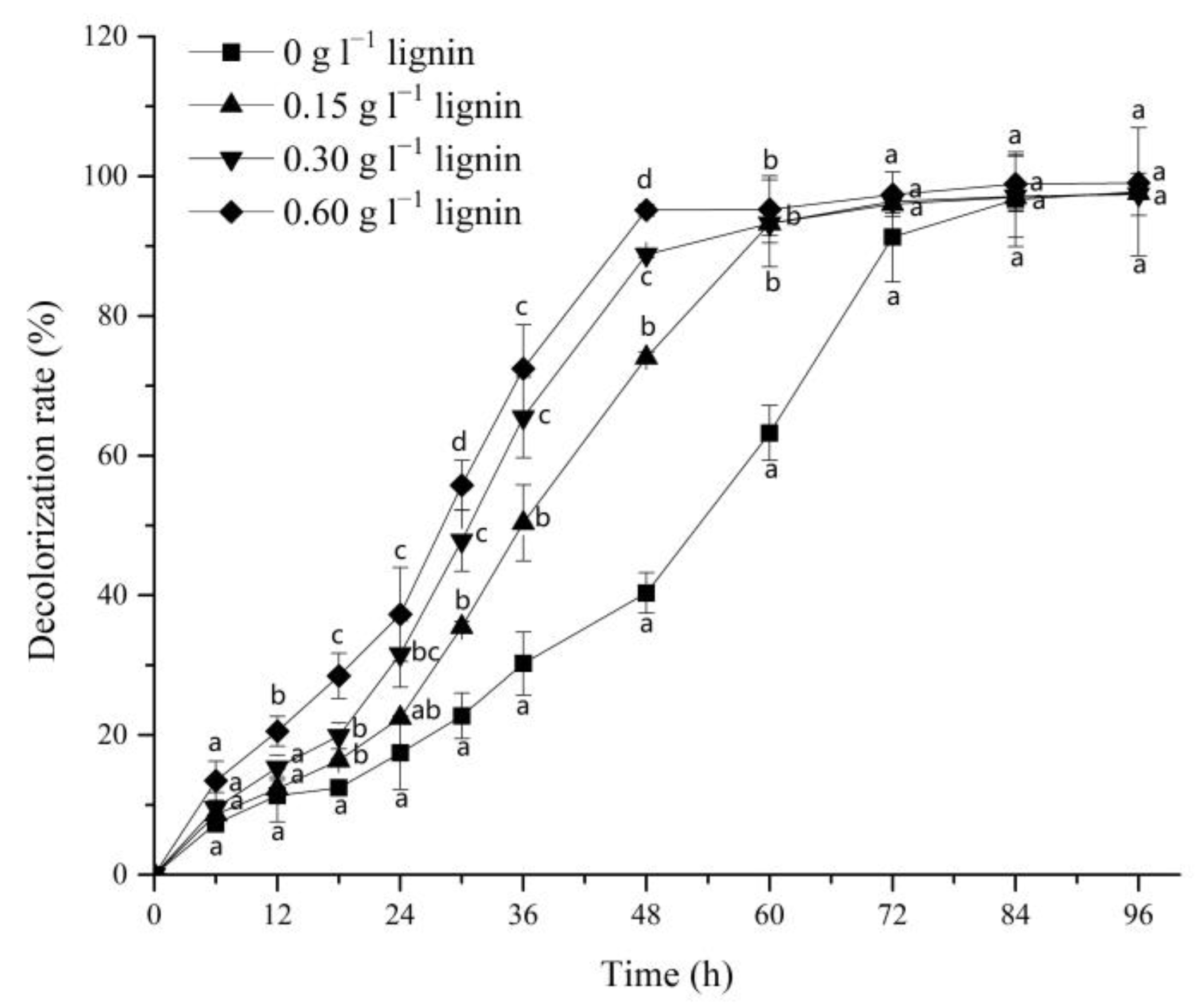
3.1. Effect of Alkali Lignin on Dye Decolorization

The effect of alkali lignin and the decolorization efficiency of G. lucidum EN2 on azo dye DR5B was monitored by adding different amounts of alkali lignin to the dye decolorization medium. The DR5B removal percentage by G. lucidum EN2 was significantly increased by the introduction of alkali lignin, and this acceleration showed a positive correlation with the increasing dosage of alkali lignin. These results showed that the decolorization rates of the 3 experimental groups could increase up to 74.12%, 89.23%, and 95.16% with alkali lignin (i.e., 0.15, 0.30, and 0.60 g L−1, respectively) within 48 h (Figure 1). However, only 40.34% decolorization was achieved in the control group without alkali lignin. The most significant differences in the DR5B decolorization percentages compared with the group without lignin were observed at 48 h. Before 72 h, the decolorization rates of the three experimental groups still exceeded that of the control group. These results specified that alkali lignin could enhance the decolorization of azo dye DR5B by G. lucidum EN2, which is consistent with previous studies using lignocellulosic waste for textile dye bioremediation. This result is also supported by a previous study, in which the consumption of lignocellulosic wastes as a co-substrate considerably increased the azo dye decolorization efficiency of Bjerkandera sp. BOL 13 [49]. Similarly, another study conducted in vivo also reported an enhancement in the azo dye bio-decolorization by Echinodontium taxodii when co-cultured with lignin [15].

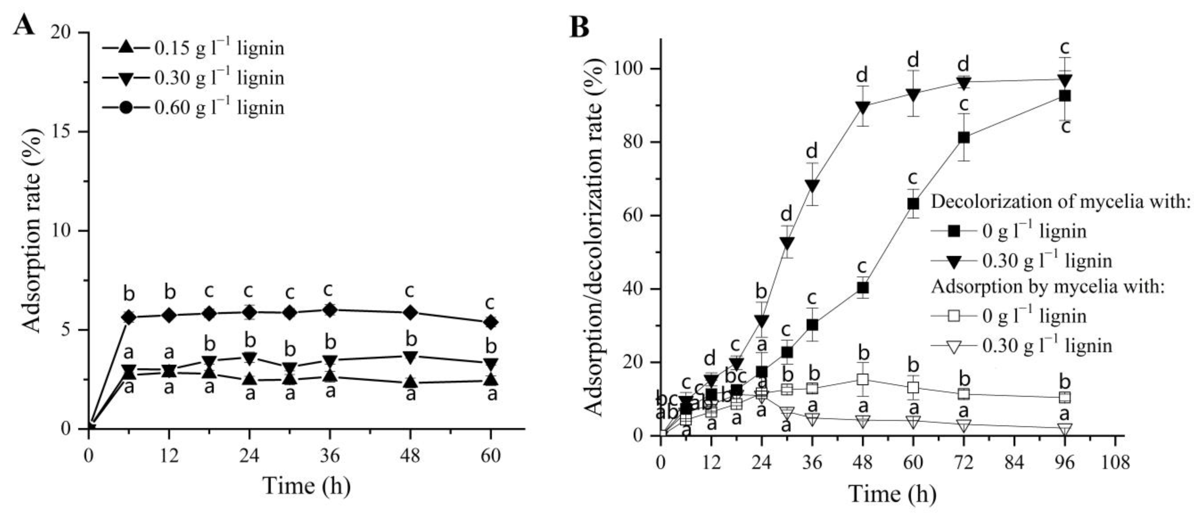
3.2. Physical Adsorption of DR5B by Alkali Lignin and Fungal Mycelia

The mycelium adsorption capacities for dyes were previously reported in many studies [50], some of which could also relate to the increased adsorption capacity in the presence of alkali lignin [51]. Therefore, it was necessary to study the effect of alkali lignin and fungal mycelia on the physical adsorption of DR5B. Initially, the DR5B was incubated with the alkali lignin to study its effect on the physical adsorption of dye DR5B. The maximum adsorption percentage (<6%) was achieved within 6 h when the highest alkali lignin content (0.60 g L−1) was added, and there was no significant increase or decrease thereafter (Figure 2A). In contrast, the dye decolorization rate reached 95.2% at 60 h after adding 0.6g/L of lignin, which was significantly different from the highest decolorization rate (<6%) caused by lignin adsorption (Figure S1). This dye adsorption assay showed that the increased dye decolorization efficiency with the alkali lignin addition during DR5B biodegradation was not mainly caused by the physical adsorption of alkali lignin.
Furthermore, the physical adsorption ability of G. lucidum EN2 mycelia for the dye DR5B was also investigated in both experimental conditions (i.e., in the presence and absence of alkali lignin) during the biodegradation process (Figure 2B). The adsorption and total decolorization of dyes were very comparable during the first 18 h, suggesting that the initial dye decolorization by the fungus was indeed mainly the result of adsorption during that period. In the presence of alkali lignin, G. lucidum EN2 was able to decolorize up to 65.48% of the azo dye within 36 h (0.30 g L−1), followed by an increase of up to 88.78% within 48 h (Figure 2B). However, the physical adsorption of dye by the mycelium remained at a relatively stable level after 36 h (approx. 2%) when compared with the control group (approx. 10%) (Figure 2B). This suggests that physical adsorption had no significant effect on the total decolorization during the whole DR5B removal process. These results clearly showed that the enhanced dye degradation rate after the addition of alkali lignin was mainly the result of the biodegradation of G. lucidum EN2 when compared with the physical adsorption by alkali lignin or mycelium.

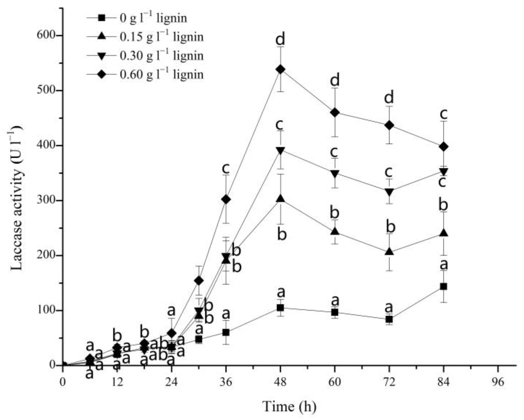
3.3. Effect of Alkali Lignin on Laccase Activities during the Degradation of DR5B by G. lucidum EN2

Ligninolytic enzymes, such as laccase, LiP, MnP, and VP, mainly secreted by white-rot fungi, can effectively degrade lignin and other persistent organic pollutants [52,53]. In this study, laccase was the only ligninolytic enzyme that could be detected in the utilized dye degradation system with G. lucidum EN2. As visible from Figure 3, the laccase enzyme activity was only 105.00 U L−1 in the absence of alkali lignin after the incubation of G. lucidum EN2 for 48 h in DR5B. By increasing the lignin content from 0 to 0.15, 0.30, and 0.60 g L−1, the laccase activity was significantly increased from 105.00 to 302.50, 392.50, and 539.00 U L−1 at 48 h, respectively. This increase in the extracellular laccase activity was about 2.5–4 times that of the control group and was thus consistent with a previous study which also reported a similar increase in the laccase activity by using various lignocellulose residues [54].
At a later stage of the lignin addition system, laccase activity also decreased and was accompanied by the complete decolorization of DR5B at 72 h. The rapid increase in the dye decolorization rates (74.12, 89.23, and 95.16% with different contents of alkali lignin added at 48 h) followed a similar trend to the variation in laccase activity. The elevation in laccase activity and the synthesis of various aromatic compounds by white-rot fungi from lignin degradation have also been reported in many previous studies [55,56]. Yang et al. [57] in 2013 reported that lignin model compounds and other related lignin derivatives increase the activity of ligninolytic enzymes in fungi. The result showed that the addition of alkali lignin (or compounds released from degraded alkali lignin) may stimulate laccase activity in G. lucidum EN2, and the augmented laccase activity may also deliver a significant contribution to the bio-decolorization of DR5B.

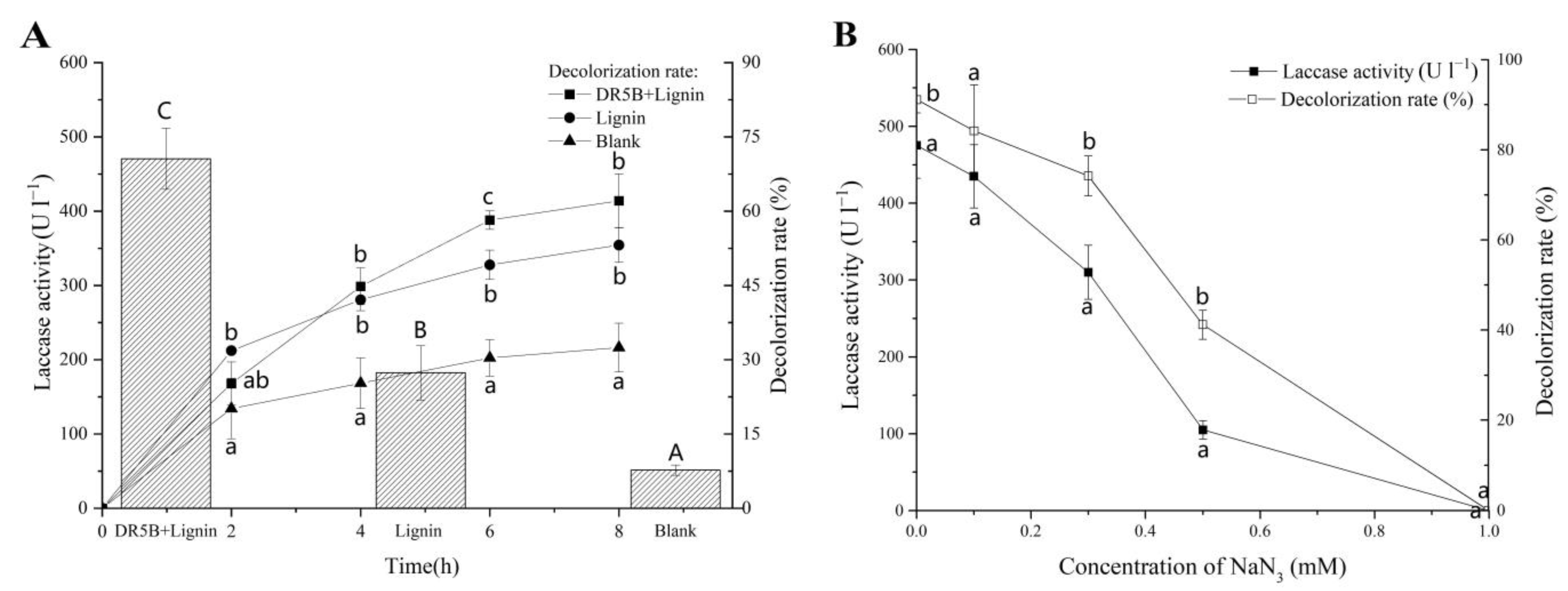
3.4. Effect of Alkali Lignin Degradation of Extracellular Liquid on DR5B Removal

To identify whether the growing laccase induced by alkali lignin was the main reason for the effective decolorization, another DR5B decolorization experiment with cell-free extracellular liquid was performed.
DR5B (final concentration 25 mg L−1) was incubated with cell-free extracellular liquid harvested from different fermentation culture media (including the groups “Lignin+DR5B”, “Lignin”, and “blank”). As shown in Figure 4A, the laccase activities in the groups Lignin+DR5B and Lignin were significantly higher than that in the Blank group. The DR5B decolorization assay showed that the lignin-containing degradation samples of extracellular liquid (62.1% in the “Lignin+DR5B” group and 55.1% in the “Lignin” group) resulted in remarkable decolorization compared with the Blank group (32.4%) after 8 h of incubation (Figure 4A). The lignin co-substrate promote the laccase activity of G. lucidum EN2, and the lignin-containing degradation of extracellular liquid significantly promoted the decolorization of DR5B (Figure 3 and Figure 4A).
To further determine the role of laccase in DR5B decolorization, different concentrations of the laccase-specific inhibitor NaN3 were added to the extracellular liquid of each group, and the resulting abilities for dye removal were tested. Laccase activity decreased with an increasing concentration of NaN3. Meanwhile, the DR5B decolorization ability was reduced with a decrease in laccase activity. As the concentration of NaN3 reached 1 mM, the laccase activity of extracellular fluid was completely suppressed and no more decolorization of DR5B could be observed (Figure 4B). Therefore, this result strongly indicated that laccase plays a prominent role in the degradation of DR5B, and the addition of alkali lignin increased the laccase activity, which may be one method to promote the dye decolorization rate. This importance of laccase could also relate to many previously reported studies [58,59] and is also consistent with our recently reported study, which showed that the improvement of laccase activity increased the dye decolorization efficiently [60].

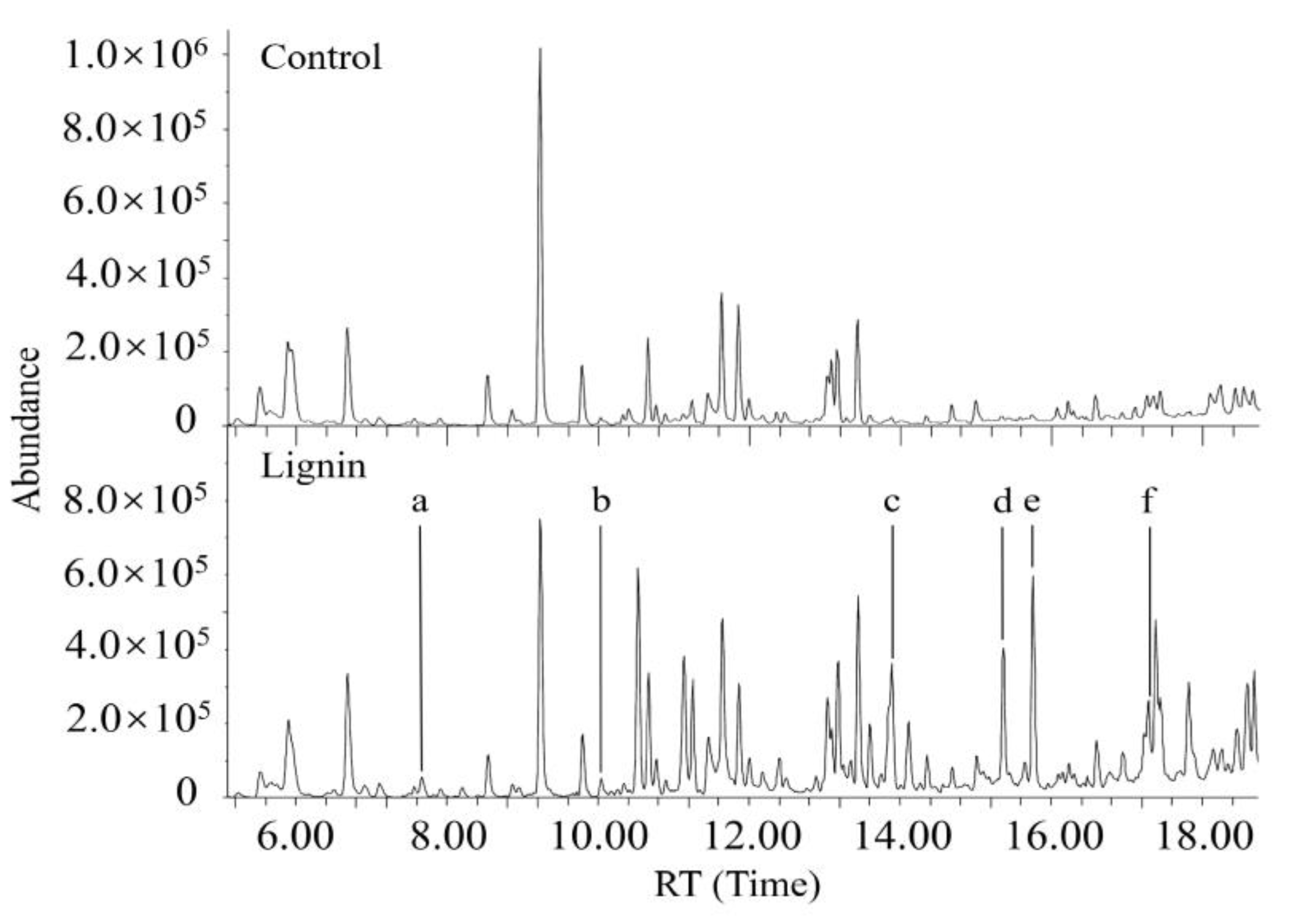
3.5. Characterization of Metabolites Resulting from Degradation of Both DR5B and Alkali Lignin by G. lucidum EN2

The increased laccase activity with the alkali lignin addition was demonstrated using enzymatic assays. However, insoluble lignin is not accessible and, thus, cannot induce enzyme activity; moreover, it also cannot regulate dye degradation [61,62]. Considerable research has shown that many low-molecular aromatic compounds derived from lignin can increase the enzyme activity of laccase in fungal systems and can also act as mediators of lignin-degrading enzymes to accelerate the degradation of dyes [15,63,64]. The comparative GC/MS analysis results further confirmed the degradation of alkali lignin and identified the released aromatic compounds during the degradation of alkali lignin by G. lucidum EN2. Ethyl-acetate-extraction samples obtained from the supernatant of DR5B fermentation by G. lucidum EN2 containing alkali lignin were compared with the lignin-free samples. However, it was difficult to capture the trace and transient amounts of lignin metabolic intermediates throughout the short-term decolorization. Several low molecular weight aromatic compounds (LMCs), such as methoxybenzene (RT 7.7), benzaldehyde (RT 10.0), guaiacol (RT 13.9), phenol (RT 15.4), 4-vinyl guaiacol (RT 15.7) and ethyl guaiacol (RT 17.2), were still captured and detected from the sample with alkali lignin (Figure 5). These recovered low aromatic compounds have been identified as lignin degradation intermediates, which strongly suggests that alkali lignin was degraded by G. lucidum EN2 accompanied by the azo dye degradation process. Additionally, several of these lignin-derived aromatic compounds, including phenols and guaiacols, are considered potential laccase substrates and may also act as natural and proficient laccase mediators for the oxidation of xenobiotics including numerous kinds of dyes [24,33,65,66,67]. In the laccase–mediator system, aromatic compounds could be oxidized into stable radicals by enzymes and act as redox mediators to oxidize other compounds. Hence, the lignin metabolic intermediates were further evaluated for dye decolorization.

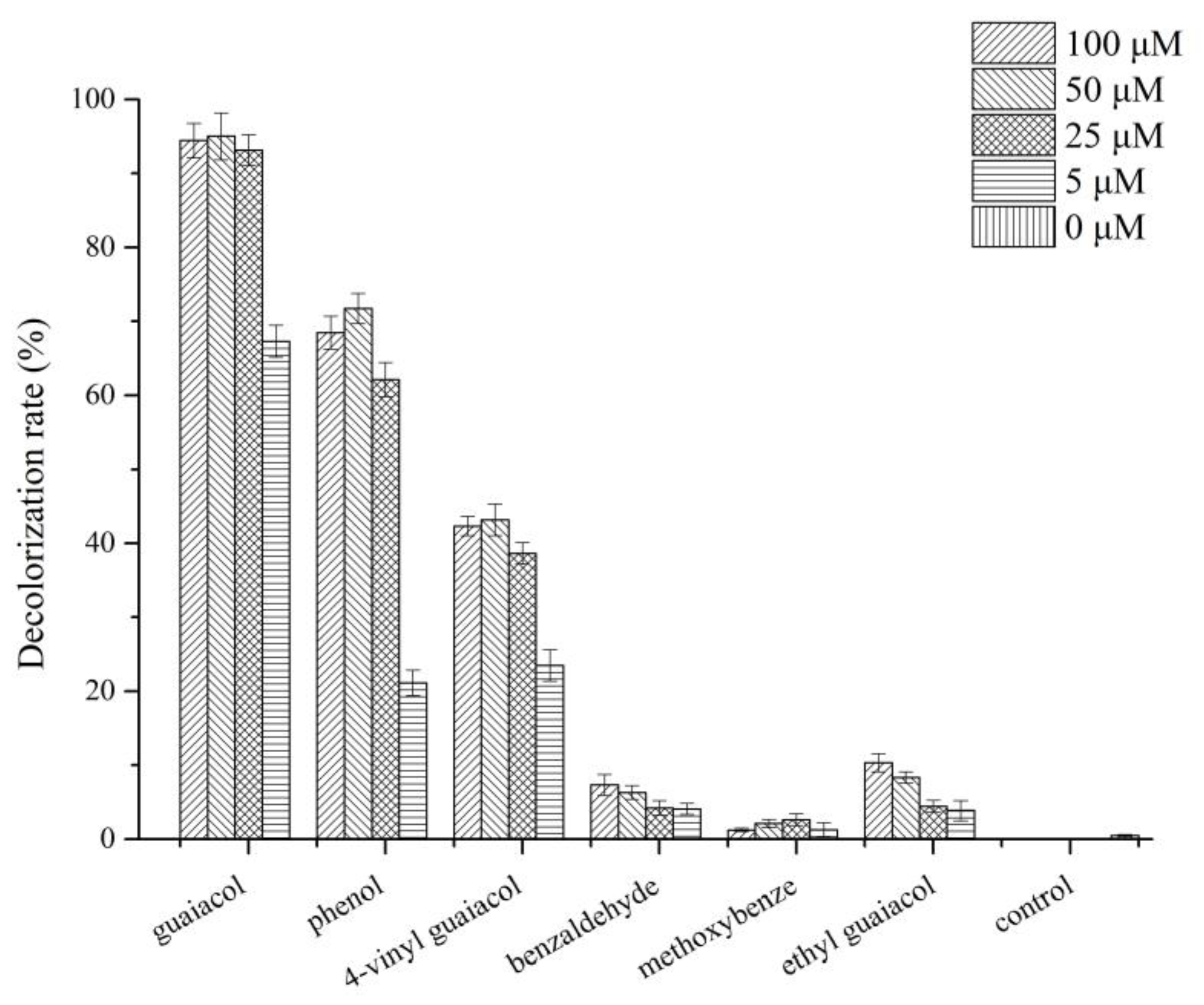
3.6. Effect of Different Lignin-Derived Aromatic Compounds on Dye Decolorization and Laccase Activity

Laccase present a promising role in the degradation of azo dye DR5B according to the presented results (Figure 4). The laccase from G. lucidum EN2 cultures was purified by fractionation with ammonium sulfate, followed by hydrophobic interaction and ion-exchange chromatography for further purification (Figure S2). Six lignin-derived model compounds (including guaiacol, phenol, 4-vinyl guaiacol, benzaldehyde, methoxybenzene, and ethyl guaiacol) were selected, and their effects on the decolorization of azo dyes were examined with the purified laccase (Figure 6). The purified laccase slowly decolorized DR5B in the absence of the aforementioned screened lignin metabolite by 3% after 2 h of incubation. However, the decolorization of DR5B could be accelerated with the presence of lignin-derived compounds including guaiacol (2-methyloxy phenol), 4-vinyl guaiacol (4-vinyl-2-methoxy phenol), phenol, ethyl guaiacol (4-ethyl-2-methoxy phenol), and benzaldehyde at different concentrations (Figure 6). Among these, guaiacol achieved the highest reaction and decolorization rate (>90%) within 1 h at 25 µM. Decolorization of DR5B was also significantly enhanced by different concentrations of phenol (21.12, 62.13, 71.72, and 68.47%) and 4-vinyl guaiacol (23.49, 38.62, 43.17, and 42.32%) (i.e., 5, 25, 50, and 100 µM) (Figure 6). The same phenomenon of DR5B decolorization was also observed in the screened aromatic compounds added to the G. lucidum EN2 degradation systems. This result was consistent with an earlier study where phenol-type derivatives demonstrated a significant induction effect on laccase expression in many species of white-rot fungi [60,68,69,70]. Additionally, for laccase oxidation reactions, these low-molecular-weight phenol derivatives could also act as natural mediators in the degradation of polycyclic aromatic hydrocarbons by laccase–mediator systems [15,33,70].
The degradation of lignin may yield numerous aromatic compounds [71,72]. However, the lower quantity of lignin intermediates made these relatively difficult to detect because of the low amount of lignin addition, existing transients, and the interference of its aromatic compound secretion by the white-rot fungi. Nevertheless, parts of the lignin-degraded compounds were still captured. Several phenolic mediators tested here were also verified to enhance the removal of DR5B as well as mediate laccase to accelerate the azo dye decolorization. Based on the aforementioned results, the addition of alkali lignin promoted the DR5B decolorization by G. lucidum EN2 through two pathways: firstly, the addition of alkali lignin enhanced the laccase activity by inducing laccase expression to promote dye decolorization; secondly, a number of low molecular weight compounds derived from lignin degradation served as mediators and increased the dye decolorization efficiency in the G. lucidum EN2 system.

4. Conclusions

This study shows the potential of lignin in promoting the decolorization of the azo dye DR5B and enhancing the activity of lignin-modifying enzymes. The practice of using alkali lignin resulted in the efficient removal of DR5B from water by G. lucidum EN2. The adsorption experiments showed that the enhanced decolorization of DR5B was primarily through biodegradation rather than the physical adsorption of lignin or mycelia. Laccase played a major role in DR5B decolorization. Moreover, if added to the proposed dye biodegradation system, alkali lignin could improve DR5B decolorization through two pathways. Firstly, alkali lignin could be degraded into small molecular aromatic compounds, which increased laccase activity to promote the DR5B decolorization; secondly, several of the small molecular aromatic compounds, especially phenol and guaiacol, acted as mediators of laccase and, thus, significantly enhanced the dye degradation by G. lucidum EN2. These findings not only provide new insights into the underlying mechanisms of significantly enhanced dye decolorization in the presence of alkali lignin, but they will also profoundly impact the development of sustainable solutions for the effective environmental remediation of azo dyes.

Supplementary Materials

The following supporting information can be downloaded at https://www.mdpi.com/article/10.3390/jof9010091/s1, Figure S1: The comparison of DR5B adsorption and the DR5B decolorization of G. lucidum EN2 in the presence of different contents of alkali lignin (0.15, 0.30, and 0.60 g L−1) at 60 h. Figure S2: SDS-PAGE analyses of purified laccase of G. lucidum EN2. Lane 1, purified laccase; Lane 2, protein molecular mass marker.

Author Contributions

Conceptualization and supervision, S.S.; methodology, S.S., P.L. and M.U.; resources, S.S.; writing—original draft preparation, S.S. and P.L.; writing—review and editing, S.S., P.L. and M.U.; funding acquisition, S.S. All authors have read and agreed to the published version of the manuscript.

Funding

This work was supported by the National Natural Science Foundation of China (Grant number 31900081), the Natural Science Foundation of Hubei Province (Grant number 2022CFB507) and the Scientific Research Project of the Department of Education of Hubei Province (Grant number B2022398).

Institutional Review Board Statement

Not applicable.

Informed Consent Statement

Not applicable.

Data Availability Statement

Not applicable.

Acknowledgments

The authors gratefully acknowledge the Key Laboratory of Molecular Biophysics of MOE, College of Life Science and Technology for its support through the infrastructure of the Experimental Chemistry Laboratory.

Conflicts of Interest

The authors declare no conflict of interest.

References

  1. Haarstrick, A.; Bahadir, M. Water and its Global Meaning. In Water and Wastewater Management; Bahadir, M., Haarstrick, A., Eds.; Springer: Cham, Switzerland, 2022; pp. 3–14. ISBN 978-3-030-95288-4. [Google Scholar]
  2. Boretti, A.; Rosa, L. Reassessing the Projections of the World Water Development Report. npj Clean Water 2019, 2, 15–19. [Google Scholar] [CrossRef] [Green Version]
  3. Al-Tohamy, R.; Ali, S.S.; Li, F.; Okasha, K.M.; Mahmoud, Y.A.-G.; Elsamahy, T.; Jiao, H.; Fu, Y.; Sun, J. A Critical Review on the Treatment of Dye-Containing Wastewater: Ecotoxicological and Health Concerns of Textile Dyes and Possible Remediation Approaches for Environmental Safety. Ecotoxicol. Environ. Saf. 2022, 231, 113160–113176. [Google Scholar] [CrossRef]
  4. Dhruv Patel, D.; Bhatt, S. Environmental Pollution, Toxicity Profile, and Physico-Chemical and Biotechnological Approaches for Treatment of Textile Wastewater. Biotechnol. Genet. Eng. Rev. 2022, 38, 33–86. [Google Scholar] [CrossRef] [PubMed]
  5. Kuppusamy, S.; Sethurajan, M.; Kadarkarai, M.; Aruliah, R. Biodecolourization of Textile Dyes by Novel, Indigenous Pseudomonas Stutzeri MN1 and Acinetobacter Baumannii MN3. J. Environ. Chem. Eng. 2017, 5, 716–724. [Google Scholar] [CrossRef]
  6. Yusuf, M. Synthetic Dyes: A Threat to the Environment and Water Ecosystem. In Textiles and Clothing: Environmental Concerns and Solutions, 1st ed.; Shabbir, M., Ed.; Scrivener Publishing LLC: New York, NY, USA, 2019; pp. 11–26. ISBN 9781119526599. [Google Scholar]
  7. Pinheiro, L.R.; Gradíssimo, D.G.; Xavier, L.P.; Santos, A.V. Degradation of Azo Dyes: Bacterial Potential for Bioremediation. Sustainability 2022, 14, 1510. [Google Scholar] [CrossRef]
  8. Blánquez, A.; Rodríguez, J.; Brissos, V.; Mendes, S.; Martins, L.O.; Ball, A.S.; Arias, M.E.; Hernández, M. Decolorization and Detoxification of Textile Dyes Using a Versatile Streptomyces Laccase-Natural Mediator System. Saudi J. Biol. Sci. 2019, 26, 913–920. [Google Scholar] [CrossRef]
  9. Solís, M.; Solís, A.; Pérez, H.I.; Manjarrez, N.; Flores, M. Microbial Decolouration of Azo Dyes: A Review. Process Biochem. 2012, 47, 1723–1748. [Google Scholar] [CrossRef]
  10. Deng, D.; Guo, J.; Zeng, G.; Sun, G. Decolorization of Anthraquinone, Triphenylmethane and Azo Dyes by a New Isolated Bacillus Cereus Strain DC11. Int. Biodeterior. Biodegrad. 2008, 62, 263–269. [Google Scholar] [CrossRef]
  11. Caritá, R.; Marin-Morales, M.A. Induction of Chromosome Aberrations in the Allium Cepa Test System Caused by the Exposure of Seeds to Industrial Effluents Contaminated with Azo Dyes. Chemosphere 2008, 72, 722–725. [Google Scholar] [CrossRef]
  12. Kishor, R.; Purchase, D.; Saratale, G.D.; Saratale, R.G.; Ferreira, L.F.R.; Bilal, M.; Chandra, R.; Bharagava, R.N. Ecotoxicological and Health Concerns of Persistent Coloring Pollutants of Textile Industry Wastewater and Treatment Approaches for Environmental Safety. J. Environ. Chem. Eng. 2021, 9, 105012–105029. [Google Scholar] [CrossRef]
  13. Zhuo, R.; Fan, F. A Comprehensive Insight into the Application of White Rot Fungi and Their Lignocellulolytic Enzymes in the Removal of Organic Pollutants. Sci. Total Environ. 2021, 778, 146132–146149. [Google Scholar] [CrossRef]
  14. Kunjadia, P.D.; Sanghvi, G.V.; Kunjadia, A.P.; Mukhopadhyay, P.N.; Dave, G.S. Role of Ligninolytic Enzymes of White Rot Fungi (Pleurotus Spp.) Grown with Azo Dyes. Springerplus 2016, 5, 1487. [Google Scholar] [CrossRef] [Green Version]
  15. Han, Y.; Shi, L.; Meng, J.; Yu, H.; Zhang, X. Azo Dye Biodecolorization Enhanced by Echinodontium Taxodii Cultured with Lignin. PLoS ONE 2014, 9, e109786. [Google Scholar] [CrossRef] [Green Version]
  16. Rekik, H.; Zaraî Jaouadi, N.; Bouacem, K.; Zenati, B.; Kourdali, S.; Badis, A.; Annane, R.; Bouanane-Darenfed, A.; Bejar, S.; Jaouadi, B. Physical and Enzymatic Properties of a New Manganese Peroxidase from the White-Rot Fungus Trametes Pubescens Strain I8 for Lignin Biodegradation and Textile-Dyes Biodecolorization. Int. J. Biol. Macromol. 2019, 125, 514–525. [Google Scholar] [CrossRef]
  17. Koyani, R.D.; Sanghvi, G.V.; Sharma, R.K.; Rajput, K.S. Contribution of Lignin Degrading Enzymes in Decolourisation and Degradation of Reactive Textile Dyes. Int. Biodeterior. Biodegrad. 2013, 77, 1–9. [Google Scholar] [CrossRef]
  18. Samir Ali, S.; Al-Tohamy, R.; Khalil, M.A.; Ho, S.-H.; Fu, Y.; Sun, J. Exploring the Potential of a Newly Constructed Manganese Peroxidase-Producing Yeast Consortium for Tolerating Lignin Degradation Inhibitors While Simultaneously Decolorizing and Detoxifying Textile Azo Dye Wastewater. Bioresour. Technol. 2022, 351, 126861–126874. [Google Scholar] [CrossRef] [PubMed]
  19. Al-Tohamy, R.; Sun, J.; Fareed, M.F.; Kenawy, E.-R.; Ali, S.S. Ecofriendly Biodegradation of Reactive Black 5 by Newly Isolated Sterigmatomyces Halophilus SSA1575, Valued for Textile Azo Dye Wastewater Processing and Detoxification. Sci. Rep. 2020, 10, 12370–12386. [Google Scholar] [CrossRef]
  20. Mir-Tutusaus, J.A.; Baccar, R.; Caminal, G.; Sarrà, M. Can White-Rot Fungi Be a Real Wastewater Treatment Alternative for Organic Micropollutants Removal? A Review. Water Res. 2018, 138, 137–151. [Google Scholar] [CrossRef] [PubMed]
  21. Shi, K.; Liu, Y.; Chen, P.; Li, Y. Contribution of Lignin Peroxidase, Manganese Peroxidase, and Laccase in Lignite Degradation by Mixed White-Rot Fungi. Waste Biomass Valorization 2021, 12, 3753–3763. [Google Scholar] [CrossRef]
  22. Othmani, A.; John, J.; Rajendran, H.; Mansouri, A.; Sillanpää, M.; Velayudhaperumal Chellam, P. Biochar and Activated Carbon Derivatives of Lignocellulosic Fibers towards Adsorptive Removal of Pollutants from Aqueous Systems: Critical Study and Future Insight. Sep. Purif. Technol. 2021, 274, 119062–119074. [Google Scholar] [CrossRef]
  23. Robinson, T.; Nigam, P.S. Remediation of Textile Dye Waste Water Using a White-Rot Fungus Bjerkandera Adusta Through Solid-State Fermentation (SSF). Appl. Biochem. Biotechnol. 2008, 151, 618. [Google Scholar] [CrossRef]
  24. Yan, K.; Wang, H.; Zhang, X. Biodegradation of Crystal Violet by Low Molecular Mass Fraction Secreted by Fungus. J. Biosci. Bioeng. 2009, 108, 421–424. [Google Scholar] [CrossRef]
  25. Han, M.-L.; Yang, J.; Liu, Z.-Y.; Wang, C.-R.; Chen, S.-Y.; Han, N.; Hao, W.-Y.; An, Q.; Dai, Y.-C. Evaluation of Laccase Activities by Three Newly Isolated Fungal Species in Submerged Fermentation with Single or Mixed Lignocellulosic Wastes. Front. Microbiol. 2021, 12, 682679–682689. [Google Scholar] [CrossRef]
  26. Raj, T.; Chandrasekhar, K.; Naresh Kumar, A.; Rajesh Banu, J.; Yoon, J.-J.; Kant Bhatia, S.; Yang, Y.-H.; Varjani, S.; Kim, S.-H. Recent Advances in Commercial Biorefineries for Lignocellulosic Ethanol Production: Current Status, Challenges and Future Perspectives. Bioresour. Technol. 2022, 344, 126292–126303. [Google Scholar] [CrossRef]
  27. Intasit, R.; Khunrae, P.; Meeinkuirt, W.; Soontorngun, N. Fungal Pretreatments of Napier Grass and Sugarcane Leaves for High Recovery of Lignocellulosic Enzymes and Methane Production. Ind. Crops Prod. 2022, 180, 114706–114715. [Google Scholar] [CrossRef]
  28. dos Santos Bazanella, G.C.; de Souza, D.F.; Castoldi, R.; Oliveira, R.F.; Bracht, A.; Peralta, R.M. Production of Laccase and Manganese Peroxidase by Pleurotus Pulmonarius in Solid-State Cultures and Application in Dye Decolorization. Folia Microbiol. (Praha.) 2013, 58, 641–647. [Google Scholar] [CrossRef] [PubMed]
  29. Dashtban, M.; Schraft, H.; Qin, W. Fungal Bioconversion of Lignocellulosic Residues; Opportunities & Perspectives. Int. J. Biol. Sci. 2009, 5, 578–595. [Google Scholar] [CrossRef] [PubMed]
  30. Hernandez-Raquet, G.; Durand, E.; Braun, F.; Cravo-Laureau, C.; Godon, J.-J. Impact of Microbial Diversity Depletion on Xenobiotic Degradation by Sewage-Activated Sludge. Environ. Microbiol. Rep. 2013, 5, 588–594. [Google Scholar] [CrossRef]
  31. Mohapatra, B.; Phale, P.S. Microbial Degradation of Naphthalene and Substituted Naphthalenes: Metabolic Diversity and Genomic Insight for Bioremediation. Front. Bioeng. Biotechnol. 2021, 9, 602445–602473. [Google Scholar] [CrossRef]
  32. Elisashvili, V.; Kachlishvili, E.; Khardziani, T.; Agathos, S.N. Effect of Aromatic Compounds on the Production of Laccase and Manganese Peroxidase by White-Rot Basidiomycetes. J. Ind. Microbiol. Biotechnol. 2010, 37, 1091–1096. [Google Scholar] [CrossRef]
  33. Susana, C.; David, I.; Jesús, M.M.; Martínez, Á.T. Lignin-Derived Compounds as Efficient Laccase Mediators for Decolorization of Different Types of Recalcitrant Dyes. Appl. Environ. Microbiol. 2005, 71, 1775–1784. [Google Scholar] [CrossRef] [Green Version]
  34. Ralph, J.; Lapierre, C.; Boerjan, W. Lignin Structure and Its Engineering. Curr. Opin. Biotechnol. 2019, 56, 240–249. [Google Scholar] [CrossRef] [PubMed]
  35. Cline, S.P.; Smith, P.M. Opportunities for Lignin Valorization: An Exploratory Process. Energy Sustain. Soc. 2017, 7, 26–37. [Google Scholar] [CrossRef] [Green Version]
  36. Zheng, L.; Lu, G.; Pei, W.; Yan, W.; Li, Y.; Zhang, L.; Huang, C.; Jiang, Q. Understanding the Relationship between the Structural Properties of Lignin and Their Biological Activities. Int. J. Biol. Macromol. 2021, 190, 291–300. [Google Scholar] [CrossRef] [PubMed]
  37. Melro, E.; Filipe, A.; Sousa, D.; Medronho, B.; Romano, A. Revisiting Lignin: A Tour through Its Structural Features, Characterization Methods and Applications. New J. Chem. 2021, 45, 6986–7013. [Google Scholar] [CrossRef]
  38. Yuan, Y.; Jiang, B.; Chen, H.; Wu, W.; Wu, S.; Jin, Y.; Xiao, H. Recent Advances in Understanding the Effects of Lignin Structural Characteristics on Enzymatic Hydrolysis. Biotechnol. Biofuels 2021, 14, 205–224. [Google Scholar] [CrossRef]
  39. Huang, J.; Fu, S.; Gan, L. Structure and Characteristics of Lignin. In Lignin Chemistry and Applications; Huang, J., Fu, S., Gan, L., Eds.; Elsevier: Amsterdam, The Netherlands, 2019; Chapter 2; pp. 25–50. ISBN 978-0-12-813941-7. [Google Scholar]
  40. del Cerro, C.; Erickson, E.; Dong, T.; Wong, A.R.; Eder, E.K.; Purvine, S.O.; Mitchell, H.D.; Weitz, K.K.; Markillie, L.M.; Burnet, M.C.; et al. Intracellular Pathways for Lignin Catabolism in White-Rot Fungi. Proc. Natl. Acad. Sci. USA 2021, 118, e2017381118. [Google Scholar] [CrossRef]
  41. Ilić Đurđić, K.; Ostafe, R.; Prodanović, O.; Đurđević Đelmaš, A.; Popović, N.; Fischer, R.; Schillberg, S.; Prodanović, R. Improved Degradation of Azo Dyes by Lignin Peroxidase Following Mutagenesis at Two Sites near the Catalytic Pocket and the Application of Peroxidase-Coated Yeast Cell Walls. Front. Environ. Sci. Eng. 2020, 15, 21419–21428. [Google Scholar] [CrossRef]
  42. Meng, X.; Scheidemantle, B.; Li, M.; Wang, Y.; Zhao, X.; Toro-González, M.; Singh, P.; Pu, Y.; Wyman, C.E.; Ozcan, S.; et al. Synthesis, Characterization, and Utilization of a Lignin-Based Adsorbent for Effective Removal of Azo Dye from Aqueous Solution. ACS Omega 2020, 5, 2865–2877. [Google Scholar] [CrossRef]
  43. Kirk, T.K.; Schultz, E.; Connors, W.J.; Lorenz, L.F.; Zeikus, J.G. Influence of Culture Parameters on Lignin Metabolism by Phanerochaete Chrysosporium. Arch. Microbiol. 1978, 117, 277–285. [Google Scholar] [CrossRef]
  44. Yan, K.; Wang, H.; Zhang, X.; Yu, H. Bioprocess of Triphenylmethane Dyes Decolorization by Pleurotus Ostreatus BP under Solid-State Cultivation. J. Microbiol. Biotechnol. 2009, 19, 1421–1430. [Google Scholar] [CrossRef] [PubMed]
  45. Shi, L.; Yu, H.; Dong, T.; Kong, W.; Ke, M.; Ma, F.; Zhang, X. Biochemical and Molecular Characterization of a Novel Laccase from Selective Lignin-Degrading White-Rot Fungus Echinodontium Taxodii 2538. Process Biochem. 2014, 49, 1097–1106. [Google Scholar] [CrossRef]
  46. Hakala, T.K.; Lundell, T.; Galkin, S.; Maijala, P.; Kalkkinen, N.; Hatakka, A. Manganese Peroxidases, Laccases and Oxalic Acid from the Selective White-Rot Fungus Physisporinus Rivulosus Grown on Spruce Wood Chips. Enzyme Microb. Technol. 2005, 36, 461–468. [Google Scholar] [CrossRef]
  47. Qin, X.; Su, X.; Luo, H.; Ma, R.; Yao, B.; Ma, F. Deciphering Lignocellulose Deconstruction by the White Rot Fungus Irpex Lacteus Based on Genomic and Transcriptomic Analyses. Biotechnol. Biofuels 2018, 11, 58–71. [Google Scholar] [CrossRef] [PubMed] [Green Version]
  48. He, X.; Song, C.; Li, Y.; Wang, N.; Xu, L.; Han, X.; Wei, D. Efficient Degradation of Azo Dyes by a Newly Isolated Fungus Trichoderma Tomentosum under Non-Sterile Conditions. Ecotoxicol. Environ. Saf. 2018, 150, 232–239. [Google Scholar] [CrossRef] [PubMed]
  49. Jonstrup, M.; Kumar, N.; Guieysse, B.; Murto, M.; Mattiasson, B. Decolorization of Textile Dyes by Bjerkandera Sp. BOL 13 Using Waste Biomass as Carbon Source. J. Chem. Technol. Biotechnol. 2013, 88, 388–394. [Google Scholar] [CrossRef]
  50. Sen, S.K.; Raut, S.; Bandyopadhyay, P.; Raut, S. Fungal Decolouration and Degradation of Azo Dyes: A Review. Fungal Biol. Rev. 2016, 30, 112–133. [Google Scholar] [CrossRef]
  51. Rybczyńska-Tkaczyk, K.; Korniłłowicz-Kowalska, T. Biosorption Optimization and Equilibrium Isotherm of Industrial Dye Compounds in Novel Strains of Microscopic Fungi. Int. J. Environ. Sci. Technol. 2016, 13, 2837–2846. [Google Scholar] [CrossRef] [Green Version]
  52. Kaushik, P.; Malik, A. Fungal Dye Decolourization: Recent Advances and Future Potential. Environ. Int. 2009, 35, 127–141. [Google Scholar] [CrossRef]
  53. Abdel-Hamid, A.M.; Solbiati, J.O.; Cann, I.K.O. Insights into Lignin Degradation and Its Potential Industrial Applications. Adv. Appl. Microbiol. 2013, 82, 1–28. [Google Scholar] [CrossRef]
  54. Rodrigues, E.M.; Karp, S.G.; Malucelli, L.C.; Helm, C.V.; Alvarez, T.M. Evaluation of Laccase Production by Ganoderma Lucidum in Submerged and Solid-State Fermentation Using Different Inducers. J. Basic Microbiol. 2019, 59, 784–791. [Google Scholar] [CrossRef] [PubMed]
  55. Couto, S.R.; Sanromán, M.A.; Guebitz, G.M. Influence of Redox Mediators and Metal Ions on Synthetic Acid Dye Decolourization by Crude Laccase from Trametes Hirsuta. Chemosphere 2005, 58, 417–422. [Google Scholar] [CrossRef] [PubMed]
  56. Zhuo, R.; Yuan, P.; Yang, Y.; Zhang, S.; Ma, F.; Zhang, X. Induction of Laccase by Metal Ions and Aromatic Compounds in Pleurotus Ostreatus HAUCC 162 and Decolorization of Different Synthetic Dyes by the Extracellular Laccase. Biochem. Eng. J. 2017, 117, 62–72. [Google Scholar] [CrossRef]
  57. Yang, S.; Hai, F.I.; Nghiem, L.D.; Price, W.E.; Roddick, F.; Moreira, M.T.; Magram, S.F. Understanding the Factors Controlling the Removal of Trace Organic Contaminants by White-Rot Fungi and Their Lignin Modifying Enzymes: A Critical Review. Bioresour. Technol. 2013, 141, 97–108. [Google Scholar] [CrossRef] [PubMed] [Green Version]
  58. Zilly, A.; da Silva Coelho-Moreira, J.; Bracht, A.; Marques de Souza, C.G.; Carvajal, A.E.; Koehnlein, E.A.; Peralta, R.M. Influence of NaCl and Na2SO4 on the Kinetics and Dye Decolorization Ability of Crude Laccase from Ganoderma Lucidum. Int. Biodeterior. Biodegrad. 2011, 65, 340–344. [Google Scholar] [CrossRef] [Green Version]
  59. Liu, S.-H.; Tsai, S.-L.; Guo, P.-Y.; Lin, C.-W. Inducing Laccase Activity in White Rot Fungi Using Copper Ions and Improving the Efficiency of Azo Dye Treatment with Electricity Generation Using Microbial Fuel Cells. Chemosphere 2020, 243, 125304–125310. [Google Scholar] [CrossRef]
  60. Liu, J.; Sun, S.; Han, Y.; Meng, J.; Chen, Y.; Yu, H.; Zhang, X.; Ma, F. Lignin Waste as Co-Substrate on Decolorization of Azo Dyes by Ganoderma Lucidum. J. Taiwan Inst. Chem. Eng. 2021, 122, 85–92. [Google Scholar] [CrossRef]
  61. Cancel, A.M.; Orth, A.B.; Tien, M. Lignin and Veratryl Alcohol Are Not Inducers of the Ligninolytic System of Phanerochaete Chrysosporium. Appl. Environ. Microbiol. 1993, 59, 2909–2913. [Google Scholar] [CrossRef] [Green Version]
  62. Faison, B.D.; Kirk, T.K. Factors Involved in the Regulation of a Ligninase Activity in Phanerochaete Chrysosporium. Appl. Environ. Microbiol. 1985, 49, 299–304. [Google Scholar] [CrossRef] [Green Version]
  63. Piscitelli, A.; Giardina, P.; Lettera, V.; Pezzella, C.; Sannia, G.; Faraco, V. Induction and Transcriptional Regulation of Laccases in Fungi. Curr. Genom. 2011, 12, 104–112. [Google Scholar] [CrossRef]
  64. Giatti Marques De Souza, C.; Kirst Tychanowicz, G.; Farani De Souza, D.; Peralta, R.M. Production of Laccase Isoforms by Pleurotus Pulmonarius in Response to Presence of Phenolic and Aromatic Compounds. J. Basic Microbiol. An Int. J. Biochem. Physiol. Genet. Morphol. Ecol. Microorg. 2004, 44, 129–136. [Google Scholar] [CrossRef]
  65. Unuofin, J.O.; Okoh, A.I.; Nwodo, U.U. Aptitude of Oxidative Enzymes for Treatment of Wastewater Pollutants: A Laccase Perspective. Molecules 2019, 24, 2064. [Google Scholar] [CrossRef] [Green Version]
  66. Mäkelä, M.R.; Tuomela, M.; Hatakka, A.; Hildén, K. Fungal laccases and their potential in bioremediation applications. In Microbiology Monographs: Laccases in Bioremediation and Waste Valorisation; Schlosser, D., Ed.; Springer International Publishing: Cham, Germany, 2020; pp. 1–25. ISBN 978-3-030-47906-0. [Google Scholar]
  67. Raj, A.; Reddy, M.M.K.; Chandra, R. Identification of Low Molecular Weight Aromatic Compounds by Gas Chromatography–Mass Spectrometry (GC–MS) from Kraft Lignin Degradation by Three Bacillus sp. Int. Biodeterior. Biodegrad. 2007, 59, 292–296. [Google Scholar] [CrossRef]
  68. Gnanamani, A.; Jayaprakashvel, M.; Arulmani, M.; Sadulla, S. Effect of Inducers and Culturing Processes on Laccase Synthesis in Phanerochaete Chrysosporium NCIM 1197 and the Constitutive Expression of Laccase Isozymes. Enzyme Microb. Technol. 2006, 38, 1017–1021. [Google Scholar] [CrossRef]
  69. Manavalan, A.; Manavalan, T.; Murugesan, K.; Kutzner, A.; Thangavelu, K.P.; Heese, K. Characterization of a Solvent, Surfactant and Temperature-Tolerant Laccase from Pleurotus Sp. MAK-II and Its Dye Decolorizing Property. Biotechnol. Lett. 2015, 37, 2403–2409. [Google Scholar] [CrossRef]
  70. Hu, M.R.; Chao, Y.P.; Zhang, G.Q.; Xue, Z.Q.; Qian, S. Laccase-Mediator System in the Decolorization of Different Types of Recalcitrant Dyes. J. Ind. Microbiol. Biotechnol. 2009, 36, 45–51. [Google Scholar] [CrossRef]
  71. Azubuike, C.C.; Allemann, M.N.; Michener, J.K. Microbial Assimilation of Lignin-Derived Aromatic Compounds and Conversion to Value-Added Products. Curr. Opin. Microbiol. 2022, 65, 64–72. [Google Scholar] [CrossRef]
  72. Nguyen, L.T.; Phan, D.-P.; Sarwar, A.; Tran, M.H.; Lee, O.K.; Lee, E.Y. Valorization of Industrial Lignin to Value-Added Chemicals by Chemical Depolymerization and Biological Conversion. Ind. Crops Prod. 2021, 161, 113219–113233. [Google Scholar] [CrossRef]
Figure 1. Decolorization rate of Direct Red 5B (DR5B) by G. lucidum EN2 in the presence of different contents of alkali lignin (0, 0.15, 0.30, and 0.60 g L−1). The vertical line on each bar indicates the standard deviation for three replicates (SD, n = 3). Different letters represent significant differences between treatment groups at each time point, as determined with two-way ANOVA followed by LSD or Tukey multiple comparison tests (p < 0.05).
Jof 09 00091 g001
Figure 2. Physical adsorption of DR5B by alkali lignin and fungal mycelia. (A) Physical adsorption rate of DR5B when it was incubated with alkali lignin at contents of 0.15, 0.30, and 0.60 g L−1. (B) Physical adsorption rate of DR5B by G. lucidum EN2 mycelia in the presence of 0 and 0.30 g L−1 alkali lignin. Decolorization by G. lucidum EN2 mycelia of DR5B in the presence of 0 and 0.30 g L−1 alkali lignin. The vertical line on each bar indicates the standard deviation for three replicates (SD, n = 3). Different letters represent significant differences between treatment groups at each time point, as determined with two-way ANOVA followed by LSD or Tukey multiple comparison tests (p < 0.05).
Jof 09 00091 g002
Figure 3. Laccase activities during the DR5B decolorization process by G. lucidum EN2 in the presence of different contents of alkali lignin (0, 0.15, 0.30, and 0.60 g L−1). The vertical line on each bar indicates the standard deviation for three replicates (SD, n = 3). Different letters represent significant differences between treatment groups at each time point, as determined with two-way ANOVA followed by LSD or Tukey multiple comparison tests (p < 0.05).
Jof 09 00091 g003
Figure 4. Effect of cell-free extracellular liquid (CFEL) and inhibition of laccase on DR5B decolorization. (A) Decolorization of DR5B (25 mg L−1) by the CFEL extracted from G. lucidum EN2 incubated with or without lignin and DR5B for 48 h. “DR5B+Lignin”: CFEL obtained from G. lucidum EN2 incubated with lignin and DR5B and added Kirk medium; “Lignin”: CFEL extracted from G. lucidum EN2 fermented with lignin and added Kirk medium; “Blank”: CFEL extracted from G. lucidum EN2 fermented with Kirk medium only; (B) Inhibition of laccase and decreased decolorization with increasing sodium azide concentration. The vertical line on each bar indicates the standard deviation for three replicates (SD, n = 3). Different letters represent significant differences between treatment groups at each time point, as determined with a two-way ANOVA followed by LSD or Tukey multiple comparison tests (p < 0.05).
Jof 09 00091 g004
Figure 5. Total ion chromatogram of the compounds extracted from dye degradation in the absence (up) or presence (down) of alkali lignin. a-f show the peaks of chemicals present in the lignin culture. a: methoxybenzene (RT 7.7); b: benzaldehyde (RT 10.0); c: guaiacol (RT 13.9); d: phenol (RT 15.4); e: 4-vinyl guaiacol (RT 15.7); f: ethyl guaiacol (RT 17.2).
Jof 09 00091 g005
Figure 6. Evaluating lignin-derived compounds based on decolorization of DR5B within 1 h. Decolorization of DR5B (100 mg L−1) within 1 h with purified laccase (final concentration: 1 U mL−1) and intermediators (0, 5, 25, 50, and 100 µM).
Jof 09 00091 g006
Disclaimer/Publisher’s Note: The statements, opinions and data contained in all publications are solely those of the individual author(s) and contributor(s) and not of MDPI and/or the editor(s). MDPI and/or the editor(s) disclaim responsibility for any injury to people or property resulting from any ideas, methods, instructions or products referred to in the content.

