Article

Antifungal Activity and Biocontrol Potential of Simplicillium lamellicola JC-1 against Multiple Fungal Pathogens of Oilseed Rape

State Key Laboratory of Agricultural Microbiology, Huazhong Agricultural University, Wuhan 430070, China
*
Author to whom correspondence should be addressed.
These authors contributed equally to this work.
J. Fungi 2023, 9(1), 57; https://doi.org/10.3390/jof9010057
Submission received: 8 December 2022 / Revised: 26 December 2022 / Accepted: 27 December 2022 / Published: 30 December 2022
(This article belongs to the Special Issue The Role of Fungi in Plant Defense Mechanisms)

Abstract

:
A fungal strain (JC-1) of Simplicillium was isolated from a pod of oilseed rape (Brassica napus) infested with the blackleg pathogen Leptosphaeria biglobosa. This study was done to clarify its taxonomic identity using morphological and molecular approaches, to characterize its antifungal activity through bioassays and genome-based identification of antifungal metabolites, and to determine its efficacy in inducing systemic resistance (ISR) in oilseed rape. The results showed that JC-1 belongs to Simplicillium lamellicola. It displayed a strong antagonistic relationship with L. biglobosa, Botrytis cinerea (gray mold) and Sclerotinia sclerotiorum (stem rot). The cultural filtrates of JC-1 showed a high efficacy in suppressing infection by S. sclerotiorum on detached leaves of oilseed rape. Genome analysis indicated that JC-1 has the capability of producing multiple antifungal metabolites, including aureobasidin A1, squalestatin S1 and verlamelin. Inoculation of JC-1 on seeds of oilseed rape caused a suppressive effect on infection by L. biglobosa on the cotyledons of the resulting seedlings, suggesting that JC-1 can trigger ISR. Endophytic growth, accumulation of anthocyanins, up-regulated expression of CHI (for chalcone isomerase) and PR1 (for pathogenesis-related protein 1), and down-regulated expression of NECD3 (for 9-cis-epoxycarotenoid dioxygenase) were detected to be associated with the ISR. This study provided new insights into the biocontrol potential and modes of action of S. lamellicola.

1. Introduction

Simplicillium (Cordycipitaceae, Hypocreales and Ascomycota) is a genus established by Zare and Gams in 2001, and the fungi of this genus are characterized by the formation of simple (solitary), unbranched conidiophores (phialides) and short-ellipsoidal conidia in globose slimy heads at the apex of the conidiophores [1]. Simplicillium was previously treated as Verticillium section Prostrata; therefore, it is closely related to Verticillium. In 2001, Simplicillium only accommodated three species, namely S. lamellicola, S. lanosoniveum, and S. obclavatum [1]. Since then, the taxonomy of Simplicillium has attracted the attention of many researchers, and substantial progress has been made in this aspect. So far, one new variety of S. lanosoniveum and at least 18 novel species of Simplicillium have been described, including S. album, S. aogashimaense, S. calcicola, S. chinensis, S. cicadellidae, S. coccinellidae, S. cylindrosporum, S. filiforme, S. formicae, S. formicidae, S. humicola, S. hymenopterorum, S. lanosoniveum var. tianjinensis, S. lepidopterorum, S. neolepidopterorum, S. niveum, S. scarabaeoidea, S. subtropicum and S. sympodiophorum [2,3,4,5,6,7,8,9,10,11]. These Simplicillium species have diversified ecological niches, some live with fungi as mycoparasites [1,12,13,14,15,16,17], some live with insects as entomolopathogens [2,3,9,18,19], some live with plants as endophytes or pathogens [20,21,22], and some live as saprophytes in fresh water [7], in soil [8], on animal faeces [11], and even in the deep sea [23].
Previous studies showed several species of Simplicillium with the mycoparasitic lifestyle can potentially be used for the control of plant fungal diseases [15,17,19,24,25]. Choi et al. (2008) found that S. lamellicola BCP (=Acremonium strictum BCP) is a mycoparasite of the gray mold fungus Botrytis cinerea [26], and it has been developed as a biofungicide for control of gray mold in tomato and ginseng [25]. Ward et al. (2011) and Gauthier et al. (2014) reported that S. lanosoniveum is a mycoparasite of the soybean rust fungus Phakopsora pachyrhizi, and it showed a high suppressive efficacy against soybean rust through suppression of the set-up of the populations of P. pachyrhizi on soybean leaves [13,24]. Wang et al. (2020) found that S. obclavatum is a mycoparasite of the stripe rust fungus Puccinia striiformis f. sp. tritici (Pgt), and it displayed a high efficacy in the suppression of the production of urediniospores by Pgt and the germination rates of the Pgt urediniospores on wheat leaves [15]. Recently, Zhu et al. (2022) reported that S. aogashimaense is a mycoparasite of the powdery mildew fungus Blumeria graminis f. sp. tritici (Bgt), and it had a high efficacy in the suppression of powdery mildew through impairing colony formation by Bgt on wheat leaves [17].
Besides mycoparasitism, Simplicillium species have other modes of action against plant pathogens, including antibiosis through the production of antimicrobial metabolites [27,28]. Previous studies showed that the BCP strain of S. lamellicola can produce verlamelin (a cyclic lipopeptide) and mannosyl lipids [27,28]. Verlamelin showed antifungal activity against many plant pathogenic fungi, including Alternaria alternata, Bipolaris maydis, Botrytis cinerea, Fusarium oxysporum and Magnaporthe grisea, and this metabolite (100 μg/mL) displayed an effective suppression of the development of barley powdery mildew caused by B. graminis f. sp. hordei (Bgh) on barley leaves. Moreover, the gene cluster (vlmS, vlmA, vlmB, vlmC) responsible for the biosynthesis of verlamelin in Lecanicillium sp. HF627 (a close relative of Verticillium) has been elucidated [29]. Mannosyl lipids had antibacterial activity against two plant pathogenic bacteria, including Agrobacterium tumefaciens (crown gall) and Ralstonia solanacearum (bacterial wilt) [28]. Another study by Liang et al. (2017) indicated that strain EIODSF 020 of S. obclavatum from the deep sea could produce other cyclic peptides with antifungal activity against Aspergillus versicolor and Curvularia australiensis [23].
Additionally, Zhang et al. (2014), Shen et al. (2012) and Teasdale et al. (2018) isolated Simplicillium from plant tissues; the resulting strains of Simplicillium were thus considered to be plant endophytes [21,22,30]. However, the endophytic capability of both species of Simplicillium has not been experimentally confirmed. Moreover, whether or not these two species can trigger a plant defense response against infection by plant pathogenic fungi remains unknown.
Li (2019) obtained the JC-1 strain of Simplicillium from a diseased pod of oilseed rape (Brassica napus) infested with Leptosphaeria biglobosa, the causal agent of blackleg in oilseed rape [31]. The peculiar association of this strain of Simplicillium with L. biglobosa prompted us to pose the following two questions: (i) could the JC-1 strain potentially be used to control L. biglobosa and other fungal pathogens of oilseed rape (e.g., Botrytis cinerea, Sclerotinia sclerotiorum)? and (ii) what mechanisms does Simplicillium JC-1 own to suppress these fungal pathogens? To address these two questions, we conducted this study to identify Simplicilliums JC-1, determine its antifungal activity and capability of producing antifungal metabolites, and detect its efficacy in inducing systemic resistance against infection by L. biglobosa.

2. Materials and Methods

2.1. Fungal Strains and Cultural Media

Four fungal strains were used in this study, including Simplicillium JC-1, Botrytis cinerea B05.10, Leptosphaeria biglobosa W10 and Sclerotinia sclerotiorum RAP-1. JC-1 was isolated from a pod of oilseed rape (Brassica napus L.) [31]. B05.10 was kindly provided by Prof. Zhonghua Ma of Zhejiang University (Hangzhou, China); it was originally isolated from table grapes (Vitis vinifera L.) [32]. W10 was isolated from a diseased plant oilseed rape collected from Wuxue County, Hubei Province, China [33]. RAP-1 was isolated from a sclerotium collected from oilseed rape [31].
Two cultural media, including potato dextrose agar (PDA) and potato dextrose broth (PDB), were used to incubate the fungal strains mentioned above. They were prepared with peeled potato tubers following the routine procedures.

2.2. Fungal Identification

The JC-1 strain was identified based on morphological observation and phylogenetic analysis. For morphological observation, mycelial agar plugs (5 mm in diameter) were removed from the colony margin of a PDA culture of JC-1 and inoculated on PDA in Petri dishes (9 cm in diameter), one mycelial agar plug in each dish. The cultures were incubated at 20 °C in the dark for 16 d. Colony morphology (size, color and texture) was observed with the naked eye. To observe the intact conidiophores, a sterilized cover slip (24 × 24 mm, length × width) was inserted into the PDA in front of the colony margin of JC-1 at an angle of ~45°. The culture was incubated at 20 °C until JC-1 grew large enough to reach the cover slip. The cover slip was then carefully taken out, and a drop of lactophenol cotton blue was pipetted onto the cover slip to stain the hyphae, conidiophores and conidia on the cover slip. After staining for 5 min, the cover slip was placed on a glass slide, which was finally placed under a compound light microscope for examination of the morphology of the hyphae, conidiophores and conidia.
In the molecular identification, the JC-1 strain was inoculated on cellophane-film overlays on PDA, and the cultures were incubated at 20 °C for 15 d. The mycelia were harvested using a sterilized aluminum spatula. Genomic DNA was extracted from the mycelial masses using the CTAB method [34]. It was then used as a template to amplify the DNA sequences of the internal transcribed spacer region (ITS), 28S large subunit of rDNA (LSU), small subunit of ribosomal DNA (SSU) and TEF1α (for translation elongation factor 1ɑ) with the primer pairs ITS4/ITS5, LROR/LR7, NS1/NS4 and EF1-983F/EF1-2218R, respectively (Table S1). Details of the PCR reaction system and the thermal program for the PCR were described in Tables S2 and S3, respectively. The resulting DNA amplicons were loaded into agarose gels, which were subjected to electrophoresis. After that, the target DNA bands were separately purified from agarose gels, cloned into E. coli DH5α using the vector pMD18-T (TaKaRa Biotechnol. Co., Ltd., Dalian, China), and sequenced. The resulting DNA sequences were submitted to the GenBank public database (http://www.ncbi.nlm.nih.gov, accessed on 5 February 2021) to obtain accession numbers (Table 1). The DNA sequences of ITS alone for 26 fungal taxa and the concatenated sequences of ITS, LSU, SSU and TEF1α for fungal 19 taxa (Table 1) were aligned using Clustal W in MEGA X [35]. Phylogenetic trees were inferred based on these two datasets using the procedures described by Guo et al. [36].

2.3. Genome Sequencing and Identification of Biosynthetic Gene Clusters for the Secondary Metabolites
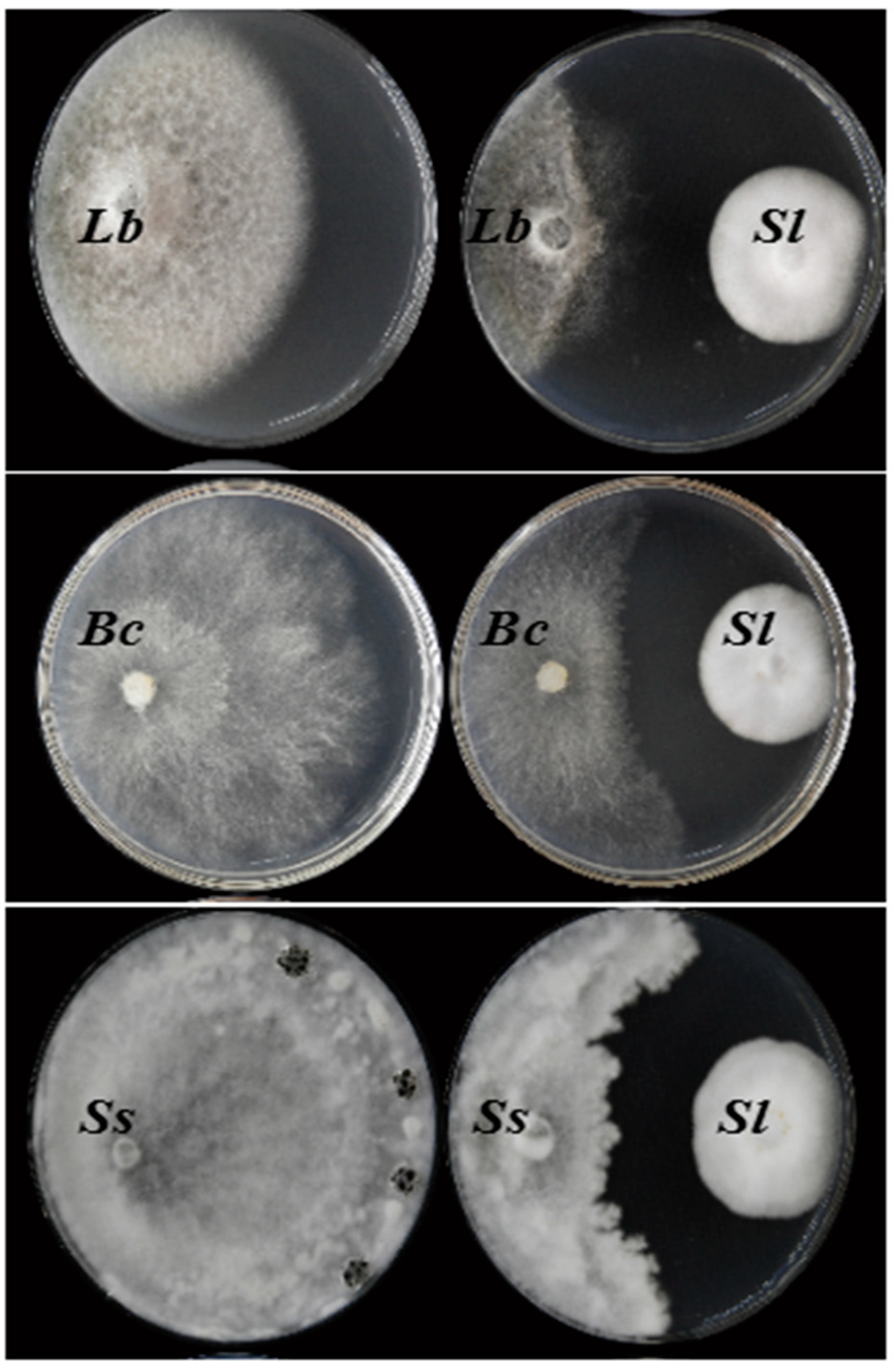
The genome-assisted approach was used to determine the capability of producing antifungal metabolites by the JC-1 strain [17]. Genomic DNA was extracted from the mycelial masses of JC-1 using the CTAB method [36]. It was then sequenced at Frasergen Bioinformatics Co., Ltd. (Wuhan, China) using a PacBio Sequel II System and MGISEQ-2000 platform, as well as at Annoroad Gene Technology Co., Ltd. (Zhejiang, China) using the Illumina Novaseq 6000 platform. De novo genome assembly was performed with PacBio long reads using MECAT2 [37]. For correction of the errors, sequence polishing was done with the MGISEQ-2000 and Illumina short reads using the Pilon package v1.23 [38]. Genome annotation was conducted following the pipeline in BRAKER v2.1.5 using the softwares GeneMark-ES v4.68 and Augustus v3.3.3 [39,40,41]. The sequences of the contigs, together with the raw sequencing data, were submitted to the China National GeneBank (CNGB) DataBase with the accession number CNP0003490. The completeness of genome annotation was estimated using BUSCO v4.1.4 with the dataset for fungi in the order Hypocreales [42]. The genome was submitted to the website https://fungismash.secondarymetabolites, and the biosynthetic gene clusters for the secondary metabolites (SM) were then searched using antiSMASH v. 6.1.1 [43]. To detect expression of the backbone genes in the SM gene clusters, total RNA was extracted from the JC-1 mycelia in 3- and 9-day-old PDB cultures (20 °C) using Fungal RNA Kit 200 (OMEGA Bio-Tek, Norcross, GA, USA). RNA profiling was done on the MGI-SEQ 2000 platform at Frasergen Bioinformatics Co. Ltd. The resulting reads for the backbone genes were counted as FPKM (fragments per kilobase of exon model per million mapped fragments).

2.4. Dual Culturing

To detect the antifungal activity of JC-1, mycelial agar plugs (5 mm in diameter) of JC-1 were inoculated to one side of Petri dishes (9 cm in diameter), each containing 20 mL PDA and one mycelial agar plug (1 cm from the dish rim). Meanwhile, mycelial agar plugs (5 mm in diameter) from fresh PDA were placed in Petri dishes and treated as control. The dishes were placed in an incubator (20 °C) for 10 d, and then, mycelial agar plugs (5 mm in diameter) of each target fungus (B. cinerea, L. biglobosa and S. sclerotiorum) were inoculated on the opposite side of the mycelial agar plug of JC-1 or the control agar plug (1 cm from the dish rim). There were three dishes (replicates) for each dual culture with JC-1 and a target fungus as well as for each single culture with the target fungus alone. The cultures were again incubated at 20 °C until the control dishes for the single cultures were almost colonized by the target fungi. The width of the clear zone between the colonies of JC-1 and a target fungus in each dual culture was measured. The trial was repeated three times.

2.5. Determination of the Antifungal Activity of the JC-1 Cultures

Two trials were done to determine the antifungal activity of the PDB cultures of JC-1, with S. sclerotiorum as the indicator for the antifungal activity. The first trial was a time-course experiment. Mycelial agar plugs (5 mm in diameter) of JC-1 were inoculated in 250-mL Erlenmeyer flasks, each containing 50 mL PDB, with one mycelial agar plug in each flask; there were 15 flasks in total. The flasks were mounted on a rotary shaker and incubated at 20 °C and 150 rpm for 3, 6, 9, 12 and 15 d. At each time point, three flasks were randomly selected and taken off the shaker. The culture in each flask was centrifuged at 9000 rpm at room temperature for 15 min to obtain the supernatant and the mycelial mass precipitate. The mycelial mass was dried at 50 °C for 48 h and weighed. The pH value of the supernatant was measured using a pH meter. Meanwhile, the supernatant was incorporated into PDA at 10% (v/v), and in the control treatment, non-utilized fresh PDB was also incorporated into PDA at 10% (v/v). The PDA of both treatments was loaded separately into Petri dishes, 20 mL per dish, and three dishes (replicates) for each treatment. Mycelial agar plugs of S. sclerotiorum were inoculated into the Petri dishes, one mycelial agar plug in each dish. The cultures were incubated at 20 °C for 2 d, the diameter of the colony in each dish was measured, and the percentage of inhibition of mycelial growth was calculated based on the colony diameters in the two treatments [44].
The second trial was a dose-effect experiment, and the 15-day-old PDB cultures of JC-1 were used in this trial. The supernatant was obtained from the PDB cultures by centrifuging, and it was supplemented in PDA by 1%, 2%, 5%, 10%, 15% and 20% (v/v), and in the control treatment, PDA was supplemented with water alone by 1% to 20% (v/v). S. sclerotiorum was inoculated on PDA with different treatments, and the cultures were incubated at 20 °C for 2 d, colony diameters were measured, and percentages of inhibition of mycelial growth were calculated [44].
Moreover, the basic properties of the antifungal metabolites in the PDB cultures of JC-1 (20 °C, 15 d) were determined, including the therm-stability (40 °C to 121 °C for 10 to 60 min), resistance to UV-irradiation (5 to 100 min), and response to ambient pH (pH 2 to 13 for 24 h). S. sclerotiorum was used as the indicator fungus. The details of the experimental procedures for these tests were described in our previous study [45].

2.6. Extraction of the Antifungal Metabolites

The procedures to extract the antifungal metabolites from the PDB cultures of JC-1 (20 °C, 150 rpm, 15 d) were outlined in Figure S1. The cultures were centrifuged to remove the mycelial masses. The resulting supernatants were pooled, and acetone was added at a volume ratio of 1:1. The two phases were thoroughly mixed, and the mixture was filtered through four-layered gauze fabric to remove the flocculent metabolites. The filtrate was evaporated in a rotavapor to remove the acetone. Then, ethyl acetate was added to the remaining solution, also at a volume ratio of 1:1, and the two phases were thoroughly mixed three times. After standing at room temperature for 20 min, the mixture was again separated into two phases: the clear organic phase and the turbid water phase. The organic phase was collected with the aid of a separatory funnel and dried by rotary evaporation. The dried extract in the flask was washed with a small amount of methanol, water was added to the resulting solution, and the mixture was freeze-dried. Finally, a yellowish powder was obtained, and it was considered to be the crude extract (CE) of the antifungal metabolites of JC-1.
To determine antifungal activity, the CE was dissolved in water to generate a mother solution at 10 mg/mL, it was supplemented into PDA to the final concentrations of 12.5, 25, 50, 100, 200, 400, or 800 μg/mL, and PDA alone was treated as a control. The PDA for different treatments were separately loaded in Petri dishes, three dishes (replicates) for each treatment, and S. sclerotiorum was inoculated on the PDA in these dishes. The cultures were incubated at 20 °C for 2 d, the diameter of the colony in each dish was measured, and the data was used to calculate the percentages of inhibition of mycelial growth [44].

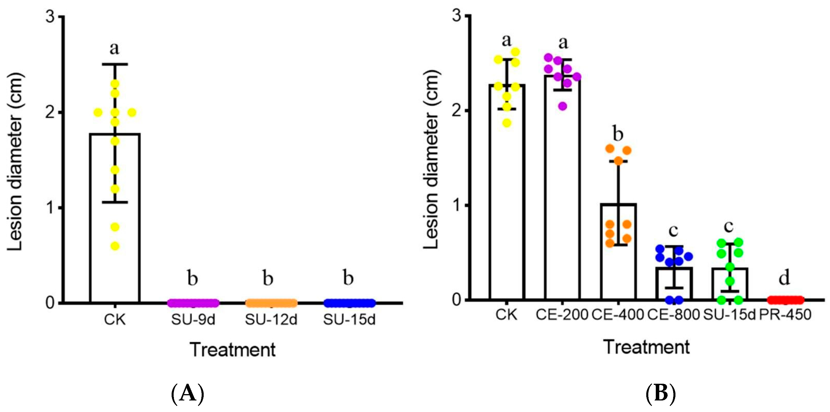
2.7. Determination of Control Efficacy of the Antifungal Metabolites

Seeds of oilseed rape (B. napus cultivar “Zhongshuang No. 9”) were pre-germinated at 20 °C on moisturized filter papers for 4 d. The germinated seeds were then sown in Organic Culture Mix (Zhenjiang Pei Lei Organic Fertilizer Co. Ltd., Zhenjiang, China, N:P:K = 1:1:1, pH 6) in plastic pots (10 × 10 cm, diameter × height), with five seeds per pot. The pots were maintained in the growth room (20 °C, 12 h light/12 h dark) for 30 d. True leaves (~10 × 8 cm, length × width) were excised from the plants. The detached leaves were randomly selected and placed on moist towels in plastic trays (45 × 30 × 2.5 cm, length × width × height) for the following two assays: (i) the supernatant assay with four treatments, including three treatments with the supernatants of JC-1 from 9-, 12- and 15-day-old PDB cultures (e.g., SU-9d, SU-12d and SU-15d, respectively) and a control treatment with water alone (control); (ii) the crude extract assay with six treatments, including three treatments with the JC-1 CE at 200, 400 and 800 μg/mL (e.g., CE-200, CE-400 and CE-800, respectively), the supernatant treatment with SU-15d of JC-1, the fungicide treatment with prochloraz at 450 μg a.i./mL (e.g., PR-450), and the control treatment with water alone. Prochloraz EW (MIAOLIANGTM) was purchased from Henan Guangnong Pesticide Factory (Mengzhou, Henan province, China). The solutions (SU-9d, SU-12d, SU-15d, CE-200, CE-400, CE-800, and PR-450) and water alone were separately amended with Tween 20 at 0.2% (v/v) and applied onto the leaves in the trays at ~1 mL per leaf. The trays were left open in a laminar flow hood for 3 h to allow evaporation of the excess water on the leaf surface. Mycelial agar plugs (5 mm in diameter) of S. sclerotiorum were inoculated on the leaves with the mycelia facing the leaves, two mycelial agar plugs on each leaf and four leaves for each treatment. The trays were individually covered with transparent plastic films (0.1 mm thick) to maintain high humidity and placed in a growth chamber at 20 °C under fluorescent light (12 h per day) for 2 d. The lesion diameter around each mycelial agar plug was measured. The assays were repeated three times.

2.8. Detection of Endophytic Growth

Seeds of oilseed rape “Zhongyou No. 9” were pre-germinated for 4 d, and then, they were sown in the moisturized Organic Culture Mix in seedling trays, which were maintained for 10 d in the growth room (20 °C). Then, the seedlings were drenched with the conidial suspension of JC-1 (1 × 10⁷ conidia/mL) or water alone (control), 20 mL per seedling. After incubation for another 30 days, six seedlings for each treatment were uprooted and washed under running water to remove the culture mix. The seedlings were blot-dried, the taproots and stems were cut into segments of 1 to 2 mm long, and the true leaves were cut into small square-shaped pieces (~5 × 5 mm, length × width). The segments of taproots/stems and the leaf pieces were surface-sterilized with 75% ethanol (30 s) and 5% NaClO (2 min). After washing in sterilized water (three times, for 30 s each time), the plant tissues were placed on PDA amended with lactic acid to inhibit bacterial contamination, and the cultures were incubated at 20 °C for 20 d. Colonies of JC-1 formed from the plant tissues were identified based on the particular morphological features of this fungus. The number of segments of taproots/stems and the leaf pieces with the emergence of JC-1 was recorded.

2.9. Induction of Systemic Resistance

Seeds of the cultivar “Zhongyou No. 9” were surface-sterilized with NaClO, followed by immersing in the conidial suspension of JC-1 (1 × 10⁷ conidia/mL) or water alone (control) for 12 h (Figure S2). They were then transferred to moisturized filter papers in Petri dishes, which were placed for 4 d in a growth room (20 °C, 16 h of light/8 h of darkness). The germinated seeds of both treatments were sown in the moisturized Organic Culture Mix in 32-hole seedling trays, one seed in each hole. The trays were placed in the growth room for 4 d, and the seedlings were drenched with the conidial suspension of JC-1 or water alone, 10 mL per seedling. Eight days later, the cotyledons were wounded on both sides of the main veins using a sharp needle. Aliquots of the conidial suspension of L. biglobosa W10 (1 × 10⁷ conidia/mL) were pipetted onto the cotyledons, 10 μL on each wound, 14 cotyledons on seven seedlings for each treatment. The treated seedlings were incubated at 20 °C for 8 d under humid conditions (R.H. > 90%). The diameter of the necrotic lesion around each wound was measured. The assay was repeated three times as three independent trials.

2.10. Quantification of Anthocyanins

Seeds of oilseed rape “Zhongyou No. 9” were treated with the conidial suspensions of JC-1 (1 × 10⁵–7 conidia/mL) or water alone (control), and they were separately placed on moisturized filter papers for 4 d (20 °C). The germinated seeds of each treatment were selected, and the hypocotyl together with the cotyledons on each seedling were removed. The plant samples with the hypocotyls and cotyledons (0.3 g) for different treatments were separately transferred to 2-mL-centrifuge tubes. Aliquots of the HCl-acidified methanol were added to the tubes (1 mL per tube) to extract the anthocyanins from the plant samples by shaking the tubes on a rotary shaker (70 rpm) at room temperature for 18 h [46]. The mixtures in the tubes were centrifuged at room temperature at 12,000 rpm, the supernatants (0.4 mL) were pipetted out and transferred to other 2-mL tubes, and aliquots (0.6 mL) of the acidified methanol were added. The optical density (OD) values of the resulting mixtures were measured at wavelengths of 530 nm and 657 nm in a spectrophotometer. The content of anthocyanins (CA) was calculated with the following formula:
CA   ( nmol / g ) = O D 530 0.25 × O D 657 ϵ   ×   v m   × 1 , 000 , 000
where OD530 and OD657 represent the OD values at 530 nm and 657 nm, respectively, ϵ is a constant (4.62 × 10⁶), v represents the volume of the mixture for measurement (1 mL), m represents the weight of each plant sample (0.3 g). The measurement was repeated three times.

2.11. Quantitative Detection of Expression of the Defense-Related Genes

Seeds of oilseed rape “Zhongyou No. 9” were surface-sterilized and immersed for 12 h in the conidial suspensions of JC-1 (1 × 10⁵–7 conidia/mL) or in water alone (control), and they were then transferred to moisturized paper filters in Petri dishes. After incubation for 4 d (20 °C), the hypocotyls and radicles (0.7 g) for each treatment were sampled and ground into a fine powder in liquid nitrogen. Total RNA was extracted from the powder using the PLANTpure Plant RNA Kit (Aidlab Biotechnol. Co., Ltd., Beijing, China), and DNA in the extract was eliminated using RNAase-free DNase I (Table S4). The RNA was then used as a template for the synthesis of cDNA under the catalysis of PrimeScript II RTase. Finally, the cDNA was used as a template to quantify the transcripts of CHI (for chalcone isomerase), NCED3 (for 9-cis-epoxycarotenoid dioxygenase), PR1 (for pathogenesis-related protein 1) and ACT7 (for actin, reference) in quantitative RT-PCR (qRT-PCR) using the primer pairs CHI/CHIR, NCED3F/NCED3R, PR-1F/PR-1R and ACT7F/ACT7R (Tables S1, S5 and S6), respectively. The transcript level of each gene was calculated using the ΔΔCt method [47]. For normalization of the data, the transcript level of each gene in the control treatment was considered to be 1.0, and the scale was used to calibrate the transcript level of that gene in the treatments of JC-1. The qRT-PCR was repeated independently three times.

2.12. Data Analysis

Data on percentages of inhibition of mycelial growth, leaf lesion diameters, content of anthocyanins, as well as the relative expression levels of CHI, NCED3 and PR1, was analyzed using the procedure of analysis of variance (ANOVA) in the SAS software (SAS Institute, Cary, NC, USA, v. 8.0, 1999). The means for different treatments in an experiment or a trial were separated using the least significant difference (LSD) test at α = 0.05. Before ANOVA, the percentage data was arcsine-transformed to angular values, and after analysis, the average angular values were individually back-transformed to numerical percentages. Data on cotyledon lesion diameters caused by L. biglobiosa in the resistance-inducing trials were analyzed using the procedure of univariate analysis in SAS. The means for the treatments of JC-1 and control in each trial were compared using a Student’s t test at α = 0.05, 0.01 or 0.001.

3. Results

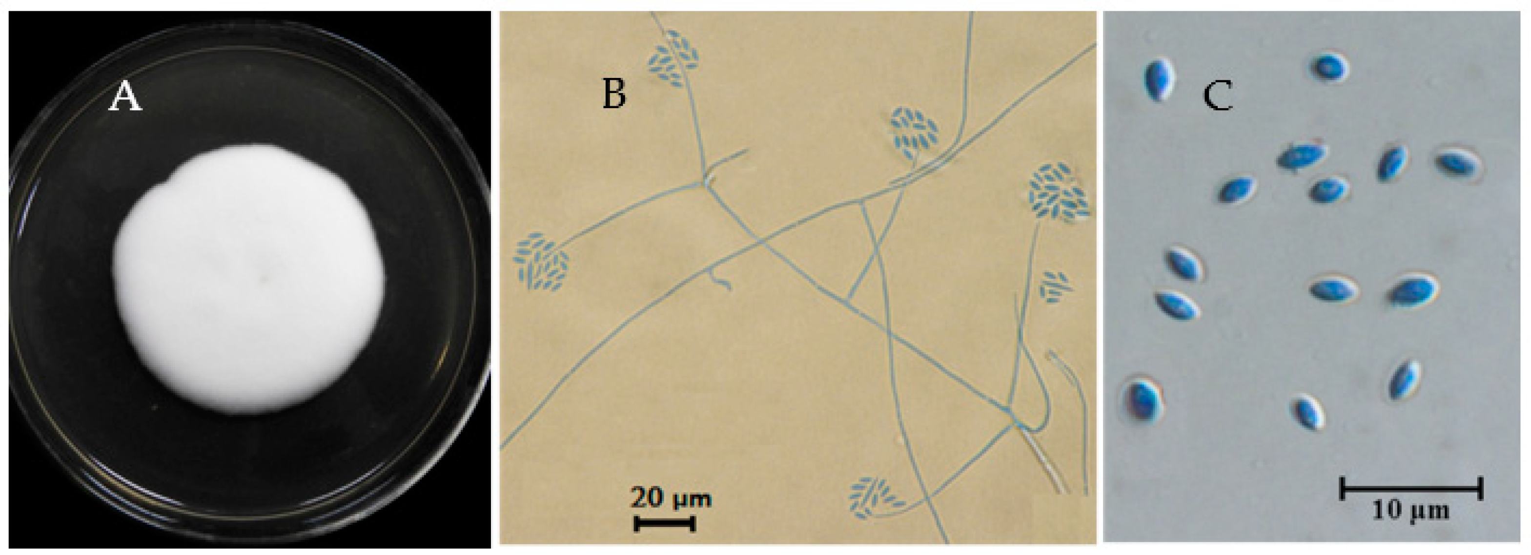
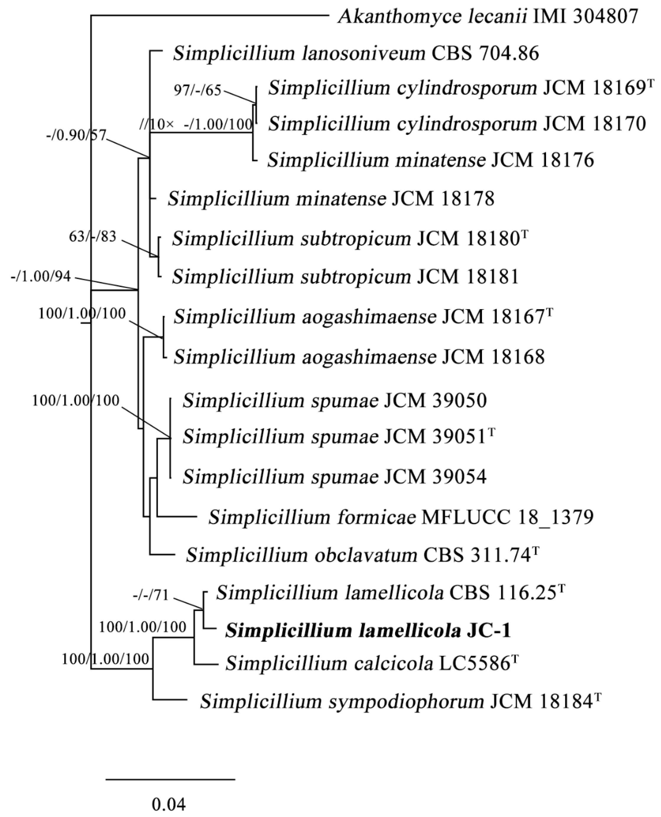
3.1. Taxonomic Identity

The JC-1 strain formed white floccose colonies on PDA at 20 °C with a villiform appearance on the surface (Figure 1A) and a pale yellow color on the colony back. Conidiophores (phialides) arose from the hyphae and tapered towards the apex; they are hyaline, aseptate, 12 to 30 μm long (averagely 23 μm long), mostly solitary, and some in pairs or in simple whorls. Globose to subglobose conidial masses (heads) were produced at the apex of the conidiophores (Figure 1B). Conidia are unicellular, hyaline, oval in shape, sharp at both ends, and 3.3–4.5 μm × 2.1–3.3 μm in size (Figure 1C). These morphological characteristics matched descriptions for the fungi in the genus Simplicillium [1], suggesting that the JC-1 strain belongs to a species of Simplicillium.
The partial DNA sequences of ITS, LSU, SSU and TEF1α in JC-1 were obtained by PCR, and they were submitted to GenBank and assigned with the GenBank accession numbers: MT807906, MT807907, MT807908 and MT826785, respectively (Table 1). Phylogenetic analysis based on ITS alone and the concatenated sequence of ITS/LSU/SSU/TEF1α showed that JC-1 is closely related to S. lamellicola CBS 116.25T and CBS 454.70 (Figure 2 and Figure S3). Therefore, the JC-1 strain belongs to Simplicillium lamellicola (Smith) Zare & Gams.

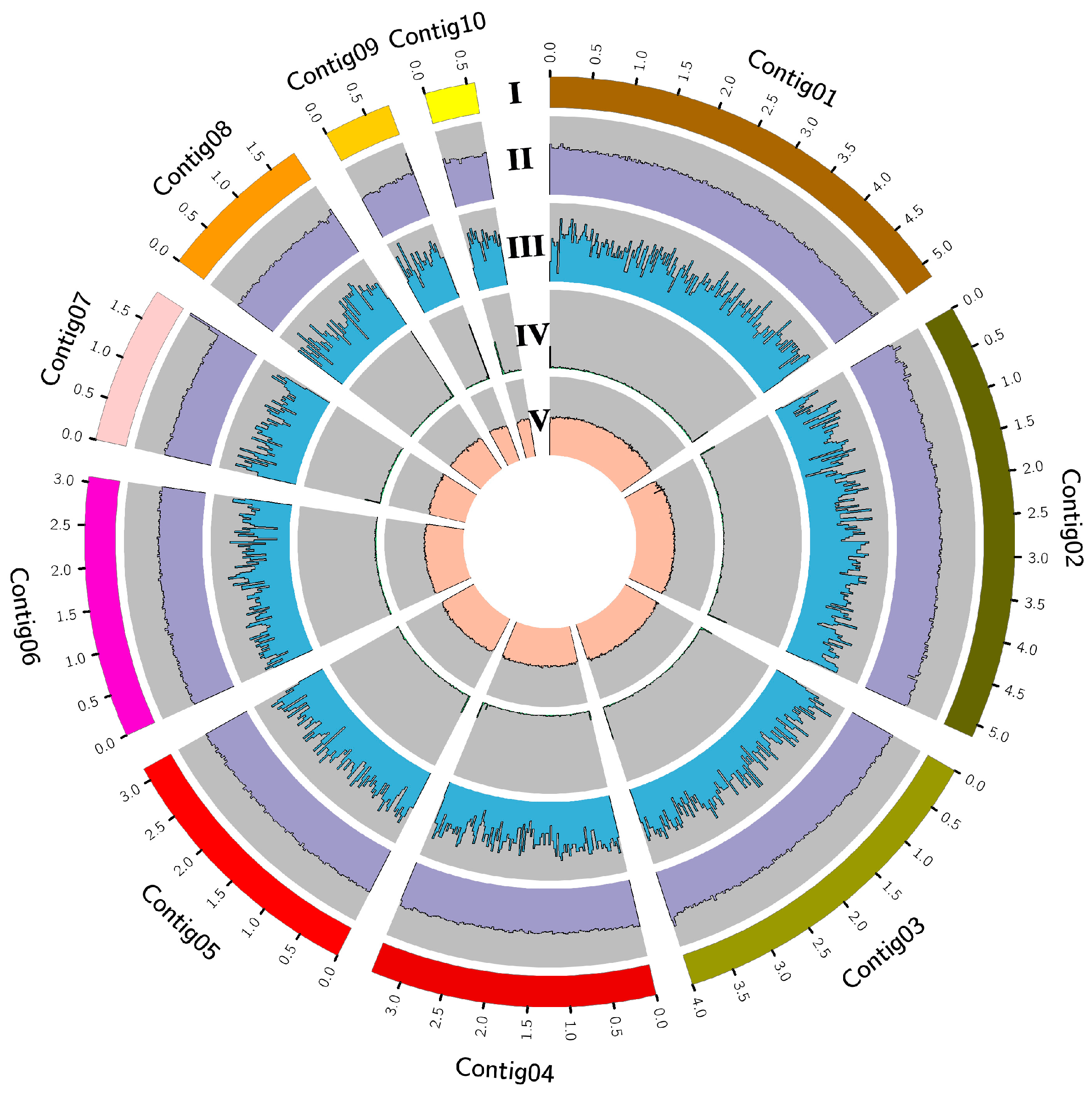
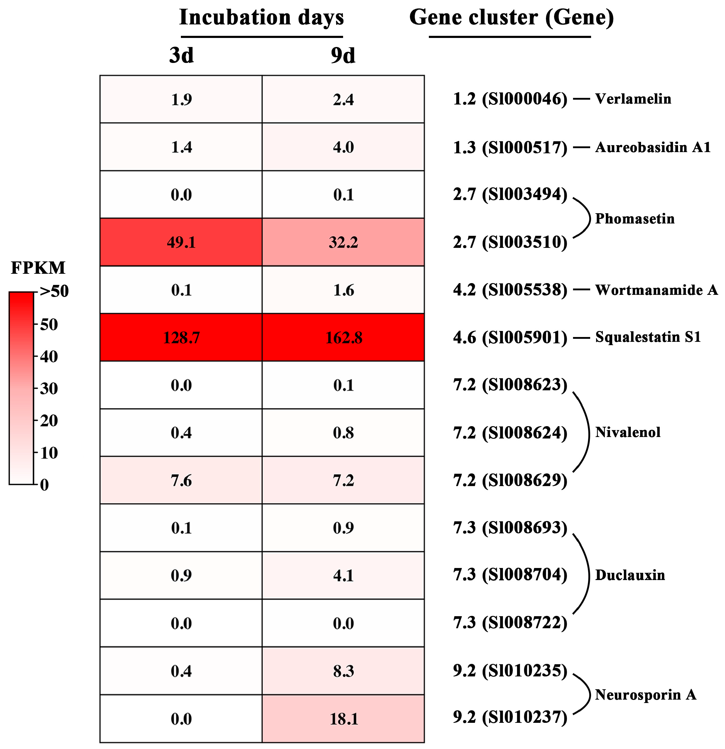
3.2. The Genome of JC-1 and the Gene Clusters for the Secondary Metabolites

The genome of S. lamellicola JC-1 was sequenced, the PacBio long reads (12.47 Gb) were used for genome assembly, and the MGISEQ-2000 and Illumina short reads (3.5 and 3.1 Gb, respectively) were used for correction of the genome. Finally, a genome was assembled on 22 contigs with a total length of 29.14 Mb (Table 2), including 28.98 Mb on 10 major contigs accounting for 99.45% of the total genome (Figure 3) and 0.16 Mb on 12 minor contigs accounting for 0.55% of the total genome. A total of 10,488 protein-coding genes were predicted in the genome of JC-1 (Table 2). The genome data was deposited in the China National GeneBank (CNGB) DataBase with the accession number CNP0003490.
Results from the antiSMASH search identified 37 gene clusters for the biosynthesis of the secondary metabolites or SMs (Table S7). They were grouped into eight biosynthetic types, including NRPS/NRPS-like/NPRS-PKS (21), PKS (8), terpene (4), β-lactone (1), NAPAA/PKS (1), PKS/indole (1), and RiPP/terpene (1) (Table S7). Eight out of the 37 putative SMs are known metabolites, including aureobasidin A1, duclauxin, neurosporin A, nivalenol, phomasetin, squalestatin S1, verlamelin and wortmanamide A (Figure S4), and the remaining 29 SMs are unknown metabolites. Three of the known SMs (e.g., aureobasidin A1, squalestatin S1 and verlamelin) have been reported to have antifungal activity [27,48,49]. The previously-characterized biosynthetic gene clusters (BGCs) for these three SMs [27,50,51] were used as references for identification of the homologous SMs in the genome of JC-1. The BGC for aureobasidin A1 was detected in Contig No. 1 (Figure S4, Table S7), and the amino acids encoded by the backbone gene (Sl000517) are 52% identical to those of the aureobasidin A1 biosynthesis complex (aba1) in Aureobasidium pullulans [51]. The BGC for verlamelin was detected in Contig No. 1 and No. 4, and it accommodates four genes, including Sl000045, Sl000044 and Sl000046 in Contig No. 1 as well as Sl005221 in Contig No. 4. These four genes are homologous to vlmS, vlmB, vlmC and vlmA, respectively, in Lecanicillium HF627 [29]. The nucleotides of Sl005221, Sl000045, Sl000044 and Sl000046 are identical by 51%, 92%, 90% and 90% to those of vlmA, vlmB, vlmC and vlmS, respectively. The amino acids encoded by Sl000045, Sl000044 and Sl000046 are identical by 47%, 96%, 96% and 92% to those encoded by vlmA, vlmB, vlmC and vlmS, respectively (Table S8). The BGC for squalestatin S1 in JC-1 was detected in Contig No. 4 (Figure S4, Table S7), and the backbone gene (Sl005901) codes for the amino acids with 58% identity to that of the squalene synthetase encoded by mfR6 in Phoma MF5453, a well-characterized producer of squalestatin S1 [50].
Transcriptome profiling showed that in the 3- and 9-day-old PDB cultures, the backbone genes for the eight known BGCs had expression, although they varied greatly in expression level as evaluated by FPKM values (Figure 4). The backbone gene Sl005901 for squalestatin S1 is highly expressed, with a FPKM higher than 100, followed by the backbone gene Sl003510 for phomasetin, with an average FPKM of 41. On the other hand, the backbone gene Sl005538 for wortmanamide A had negligible expression, with a FPKM lower than 1.0. The verlamelin backbone gene Sl000046 and the aureobasidin A1 backbone gene Sl000517 had FPKM values ranging from 1.4 to 4.0 (Figure 4).

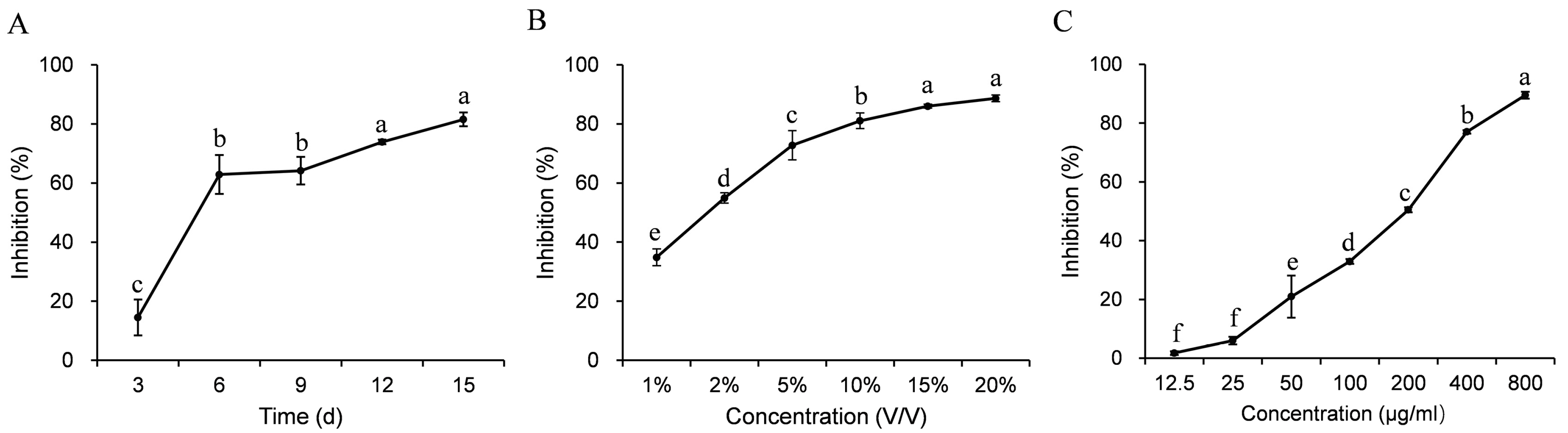
3.3. Antifungal Activity

Results of the dual culturing test showed that the JC-1 strain strongly inhibited the mycelial growth of B. cinerea, L. biglobosa and S. sclerotiorum, as indicated by the formation of inhibition zones (clear zones) between the colonies of JC-1 and these three target fungi in the dual cultures (Figure 5). The average width of the inhibition zones (n = 3) was 1.6, 1.7 and 1.5 cm between JC-1 and L. biglobosa, JC-1 and B. cinerea, and JC-1 and S. sclerotiorum, respectively.
The antifungal metabolites in the PDB cultures were extracted with acetone and ethyl acetate (Figure S2). The crude extract showed antifungal activity against S. sclerotiorum; the percentage of mycelial growth inhibition increased from 24% to 92% when the concentration of the crude extract was increased from 12.5 to 800 μg/mL (Figure 6C). The half-effective concentration (EC50) of the crude extract was 33.2 μg/mL.

3.4. Control Efficacy of the Cultures and the Crude Extract

In the supernatant trial, the leaves of oilseed rape treated with water alone formed large necrotic lesions around the mycelial agar plugs of S. sclerotiorum (20 °C, 2 d), the average lesion diameter reached 1.8 cm. However, the leaves of oilseed rape treated with the supernatants from the 9-, 12- and 15-day-old cultures of JC-1 (SU-9d, SU-12d and SU-15d, respectively) produced no visible lesions (Figure 7A). In the crude extract trial, while the control treatment (CK) also formed large necrotic lesions with the average lesion diameter at 2.3 cm and the fungicide treatment (e.g., PR-450) formed no visible lesions, the three treatments of the crude extract (e.g., CE-200, CE-400 and CE-800) formed lesions with average diameters ranging from 0.3 to 2.4 cm (Figure 7B). While the treatment with a low concentration of the crude extract (e.g., CE-200) produced large lesions with an average diameter of 2.4 cm, which had no significant difference (p > 0.05) from that in the control treatment, the treatments with high concentrations of the crude extract (e.g., CE-400, CE-800) produced small lesions with average diameters of 1.0 and 0.3 cm, respectively, significantly lower (p < 0.05) than that in the control treatment. Interestingly, the average lesion diameter in CE-800 had no significant difference (p > 0.05) from that in the treatment of SU-15d (0.3 cm in diameter), suggesting that the crude extract at 800 μg/mL had a control efficacy equal to that of the 15-day-old cultures.

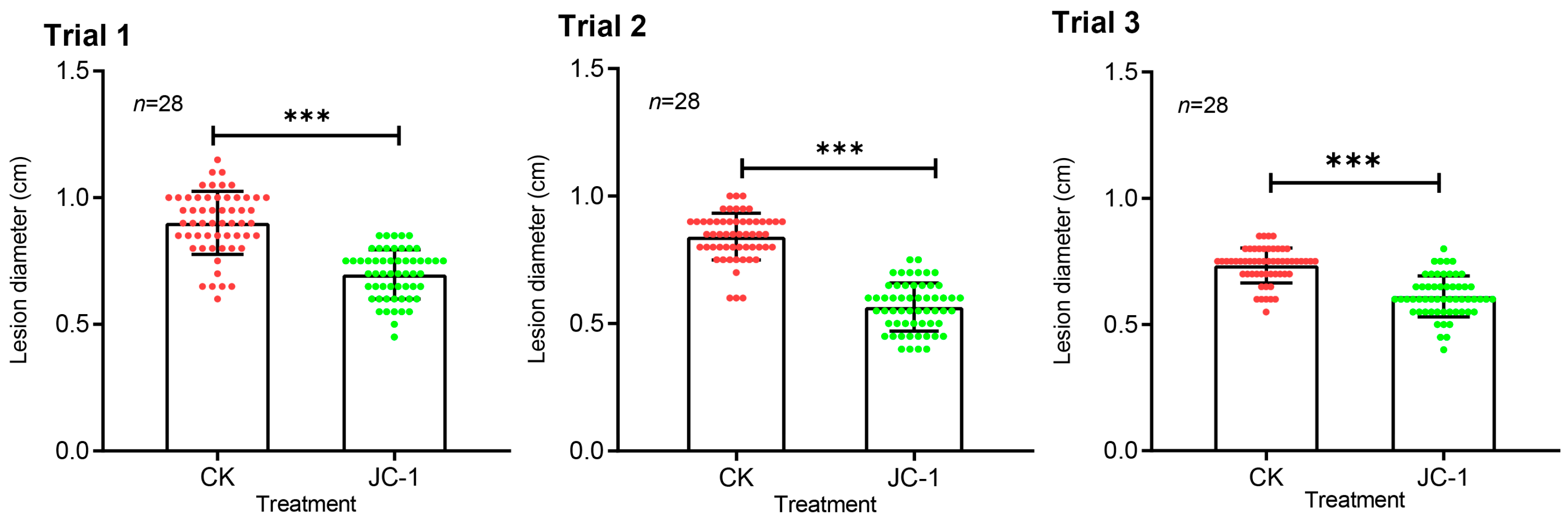
3.5. Endophytic Growth and Induced Systemic Resistance

Seeds of oilseed rape treated with JC-1 (1 × 10⁷ conidia/mL) or water alone for 12 h or 24 h germinated after incubation at 20 °C for 4 d, the percentages of the germinated seeds reached 60%–80%, and no significant difference (p > 0.05) was detected between the treatments of JC-1 and water alone (Figure S5A). However, the seedlings of the two treatments differed significantly (p < 0.05) in length of radicals (roots) and hypocotyls (shoots), the seedlings in the treatment of JC-1 formed shorter hypocotyls than those in the control treatment (Figure S6B); however, they produced longer radicals than those in the control treatment (Figure S6B,C). Moreover, the results from the seedling-drenched inoculation experiment showed that JC-1 existed in the stems and leaves of oilseed rape without causing any visible disease symptoms; 23.8% of stem segments and 14.3% of leaf pieces were detected as positive for S. lamellicola; however, the root segments were detected negative for S. lamellicola (Table 3).
Results from the cotyledon-inoculation trials showed that necrotic lesions caused by L. biglobosa appeared on the cotyledons of the seedlings drenched with the conidial suspension of JC-1 (1 × 10⁷ conidia/mL) and water alone. Statistical analysis indicated that drenching with the JC-1 conidia formed significantly (p < 0.001) smaller cotyledon lesions than drenching with water alone (Figure 8). The average cotyledon lesion diameters in the treatment of drenching with JC-1 were reduced by 22%, 16% and 32% in trials 1, 2 and 3, respectively, compared to those in the control treatment of drenching with water alone.

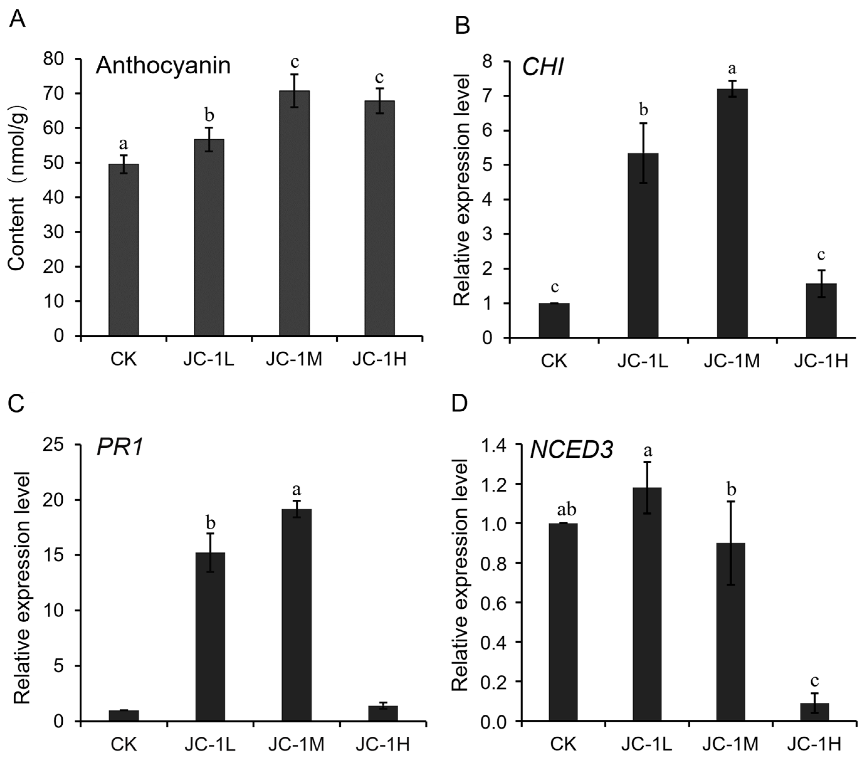
3.6. Mechanisms for the Enhanced Systemic Resistance

The seedlings of oilseed rape from the seeds treated with the JC-1 conidial suspensions (1 × 10⁵–7 conidia/mL) and water alone (control) had accumulations of anthocyanins. The treatments of JC-1 at 1 × 10⁵, 1 × 10⁶ and 1 × 10⁷ conidia/mL had the content of anthocyanins at 56.7, 70.8 and 67.9 nmol/g, respectively, and the values were significantly (p < 0.05) higher than that of 49.5 nmol/g in the control treatment (Figure 9A).
Results of qRT-PCR showed that the three defense-related genes, CHI, PR1 and NECD3, expressed in the seedlings treated with JC-1 (1 × 10⁵–7 conidia/mL) and water alone (control). An up-regulated expression of CHI and PR1 was observed in the seedlings treated with JC-1 at 1 × 10⁵ conidia/mL (5 and 15 folds of the control, respectively) and 1 × 10⁶ conidia/mL (7 and 19 folds of the control, respectively). However, the seedlings treated with JC-1 at 1 × 10⁷ conidia/mL had relative expression values of the two genes similar to those in the control treatment (Figure 9B,C). On the contrary, the seedlings of oilseed rape showed a different expression pattern for NECD3 from that of CHI and PR1 in response to JC-1 (Figure 9D). In the treatment with JC-1 at 1 × 10⁵ conidia/mL, the seedlings had a relative expression level of NECD3 similar to that in the control treatment. However, in the treatments with JC-1 at 1 × 10⁶ and 1 × 10⁷ conidia/mL, the seedlings showed a down-regulated expression of NECD3, as the relative expression values of NECD3 in these two treatments of JC-1 were significantly (p < 0.05) decreased by 10% and 91%, respectively, compared to that in the control treatment.

4. Discussion

This study revealed that the JC-1 strain belongs to Simplicillium lamellicola (F. E. W. Smith) Zare & W. Gams. According to the previous record, S. lamellicola is a common fungus with a world-wide distribution, and it has been reported to be able to infect mushrooms like Agaricus biaporus and A. bitorquis, causing gill mildew and small brown spots, respectively [1]. Moreover, S. lamellicola has the capability of living with rust fungi as a mycoparasite as well as living with cysts of Heterodera glycines and eggs of Meloidogyne arenaria as a nematode parasite [1]. The JC-1 strain of S. lamellicola was isolated from a diseased pod of oilseed rape infested with L. biglobosa. The re-inoculation experiment indicated that S. lamellicola JC-1 hardly infected the cotyledons of oilseed rape as L. biglobosa did. Therefore, S. lamellicola JC-1 is not a pathogen of oilseed rape (Li, 2019). Moreover, mixed inoculation of the conidia of S. lamellicola (Sl) and L. biglobosa (Lb) on oilseed rape (even at a ratio as high as 8:2, Sl:Lb) still caused large necrotic lesions of similar sizes to those inoculated with L. biglobosa alone [52]. This result suggests that S. lamellicola JC-1 could not effectively inhibit infection by L. biglobosa through physical contact, implying that it is at least not a specific and highly-aggressive mycoparasite of L. biglobosa.
Zhang et al. (2014) obtained three strains of S. lamellicola from healthy plants of oilseed rape (Brassica napus), and these strains were considered endophytes of oilseed rape, although the endophytic lifestyle of these strains was not confirmed by re-inoculation and re-isolation [30]. In this study, we re-inoculated S. lamellicola JC-1 on the roots of oilseed rape; fortunately, it was re-isolated from the leaves and stems of the inoculated seedlings without showing any visible disease symptoms. This result suggests that S. lamellicola JC-1 is really an endophyte of oilseed rape. Further study to trace the infection and spread of S. lamellicola JC-1 in the tissues of oilseed rape using certain marker genes such as the green fluorescent protein gene (gfp) is warranted.
The present study demonstrated that seed/root-inoculation with the conidial suspension of S. lamellicola JC-1 caused a significant (p < 0.001) reduction in cotyledon size of lesions caused by L. biglobosa on the resulting seedlings of oilseed rape. This result indicated that S. lamellicola JC-1 could induce systemic resistance (ISR) against infection by L. biglobosa. The possible mechanisms underlying the ISR may lie in endophytic growth in plant tissues of oilseed rape and the consequent enhanced accumulation of anthocyanins, as well as up-regulated expression of CHI and PR1. It is well recognized that the PR1 gene is associated with the salicylic acid (SA) pathway, and up-regulated expression of the SA pathway can activate the biosynthesis of some phenol compounds and analogs, such as anthocyanins, phytoalexins and lignin, which act as physical and physiological barriers to defend against pathogen infection [53]. On the other hand, the NCED3 gene is associated with the biosynthesis of abscisic acid (ABA), and up-regulation of the ABA pathway usually accelerates plant senescence. Lowe et al. (2014) reported that L. biglobosa infects oilseed rape in a necrotrophic way [54], and down-regulation of expression of NCED3 may lower the ABA level, thereby enhancing resistance against infection by L. biglobosa possibly through slowing down the senescence of the cotyledons of oilseed rape. This result also suggests that S. lamellicola JC-1 can be potentially used as a seed coating agent to suppress infection by soilborne pathogens such as Plasmodiophora brassicae (clubroot), Rhizoctonia solani (damping-off), as well as some airborne pathogens such as Peronospora parasitica (powdery mildew). Further field evaluation of the biocontrol efficacy of seed coating with S. lamellicola JC-1 against these diseases of oilseed rape is needed.
Kim et al. (2002) reported that S. lamellicola is a strong antagonist with the capability of producing the antifungal cyclic lipopeptide verlamelin [27]. A similar result was observed in this study, S. lamellicola JC-1 displayed strong antifungal activity against B. cinerea, L. biglobosa and S. sclerotiorum. So far, the antifungal metabolites produced by S. lamellicola JC-1 were not purified and identified. As an alternative, we analyzed the genome of S. lamellicola JC-1 to elucidate its capability of producing antifungal metabolites. We found that this particular fungus can produce verlamelin, aureobasidin A1 and squalestatin S1, which might be at least partially responsible for the antifungal activity of S. lamellicola JC-1. Moreover, genome analysis revealed that S. lamellicola JC-1 has the capability of producing 29 unknown metabolites, suggesting that S. lamellicola JC-1 is a prolific producer of antifungal metabolites. Further purification and identification are worthwhile to clarify the known and novel metabolites produced by JC-1.
Based on the reference biosynthetic gene cluster (BGC) for verlamelin in Lecanicillium HF627 [29], this study identified four genes (Sl000044, Sl000045, Sl000046 and Sl005221) putative for biosynthesis of verlamelin in S. lamellicola JC-1. Three (Sl000045, Sl000044 and Sl000046) of the four genes are clustered in Contig. No. 1, and they are highly identical to vlmB, vlmC and vlmS, respectively (≥90%) in Lecanicillium HF627 [29]. However, the other gene (Sl005221) locates in Contig No. 4 with a low identity (51% by nucleotides) to vlmA in Lecanicillium HF627 [29]. This comparison suggests that the verlamelin BGC in S. lamellicola JC-1 differs from that in Lecanicillium HF627, as Sl005221 is physically separated from Sl000045, Sl000044 and Sl000046 in S. lamellicola JC-1, whereas the four verlamelin BGC genes (vlmA, vlmB, vlmC and vlmS) in Lecanicillium HF627 are tightly clustered [29]. Further study is worthwhile to characterize the biosynthesis of verlamelin in S. lamellicola JC-1 using gene manipulation techniques (e.g., gene knockout and complementation).
The cultural filtrates of S. lamellicola JC-1 showed a high efficacy in suppressing infection by S. sclerotiorum on leaves of oilseed rape. Moreover, the antifungal activity of the cultural filtrates was stable under heat (121 °C/30 min) and the extreme acidic (pH 2)/alkaline (pH 13) conditions, as well as under UV irradiation. These results suggest that the metabolites in the cultures of S. lamellicola JC-1 have the potential to be used by spray on air parts of plants for control of sclerotinia stem rot as well as other fungal diseases of oilseed rape. This study tried to extract the antifungal metabolites from the cultures of S. lamellicola JC-1, the crude extract at 800 μg/mL displayed the same control efficacy as the cultural filtrates did. This result indicated that the crude extract had an unexpectedly low control efficacy, possibly due to the loss of some important components in the process of extraction using acetone and ethyl acetate. Therefore, it is necessary to optimize the procedure to extract the active antifungal metabolites from the cultures of S. lamellicola JC-1 in the future, as this is very important for the formulation of the cultures of this fungus as a biocontrol agent.
In summary, this study revealed that the JC-1 strain belongs to Simplicillium lamellicola, it is a strong fungal antagonist through the production of antifungal metabolites, and that the filtrates of JC-1 in the PDB cultures showed a high efficacy in suppressing infection by S. sclerotiorum on leaves of oilseed rape. Genome analysis indicated that JC-1 owns the capability of producing multiple antifungal metabolites, including aureobasidin A1, squalestatin S1 and verlamelin. Inoculation of JC-1 on seeds of oilseed rape caused a suppressive effect on infection by L. biglobosa on the cotyledons of the resulting seedlings, suggesting that JC-1 can induce systemic resistance (ISR). Endophytic growth, accumulation of anthocyanins, up-regulated expression of CHI and PR1, and down-regulated expression of NECD3 were found to be possibly responsible for the ISR. Therefore, S. lamellicola JC-1 is a promising and multifunctional biocontrol agent.

Supplementary Materials

The following supporting information can be downloaded at: https://www.mdpi.com/article/10.3390/jof9010057/s1, Figure S1: The procedure for obtaining the crude extract from the cultures of Simplicillium lamellicola JC-1 in potato dextrose broth; Figure S2: The procedure for determination of induced systemic resistance of S. lamellicola JC-1 against infection by L. biglobosa; Figure S3: Phylogenetic tree of 26 fungal taxa in the genus Simplicillium and Akanthomyces lecanii (out-group); Figure S4: Diagram showing composition of the eight biosynthetic gene clusters for known metabolites in Simplicillium lamellicola JC-1 based on antiSMASH; Figure S5: Effect of treatment of seeds of oilseed rape with the conidia of S. lamellicola JC-1 on seed germination rate and elongation of the radicles and hypotyls of the resulting seedlings; Figure S6: Detection of S. lamellicola JC-1 in stems (right dish) and leaves (middle and left dishes) of oilseed rape; Table S1: PCR primers and their oligonucleotide sequences [55,56,57]; Table S2: Reagents for PCR amplification of ITS, LSU, SSU and TEF1α; Table S3: Thermal program for PCR amplification of ITS, SSU, LSU and TEF1α; Table S4: Reaction system for DNA elimination in the RNA extracts from seedlings of oilseed; Table S5: Reagents for RT-PCR to detect expression of defense-related genes in seedlings of oilseed rape; Table S6: Thermal program for quantitative RT-PCR to detect expression of defense-related genes in seedlings of oilseed rape; Table S7: Gene clusters for biosynthesis of secondary metabolites (SMs) in the S. lamellicola JC-1 strain; Table S8: Putative genes of the verlamelin biosynthesis gene cluster in S. lamellicola JC-1.

Author Contributions

Conceptualization, W.L., J.L. and G.L.; methodology, W.L., T.L., J.Z. and M.W.; software, T.L.; validation, W.L., T.L., L.Y. and G.L.; investigation, W.L., T.L. and J.L.; resources, G.L. and L.Y.; data analysis, W.L. and T.L.; writing—original draft preparation, W.L. and G.L.; writing—review and editing, W.L. and G.L.; visualization, W.L. and G.L.; supervision, G.L.; funding acquisition, G.L., L.Y., J.Z. and M.W. All authors have read and agreed to the published version of the manuscript.

Funding

This study was financially supported China’s Agricultural Research System (CARS-12).

Institutional Review Board Statement

Not applicable.

Informed Consent Statement

Not applicable.

Data Availability Statement

The sequences of the contigs together with the raw sequencing data for S. lamellicola JC-1 were submitted to the China National GeneBank (CNGB) DataBase with the accession number CNP0003490.

Conflicts of Interest

The authors declare no conflict of interest.

References

  1. Zare, R.; Gams, W. A revision of Verticillium section Prostrata. IV. The genera Lecanicillium and Simplicillium gen. nov. Nova Hedwigia 2001, 73, 1–50. [Google Scholar] [CrossRef]
  2. Chen, W.H.; Liu, C.; Han, Y.F.; Liang, J.D.; Tian, W.Y.; Liang, Z.Q. Three novel insect-associated species of Simplicillium (Cordycipitaceae, Hypocreales) from Southwest China. MycoKeys 2019, 58, 83–102. [Google Scholar] [CrossRef] [PubMed] [Green Version]
  3. Chen, W.H.; Han, Y.F.; Liang, J.D.; Liang, Z.Q. Taxonomic and phylogenetic characterizations reveal four new species of Simplicillium (Cordycipitaceae, Hypocreales, Ascomycota) from Guizhou, China. Sci. Rep. 2021, 11, 15300. [Google Scholar] [CrossRef] [PubMed]
  4. Crous, P.W.; Luangsa-ard, J.J.; Wingfield, M.J.; Carnegie, A.J.; Hernández-Restrepo, M.; Lombard, L.; Roux, J.; Barreto, R.W.; Baseia, I.G.; Cano-Lira, J.F.; et al. Fungal Planet description sheets: 785–867. Persoonia 2018, 41, 238–417. [Google Scholar] [CrossRef]
  5. Crous, P.W.; Cowan, D.A.; Maggs-Kölling, G.; Yilmaz, N.; Thangavel, R.; Wingfield, M.J.; Noordeloos, M.E.; Dima, B.; Brandrud, T.E.; Jansen, G.M.; et al. Fungal Planet description sheets: 1182–1283. Persoonia 2021, 46, 313–528. [Google Scholar] [CrossRef] [PubMed]
  6. Dong, Q.L.; Dong, R.Z.; Xing, X.Y.; Li, Y.K. A new antibiotic produced by the cyanobacterium-symbiotic fungus Simplicillium lanosoniveum. Nat. Prod. Res. 2018, 32, 1348–1352. [Google Scholar] [CrossRef]
  7. Liu, F.; Cai, L. Morphological and molecular characterization of a novel species of Simplicillium from China. Cryptogamie Mycologie 2012, 33, 137–144. [Google Scholar] [CrossRef]
  8. Nonaka, K.; Kaifuchi, S.; Omura, S.; Masuma, R. Five new Simplicillium species (Cordycipitaceae) from soils in Tokyo, Japan. Mycoscience 2013, 54, 42–53. [Google Scholar] [CrossRef]
  9. Wei, D.P.; Wanasinghe, D.N.; Hyde, K.D.; Mortimer, P.E.; Xu, J.C.; Xiao, Y.P.; Bhunjun, C.S.; To-anun, C. The genus Simplicillium. MycoKeys 2019, 60, 69–92. [Google Scholar] [CrossRef] [Green Version]
  10. Zhang, Z.F.; Liu, F.; Zhou, X.; Liu, X.Z.; Liu, S.J.; Cai, L. Culturable mycobiota from Karst caves in China, with descriptions of 20 new species. Persoonia 2017, 39, 1–31. [Google Scholar] [CrossRef]
  11. Zhang, Z.F.; Zhou, S.Y.; Eurwilaichitr, L.; Ingsriswang, S.; Raza, M.; Chen, Q.; Zhao, P.; Liu, F.; Cai, L. Culturable mycobiota from Karst caves in China II, with descriptions of 33 new species. Fung. Divers. 2021, 106, 29–136. [Google Scholar] [CrossRef]
  12. Baiswar, P.; Ngachan, S.V.; Rymbai, H.; Chandra, S. Simplicillium lanosoniveum, a hyperparasite on Aecidium elaeagni-latifoliae in India. Austral. Plant Dis. Notes 2014, 9, 144. [Google Scholar] [CrossRef] [Green Version]
  13. Gauthier, N.W.; Maruthachalam, K.; Subbarao, K.V.; Brown, M.; Xiao, Y.; Robertson, C.L.; Schneider, R.W. Mycoparasitism of Phakopsora pachyrhizi, the soybean rust pathogen, by Simplicillium lanosoniveum. Biol. Control 2014, 76, 87–94. [Google Scholar] [CrossRef]
  14. Sun, J.Z.; Liu, X.Z.; McKenzie, E.H.C.; Jeewon, R.; Liu, J.K.; Zhang, X.L.; Zhao, Q.; Hyde, K.D. Fungicolous fungi: Terminology, diversity, distribution, evolution, and species checklist. Fung. Divers. 2019, 95, 337–430. [Google Scholar] [CrossRef]
  15. Wang, N.; Fan, X.; Zhang, S.; Liu, B.; He, M.; Chen, X.; Tang, C.; Kang, Z.; Wang, X. Identification of a hyperparasitic Simplicillium obclavatum strain affecting the infection dynamics of Puccinia striiformis f. sp. tritici on wheat. Front. Microbiol. 2020, 11, 1277. [Google Scholar] [CrossRef]
  16. Ward, N.A.; Schneider, R.W.; Aime, M.C. Colonization of soybean rust sori by Simplicillium lanosoniveum. Fung. Ecol. 2011, 4, 303–308. [Google Scholar] [CrossRef]
  17. Zhu, M.; Duan, X.; Cai, P.K.; Li, Y.F.; Qiu, Z.B. Deciphering the genome of Simplicillium aogashimaense to understand its mechanisms against the wheat powdery mildew fungus Blumeria graminis f. sp. tritici. Phytopathol. Res. 2022, 4, 16. [Google Scholar] [CrossRef]
  18. Lim, S.Y.; Lee, S.; Kong, H.G.; Lee, J.K. Entomopathogenicity of Simplicillium lanosoniveum isolated in Korea. Mycobiology 2014, 42, 317–321. [Google Scholar] [CrossRef] [Green Version]
  19. Zhao, D.; Liu, B.; Li, L.Y.; Zhu, X.F.; Wang, Y.Y.; Wang, Q.J.; Duan, Y.X.; Chen, L.J. Simplicillium chinense: A biological control agent against plant parasitic nematodes. Biocontrol Sci. Technol. 2013, 23, 980–986. [Google Scholar] [CrossRef]
  20. Chen, R.S.; Huang, C.C.; Li, J.C.; Tsay, J.G. First report of Simplicillium lanosoniveum causing brown spot on Salvinia auriculata and S. molesta in Taiwan. Plant Dis. 2008, 92, 1589. [Google Scholar] [CrossRef]
  21. Shen, X.Y.; Zheng, D.Q.; Gao, J.; Hou, C.L. Isolation and evaluation of endophytic fungi with antimicrobial ability from Phyllostachys edulis. Bangladesh J. Pharmacol. 2012, 7, 249–257. [Google Scholar] [CrossRef]
  22. Teasdale, S.E.; Caradus, J.R.; Johnson, L.J. Fungal endophyte diversity from tropical forage grass Brachiaria. Plant Ecol. Divers. 2018, 11, 611–624. [Google Scholar] [CrossRef]
  23. Liang, X.; Nong, X.H.; Huang, Z.H.; Qi, S.H. Antifungal and antiviral cyclic peptides from the deep-sea-derived fungus Simplicillium obclavatum EIODSF 020. J. Agric. Food Chem. 2017, 65, 5114–5121. [Google Scholar] [CrossRef]
  24. Ward, N.A.; Robertson, C.L.; Chanda, A.K.; Schneider, R.W. Effects of Simplicillium lanosoniveum on Phakopsora pachyrhizi, the soybean rust pathogen, and its use as a biological control agent. Phytopathology 2012, 102, 749–760. [Google Scholar] [CrossRef] [Green Version]
  25. Shin, T.S.; Yu, N.H.; Lee, J.; Choi, G.J.; Kim, J.C.; Shin, C.S. Development of a biofungicide using a mycoparasitic fungus Simplicillium lamellicola BCP and its control efficacy against gray mold diseases of tomato and ginseng. Plant Pathol. J. 2017, 33, 337–344. [Google Scholar] [CrossRef] [PubMed] [Green Version]
  26. Choi, G.J.; Kim, J.C.; Jang, K.S.; Cho, K.Y.; Kim, H.T. Mycoparasitism of Acremonium strictum BCP on Botrytis cinerea, the gray mold pathogen. J. Microbiol. Biotechnol. 2008, 18, 167–170. [Google Scholar] [PubMed]
  27. Kim, J.C.; Choi, G.J.; Kim, H.J.; Kim, H.T.; Ahn, J.W.; Cho, K.Y. Verlamelin, an antifungal compound produced by a mycoparasite, Acremonium strictum. Plant Pathol. J. 2002, 18, 102–105. [Google Scholar] [CrossRef]
  28. Le Dang, Q.; Shin, T.S.; Park, M.S.; Choi, Y.H.; Choi, G.J.; Jang, K.S.; Kim, I.S.; Kim, J.C. Antimicrobial activities of novel mannosyl lipids isolated from the biocontrol fungus Simplicillium lamellicola BCP against phytopathogenic bacteria. J. Agric. Food Chem. 2014, 62, 3363–3370. [Google Scholar] [CrossRef]
  29. Ishidoh, K.; Kinoshita, H.; Nihira, T. Identification of a gene cluster responsible for the biosynthesis of cyclic lipopeptide verlamelin. Appl. Microbiol. Biotechnol. 2014, 98, 7501–7510. [Google Scholar] [CrossRef]
  30. Zhang, Q.H.; Zhang, J.; Yang, L.; Zhang, L.; Jiang, D.H.; Chen, W.D.; Li, G.Q. Diversity and biocontrol potential of endophytic fungi in Brassica napus. Biol. Control 2014, 72, 98–108. [Google Scholar] [CrossRef]
  31. Li, J.C. Fixed-Site Monitoring of Occurrence of Blackleg of Oilseed Rape and Characterization of Infection by Conidia of the Pathogen. Master’s Thesis, Huazhong Agricultural University, Wuhan, China, 2019; pp. IV + 61. [Google Scholar]
  32. Büttner, P.; Koch, F.; Voigt, K.; Quidde, T.; Risch, S.; Blaich, R.; Bruckner, B.; Tudzynski, P. Variations in ploidy among isolates of Botrytis cinerea: Implications for genetic and molecular analyses. Curr. Genet. 1994, 25, 445–450. [Google Scholar] [CrossRef] [PubMed]
  33. Cai, X.; Huang, Y.J.; Jiang, D.H.; Fitt, B.D.L.; Li, G.Q.; Yang, L. Evaluation of oilseed rape seed yield losses caused by Leptosphaeria biglobosa in central China. Eur. J. Plant Pathol. 2018, 150, 179–190. [Google Scholar] [CrossRef] [Green Version]
  34. Du, R.; Huang, Y.J.; Zhang, J.; Yang, L.; Wu, M.D.; Li, G.Q. LAMP detection and identification of the blackleg pathogen Leptosphaeria biglobosa ‘brassicae’. Plant Dis. 2021, 105, 3192–3200. [Google Scholar] [CrossRef] [PubMed]
  35. Kumar, S.; Stecher, G.; Tamura, K. MEGA7: Molecular evolutionary genetics analysis version 7.0 for bigger datasets. Mol. Evol. Genet. Anal. 2016, 33, 1870–1874. [Google Scholar] [CrossRef] [PubMed] [Green Version]
  36. Guo, Y.S.; Crous, P.W.; Bai, Q.; Fu, M.; Yang, M.M.; Wang, X.H.; Du, Y.M.; Hong, N.; Xu, W.X.; Wang, G.P. High diversity of Diaporthe species associated with pear shoot canker in China. Persoonia 2020, 45, 132–162. [Google Scholar] [CrossRef]
  37. Xiao, C.L.; Chen, Y.; Xie, S.Q.; Chen, K.N.; Wang, Y.; Han, Y.; Luo, F.; Xie, Z. MECAT: Fast mapping, error correction, and de novo assembly for single-molecule sequencing reads. Nat. Methods 2017, 14, 1072–1074. [Google Scholar] [CrossRef]
  38. Walker, B.J.; Abeel, T.; Shea, T.; Priest, M.; Abouelliel, A.; Sakthikumar, S.; Cuomo, C.A.; Zeng, Q.; Wortman, J.; Young, S.K.; et al. Pilon: An integrated tool for comprehensive microbial variant detection and genome assembly improvement. PLoS ONE 2014, 9, e112963. [Google Scholar] [CrossRef]
  39. Hoff, K.J.; Lomsadze, A.; Borodovsky, M.; Stanke, M. Whole-genome annotation with BRAKER. Methods Mol. Biol. 2019, 1962, 65–95. [Google Scholar]
  40. Borodovsky, M.; Lomsadze, A. Eukaryotic gene prediction using GeneMark.hmm-E and GeneMark-ES. Curr. Protoc. Bioinf. 2011, 35, 4.6.1–4.6.10. [Google Scholar] [CrossRef] [Green Version]
  41. Stanke, M.; Diekhans, M.; Baertsch, R.; Haussler, D. Using native and syntenically mapped cDNA alignments to improve de novo gene finding. Bioinformatics 2008, 24, 637–644. [Google Scholar] [CrossRef] [Green Version]
  42. Waterhouse, R.M.; Seppey, M.; Simao, F.A.; Manni, M.; Ioannidis, P.; Klioutchnikov, G.; Kriventseva, E.V.; Zdobnov, E.M. BUSCO applications from quality assessments to gene prediction and phylogenomics. Mol. Biol. Evol. 2018, 35, 543–548. [Google Scholar] [CrossRef] [PubMed]
  43. Blin, K.; Shaw, S.; Kloosterman, A.M.; Charlop-Powers, Z.; Wezel, G.P.; Medema, M.H.; Weber, T. antiSMASH 6.0: Improving cluster detection and comparison capabilities. Nucleic Acids Res. 2021, 49, 29–35. [Google Scholar] [CrossRef] [PubMed]
  44. Yang, R.; Han, Y.C.; Li, G.Q.; Jiang, D.H.; Huang, H.C. Suppression of Sclerotinia sclerotiorum by antifungal substances produced by the mycoparasite Coniothyrium minitans. Eur. J. Plant Pathol. 2007, 119, 411–420. [Google Scholar] [CrossRef]
  45. Yang, R.; Han, Y.C.; Yang, L.; Zhang, J.; Jiang, D.H.; Li, G.Q. Characterization of the antifungal substances produced by Coniothyrium minitans strain Chy-1. Chin. J. Biol. Control 2014, 30, 520–527. [Google Scholar]
  46. Methrtens, F.; Kranz, H.; Bednarek, P.; Weisshaar, B. The Arabidopsis transcription factor MYB12 is a flavonol-specific regulator of phenylpropanoid biosynthesis. Plant Physiol. 2005, 138, 1083–1096. [Google Scholar] [CrossRef] [Green Version]
  47. Zhou, Y.J.; Yang, L.; Wu, M.D.; Chen, W.D.; Li, G.Q.; Zhang, J. A single-nucleotide deletion in the transcription factor gene bcsmr1 causes sclerotial-melanogenesis defificiency in Botrytis cinerea. Front. Microbiol. 2017, 8, 2492. [Google Scholar] [CrossRef]
  48. Dawson, M.J.; Farthing, J.E.; Marshall, P.S.; Middleton, R.F.; O’Neill, M.J.; Shuttleworth, A.; Stylli, C.; Tait, R.M.; Taylorm, P.M.; Wildman, H.G.; et al. Squalestatins, novel inhibitors of squalene synthase produced by a species of Phoma I. Taxonomy, fermantation isolation physico-chemical properties and biological activity. J. Antibiot. 1992, 45, 614–622. [Google Scholar] [CrossRef] [Green Version]
  49. Takesako, K.; Ikai, K.; Haruna, F.; Endo, M.; Shimanaka, K.; Sonon, E.; Nakamura, T.; Kato, I. Aureobasidins, new antifungal antibiotics. taxonomy, fermentation, isolation and properties. J. Antibiot. 1991, 44, 919–924. [Google Scholar] [CrossRef]
  50. Bonsch, B.; Belt, V.; Bartel, C.; Duensing, N.; Koziol, M.; Lazarus, C.M.; Bailey, A.M.; Simpson, T.J.; Cox, R.J. Identification of genes encoding squalestatin S1 biosynthesis and in vitro production of new squalestatin analogues. Chem. Comm. 2016, 52, 6777–6780. [Google Scholar] [CrossRef] [Green Version]
  51. Slightom, J.L.; Metzger, B.P.; Luu, H.T.; Elhammer, A.P. Cloning and molecular characterization of the gene encoding the aureobasidin A biosynthesis complex in Aureobasidium pullulans BP-1938. Gene 2009, 431, 67–79. [Google Scholar] [CrossRef]
  52. Li, W.T. Characterization of the Taxonomic Identity, Biocontrol Potential and Mechanisms of Simplicillium sp. JC-1. Master’s Thesis, Huazhong Agricultural University, Wuhan, China, 2020; pp. IV + 103. [Google Scholar]
  53. Peng, G.; Liu, X.J.; McLaren, D.L.; McGregor, L.; Yu, F.Q. Seed treatment with the fungicide fluopyram limits cotyledon infection by Leptosphaeria maculans and reduces blackleg of canola. Can. J. Plant Pathol. 2020, 42, 480–492. [Google Scholar] [CrossRef]
  54. Lowe, R.G.T.; Cassin, A.; Grandaubert, J.; Clark, B.L.; Van de Wouw, A.P.; Rouxel, T.; Howlett, B.J. Genomes and transcriptomes of partners in plant-fungal-interactions between canola (Brassica napus) and two Leptosphaeria species. PLoS ONE 2014, 9, e103098. [Google Scholar] [CrossRef] [PubMed]
  55. Rehner, S.A.; Buckley, E. A Beauveria phylogeny inferred from nuclear ITS and EF1-alpha sequences: Evidence for cryptic diversification and links to Cordyceps teleomorphs. Mycologia 2005, 97, 84–98. [Google Scholar] [PubMed]
  56. Vilgalys, R.; Hester, M. Rapid genetic identification and mapping of enzymatically amplified ribosomal DNA from several Cryptococcus species. J. Bacteriol. 1990, 172, 4238–4246. [Google Scholar] [CrossRef]
  57. White, T.J. PCR Protocols: A Guide to Methods and Applications; Innis, M.A., Ed.; Academic Press: San Diego, CA, USA, 1990; pp. 315–322. [Google Scholar]
Figure 1. Morphology of Simplicillium lamellicola JC-1. (A) A colony with a white villiform appearance (PDA, 20 °C, 16 d) in a Petri dish (9 cm in diameter); (B) solitary and slender conidiophores (phialides) on hyphae with conidial masses at the apex (stained with cotton blue); (C) unicellular and oval-shaped conidia (stained with cotton blue).
Jof 09 00057 g001
Figure 2. Phylogenetic tree of 19 fungal taxa in the genus Simplicillium and Akanthomyces lecanii (outgroup). The tree was constructed using concatenated sequences of ITS, LSU, SSU, and TEF1α (see Table 1 for GenBank accession numbers for these loci). RAxML bootstrap support values (ML) higher than 50, Bayesian posterior probability (PP) higher than 0.90, and maximum parsimony (MP) bootstrap support values higher than 50 were shown at the nodes (ML/PP/MP). The scale bar indicates sequence divergence. -, ML < 50 or PP < 0.90. Type strains were labeled with “T” at the top-right corner.
Jof 09 00057 g002
Figure 3. Circos-plot showing the genomic features of Simplicillium lamellicola JC-1. I, contigs larger than 0.5 Mb in length; II, frequency of distribution of reading coverage (scale: 0 to 200×); III, gene density (scale: 0 to 20); IV, repeat density (scale: 0 to 100%); and V, G + C content (scale: 0 to 100%). All statistics are based on non-overlapping windows (size = 30 kb).
Jof 09 00057 g003
Figure 4. Heat map showing expression of the backbone genes for biosynthesis of eight known secondary metabolites in Simplicillium lamellicola JC-1 based on RNA profiling.
Jof 09 00057 g004
Figure 5. Dual cultures on potato dextrose agar showing the antagonistic interaction of Simplicillium lamellicola (Sl) JC-1 against L. biglobosa (Lb, 20 °C, 10 d), B. cinerea (Bc, 20 °C, 3 d) and Sclerotinia sclerotiorum (Ss, 20 °C, 3 d).
Jof 09 00057 g005
Figure 6. Antifungal activity of the cultures and the crude extract of Simplicillium lamellicola JC-1. The JC-1 strain of S. lamellicola was shake-incubated (20 °C, 150 rpm) in potato dextrose broth (PDB) for 3 to 15 d, and the cultures were centrifuged at 9000 rpm to obtain the supernatant. (A) Time-course of the antifungal activity of the 3- to 15-day-old DB cultures; (B) dose effect of the antifungal activity of the 15-day old PDB cultures; (C) antifungal activity of the crude extract of JC-1 from the 15-day-old PDB cultures. In each graph, means ± S.E. labeled with the same letters are not significantly different (p > 0.05) according to the least significant difference test.
Jof 09 00057 g006
Figure 7. Efficacy of the cultures of Simplicillium lamellicola JC-1 and the crude extract in the suppression of infection by Sclerotinia sclerotiorum on detached leaves of oilseed rape (20 °C, 2 d). CK, control; SU-9d, SU-12d and SU-15d, supernatants from 9-, 12- and 15-day-old PDB cultures, respectively; (A) CE-200, CE-400 and CE-800, the crude extract of JC-1 at 200, 400 and 800 μg/mL, respectively; PR-450, the fungicide prochloraz at 450 μg a.i./mL (B). In each histogram, bars (means ± S.E.) labeled with the same letters are not significantly different (p > 0.05) according to the least significant difference test.
Jof 09 00057 g007
Figure 8. Histograms showing cotyledon lesion diameters on seedlings of oilseed rape treated with water alone (CK) and S. lamellicola (JC-1). The three trials were independently performed. ***, significantly different between CK and JC-1 at p < 0.001 according to a Student’s t test.
Jof 09 00057 g008
Figure 9. Content of anthocyanins (A) and relative expression levels of CHI (B) PR1 (C) and NECD3 (D) in the treatments of water alone (CK) as well as S. lamellicola at 1 × 10⁵ conidia/mL (Low conidial concentration or JC-1L), 1 × 10⁶ conidia/mL (Medium conidial concentration or JC-1M) and 1 × 10⁷ conidia/mL (High conidial concentration or JC-1H). In each graph, bars (means ± S.E., n = 3) labeled with the same letters are not significantly different (p > 0.05) according to the least significant difference test. CHI, PR1 and NECD3 code for chalcone isomerase, pathogenesis-related protein 1 and 9-cis-epoxycarotenoid dioxygenase, respectively.
Jof 09 00057 g009
Table 1. Strains and the selected genes used in the phylogenetic analysis.
SpeciesCollection No.Host or NicheCountry or RegionGenBank Acc. No.
ITSSSULSUTEF1a
Akanthomyces lecaniiCBS 101,247 = IMI 304807Coccus viridisWest IndiesAJ292382AF339604AF339555DQ522359
S. aogashimaenseJCM 18167SoilJapanAB604002LC496889LC496874LC496904
S. aogashimaenseJCM 18168SoilJapanAB604004LC496890LC496875LC496905
S. calcicolaLC5586 = CGMCC3.17943CalcaireChinaKU746706KY883301KU746752KX855252
S. chinenseLC 1342FreshwaterChinaJQ410323-JQ410321-
S. chinenseLC 1345FreshwaterChinaJQ410324-JQ410322-
S. coffeanumCDA 734Coffea arabicaBrazilMF066034-MF066032-
S. cylindrosporumJCM 18169SoilJapanAB603989LC496891LC496876LC496906
S. cylindrosporumJCM 18170SoilJapanAB603994LC496892LC496877LC496907
S. filiformeURM 7918Citrullus lanatusBrazilMH979338-MH979399-
S. formicaeMFLUCC 18-1379FormicidaeThailandNR_168789NG_070121NG_068624MK926451
S. lamellicolaJC-1Brassica napusChinaMT807906MT807908MT807907MT826785
S. lamellicolaCBS 116.25 TAgaricus bisporusUKMH854806AF339601AF339552DQ522356
S. lamellicolaCBS 454.70-USAMH859793-MH871559-
S. lanosoniveumCBS 704.86Hemileia vastatrixVenezuelaAJ292396AF339602AF339553DQ522358
S. lanosoniveum var. tianjinienssCGMCC 4460Blue-green algaChinaHM989951---
S. minatenseJCM 18176SoilJapanAB603992LC496893LC496878LC496908
S. minatenseJCM 18178SoilJapanAB603993LC496894LC496879LC496909
S. obclavatumCBS 311.74AirIndiaAJ292394AF339567AF339517EF468798
S. obclavatumJCM 18179SoilJapanAB604000---
S. spumaeJCM 39050FoamJapanLC496869LC496898LC496883LC496913
S. spumaeJCM 39051FoamJapanLC496870LC496899LC496884LC496914
S. spumaeJCM 39054FoamJapanLC496871LC496902LC496887LC496917
S. subtropicumJCM 18180SoilJapanAB603990LC496895LC496880LC496910
S. subtropicumJCM 18181SoilJapanAB603995LC496896LC496881LC496911
S. sympodiophorumJCM 18184SoilJapanAB604003LC496897LC496882LC496912
Note: ITS = internal transcribed spacer (ITS1-5.8S rDNA-ITS2), SSU = small subunit ribosomal RNA gene, LSU = large subunit ribosomal RNA gene, TEF1α = translation elongation factor 1, -, not available.
Table 2. Summary statistics of the Simplicillium lamellicola the JC-1 strain genome.
VariableStatistics
Genome size29.14 Mb
Genome coverage427.93×
Number of scaffolds22
Average contig length1.32 Mb
Length of the largest contig5.180 Mb
N503.35 Mb
GC content48.37%
Protein coding genes10,488
Gene clusters for secondary metabolites (SM)38
Known SM gene clusters8
Note: The genome data was deposited in the China National GeneBank (CNGB) DataBase with the accession number CNP0003490.
Table 3. Number and percentages of tissues of oilseed rape (roots, stems and leaves) colonized by S. lamellicola JC-1.
Treatment 1No. Tissues Colonized by S. lamellicola/Total Tissues (Percentage)
Root SegmentsStem SegmentsLeaf Pieces
Water alone0/21 (0)0/21 (0)0/21 (0)
Conidial suspension of JC-10/21 (0)5/21 (23.8%)3/21 (14.3%)
1 Ten-day-old seedlings of oilseed rape were inoculated with the conidial suspension of the JC-1 strain or with water alone. The seedlings were incubated for another 30 days, and then, colonization of roots, stems and leaves by S. lamellicola was then detected by tissue isolation on PDA.
Disclaimer/Publisher’s Note: The statements, opinions and data contained in all publications are solely those of the individual author(s) and contributor(s) and not of MDPI and/or the editor(s). MDPI and/or the editor(s) disclaim responsibility for any injury to people or property resulting from any ideas, methods, instructions or products referred to in the content.

