Article

Reappraisal of the Genus Exsudoporus (Boletaceae) Worldwide Based on Multi-Gene Phylogeny, Morphology and Biogeography, and Insights on Amoenoboletus

1
Institute of Evolution, University of Haifa, Aba Khoushi Ave. 199, Mt. Carmel, Haifa 3498838, Israel
2
Institute of Biochemistry, Biological Research Center, Eötvös Lóránd Research Network, Temesvari Blvd. 62, H-6726 Szeged, Hungary
3
Mycological Society of Israel, P.O. Box 164, Pardesiya 42815, Israel
4
Jodrell Laboratory, Royal Botanic Gardens, Kew, Richmond TW9 3DS, UK
5
Independent Researcher, Via dei Barattoli 3A, Anguillara Sabazia, I-00061 Rome, Italy
6
Department of Plant Pathology, University of Florida, Gainesville, FL 32611, USA
7
Independent Researcher, Via Bell’aria 8, I-42121 Reggio Emilia, Italy
8
Independent Researcher, 392-3 Yashikida, Nagano 381-0055, Japan
9
Laboratorio di Isto-Cito-Patologia s.n.c., Via del Bosco 96, I-95125 Catania, Italy
10
M.G. Kholodny Institute of Botany, National Academy of Sciences of Ukraine, Tereshchenkivska St. 2, 01601 Kiev, Ukraine
11
Institute of Microbiology, University of Innsbruck, Technikerstrasse 25, A-6020 Innsbruck, Austria
12
Independent Researcher, P.O. Box 98, Batavia, IL 60510, USA
13
Department of Life Sciences and Systems Biology, University of Torino, Viale P.A. Mattioli 25, I-10125 Torino, Italy
*
Authors to whom correspondence should be addressed.
These authors contributed equally to this work.
J. Fungi 2022, 8(2), 101; https://doi.org/10.3390/jof8020101
Submission received: 28 December 2021 / Revised: 12 January 2022 / Accepted: 14 January 2022 / Published: 21 January 2022

Abstract

:
The boletoid genera Butyriboletus and Exsudoporus have recently been suggested by some researchers to constitute a single genus, and Exsudoporus was merged into Butyriboletus as a later synonym. However, no convincing arguments have yet provided significant evidence for this congeneric placement. In this study, we analyze material from Exsudoporus species and closely related taxa to assess taxonomic and phylogenetic boundaries between these genera and to clarify species delimitation within Exsudoporus. Outcomes from a multilocus phylogenetic analysis (ITS, nrLSU, tef1-α and rpb2) clearly resolve Exsudoporus as a monophyletic, homogenous and independent genus that is sister to Butyriboletus. An accurate morphological description, comprehensive sampling, type studies, line drawings and a historical overview on the nomenclatural issues of the type species E. permagnificus are provided. Furthermore, this species is documented for the first time from Israel in association with Quercus calliprinos. The previously described North American species Exsudoporus frostii and E. floridanus are molecularly confirmed as representatives of Exsudoporus, and E. floridanus is epitypified. The eastern Asian species Leccinum rubrum is assigned here to Exsudoporus based on molecular evidence, and a new combination is proposed. Sequence data from the original material of the Japanese Boletus kermesinus were generated, and its conspecificity with L. rubrum is inferred as formerly presumed based on morphology. Four additional cryptic species from North and Central America previously misdetermined as either B. frostii or B. floridanus are phylogenetically placed but remain undescribed due to the paucity of available material. Boletus weberi (syn. B. pseudofrostii) and Xerocomus cf. mcrobbii cluster outside of Exsudoporus and are herein assigned to the recently described genus Amoenoboletus. Biogeographic distribution patterns are elucidated, and a dichotomous key to all known species of Exsudoporus worldwide is presented.

1. Introduction

Red-pored boletes were originally placed in Boletus Fr. sect. Luridi Fr. emend. Lannoy and Estadès, a species-rich complex typified by Boletus luridus Schaeff. of apparently similar taxa mostly sharing an orange to reddish colored hymenophoral surface, variably bluing oxidation reaction of tissues and mild taste [1,2,3,4,5,6]. Several informal infrageneric groupings (subsections, stirps, series, etc.) were mainly proposed by North American and European authors based on different combinations of morphological, chemotaxonomical and ecological traits [4,7,8,9,10,11,12]. At least two additional validly published sections, Boletus sect. Rubropori M. Zang [13] and Boletus sect. Erythropodes Galli [14], were erected to include boletes with reddish tube dissepiments. However, due to the obvious diversity and phenotypic variability of this large assemblage of boletoid mushrooms, it has long been challenging to address their true phylogenetic affinities based solely on conventional research techniques. Molecular phylogenetic data are essential to determine the evolutionary relationships within the family Boletaceae. Based on recent phylogenetic analyses, the historical and long-established genera of Boletaceae, including Boletus, Leccinum, Pulveroboletus, Tylopilus, Xerocomus, etc., have been shown to be polyphyletic [15,16,17,18]. These genera contain species from multiple unrelated lineages and therefore have recently undergone dramatic taxonomic reassessments.
Among several newly described genera recently segregated from Boletus s.l. [18,19,20,21,22,23,24,25], Exsudoporus Vizzini, Simonini and Gelardi, typified by Boletus permagnificus Pöder, was established to accommodate species sharing bright red colors overall, reddish pore surface typically beaded with golden droplets when young and fresh, prominently reticulate to reticulate-alveolate stipe surface, tissues bruising blue on injury, mild to acidic taste, olive-brown spore print, ellipsoid-fusiform, smooth basidiospores, trichodermal to ixotrichodermal or ixocutis pileipellis, hymenophoral trama bilaterally divergent of the “Boletus-type”, fertile caulohymenium and gymnocarpic ontogenetic development [26,27,28]. Prior to the establishment of Exsudoporus as a genus on its own, the clade encompassing the American species Boletus frostii J.L. Russell and B. floridanus Singer was already inferred as distantly related to Boletus s. str. and separate from other boletoid clades [16,17,29,30,31,32,33]. In addition, further evidence supporting Exsudoporus was provided by Zhao et al. [23,34], Gelardi et al. [25], Smith et al. [35], Henkel et al. [36], Crous et al. [37], Bozok et al. [38] and Loizides et al. [39]. Based on the most recent comprehensive phylogenetic classification, Exsudoporus is nested within the informal “Pulveroboletus group” [17], an unresolved heterogeneous assemblage dominated by boletoid species but also including sequestrate and lamellate taxa. The taxonomic status of Exsudoporus has recently been disputed and Wu et al. [18] proposed that Exsudoporus be synonymized with Butyriboletus D. Arora and J.L. Frank. Moreover, the genus was not evaluated in recent taxonomic overviews of the Basidiomycota by He et al. [40] and Wijayawardene et al. [41].
Wu et al. [42] recently described a new genus of Amoenoboletus G. Wu, E. Horak and Zhu L. Yang, which has certain morphological similarity with Exsudoporus based on size and color of basidiomes and stipe surface ornamentation.
In the present study, several DNA sequences have been generated from representative voucher specimens from across the Northern Hemisphere, including the holotype material of the type of the genus E. permagnificus, in order to (1) establish the generic limits of Exsudoporus and resolve the taxonomic issue related to the proposed synonymy of Exsudoporus with Butyriboletus by inferring their phylogenetic relationship, (2) ascertain species-level diversity within Exsudoporus on a global scale, (3) elucidate the phylogenetic infrageneric affiliations of species within Exsudoporus, (4) clarify the morphological variability of E. permagnificus and (5) assess the ecological requirements and biogeographic distribution of Exsudoporus species worldwide.

2. Materials and Methods

2.1. Collection Site and Sampling

Specimens were collected at several different localities in Italy, Israel, the USA and Japan and are deposited in BTS, EMAC, F, FH, FLAS, HAI, IB, K-M, LE, MCVE, NY, TO and TNS-F (acronyms from Thiers [43]), while “MG”, “AB”, “GS” and “FM” refer to the personal herbaria of Matteo Gelardi, Alona Yu. Biketova, Giampaolo Simonini, and Francesco Mondello, respectively. With the only exception of a single collection of E. permagnificus from Italy, herbarium numbers are cited for all samples from which morphological features were examined. Author citations follow the Index Fungorum, Authors of Fungal Names [44]. Novel combinations are registered with MycoBank [45] and the epitype of E. floridanus—with Index Fungorum [44]. The distribution range of North American species has also been checked on MyCoPortal [46]. Since E. ruber is a rare species in Japan, geographic grid references are not reported in the examined material of that species in order to better preserve its occurrence localities.

2.2. Morphological Study

Macroscopic descriptions, macro-chemical reactions (25% NH3, 30% KOH, FeSO4) and ecological information, such as habitat notations, time of fruiting and associated plant communities accompanied the detailed field notes of the fresh basidiomes. For some collections, macro-morphological characteristics of the specimens were also examined using Carl Zeiss Stemi DV4 stereo microscope. In the field, latitude, longitude and elevation were determined with a global positioning system (GPS) receiver. Color terms in capital letters (e.g., White, Plate LIII) are from Ridgway [47]. Microscopic anatomical features were observed and recorded from revived dried material; sections were rehydrated either in water, 5% potassium hydroxide (5% KOH), 3% NH3 or in anionic solution saturated with Congo red. All anatomical structures were measured from preparations in anionic Congo red. Colors and pigments were described after examination in water and 5% KOH. Measurements were made at 1000× using a calibrated ocular micrometer (Nikon Eclipse E200 (Tokyo, Japan) and Carl Zeiss Axiostar 1122-100 (Germany) light microscopes). Basidiospores were measured directly from the hymenophore of mature basidiomes, average sizes were calculated for each collection and used in the description, dimensions are given as (minimum) average ± standard deviation (maximum), Qm = average quotient (length/width ratio) ± standard deviation with extreme values (minimum and maximum) in parentheses, and average spore volume was approximated as a rotation ellipsoid (V = (π × L × W2)/6 ± standard deviation). The notation (n/m/p) indicates that measurements were made on “n” randomly selected basidiospores from “m” basidiomes of “p” collections. The morphometric variables “spore length” and “spore width” were measured and statistically analyzed. Spore size distribution (length and width) with Gauss’ bivariate confidence ellipse of Exsudoporus permagnificus. In the field of the variables “spore length” and “spore width” the ellipse represents the pairs of the variable values that have an identical probability (equal to 68%) to occur. All the points inside the ellipse represent pairs of the variables having a probability of occurrence greater than 68%. As for the other anatomical elements aside from spores, absolute sizes are given. The width of each basidium was measured at the widest part, and the length was measured from the apex (sterigmata excluded) to the basal septum. Radial and/or vertical sections of the pileipellis were taken midway between the center and margin of the pileus. Sections of the stipitipellis were taken from the middle part along the longitudinal axis of the stipe. Metachromatic, cyanophilic and iodine reactions were tested by staining the basidiospores in Brilliant Cresyl blue, Cotton blue and Melzer’s reagent, respectively. Line drawings of microstructures were traced in free hand based on digital photomicrographs of rehydrated material.

2.3. DNA Extraction, PCR Amplification and DNA Sequencing

Total genomic DNA was extracted from dried basidiomes using NucleoSpin Plant II kit with minor modifications. The following primers were used: ITS1F, ITS1, ITS4B, ITS4 and ITS2 for internal transcribed spacer (ITS) [48,49], LROR, LR5 and LR7 for nuclear large subunit ribosomal DNA (nrLSU) [50,51], EF1-983F, EF1-1567R and EF1-2218R for translation elongation factor 1-α gene (tef1-α) [52] and RPB2-BF2, bRPB2-7R2 and RPB2-BR for DNA-directed RNA polymerase II subunit 2 gene (rpb2) [53].
For ITS and nrLSU, PCR was carried out under the following cycling parameters: initial denaturation: 95 °C for 5 min, followed by 35 cycles: 95 °C for 30 s, 55 (or 53) °C for 30 s, 72 °C for 1 min, and final extension at 72 °C for 7 min. For tef1-α and rpb2, PCR conditions were as follows: initial denaturation at 95 °C for 5 min, followed by 40 cycles: 95 °C for 30 s, 55 °C for 45 s, 72 °C for 45 s, and final extension at 72 °C for 7 min.
Sequences were manually edited and assembled using Sequencher 4.1.4 program (Gene Codes Corporation, Ann Arbor, MI, USA). Sequences generated for this study were submitted to GenBank and their accession numbers are cited in Table 1.

2.4. Sequence Alignment, Data Set Assembly and Phylogenetic Analysis

A total of 208 sequences from 95 specimens, including 48 newly generated (19 ITS, 8 nrLSU, 6 ITS-nrLSU, 7 tef1-α, 8 rpb2) and 160 retrieved from GenBank database (49 ITS, 52 nrLSU, 8 ITS-nrLSU, 36 tef1-α, 23 rpb2), were used in the phylogenetic analysis (Table 1). Rubroboletus sinicus was chosen as an outgroup taxon.
Three alignments were generated for combined ITS-nrLSU (algorithm Q-INS-i), tef1-α (algorithm E-INS-i) and rpb2 (algorithm E-INS-i) datasets with MAFFT 7 [54]. Data for each locus were manually adjusted and combined in MEGA 6.06 [55]. Sites with 99% gaps (59 positions in total) were removed using trimAl v.1.2 program [56]. Phylogenetic reconstructions were performed using the maximum likelihood (ML) and Bayesian inference (BI) methods of analysis. In both ML and BI analyses, the general time reversible model with rates that vary over sites according to gamma (GTR+G) was employed.
The ML phylogenetic analysis was run in raxmlGUI 2.0 [57], which implements the search protocol of Stamatakis et al. [58], under a partitioned model (ITS1, 5.8S, ITS2, nrLSU, tef1-α, and rpb2), estimating unique parameters for each partition, using 1000 rapid bootstrap replicates and branch lengths saved in the bootstrap trees (BS brL enabled).
BI was performed with MrBayes 3.2.6 software [59], under the described model. The BI analysis was performed with two parallel searches and four chains, with three million generations and a sampling frequency of every 100th generation. Tracer 1.6.0 [60] was used to evaluate the quality of a sample from the posterior and the continuous parameters, using effective sample size (ESS). To remove the pre-stationary posterior probability distribution, a burn-in of 25% was applied.
Only bootstrap support (BS) from 50% and posterior probability (PP) values exceeding 0.7 are reported in the resulting tree (Figure 1). A clade was considered strongly supported if it received bootstrap support (BS) greater than 70% and/or posterior probability (PP) equal to or greater than 0.95. Branch lengths were estimated as mean values over the sampled trees. The final tree was edited in Inkscape v.0.92.

3. Results

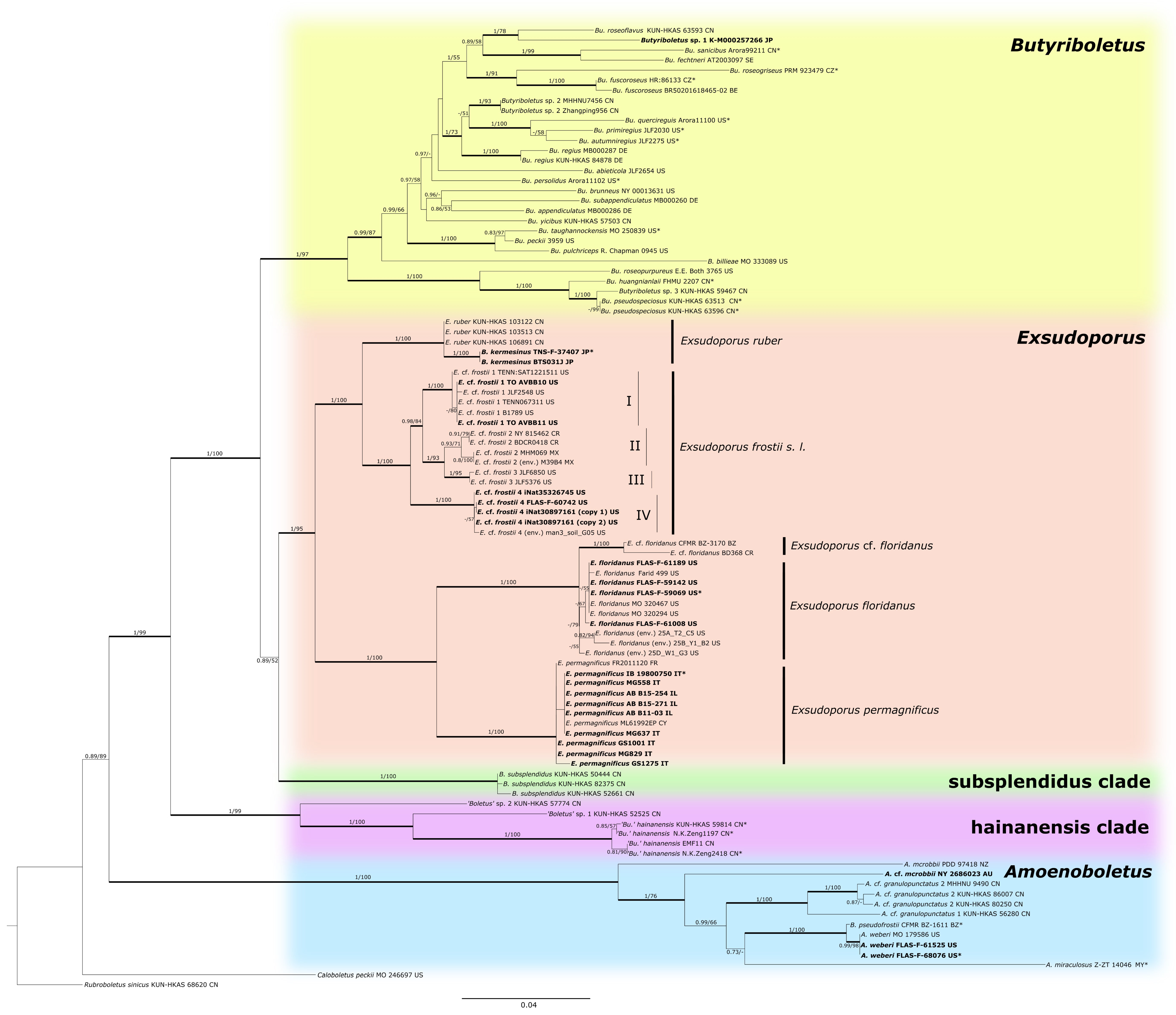
3.1. Molecular Phylogenetic Analysis
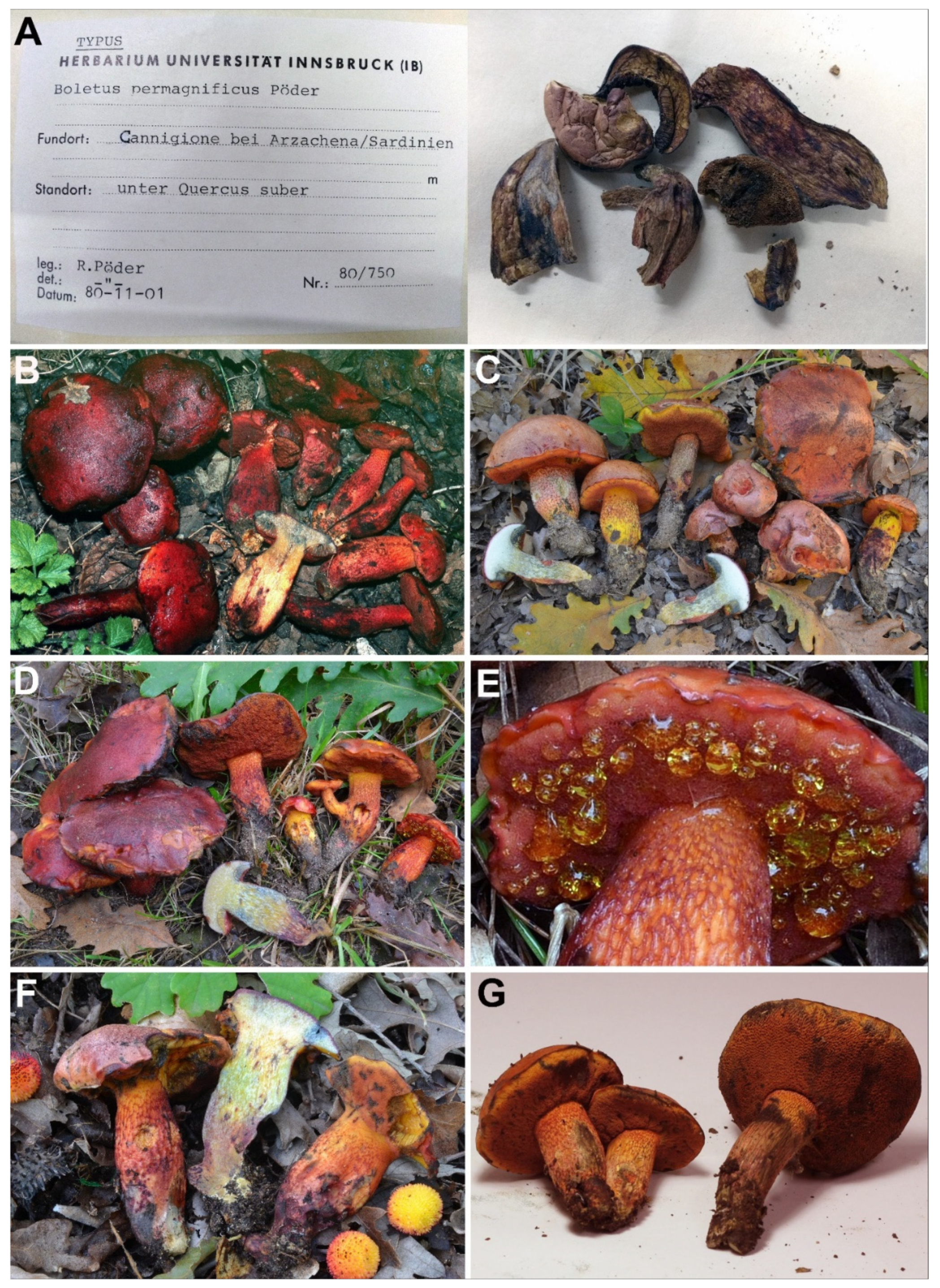
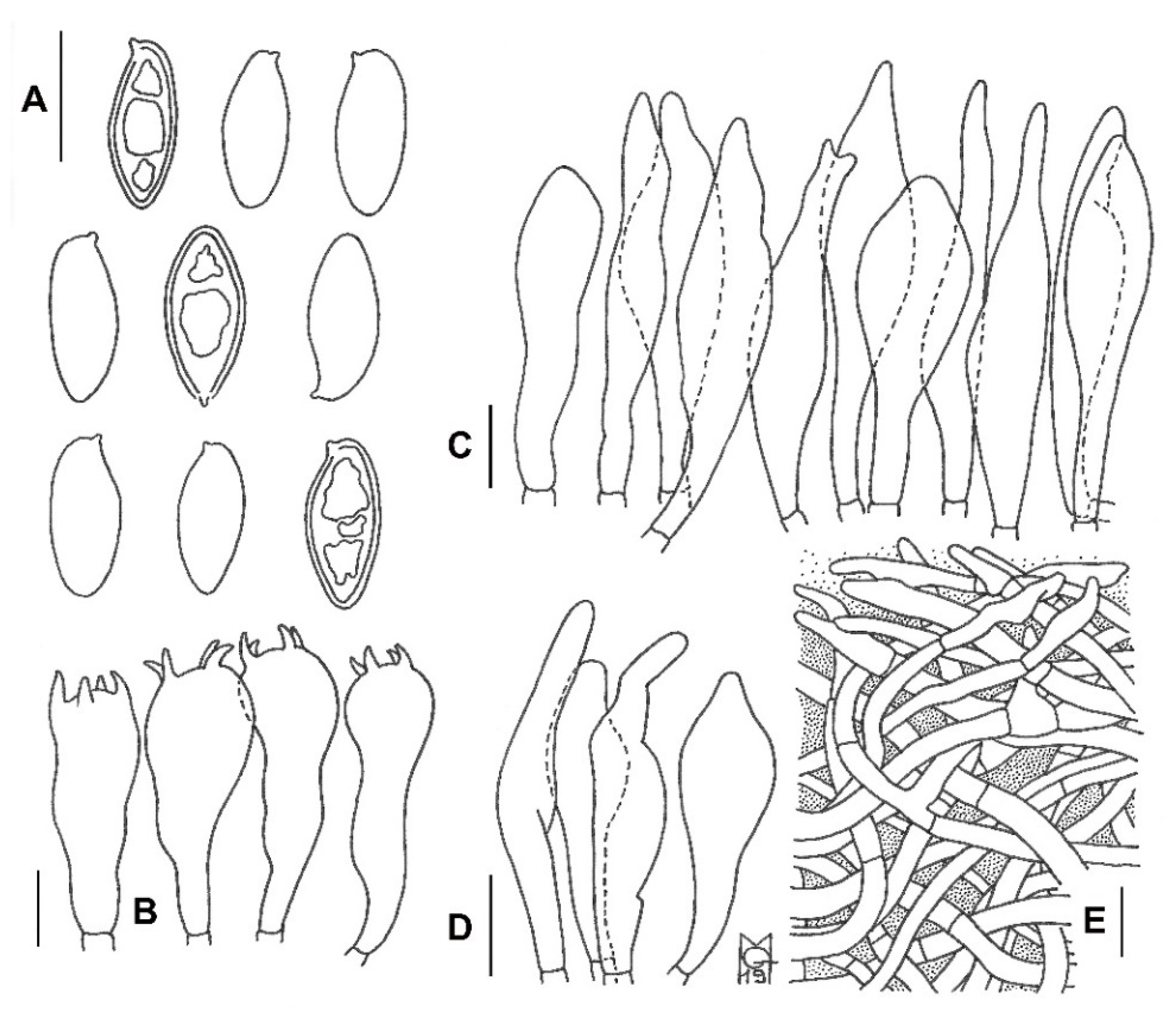
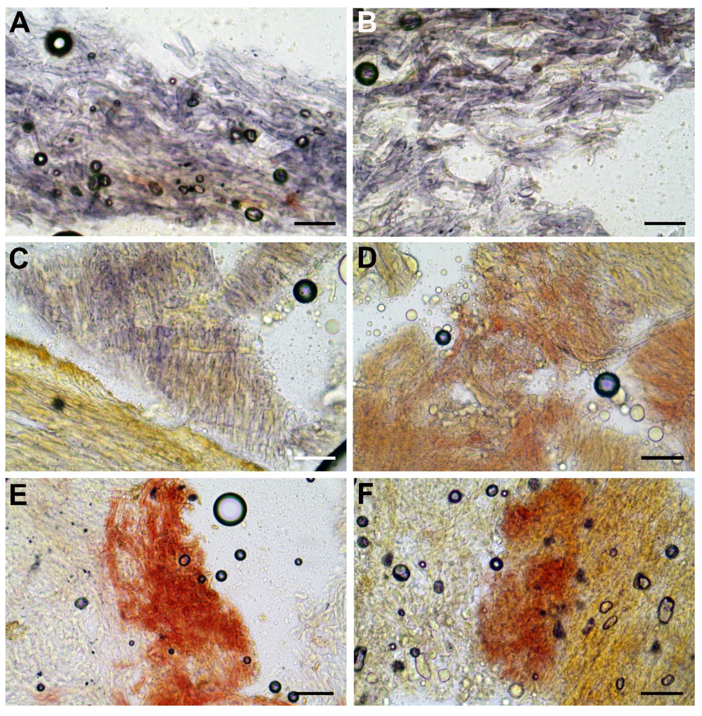
The aligned multigene matrix contained a total of 96 samples and 3302 aligned bases with gaps (Supplementary File S1). The ML and BI analyses generated almost identical tree topologies with minimal variation in statistical support values; thus, an ML tree with both BS and PP values was selected for the purposes of display (Figure 1).
Our phylogenetic analysis indicated that genera Exsudoporus and Butyriboletus form two different generic clades, both with high support and showing a considerable genetic difference. Exsudoporus (BS = 95%, PP = 1.00) and Boletus subsplendidus (BS = 100%, PP = 1.00) clades form a sister superclade (BS = 52%, PP = 0.89) to the Butyriboletus clade (BS = 97%, PP = 1.00). There are two other generic clades with strong statistical support: “hainanensis” clade (BS = 99% and PP = 1.00) and Amoenoboletus (BS = 100% and PP = 1.00).

3.2. Taxonomy

Exsudoporus Vizzini, Simonini and Gelardi 2014, emend. Biketova and Gelardi.
MYCOBANK MB 550708.
Diagnosis: Basidiome stipitate-pileate with tubular hymenophore, epigeal, evelate; pileus convex to applanate, bright blood red, crimson-red, purplish-red, reddish-pink or reddish-brown, opaque to shiny, dry to subviscid with moist weather, glabrous to subpruinose or subtomentose; hymenophore poroid, adnate or slightly depressed around stipe apex; tubes yellow to olivaceous-brown; pores pinkish-red, reddish-orange, blood red to dark red, rarely yellowish-orange or yellow, often beaded with golden yellow or amber yellow droplets when young and fresh; stipe central, solid, yellowish to concolorous with the pileus, conspicuously reticulate with elongated, red meshes or deeply reticulate-alveolate or can be covered with scaly patches; context pale yellow to bright yellow; tissues quickly turning dark blue or more rarely light blue or even unchanging when injured or exposed, then fading blackish; taste mild to acidic; spore print olive-brown; basidiospores smooth, subfusiform to ellipsoid or ellipsoid-fusoid; pleuro-, cheilo- and caulocystidia present; pileipellis an interwoven (ixo)trichoderm tending to a cutis; hymenophoral trama bilateral-divergent of the “Boletus-type”; clamp connections absent; stipe context varies from inamyloid to amyloid or dextrinoid; ontogenetic development gymnocarpic.
Generic type: Boletus permagnificus Pöder 1982.
Exsudoporus permagnificus (Pöder) Vizzini, Simonini and Gelardi, Index Fungorum 183:1, 2014.
MYCOBANK MB 550709.
Boletus permagnificus Pöder, Sydowia 34:151, 1982 (“1981”). (Basionym).
Suillellus permagnificus (Pöder) Blanco-Dios, Index Fungorum 211:1, 2015.
Misapplied names:
Boletusflammans E.A. Dick and Snell, Mycologia 57(3):453, 1965 s. Angarano non s. E.A. Dick and Snell.
Boletus frostii J.L. Russell, Bulletin of the Buffalo Society of Natural Sciences 2:102, 1874 s. Alessio non s. J.L. Roussell.
Boletus siculus Inzenga, Funghi Siciliani. Centuria 2: 57, 1869 s. Alessio non s. Inzenga (? = Alessioporus ichnusanus (Alessio, Galli and Littini) Gelardi, Vizzini and Simonini).
Holotype: Italy, Sardinia, Arzachena (OT), Cannigione, under Quercus suber with the presence of Cistus sp. And Eryngium sp., 1 November 1980, R. Pöder, IB 19800750.
Basidiomes small to medium. Pileus (2.0) 3.2–9.5 (10.0) cm broad, at first hemispherical then persistently convex and finally broadly pulvinate-flattened to slightly depressed, regularly to sometimes unevenly shaped by shallow depressions, moderately fleshy, firm at the beginning but progressively softer with age, flabby in old basidiomes; margin generally obtuse, steady to faintly wavy-lobed especially in young specimens, initially involute then curved downwards and finally completely plane or even uplifted, extending beyond the tubes up to 2 mm in primordia; surface matt, dry but slightly greasy and polished with moist weather, very finely pubescent in the early stage of development but later smooth and glabrous, not cracked; cuticle somewhat variable in color, ranging from pale pinkish, pinkish-violet to pinkish-red (Shrimp Pink, Geranium Pink, Rose Doree, pl. I; Pale Amaranth Pink, Rose Pink, pl. XII; Rose-Purple, pl. XVI) in young specimens due to the presence of a whitish pubescence which soon tends to dissolve revealing the true color below, blood red to dark carmine red, coral red or garnet red to reddish-purple (Spinel Pink, Spinel Red, pl. XXVI; Spectrum Red, Scarlet-Red, Carmine, pl. I) at maturity, often with scattered orange shades (Orange Chrome, pl. II; Scarlet, pl. I) especially towards the margin, gradually fading with age and becoming copper red to ochraceous red or even ochraceous brown (Madder Brown, Brick Red, pl. XIII; Buckthorn Brown, pl. XV) in senescence or if exposed to direct sunlight; slowly but pronouncedly turning bluish-black (Berlin Blue, pl. VIII) on handling or when injured, particularly in young and fresh basidiomes, then fading dull brownish-red (Indian Lake, Dahlia Carmine, pl. XXVI; Bordeaux, pl. XII); subcuticular layer reddish-purple (Amparo Purple, pl. XI; Lobella Violet, pl. XXXVII). Tubes at first thin then increasingly broader and shorter than the thickness of the pileus context (up to 1.8 cm long), adnate-subdecurrent at first but soon adnexed, finally depressed around the stipe apex and shortly decurrent with a tooth, pale yellow (Barita Yellow, pl. IV) at first, yellowish-olive (Yellowish Citrine, pl. XVI) to brownish-olive (Saccardo Olive, pl. XVI; Light Brownish Olive, pl. XXX; Dark Olive-Buff, pl. XL) in old fruiting bodies, bluing (Blanc’s Blue, Dusky Greenish Blue, pl. XX) when cut. Pores initially forming a flat surface, later convex to ascendant, at first very small then gradually wider (up to 2 mm in diam.), simple, roundish to barely angular and radially stretched at maturity, at first more or less evenly yellow (Lemon Chrome, pl. IV) but soon orange pinkish to blood red (Vinaceous, Deep Vinaceous, pl. XXVII; Eosine Pink, Spectrum Red, Scarlet-Red, pl. I), sometimes yellowish-orange (Flame Scarlet, Orange Chrome, pl. II) towards the margin and finally rusty red (Carmine, pl. I), quickly and intensely turning dark blue (Blanc’s Blue, Dusky Greenish Blue, pl. XX) on bruising or when injured and finally fading to blackish (Black, pl. LIII); the hymenophore of young and fresh specimens exude abundant golden yellow (Light Orange-Yellow, pl. III), markedly salty-tasting droplets which tend to disappear with age. Stipe (4.0) 4.6–11.0 (12.0) × (0.6) 0.8–3.1 (4.0) cm, slightly longer than or as long as the pileus diameter at maturity, central to slightly off-center, solid, firm, dry, straight or faintly curved, cylindrical to fusiform, rarely progressively attenuated downwards, usually swollen in the middle part and tapering towards both the apex and the base, never clavate, ending with a long pointed taproot at the very base, frequently deeply rooting; surface showing a fine to pronounced reticulum at least in the upper half or over the upper three fourths, sometimes extending down to the base, smooth and glabrous elsewhere, evelate; lemon yellow or bright yellow to occasionally pinkish (Barita Yellow, pl. III; Light Orange-Yellow, pl. II) in the upper third or in the upper half in young specimens, orange yellowish to pinkish (Capucine Yellow, Orange, pl. II; Ochraceous-Salmon, pl. XV) elsewhere, reddish-purple to purplish-brown (Aster Purple, Dahlia Purple, Pansy Purple, pl. XII) in the lower portion, entirely blood red to carmine red or garnet red (Spinel Pink, Spinel Red, pl. XXVI; Spectrum Red, Scarlet-Red, Carmine, pl. I) in mature specimens; reticulum consisting of fine to well-defined, narrow and longitudinally stretched polygonal meshes, concolorous to the ground in early stages of development but soon blood red to carmine red (Spectrum Red, Scarlet-Red, Carmine, pl. I) lengthwise; bruising dark blue (Berlin Blue, pl. VIII) when pressed then fading sordid reddish-purple to blackish (Claret Brown, pl. I; Black, pl. LIII); basal mycelium cream yellowish (Barita Yellow, pl. IV), rhizomorphs brownish (Sayal Brown, pl. XXIX). Context firm and tough when young, later soft textured and eventually flabby in the pileus (up to 3.2 cm thick in the central zone and gradually becoming thinner towards the edge), a little more fibrous in the stipe, evenly watery yellow (Citron Yellow, pl. XVI) throughout with darker tones towards the base, with a thin reddish-purple line (Amparo Purple, pl. XI; Amaranth Purple, pl. XII; Lobella Violet, pl. XXXVII) beneath the cuticle and sometimes with reddish-purple scattered spots or shades in the stipe; turning extensively blue (Yale Blue, Vanderpoel’s Blue, Blanc’s Blue, Dusky Greenish Blue, pl. XX; Grayish Violaceous Blue, pl. XXII) when exposed to air with the only exception of the stipe base in young specimens, which remains practically unchangeable, finally fading dull yellowish (Aniline Yellow, Pyrite Yellow, pl. IV) to dirty yellowish-orange (Yellow Ocher, pl. XV) or occasionally reddish (Etruscan Red, pl. XXVII); reddish-purple (Pomegranate Purple, pl. XII) where eroded by maggots and bright yellow (Strontian Yellow, pl. XVI) to reddish (Etruscan Red, pl. XXVII) in places eaten by slugs; subhymenophoral layer bright yellow (Strontian Yellow, pl. XVI); exsiccate pinkish-red, wine red to dark reddish-purple (Spinel Pink, Spinel Red, pl. XXVI; Spectrum Red, Scarlet-Red, Carmine, pl. I) on pileus and stipe, brownish (Cinnamon-Brown, pl. XV) on hymenophore, beige to dull ochraceous brown on context (Ivory Yellow, pl. XXX; Clay Color, pl. XXIX). Odor intensely fruity, agreeable. Taste mild then with a weakly acidic aftertaste. Spore print olive-brown. Macrochemical spot-test reactions: 25% NH3: Pileus cuticle fades ochraceous, no reaction elsewhere (but bluing oxidation on tissues disappears); 30% KOH: staining dark wine red on hymenophore, bright orange to wine red elsewhere; FeSO4: blackish on hymenophore, no response or pale olivaceous on context, brownish-olive on pileus and stipe. Ontogenetic development gymnocarpic.
Basidiospores (768/55/23) (12.7) 14.4 ± 0.7 (15.4) × (5.5) 5.9 ± 0.2 (6.1) μm, Qm = (2.23) 2.47 ± 0.12 (2.61), V = 263 ± 22 μm3, inequilateral, ellipsoid to ellipsoid-fusiform or less frequently broadly ellipsoid in side view, broadly ellipsoid to ellipsoid in face view, smooth, apex rounded, with a short apiculus and usually with a shallow suprahilar depression, moderately thin-walled (0.3–0.5 μm), straw-yellow colored in water and 5% KOH, having one, two or three large oil droplets when mature, less frequently pluri-guttulate, inamyloid, acyanophilic and with an orthochromatic reaction. Basidia (28) 31–47 × 9–15 μm (n = 31), subclavate to clavate, moderately thick-walled (0.3–0.8 μm), predominantly four-spored but also one-, two- or three-spored, usually bearing relatively long sterigmata (3–6 μm) (sterigmata up to 7 μm long in one- and two-spored basidia), hyaline to yellowish and containing scattered straw-yellow oil guttules in water and 5% KOH, bright yellow (inamyloid) in Melzer’s, without basal clamps; basidioles cylindrical, subclavate to clavate, similar in size to basidia. Cheilocystidia (28) 31–60 (62) × 8–13 μm (n = 17), common, moderately slender, projecting straight to sometimes flexuous, fusiform, ventricose-fusiform to sublageniform or lageniform, showing a narrow and long neck with rounded to subacute tip, smooth, moderately thick-walled (0.4–0.9 μm), nearly hyaline to yellowish in water and 5% KOH, bright yellow (inamyloid) in Melzer’s, without epiparietal incrustations. Pleurocystidia (43) 47–59 (61) × 7–11 μm (n = 12), infrequent, irregularly cylindrical or cylindrical-fusiform to narrowly fusiform or sublanceolate, on average usually slightly longer and narrower than cheilocystidia, color and chemical reactions similar to cheilocystidia. Pseudocystidia not recorded. Pileipellis an ixocutis consisting of mostly repent, subparallel to interwoven, elongated, frequently branched, filamentous and sinuous to cylindrical hyphae embedded in gelatinous matter; terminal elements 14–90 × 4–18 μm, versiform, short cystidioid or rarely slightly clavate to long and slender cylindrical, apex rounded-obtuse, moderately thick-walled (up to 1 μm), yellowish to pale pinkish-red in water and pale yellowish to bright yellow in 5% KOH, golden yellow to orange yellow (inamyloid) in Melzer’s, smooth to sometimes ornamented by a very subtle granular epiparietal incrustation; subterminal elements similar in shape, size and color to terminal elements; the subpellis consists of irregularly and loosely arranged, short, predominantly inflated to nearly globose, 6–18 μm broad, hyaline to pale yellowish cells. Stipitipellis a layer of slender, parallel to loosely intermingled and longitudinally running, smooth-walled, adpressed hyphae, 2–13 μm wide, hyaline to very pale yellowish in water and 5% KOH; the stipe apex covered by a well-developed caulohymenial layer consisting of sterile caulobasidioles, very common, predominantly four-spored, fertile caulobasidia, (24) 26–39 (41) × 9–11 (13) μm (n = 15), sterigmata up to 6 μm and very scattered projecting fusiform, ventricose-fusiform to lageniform caulocystidia similar in shape and color to hymenial cystidia but distinctly shorter, (25) 31–51 (55) × 6–14 μm (n = 10), having a wall up to 0.5 μm thick. Lateral stipe stratum under the caulohymenium present and well differentiated from the stipe trama, of the “boletoid type”, at the stipe apex a (10) 20–40 (50) μm thick layer consisting of divergent, inclined and running towards the external surface, loosely intermingled and branched hyphae remaining separate and embedded in a gelatinous substance; the stratum is clearly visible in earlier developmental stages but tends to disappear with age. Stipe trama composed of confusedly and densely arranged, subparallel to moderately interwoven, filamentous, smooth, inamyloid hyphae, 3–24 μm broad. Hymenophoral trama bilateral divergent of the “Boletus-type”, with slightly to strongly divergent, recurved-arcuate and loosely arranged, not-branched, distantly septate and generally not restricted at septa, gelatinous hyphae (lateral strata hyphae in transversal section not touching each other, (3) 4–8 (10) μm apart, 3–7 μm broad), hyaline to very pale yellowish in water and 5% KOH, inamyloid in Melzer’s; lateral strata (30) 40–60 (70) μm thick, mediostratum (10) 20–30 (40) µm thick, axially arranged, consisting of a tightly adpressed, non-gelatinous bundle of hyphae, 3–7 µm broad, distantly septate; in Congo Red the mediostratum is darker than the lateral strata. Oleipherous hyphae scattered although more frequently observed in the basal stipe trama, golden yellow to brownish in 5% KOH and Melzer’s. Clamp connections absent in all tissues. Hyphal system monomitic.
Edibility: Edible after prolonged cooking.
Ecology and phenology: Solitary or more frequently gregarious or subcaespitose to truly caespitose, sometimes with basidiomes emerging from the stipe of adjacent fruiting bodies, growing in warm Mediterranean regions preferably on slightly to decidedly acidic, clayey or sandy soil among litter associated exclusively with hardwoods in dry, warm, exposed groves of Quercus (Fagaceae), occasionally also with Castanea sativa (Fagaceae) and Cistus (Cistaceae), sometimes in mixed woods with the presence of Pinus halepensis and P. pinea (Pinaceae), Erica spp., Arbutus unedo (Ericaceae) and Eryngium sp. (Apiaceae). Summer to late fall (August to November).
Known distribution: Previously reported from southern Europe in warm countries at low altitudes, bordering the Mediterranean basin (Portugal, Spain, Italy, Slovenia, Bulgaria, Greece) east to the Asian Middle East (Cyprus, Israel) and likely extending as far north as France. It probably also occurs in Mediterranean northern Africa. Apparently widespread throughout mainland and insular Mediterranean basin but rare and localized. Northern, eastern and southern distribution limits yet to be established.
Examined material: ISRAEL, Mount Carmel: Mount Carmel National Park, near the crossroad Damon, on the ground in a clearing near Quercus calliprinos, 32°44′03″ N, 35°02′22″ E, 505 m, 30 October 2015, legit. Sh. Alter, det. A. Yu. Biketova, AB B15-254 (HAI B15-254); same loc., Nahal Oren valley, Henyon Ha’Agam, under Quercus calliprinos, 32°43′26″ N, 35°00′55″ E, 260 m, 13 November 2015, legit. Z. Shafranov, det. A. Yu. Biketova, AB B15-274 (HAI B15-274); Golan Heights: Odem Forest Reserve, under Quercus sp., 33°12′56″ N, 35°45′26″ E, 1028 m, 10 October 2011, legit. R. Kuznetsov and Z. Shafranov, det. A. Yu. Biketova, AB B11-06 (HAI B11-06); same loc., under Quercus calliprinos, 26 September 2013, legit. Z. Shafranov, det. A. Yu. Biketova, AB B13-165 (HAI B13-165); same loc., under Quercus calliprinos, 07 November 2015, legit. R. Kuznetsov and Z. Shafranov, det. A. Yu. Biketova, AB B15-271 (HAI B15-271); Upper Galilee: Hanita Forest, in a mixed wood of Quercus calliprinos and Pinus halepensis, 33°04′44″ N, 35°09′57″ E, 160 m, 6 October 2011, legit. Y. Cherniavsky, det. A. Yu. Biketova, AB B11-03 (HAI B11-03); same loc., Bar’am Forest, on the ground near Quercus calliprinos, 33°02′20″ N, 35°25′33″ E, 679 m, 27 November 2020, legit. Y. Segal, det. A. Yu. Biketova and G. Simonini, AB B20-370; ITALY, Sardinia: Arzachena (OT), Cannigione, under Quercus suber with the presence of Cistus sp. and Eryngium sp., 1 November 1980, legit. And det. R. Pöder, IB 19800750 (holotype); Piedmont: Ceresole d’Alba (CN), under Quercus sp., 22 August 1980, legit. M. Strani, det. R. Pöder, IB 19800751a and IB 19800751b (paratypes); Lazio: Nettuno (LT), Tre Cancelli, on slightly acidic, sandy soil, along a trackside in a coastal mixed broadleaved woodland dominated by Quercus robur with the presence of Quercus cerris, Quercus frainetto and Phyllirea angustifolia, 41°28′39″ N, 12°43′54″ E, 36 m, 26 September 2009, legit. M. Gelardi, G. Gelardi and C. Aita, det. M. Gelardi, MG238 (MCVE25596); same loc., 18 September 2013, legit. And det. M. Gelardi, MG558; same loc., on a clearing side, 2 October 2014, legit. And det. M. Gelardi and F. Costanzo, MG662; same loc., 22 September 2006, legit. And det. M. Gelardi (no voucher material preserved); Nettuno (LT), Torre Astura, on slightly acidic, sandy soil, in a coastal mixed broadleaved woodland under Quercus robur, Quercus frainetto, Quercus cerris with the presence of Pinus pinea, 41°25′15″ N, 12°45′33″ E, 5 m, 9 August 2014, legit. M. Gelardi, M. Tullii and R. Polverini, det. M. Gelardi, MG637; Manziana (RM), Cerreta di Manziana, on acidic soil, along a trackside in a pure stand of Quercus cerris, 42°07′12″ N, 12°06′53″ E, 320 m, 4 August 2011, legit. M. Gelardi and V. Migliozzi, det. M. Gelardi, MG418; Mounts Tolfa, Allumiere (RM), Capo Nord, on acidic volcanic soil, along a trackside in a mixed broadleaved woodland under Quercus cerris, Q. Petraea, Arbutus unedo, Erica arborea and Fraxinus ornus, 42°11′12″ N, 11°55′21″ E, 426 m, 3 October 2020, legit. And det. M. Gelardi, F. Costanzo and O. Gelardi, MG829; Emilia Romagna: Pulpiano, Viano (RE), on acidic soil under Quercus cerris, 44°31′08″ N, 10°33′13″ E, 520 m, 06 September 1983, legit. U. Bonazzi, det. G. Simonini, GS143 (MCVE17281); same loc., 3 October 1986, legit. And det. G. Simonini, GS336 (MCVE17298); same loc., 12 September 1987, legit. And det. G. Simonini, GS488 (MCVE29227); same loc., 29 October 1990, legit. And det. G. Simonini, GS784 (MCVE17556); same loc., 24 September 1994, legit. U. Bonazzi, det. G. Simonini, GS1275 (MCVE17763); same loc., 30 August 2015, legit. And det. G. Simonini, GS10151; Quattro Castella (RE), Parco di Roncolo, on basic soil under Quercus pubescens and Ostrya carpinifolia, 320 m, 19 September 1993, 44°37′20″ N, 10°29′23″ E, legit. And det. G. Simonini, GS1001 (MCVE17523); Calabria: Santa Sofia d’Epiro (CS), Serra di Zoto, on acidic soil under Quercus pubescens and Cistus sp., 550 m, 01 Sep 1995, 39°33′ N, 16°25′ E, legit. C. Lavorato, det. G. Simonini, GS1599 (MCVE18037); Santa Sofia d’Epiro (CS), Contrada Calamia, on acidic soil under Castanea sativa and Cistus sp., 800 m, 1 September 1995, 39°33′ N, 16°25′ E, legit. C. Lavorato, det. G. Simonini, GS1570 (MCVE17978); Acri (CS), Cozzo S. Angelo, on acidic soil under Castanea sativa, 900 m, 8 September 1996, 39°33′ N, 16°25′ E, legit. C. Lavorato, det. G. Simonini, GS1717 (MCVE18132); Sicily: Mounts Nebrodi, Cesarò (ME), Torti II creek, under Fagus sylvatica and Quercus pubescens, 1400 m, 1 July 2002, 37°53′32″ N, 14°38′51″ E, legit and det. A. Pappalardo, EMAC300-020701; same loc., Mount Soro, under Fagus sylvatica and Quercus pubescens, 1500 m, 11 August 2002, 37°55′12″ N, 14°39′25″ E, legit and det. S. Silviani, EMAC311-020811; same loc., Randazzo (ME), along river Flascio, under Quercus cerris, 1250 m, 20 September 2020, 37°56′29″ N, 14°52′36″ E, legit and det. G. Vasquez, EMAC1511-200920; Mounts Peloritani, Antillo (ME), on soil under Castanea sativa, 500 m, 25 September 2015, 37°58′56″ N, 15°12′57″ E, legit. And det. F. Mondello, FM 20150925_100.
Additional material examined: Unidentified Boletaceae sp. (initially identified as Boletus permagnificus): RUSSIAN FEDERATION: Belgorod Region, Krasnogvardeysky district, Valuychik village, under Quercus robur in oak grove on chalky soil, 50°23′ N, 138°15′ E, 180 m, August 1979, legit. E.P. Bedenko, det. T. Yu. Svetasheva and A. Yu. Biketova, LE 17906.
Comments: The Italian mycologists C.L. Alessio and A. Angarano were the first to struggle with the placement of Exsudoporus permagnificus in the early 1980s, assigning it three misapplied species epithets [61,62]. Alessio wrote at length and repeatedly about this species based on several collections recorded in Piedmont (northwestern Italy) in 1973 [61]. Two years later, in 1975, the species was also recorded with several specimens emerging from a common base under a single oak tree near Bologna, Emilia Romagna [63]. Alessio’s first contribution was devoted to this species which, following A. Marchand’s suggestion, was at first believed to represent the eastern North American species Boletus frostii Roussell [61]. At the same time, Angarano misidentified the species as another North American taxon, B. flammans E.A. Dick and Snell, based on his collections from Cannigione (northern Sardinia) [62]. Alessio continued to use the name B. frostii for collections of B. permagnificus [64]. However, Alessio reconsidered the best name for these collections and instead applied the binomial Boletus siculus Inzenga [65], a species described from Sicily in the second half of the 19th century [66]. However, as already argued by the French mycologists M. Bon [61] and G. Redeuilh [67], and the Italian F. Bellù [68], the collections of Alessio [61,64,65] and Angarano [62] represented a taxon without a valid name.
Indeed, Pöder [69], working mainly on the same Sardinian specimens previously misidentified by Angarano, described this taxon as new to science under the binomial Boletus permagnificus Pöder (see also Pöder [67] for an Italian translation of Pöder’s original manuscript by F. Bellù). Despite the convincing evidence of a novel species provided by the Austrian mycologist, in the subsequent years Alessio continued to reiterate his taxonomic view by considering B. permagnificus a later heterotypic synonym of B. siculus [70,71,72,73,74] and apparently did not change his opinion throughout his life. Conversely, Bellù did not concur with Alessio’s interpretations and instead agreed with Pöder that E. permagnificus was indeed a new taxon [67,75]. Furthermore, as already pointed out by Lavorato [76] and M. Contu (pers. Comm.), B. siculus might represent an older name for Alessioporus ichnusanus (Alessio, Galli and Littini) Gelardi, Vizzini and Simonini—a species described by the same Alessio and co-workers in 1984 [77]—even though the exact identity of B. siculus remains unclear [78].
In more recent times, B. permagnificus was subjected to preliminary phylogenetic analysis which led to the erection of the genus Exsudoporus encompassing E. permagnificus and its closest relatives [26]. Finally, Blanco-Dios recombined nearly all European red-pored boletes in Suillellus Murrill without providing any supporting phylogenetic evidence to justify this placement [79].
Exsudoporus permagnificus is an easily recognized species in the warm regions of southern Europe and the eastern Mediterranean region of western Asia, but it appears to be practically unknown elsewhere, especially outside Europe. It is easily recognizable based on the following diagnostic key features, which place this species in a morphologically unique position among red-pored European boletes: small to medium-sized basidiomes, overall bright red coloration, cylindrical to fusiform stipe that is never clavate but is always covered by a well-developed raised reticulum, hymenophore exuding golden or amber-yellow droplets in young and fresh specimens, yellowish context and basal mycelium, deep blue staining reaction upon bruising or injury, ellipsoid-fusiform, smooth basidiospores, ixocutis pileipellis mainly consisting of filamentous to cylindrical hyphae, subcaespitose to caespitose growth and the occurrence under broadleaved trees or shrubs in warm environments. As far as the pileipellis structure is concerned, inflated to nearly globose cells up to 18–20 μm wide are sometimes observed in the subpellis (as broad as 25 μm) [80,81,82]. Another peculiar organoleptic trait that has likely gone unnoticed but is worthy of mention is the salty taste of the droplets; this neglected feature has not been previously reported for E. permagnificus but should be examined in the remaining extraeuropean species to evaluate its taxonomic significance.
The amyloidity of tissues with Melzer’s reagent is a point that deserves further discussion. Bozok et al. reported the amyloid reaction of the hyphae of the stipe context as an additional attribute of E. permagnificus [38]. The holotype material and all collections from Israel studied in the present paper also exhibited the same positive reaction of the stipe base context: moderately amyloid in AB B11-03, AB B13-165, AB B15-271 and AB B20-370, weakly amyloid in AB B11-06 and IB 19800750 and variably amyloid and dextrinoid in AB B15-254 and AB B15-274 (Figure 5). As a matter of fact, in the original diagnosis, Pöder noted that the stipe trama was almost black with Melzer’s reagent, suggesting amyloid tissues [69]. Icard and Hurtado [80], Lannoy and Estades [5], and Horak [83] also observed a positive amyloid reaction in this species. However, other authors reported a negative reaction with Melzer’s reagent [14,82,84,85,86]. As already suggested by Assyov, contrasting results might be due to different procedures followed in testing the amyloidity of fungal tissues [84]. However, we have tested the iodine reaction of the context at the stipe base according to Imler’s procedure [4] on several of our Italian collections using the same techniques and surprisingly we observed dissimilar results depending on the studied samples; some of them showed a strong (GS1717) to weak (MG238, MG662, GS336) amyloid reaction, whereas some others exhibited an inamyloid reaction (MG558, MG637, GS784, GS10151), and a few others even displayed a dextrinoid reaction (MG418, GS1001, GS1275). Consequently, this macro-chemical reaction should be carefully re-evaluated, as it seems to be quite inconstant and variable.
A chromatographic analysis of the pigments in E. permagnificus was carried out by Davoli and Weber, which led to the isolation of variegatic acid, xerocomic acid and variegatorubin [87,88].
With regards to its ecological requirements, this species is primarily found in oak-dominated woodlands. The species occurs with deciduous and evergreen Quercus species (including Q. cerris, Q. calliprinos, Q. pubescens, Q. petraea, Q. pyrenaica, Q. virgiliana, Q. ilex, Q. suber, Q. alnifolia, Q. frainetto and the introduced-in-Europe Q. rubra), sometimes in mixed forests with the presence of pines (Pinus pinea and P. halepensis) and also sporadically under sweet chestnut (Castanea sativa), common beech (Fagus sylvatica), some Ericaceae (Erica scoparia, E. arborea and Arbutus unedo), and rockroses (Cistus spp.), preferably on acidic soil [30,38,76,82,85,89,90]. The occurrence of E. permagnificus with several host trees indicates that it is not selective in its plant symbiotic partners. Concerning geographical distribution, the natural range of E. permagnificus covers most of the warm and dry regions of southern Europe and Levant in western Asia. Aside from Italy (including Sardinia and Sicily) [14,63,69,72,76,85,89,90,91,92,93,94,95,96,97,98,99,100,101,102,103,104,105], the species has also been reported from Portugal [106,107], Spain [81,82,86,108,109,110,111,112,113,114,115], France (including Corsica) [5,80,116,117,118,119], Bulgaria [38,84,120,121], Slovenia [122], Greece [123] and Cyprus [39]; and here it is also reported for the first time from Israel. Exsudoporus permagnificus is relatively widespread throughout the Western Mediterranean basin, more rarely found in the East Mediterranean basin, and we assume it might also occur in northern and northwestern Africa. We suspect that it has not been reported from this region because it may have been overlooked. Based on the International Union for Conservation of Nature (IUCN) Red Listing protocol, E. permagnificus has most recently been assessed as “Vulnerable” (VU) in central Italy at the ecoregion scale [124]. Finally, E. permagnificus is an edible species that is commonly harvested and consumed by local mushroom pickers in the coastal areas south of Rome, although it is rarely prolific enough to be widely eaten.
A bolete sample (LE 17906) identified by Bedenko [125] as B. permagnificus from the Belgorod Region in the Russian Federation has been morphologically re-examined by T. Yu. Svetasheva and I. V. Zmitrovich and was confirmed to be another species of Boletaceae (likely a member of either the genus Rubroboletus or Neoboletus). Unfortunately, the specimen is poorly preserved and no DNA sequences could be generated from this material, so it is not possible to identify this specimen to the species level.
The most similar species to E. permagnificus is probably E. floridanus (Singer) Vizzini, Simonini and Gelardi, which is its closest relative based on phylogenetic data. However, E. floridanus is distinguished by the larger size (pileus up to 15 cm diam.), the pileus cuticle staining olivaceous black with NH3, the typically clavate stipe, slightly narrower basidiospores (13.2–16.7 × 4.5–5.0 μm) and the different geographic distribution in eastern and southeastern North America [28,126,127,128,129].
The eastern North American E. frostii differs from E. permagnificus in the larger size (pileus up to 15.5 cm diam.), stouter basidiomes, polished and shining cuticle that is viscid when wet, blood-red to candy-apple-red pileus surface, usually clavate stipe, raised and much coarser, deeply pronounced reticulate-alveolate pattern over the entire length of the stipe, yellowish reticulum on a reddish background, tissues turning light blue slowly and erratically, narrower basidiospores (12–15 × 3.7–5.0 μm, Qm = 3.1–3.6), smaller basidia (23–32 × 8–10 μm), narrower pileipellis hyphae (up to 6 μm broad) and the occurrence in North America [7,28,69,127,128,129,130,131].
Exsudoporus ruber is consistently separated from E. permagnificus by the slightly larger size (pileus up to 13 cm diam.), polished and shining cuticle that is viscid when wet, dark red, brownish-red to violet-brown pileus surface, sometimes unchangeable or very slowly and lighter bluing context on exposure, overall reddish stipe surface (even in young specimens) which tends to become disrupted into irregular scaly patches in ageing specimens especially on the lower half, longer basidiospores (15.5–18.6 × 5.6–6.6 μm, Qm = 2.8), subcylindrical, slightly narrower cheilocystidia (35–76 × 4–9.5 μm), narrower pileipellis hyphae, 2.0–6.4 μm wide, longer caulocystidia (37–85 × 4–10 μm), narrower stipe trama hyphae (up to 11 μm broad) and the occurrence in subalpine coniferous forests in association with Pinaceae (Abies, Pinus, Tsuga) in eastern Asia ([132,133,134], as “Boletus kermesinus”; [135,136]).
Another species that is similar to E. permagnificus is Boletus weberi Singer, which appears to be conspecific with Boletus pseudofrostii B. Ortiz, based on our phylogenetic reconstruction. This taxon belongs to another genus (see below) and differs by the smaller size (pileus up to 6.5 cm diam., stipe up to 5.3 × 1.7 cm), unchanging tissues on bruising, white basal mycelium, slightly smaller and shorter basidiospores (8.1–10.7 × 4.1–5.5 μm, Qm = 2.0), slightly narrower basidia (33–45 × 7–10 μm), smaller cheilocystidia (14–60 × 4–8 μm) and caulocystidia (14–29 × 8–9 μm), cylindrical, narrower pileipellis terminal cells (up to 10 μm wide) and the occurrence in Pinus-dominated or in mixed forests with Fagaceae in North (Atlantic and Gulf Coasts of the USA) and Central America (Belize) [28,126,127,128,137,138,139,140].
Exsudoporus floridanus (Singer) Vizzini, Simonini and Gelardi, Index Fungorum 183:1, 2014.
MYCOBANK MB 550711.
Boletus frostii subsp. floridanus Singer, Mycologia 37(6):799, 1945. (Basionym).
Suillellus floridanus (Singer) Murrill, Lloydia 11(1):29, 1948 (nom. inval., art. 41.5, basyonim not cited).
Boletus floridanus (Singer) Murrill, Lloydia 11(1):23, 1948 (nom. inval., art. 41.5, basyonim not cited).
Boletus floridanus (Singer) Singer, Sydowia 30(1-6):255, 1977.
Butyriboletus floridanus (Singer) G. Wu, Kuan Zhao and Zhu L. Yang, Fungal Diversity 81(1):72, 2016.
Holotype: USA, Florida, Alachua Co., Gainesville, June 1943, R. Singer, F 2428.
Epitype designated here: USA, Florida, Alachua Co., Gainesville, University of Florida Campus, Fifield Hall, under Quercus virginiana, 29°38′19″ N, 82°21′41″ W, 16 September 2014, legit. C. Ferguson, det. M. E. Smith, FLAS-F-59069, Index Fungorum 559409, GenBank: OL960514 for ITS, OL960488 for nrLSU, OL960496 for tef1-α and OL960503 for rpb2.
Selected morphological descriptions and illustrations: Murrill ([141], as “Suillellus luridus”?; [137]), Singer [126,127], Both [128], Bessette et al. [28,129,139,142], dubitatively García-Jiménez and Garza-Ocañas [143], Ortiz-Santana et al. [138], García-Jiménez [144], García-Jiménez et al. [145] and Gonzáles-Chicas et al. [146].
Edibility: Edible after prolonged cooking although with a somewhat acidic taste [129].
Ecology and phenology: Solitary to most frequently gregarious, growing in open stands, clearings or shaded lawns on sandy soil associated with various Quercus spp. (including Q. chapmanii, Q. laurifolia and Q. virginiana) (Fagaceae) or mixed with Pinus clausa (Pinaceae) and Carya (Juglandaceae). Late spring to fall (April to December), uncommon to rare.
Known distribution: Reported from eastern and southeastern USA (Tennessee and the Coastal Plain of North Carolina south to Florida and west to Texas), reported as far north as Long Island (New York) (MyCoPortal); records from Costa Rica, Mexico, Belize and Guatemala putatively belong to another related species (see below).
Examined material: USA, Florida: Alachua Co., Gainesville, University of Florida Campus, Fifield Hall, under Quercus virginiana, 29°38′19″ N, 82°21′41″ W, 1 April 2015, legit. R. Kneal and M. E. Smith, det. M. E. Smith, FLAS-F-59142; same loc., under Quercus virginiana, 16 September 2014, legit. C. Ferguson, det. M. E. Smith, FLAS-F-59069 (epitype); Putnam Co., Ordway-Swisher Biological Station, south of Goose Lake, along gravel road, under Quercus hemisphaerica, 29°41′50″ N, 81°58′53″ W, 22 June 2017, legit. G. LaPierre, det. M. E. Smith, FLAS-F-61008; same loc., NE of point C21, Q. virginiana dominated hardwood forest, with some Carya, Pinus and Serenoa repens, 29°42′00″ N, 81°59′26″ W, 1 August 2017, legit. D. Borland, L. Kaminsky, A. E. Bessette and A. R. Bessette, det. A. E. Bessette, FLAS-F-61189.
Comments: This species has been sufficiently described and illustrated. The exact collecting date of the original material is not clear and was not given in the publication or on the packet, but authentic samples from the description were collected in north Florida during June and September. Curiously, Both indicated a different collection (paratype F2204) as the type of E. floridanus [128]. This is likely because there were some confusing annotations on the specimens at FH and other herbaria and because F2428 does not appear to be present at either FH or F. An attempt was made to sequence the ITS region from two paratypes of E. floridanus from FH (F2204 and F650), but due to the age and condition of the specimens, this was unsuccessful. We consider it necessary to designate a new specimen of E. floridanus from the vicinity of the type locality as a modern epitype. Phylogenetic analysis shows that this entity might be a species complex, and E. floridanus s. l. appears to encompass two different species. One is the true E. floridanus, which is distributed in eastern and southeastern North America, including our specimens from Florida that were collected nearby the type locality. A second, morphologically similar species is recorded here from Central America, based on collections from Belize and Costa Rica. It seems likely that other collections from Belize [138] and Guatemala [147] might have been misidentified as E. floridanus s. str. but actually belong to this second species. Records from Mexico that were identified as E. floridanus s. str. [143,144,145,146] should be examined and/or have their DNA sequenced to determine which of the two species they represent. However, since many taxa from Florida extend into Mexico, it seems likely that both species may be present in Mexico.
Similar to E. permagnificus, the amyloid reaction of the hyphae of the stipe context with Melzer’s reagent varies from weakly dextrinoid (FLAS-F-61008) and dextrinoid (FLAS-F-59069) to mixed amyloid and dextrinoid (FLAS-F-59142), as well as weakly amyloid (FLAS-F-61189). Farid et al. also noticed a dextrinoid reaction in the studied specimen (Farid 499) [148].
Exsudoporus frostii (J.L. Russell) Vizzini, Simonini and Gelardi, Index Fungorum 183:1, 2014.
Figure 6C–G.
MYCOBANK MB 550710.
Boletus frostii J.L. Russell, Bulletin of the Buffalo Society of Natural Sciences 2:102, 1874. (Basionym).
Suillus frostii (J.L. Russell) Kuntze, Revisio Generum Plantarum 3(2):535, 1898.
Suillellus frostii (J.L. Russell) Murrill, Mycologia 1(1):17, 1909.
Tubiporus frostii (J.L. Russell) S. Imai, Transactions of the Mycological Society of Japan 8(3):113, 1968.
Butyriboletus frostii (J.L. Russell) G. Wu, Kuan Zhao and Zhu L. Yang, Fungal Diversity 81(1):72, 2016.
= Boletus alveolatus Berkeley and M.A. Curtis apud Frost, Bulletin of the Buffalo Society of Natural Sciences 2:102, 1874.
Holotype: USA, Vermont, Brattleboro, C.C. Frost. Preserved in VT and selected by Halling as lectotype (VT3156) [149]. According to Halling, several additional specimens currently housed in VT, FH and NYBG can be considered “original material” because these are labeled in Frost’s handwriting and were apparently identified by Frost [149].
Selected morphological descriptions and illustrations: Curtis ([150], as “Boletus purpureus”), Frost ([130], also as “Boletus alveolatus”), Peck [151,152], Murrill [141,153], Farlow and Burt [154], Coker and Beers [155], Singer [127,156], Snell and Dick [157], Smith and Thiers [7], Miller [158], Smith and Weber [159], Lincoff [160], Halling [149], Imler [161], Weber and Smith [162], Arora [163], Bessette and Sundberg [164], McKnight and McKnight [165], Phillips [166], Metzler and Metzler [167], Both [128], Krisai-Greilhuber [131], Bessette et al. [28,129,139,168], Kibby [169]; Elliott and Stephenson [170].
Edibility: Edible after prolonged cooking [129,152,157] but not recommended [7] because this taxon sometimes causes gastrointestinal upset [28].
Ecology: Solitary to scattered or gregarious, growing primarily in forests but sometimes also in open grassy places, in association with Quercus (Fagaceae) or in mixed stands with Fagus (Fagaceae), Tsuga (Pinaceae) and Arbutus menziesii (Ericaceae). Summer to autumn (June to October), occasional to fairly common.
Known distribution: Reported from eastern North America, Canada (Quebec), and USA (New England south to Florida and west to Wisconsin and Arizona), repeatedly recorded in Mexico, Guatemala and Costa Rica, but identity not confirmed to date based on molecular data. Southern limits yet to be established.
Examined material: USA, Ohio: Swanton, Kitty Todd Nature Preserve, on sandy soil in oak savannah under Quercus palustris, 41°37′45″ N, 83°47′29″ W, 206 m, 15 August 2019, legit and det. R. K. Antibus, iNat 30897161 (TO AVBB13); same loc., 26 August 2019, legit. and det. R. K. Antibus, iNat 35326745 (TO AVBB12); Portage Co. Aurora, Aurora Sanctuary State Nature Preserve, in a mixed hardwood forest under Quercus sp., Pinus sp., Fagus sp. and Acer sp., 41°37′41″ N, 83°43′10″ W, 206 m, 29 August 2018, legit. and det. L. Craig, TO AVBB11; Michigan: Oakland Co., Commerce Township Preserve, Nike Missile Base Park, in a mixed hardwood forest under Quercus sp. and Acer sp., 42°35′56″ N, 83°28′05″ W, 285 m, 29 July 2019, legit. and det. R. Abbott, TO AVBB10; Florida: Putnam Co., Ordway-Swisher Biological Station, hammock near campground, with Quercus spp., 29°42′24″ N, 81°58′0.94″ W, 5 June 2017, legit. G. LaPierre, det. M. E. Smith, FLAS-F-60742.
Comments: This species has been sufficiently described and illustrated in several monographic treatments and popular field guides over the past century (see above). Similar to the case for E. floridanus (see above), there is cryptic diversity within E. frostii s. l., including at least four distinct clades. Our molecular evidence indicates that specimens from the Eastern USA are found in three (Figure 1, E. cf. frostii 1, 3 and 4) of the four clades, so it is premature to epitypify E. frostii until further studies are carried out in New England to determine which taxon is likely to represent the type. Exsudoporus cf. frostii 1 (from New Hampshire, Ohio, Michigan, Tennessee) or E. cf. frostii 4 (from Ohio, Michigan and Florida) probably represent the original species as described by J.L. Russell from Vermont and New York [130,149].
One of the clades (Figure 1, E. cf. frostii 2) in the E. frostii complex includes specimens from Mexico and Costa Rica that likely represent a new species. It is clear that records of E. frostii s. l. from Mexico [144,145,163,171,172,173,174,175,176], Guatemala [147] and Costa Rica [163,177,178] are in need of urgent taxonomic revision because they likely represent an undescribed taxon. The occurrence of Boletus frostii in India as reported by Sharma et al. and Verma and Pandro are doubtful and likely represent a different taxon too [179,180].
Exsudoporus ruber (M. Zang) Gelardi, Biketova and Vizzini, comb. nov.
MYCOBANK MB 842306.
Leccinum rubrum M. Zang, Acta Botanica Yunnanica 8(1):11, 1986. (Basionym).
Boletus ruber M. Zang (as “rubrus”) in Yuan MS and Sun PQ, the pictorial book of mushrooms of China: 194, 2007 (nom. inval., art. 41.5, basionym not cited).
Butyriboletus ruber (M. Zang) K. Wu, G. Wu and Zhu L. Yang (as “rubrus”), Acta Edulis Fungi 27(2):96, 2020.
= Boletus kermesinus Har. Takah., Taneyama and Koyama, Mycoscience 52(6):419, 2011.
Holotype: China, Xizang Autonomous Region, Chayu Co., Linzhi City, Ridong, under Abies sp., 21 September 1982, D. Zhang, KUN-HKAS 17055.
Selected morphological descriptions and illustrations: Zang [132,133], Yoneyama ([181], named as “Miyama Aka Iguchi”), Yuan and Sun ([182], as “Boletus rubrus”), Takahashi et al. ([134], as “Boletus kermesinus”), Zang et al. [135], Mikšík ([107], as “Boletus kermesinus”).
Edibility: Assumed to be edible by Wu et al. [136], edible after cooking according to Yoneyama [181].
Ecology: Solitary to scattered, growing in subalpine coniferous forests (1800–4200 m alt.) dominated by Abies spp. (including A. chayuensis, A. georgei, A. mariesii and A. veitchii), Pinus densata and Tsuga diversifolia (Pinaceae). Summer to autumn (July to October), uncommon.
Known distribution: Reported from southwestern China (Xizang, Tibet, Sichuan and Yunnan Provinces) and Japan (Nagano).
Examined material: JAPAN, Nagano Prefecture: Minamisaku-gun, Sakuho-cho, under Abies mariesii, 29 August 2009, legit. M. Taneyama, TNS-F-37407 (holotype of B. kermesinus); same loc., under Tsuga diversifolia and Abies veitchii, 5 September 2008, legit. A. Koyama, TNS-F-36802; same loc., under Abies mariesii, 6 September 2009, legit. T. Arano, TNS-F-37408; same loc., under Abies mariesii, 29 August 2009, legit. K. Kitahara, TNS-F-37409; same loc., under Tsuga diversifolia and Abies veitchii, 25 July 2010, legit. A. Koyama, TNS-F-36804; same loc., under Tsuga diversifolia and Abies veitchii, 3 August 2010, legit. A. Koyama, TNS-F-36805; same loc., under Tsuga diversifolia and Abies veitchii, 3 October 2010, legit. A. Koyama, TNS-F-36806; Azumino-shi, under Abies mariesii, 6 September 2008, legit. K. Itahana, TNS-F-37404; Nagano-shi, under Abies mariesii, 16 July 2009, legit. T. Fujisawa, TNS-F-37405; Simotakai-gun, Yamanouchi-cho, under Abies mariesii, 8 August 2009, legit. Y. Taneyama, TNS-F-37406; Suzaka-shi, under Abies mariesii, 29 August 2009, legit. T. Fujisawa, TNS-F-37410; Ina-shi, under Tsuga diversifolia and Abies veitchii, 18 September 2006, legit. A. Koyama, TNS-F-36801; Suwa-gun, Hara-Mura, 30 August 2012, under Abies mariesii and Tsuga diversifolia, legit. M. Koike, BTS-031J; Minamisaku-gun, Minamimaki-mura, 30 August 2011, under Abies mariesii and Tsuga diversifolia, legit. Y. Imai, BTS-031i.
Comments: Exsudoporus ruber was originally described from Yunnan Province (southwestern China) by Zang [132] and was also examined in his additional publications [133,135]. This species was recently re-described based on fresh collections, taxonomically revisited in the light of molecular inference and subsequently transferred to the genus Butyriboletus [136]. Two combinations of B. rubrus nom. inval. and Bu. rubrus were published with a grammatical mistake. The epithet was intended as the masculine form of the neuter basionym epithet “rubrum”, so the correct one should be “ruber”. A more comprehensive phylogenetic analysis carried out in the present study indicates that this species is more appropriately placed in Exsudoporus. This placement is supported by the close phylogenetic relationship between E. ruber and E. frostii s. l. but is also strengthened by the evident morphological resemblance with other species of Exsudoporus. Exsudoporus ruber was incorrectly identified from Japan as B. frostii by the Mycological Society of Japan during a past investigation of the mycoflora of Mount Fuji (Y. Taneyama, personal observation). Later, it was fully characterized by Takahashi et al. under the name Boletus kermesinus Har. Takah., Taneyama and Koyama, based on collections from Nagano Prefecture [134]. There was no mention in the original diagnosis or protologue of B. kermesinus about the yellow droplets exuding from the hymenophore, since at the time of publication it could not be determined with confidence whether it was a diagnostic trait for this species. However, these droplets are clearly visible in photos of this species, including from the holotype material (Figure 6H). Similarly, hymenophoral droplets have not been reported in the literature on E. ruber. According to the original diagnosis, E. ruber differs from B. kermesinus by the squamulose-punctate stipe surface, reddish tubes and lack of cheilocystidia [132,134]. These discrepancies, however, turned out to be unreliable [136]. Molecular phylogenetic inference confirmed that the Japanese Boletus kermesinus is conspecific with E. ruber, a heterotypic synonymy previously conjectured by Wu et al. [136] based solely on morphology.
In the phylogenetic tree (Figure 1), a visible distance between our two specimens from Japan (TNS-F-37407, BTS031J) and Chinese ones (KUN-HKAS 106891, KUN-HKAS 103513 and KUN-HKAS 103122) is an artefact, caused by a disproportion of missing data in the analysis: Japanese specimens have ITS and nrLSU sequences, while Chinese specimens have nrLSU, tef1-α and rpb2. BLASTn analysis of the only overlapping locus (nrLSU) between BTS031J and Chinese specimens shows 99.8% of similarity (the longest overlapping region is with the sequence of KUN-HKAS 103122: identities = 867/869 (99.77%), 0 gaps (0%)) as well as between TNS-F-37407 and Chinese specimens—100% of similarity (the longest overlapping region is with the sequence of KUN-HKAS 103122: identities = 588/588 (100%), 0 gaps (0%)).
Unfortunately, it has not been possible to examine or sequence DNA from the holotype material of L. rubrum (KUN-HKAS 17055) or from additional samples (KUN-HKAS 33928, KUN-HKAS 32431, KUN-HKAS 36578) preserved at the Herbarium of Cryptogams, Kunming Institute of Botany. The geographic range of this eastern Asian taxon is much wider than initially thought, spanning from the eastern Himalayas and the Hengduan Mountains of southwestern China [136,183,184,185] east to Japan [134], although with a possible disjunct distribution.

Extralimital Taxa

Amoenoboletus G. Wu, E. Horak and Zhu L. Yang 2021.
MYCOBANK MB 838620.
Generic type: Boletus granulopunctatus Hongo 1967.
Amoenoboletus weberi (Singer) Biketova, M.E. Sm. and Gelardi, comb. nov.
MYCOBANK MB 842315.
Boletus weberi Singer, Mycologia 37(6): 797, 1945. (Basionym).
Suillellus weberi (Singer) Murrill, Lloydia 11: 29, 1948.
= Boletus pseudofrostii B. Ortiz, Fungal Diversity 27(2): 322, 2007.
Holotype: USA, Florida, Alachua Co., Gainesville, Campus, 28 July 1943, C. Weber, F 3036.
Selected morphological descriptions and illustrations: Singer [126,127], Murrill [137], Both [128], Ortiz-Santana et al. ([138], as “Boletus pseudofrostii); Bessette et al. [28,139].
Edibility: Unknown.
Ecology and phenology: Solitary to gregarious, growing in tropical coniferous forests under Pinus palustris, P. rigida, P. elliottii and P. caribaea (Pinaceae) or mixed with Quercus incana, Q. laurifolia, Fagus sp. (Fagaceae) and Acer sp. (Sapindaceae). Fruiting in summer (June, July and August) and autumn (September and October), uncommon to rare [127,138,140].
Known distribution: Reported from Atlantic and Gulf Coasts of the USA (New Jersey, Florida west to Texas) and Belize. Almost certainly occurring in Mexico as well.
Examined material: USA, Florida: Putnam Co., Ordway-Swisher Biological Station, Ashley Lake boat ramp, under Quercus sp. and Pinus sp., 29°42′29″ N 81°59′06″ W, 28 August 2017, legit. L. Kaminsky and D. Borland, det. M. E. Smith, FLAS-F-61525; Alachua Co., Gainesville, University of Florida campus, Natural Area Teaching Laboratory, under Quercus laurifolia and Pinus elliottii, 29°38′4.81″ N, 82°22′2.67″ W, 18 September 2020, leg. P. M. Ramos Perez, det. M. E. Smith, FLAS-F-68076 (topotype).
Amoenoboletusmcrobbii (McNabb) G. Wu, E. Horak and Zhu L. Yang 2021.
MYCOBANK MB 838624.
Xerocomus mcrobbii McNabb, New Zealand Journal of Botany 6(2): 147, 1968. (Basionym).
Boletus mcrobbii (McNabb) G. Stev., Field guide to fungi: 91, 1982.
Holotype: New Zealand, Buller District, Maruia, Jackson’s Creek, under Fuscospora fusca and Lophozonia menziesii, 23 March 1966, R.F.R. McNabb, PDD 25242.
Selected morphological descriptions and illustrations: McNabb [186], Stevenson [187], Wu et al. [42].
Edibility: Unknown.
Ecology and phenology: Gregarious or occasionally caespitose under Fuscospora fusca, F. cliffortioides, Lophozonia menziesii (Nothofagaceae), sometimes mixed with Dacrydium cupressinum (Podocarpaceae) and Libocedrus bidwillii (Cupressaceae), from summer to autumn (December to May), likely uncommon to rare [42,140,186,187].
Known distribution: Reported from New Zealand and Australia (Eastern coast of Queensland).
Examined material: NEW ZEALAND, West Coast: Buller District, Maruia, under Fuscospora fusca and Lophozonia menziesii, 23 March 1966, legit. J.A. McRobb, det. R.F.R. McNabb, K-M000025241 (paratype).
Additional material examined: Amoenoboletus cf. mcrobbii: AUSTRALIA, Queensland: Gold Coast, Springbrook National Park, Purling Brook Falls, under Eucalyptus sp. and Eucalyptus grandis, 28°11′22″ S 153°16′08″ E, 612 m, 30 April 2014, legit. R. E. Halling and N. Fechner, NY 2686023 (Halling 9916).
Notes on the genus Amoenoboletus: McNabb placed X. mcrobbii in Xerocomus sect. Pseudogyrodontes Singer [186]. Boletus weberi was tentatively placed by Singer in Boletus sect. Subpruinosi Fr., whereas there was no mention of X. mcrobbii [4]. Ortiz-Santana et al. recognized taxa that they considered in the “Boletus weberi complex” based on morphological characters [138]. Their discussion of this complex included six different species: B. weberi (North America), X. mcrobbii (Australia), B. granulopunctatus Hongo (Japan), Boletus morrisii Peck (North America), B. rubropictus Snell and A.H. Sm. (North America) and B. guatemalensis R. Flores and Simonini (Central America). The genus Amoenoboletus was described by Wu et al. and included four species: Amoenoboletus granulopunctatus (Hongo) G. Wu, E. Horak and Zhu L. Yang, A. mcrobbii, A. miraculosus E. Horak and G. Wu, and A. phoeniculus (Corner) G. Wu and Zhu L. Yang [42].
Our current study shows that the genus Amoenoboletus contains at least three additional species: A. weberi, A. cf. mcrobbii and A. cf. granulopunctatus. One of the studied collections, FLAS-F-68076, is a topotype of A. weberi, but unfortunately, we do not have a good quality photo of fresh basidiomes in order to designate it as an epitype.
The current phylogenetic analysis shows that samples from New Zealand (PDD97418) and Australia (NY 2686023), identified as X. mcrobbii, belong to two different species of the genus Amoenoboletus. Therefore, the Australian A. cf. mcrobbii represents a new, undescribed species. There is another sample PDD94435 identified as X. mcrobbii, whose ITS (JQ924297) and nrLSU (JQ924323) sequences are available in GenBank, but it has likely been misidentified and belongs to another genus. Unfortunately, our attempt to generate a sequence from the paratype collection of A. mcrobbii (K-M000025241) was unsuccessful. It should be mentioned that the type locality of A. mcrobbii in Maruia was wrongly attributed to Nelson Province by McNabb [186]. However, this province was abolished back in 1876, along with all other provinces of New Zealand [188].
Interestingly, our analysis also shows that A. granulopunctatus is a species complex. Wu et al. studied the type of A. granulopunctatus (Z-ZT 3103, isotype); however, they did not generate sequences from this collection and merged samples of two different species under the same name in the description and phylogenetic tree [42]. A specimen KUN-HKAS 56280 represents a distinct species from three other collections (MHHNU 9490, KUN-HKAS 80250 and KUN-HKAS 86007) included in the phylogenetic analysis. All of these collections were found in different regions of China, and fresh specimens from the type locality in Japan were not studied.
Boletus rubropictus and B. guatemalensis can be additional putative members of Amoenoboletus. Unfortunately, their sequences are unavailable, so the phylogenetic disposition of these taxa remains unknown. Boletus morrisii, another species of the “Boletus weberi complex” is rather distantly related to Amoenoboletus. Based on BLASTn analysis of ITS sequences in GenBank of the specimen Mushroom Observer 325450 [189], B. morrisii likely belongs in the subfamily Xerocomoideae and therefore outside of the “Pulveroboletus group”. Further type studies of all actual and potential species of the genus Amoenoboletus are required.
Members of Amoenoboletus are generally small-sized boletes (pileus up to 6.5 cm diam., stipe up to 7 × 1.7 cm) displaying a fibrillose-squamulose to areolate pileal surface, yellow tubes with orange-reddish or yellow pores, reddish squamulose to scaly or floccose stipe surface, yellow context and non-bluing tissues, hymenophoral trama of the “Pylloporus-type” or the “Boletus-type”, pileipellis composed of entangled hyphae and smooth basidiospores without a distinctive suprahilar depression [42,127,186]. This genus is morphologically similar to Exsudoporus but differs by generally larger basidiomes (pileus up to 15 cm diam.), bluing tissues, a non-squamulose and non-cracked pileus surface, a pronounced reticulum on the stipe and longer basidiospores (12–20 × 3.7–7.0 μm; Qm = 2.47–3.35) [28,38,127,129,136]. As for the Melzer’s reaction in the stipe base context in Amoenoboletus species, there are no data in the literature. Present studies of two specimens of A. weberi (FLAS-F-61525 and FLAS-F-68076) and one specimen of A. mcrobbii (K-M000025241) show an inamyloid reaction.
Hymenophoral droplets of Amoenoboletus spp. have not been reported in the published sources. However, pale greenish-yellow droplets are clearly visible in the picture of A. weberi 102459 on the Mushroom Observer website, and I.G. Safonov noted this feature in the description of the specimen [189].

4. Discussion

Over the past several years, numerous publications have treated Exsudoporus species as members of the genus Butyriboletus [18,136,190,191,192,193]. However, our integrated morphological and molecular analyses indicate that Exsudoporus is the unequivocally resolved sister group of Butyriboletus, as previously hypothesized by Vizzini [26] and confirmed by Gelardi et al. [25], Bozok et al. [38], Loizides et al. [39] and Farid et al. [148]. Since there are distinct morphological features associated with each of the two lineages and they are unambiguously autonomous evolutionary lineages, we recognize these as two distinct genera.
Boletus subsplendidus W.F. Chiu is the most closely related taxon to Exsudoporus and forms a monotypic generic clade (BS = 100%, PP = 1.00). A branch that unites these two sister clades has weak statistical support (BS = 52%, PP = 0.89). Boletus subsplendidus was also placed in Butyriboletus by Wu et al. but with the recognition of the genus Exsudoporus it will need to be transferred to a separate genus [18]. Boletus subsplendidus is a small to medium sized bolete with reddish-brown to dark brown pileus, 2.5–6 cm diam., stipe 5–9 × 0.7–2 cm, yellow on the upper part and pinkish to reddish towards the base, with a reddish reticulum covering nearly the entire length of the stipe or at least the upper half, orange-red to yellow hymenophore, tissues turning blue when exposed or bruised and basidiospores 9–12 × 3.5–4.0 μm, Qm = 2.7 [18,194,195].
Along with Exsudoporus, Liang et al. also decided to include Butyriboletus hainanensis N.K. Zeng, Zhi Q. Liang and Dong Y. [190] in the genus Butyriboletus, despite the fact that the topology of these three well-supported clades in their phylogram is similar to the current phylogenetic reconstruction. They indicated that Bu. hainanensis is phylogenetically distinct from the described Butyriboletus taxa. There are also clear morphological features that distinguish Bu. hainanensis from the other known species in Butyriboletus. These characters include a blue-red-black color change of the hymenophore and context, a thick pileal context and a thin hymenophore (similar to that in Baorangia) and a stipe with a faint reticulation. Butyriboletus hainanensis and two unknown allied species from China constitute a well-supported generic clade (BS = 99%, PP = 1.00) and further research is needed to describe this group in more detail.
In this study, we also analyzed an additional generic lineage, Amoenoboletus, which has some morphological and genetical similarity with Exsudoporus. This group forms a strongly supported clade (BS = 100%, PP = 1.00) and includes at least six different species, but some other taxa (e.g., B. rubropictus and B. guatemalensis) should be evaluated in the future based on molecular data to see whether they are phylogenetically related or not.
In the present study we have performed the first comprehensive molecular phylogenetic reconstruction of the genus Exsudoporus with the inclusion of several new sequences of multiple genetic markers. Our analysis includes accessions across the Northern Hemisphere and has verified the separation of this group from the genus Butyriboletus. Phylogenetic results clearly support the separation of these two genera, and their independence is also reinforced by consistent morphological differences. Species of Exsudoporus exhibit several critical characters that discriminate them from Butyriboletus species, including: (1) an overall reddish color of the basidiomes, (2) red pores, (3) non-stuffed pores (although Smith and Thiers [7], quoting Coker, indicated stuffed pores for E. frostii), (4) hymenophore exuding golden-yellow droplets in fresh, young specimens, (5) stipe surface strongly and coarsely reticulate to reticulate-alveolate or with scaly patches, (6) generally stronger bluing reaction on bruising (with the exception of E. ruber) and (7) hyphae of the stipe base context usually weakly to strongly amyloid, although the iodine test may also result in a negative or even “pseudoamyloid” (dextrinoid) reaction. In sharp contrast, Butyriboletus is characterized by: (1) a different combination of colors with predominantly yellow tints, (2) yellow pores, (3) stuffed pores in early developmental stages, (4) a hymenophore that never exudes droplets, (5) a very shallowly and densely reticulate stipe surface, (6) generally weaker bluing reaction and no bluing at all in the context of the mid or lower stipe and (7) consistently inamyloid hyphae of the stipe base context [16,28,32,196,197].
Based on phylogenetic evidence, the closely related eastern North American species E. frostii and E. floridanus have been recognized as congeneric to the type species, the European E. permagnificus. Moreover, the Asian Leccinum ruber is an additional representative of the genus and accordingly is transferred to Exsudoporus based on morphological affinities and molecular evidence. There also appear to be at least four additional taxa from the USA, Mexico and Central America (Belize, Guatemala, Costa Rica) that were previously misidentified as either B. frostii or B. floridanus [138,143,144,145,146,147,163,171,173,174,175,176,177,178,198]. However, the taxonomic and geographic limits of these tentative new species are in need of further investigation.
SEM images of the basidiospores of E. permagnificus were previously published by Assyov and clearly show a smooth spore wall. Although we did not take SEM pictures of the basidiospores of E. frostii, E. floridanus and E. ruber, it is likely that they also possess a smooth spore wall [120].
Krisai-Greilhuber and Takahashi et al. reported an inamyloid reaction of the hyphae for E. frostii and E. ruber, respectively [131,134]. Conversely, we observed an amyloid reaction in several specimens of E. permagnificus as well as in the herbarium material of E. frostii, E. floridanus and E. ruber. A fleeting amyloid reaction was also reported previously for E. frostii by Smith and Thiers [7]. Moreover, a dextrinoid reaction was also observed in E. floridanus and E. permagnificus.
Results obtained in this study highlight a Holarctic distribution of Exsudoporus. Species in the genus are widespread across temperate to subtropical latitudes in both the Western Hemisphere (E. frostii and E. floridanus in eastern North America) and the Eastern Hemisphere (E. ruber in East Asia). The genus also extends into warm climatic regions, including two undescribed neotropical species reported from Central America and E. permagnificus, which occurs in dry and warm Mediterranean environments in Europe and the Middle East. It is worth noting that, with the exception of E. ruber, all other Exsudoporus species appear to be associated with angiosperms, especially with diverse species of oaks. Conversely, E. ruber seems to be restricted to the subalpine belt in association with coniferous trees such as Abies, Pinus and Tsuga. However, the evolutionary paleogeographic dynamics that led to the present distribution and ecological preferences of Exsudoporus species remain unknown.
Further research will be required to better assess the ecological requirements and distribution limits of the known species to formally describe cryptic species detected in this study and also to ascertain whether or not there are other overlooked species of Exsudoporus that remain undescribed from other parts of the world.

Key to the Described Species of Exsudoporus

1. Species occurring in East Asia in subalpine coniferous forests associated with Pinaceae (mainly Abies) at high altitudes (1800–4200 m a.s.l.), stipe surface typically disrupting into scaly patches at maturity………………………………………………………………………E. ruber
1. Species occurring in Europe, West Asia or the Americas, in temperate, tropical or Mediterranean broadleaved forests, associated mostly with Fagaceae (especially Quercus) at lower altitudes, stipe surface reticulate to alveolate-reticulate……………………………………………2
2. Stipe conspicuously reticulate-alveolate throughout with longitudinally stretched meshes, occurring in eastern North America…………………………………………E. frostii
2. Stipe finely to coarsely reticulate but predominantly in the upper half, occurring in eastern and southeastern North America or in Europe and West Asia ………………………3
3. Basidiospores slightly narrower, (13.0) 13.2–16.7 (18.0) × (4.0) 4.5–5.0 (5.3) μm, occurring in eastern and southeastern North America………………………………………E. floridanus
3. Basidiospores slightly broader, (12.7) 13.7–15.1 (15.4) × (5.5) 5.7–6.1 μm, occurring in Europe and West Asia……………………………………………………………E. permagnificus

Supplementary Materials

The following supplementary materials are available online at https://www.mdpi.com/article/10.3390/jof8020101/s1, File S1: aligned and concatenated dataset of Exsudoporus and closely related genera (ITS, nrLSU, tef1-α, rpb2). File S2: partition file for RAxML analysis.

Author Contributions

Conceptualization, M.G., A.Yu.B., G.S. and A.V.; methodology, A.Yu.B., M.G., G.S., A.V. and L.G.N.; sampling and morpho-anatomical investigation, G.S., M.G., A.Yu.B., M.E.S., Y.T., G.V., B.A.B. and A.V.; molecular experiments and phylogenetic data analysis, A.Yu.B., M.E.S., R.A.H., Á.K. and A.V.; resources, S.P.W., E.N., G.S., A.V., L.G.N., M.E.S. and U.P.; writing—original draft preparation, M.G., A.Yu.B. and G.S.; writing—review and editing, A.Yu.B., M.G., M.E.S., G.S., R.A.H., A.V., Y.T., U.P., L.G.N., B.A.B. and S.P.W.; visualization, A.Yu.B., M.G., A.V. and G.S.; project administration, M.G., A.Yu.B. and A.V.; funding acquisition, A.Yu.B., S.P.W., E.N., M.E.S., L.G.N., G.S. and A.V. All authors have read and agreed to the published version of the manuscript.

Funding

This work was supported in part by the University of Haifa (to A.Yu.B., E.N. and S.P.W.), the Mycological Society of Israel (to A.Yu.B.), US National Science Foundation grant DEB-2106130 (to M.E.S.), the Institute of Food and Agricultural Sciences (IFAS) at the University of Florida, the Florida Agricultural Experiment Station’s Ordway-Swisher research grants and the USDA NIFA (Hatch 1001991, McIntire-Stennis 1011527) (to M.E.S. and R.A.H.).

Institutional Review Board Statement

Not applicable.

Informed Consent Statement

Not applicable.

Data Availability Statement

Data is contained within the article and Supplementary Materials. Also some data can be found in publicly available datasets: https://www.ncbi.nlm.nih.gov/; http://www.mycobank.org/; http://www.indexfungorum.org/, accessed on 24 December 2021.

Acknowledgments

The authors would like to express their sincere gratitude to our friends and colleagues who contributed collections and color pictures: Regina Kuhnert (Austria); Shani Alter, Yonah Cherniavsky, Roman Kuznetsov, Yaniv Segal and Zohar Shafranov (Israel); Claudia Aita, Ulderico Bonazzi, Federica Costanzo, Giuseppe Gelardi, Olivia Gelardi, Carmine Lavorato, Francesco Mondello, Andrea Pappalardo, Roberto Polverini, Salvatore Silviani and Maria Tullii (Italy); Motokazu Koike and Yoshiyuki Imai (Japan); Robert K. Antibus (Bluffton University, Ohio, USA); Arian Farid (University of South Florida, USA); David Borland, Christopher Ferguson, Laurel Kaminsky, Gage LaPierre and Paula M. Ramos Perez (University of Florida, USA); Renee Abbott, Alan E. Bessette, Arleen R. Bessette, L. Craig and Richard Kneal (USA). We are very grateful to Yula Vilozni (Mycological Society of Israel, Pardesiya, Israel) and Zohar Shafranov for organizing collection trips to Mt. Carmel National Park and Odem Forest Reserve, Israel. Tatiana Yu. Svetasheva (Tula State Lev Tolstoy Pedagogical University, Tula; and Komarov Botanical Institute of Russian Academy of Sciences, St. Petersburg, Russian Federation) and Ivan V. Zmitrovich (Komarov Botanical Institute RAS), who kindly provided us anatomical and macrochemical features of the specimens LE 17906 and data about findings of E. permagnificus in Russia. We wish to express our gratitude to Raffaella Trabucco (Natural History Museum of Venice, Italy; MCVE), Alexander E. Kovalenko and Olga V. Morozova (Komarov Botanical Institute RAS; LE), Begoña Aguirre-Hudson and Angela Bond (Fungarium, Royal Botanic Gardens, Kew, UK; K-M) and Roy E. Halling and Laura Briscoe (Cryptogamic Herbarium, New York Botanical Garden, New York, USA; NY) for providing collections on loan and permission for molecular study. We are very grateful to Federica Costanzo (Anguillara Sabazia, Italy) who first noted the salty taste of the droplets in E. permagnificus; to Naoki Yoshimura (Tokyo, Japan) who observed golden yellow droplets exuding from the hymenophore of E. ruber and provided a color picture; and to Konstanze Bensch (Mycobank; and Botanische Staatssammlung München, Munich, Germany) who pointed out a grammar mistake of the species epithet “rubrus”. We would like to acknowledge Philipp Dresch (Austria) for help with the molecular work on some specimens from the mycological collection of the University of Innsbruck (IB). Many thanks to Arian Farid and Gang Wu (Kunming Institute of Botany, Chinese Academy of Sciences, Kunming, China) for providing sequences from their articles before they were released in GenBank. A special thanks goes to Marco Contu (Tempio Pausania, Sardinia, Italy) for the invaluable discussions on taxonomic and nomenclatural issues. We would also like to thank Ekaterina F. Malysheva and Vera F. Malysheva (Komarov Botanical Institute RAS) for their critical comments on phylogenetic analysis. Finally, thanks are also given to the anonymous reviewers for their valuable comments and suggestions.

Conflicts of Interest

The authors declare no conflict of interest. The funders had no role in the design of the study, in the collection, analyses or interpretation of data, in the writing of the manuscript or in the decision to publish the results.

References

  1. Fries, E.M. Epicrisis Systematis Mycologici Seu Synopsis Hymenomycetum; Typographia Academica: Uppsala, Sweden, 1838. [Google Scholar]
  2. Horak, E. Synopsis generum Agaricalium (Die Gattungstypen der Agaricales). In Beiträge zur Kryptogamenflora der Schweiz; Waber: Bern, Germany, 1968; Volume 13. [Google Scholar]
  3. Moser, M.M. Die Röhrlinge und Blätterpilze (Polyporales, Boletales, Agaricales, Russulales). In Kleine Kryptogamenflora IIb/2, Basidiomyceten, XIII, 5th ed.; Gams, H., Ed.; Gustav Fischer: Stuttgart, Germany, 1983. [Google Scholar]
  4. Singer, R. The Agaricales in Modern Taxonomy, 4th ed.; Koeltz Scientific Books: Koenigstein, Germany, 1986. [Google Scholar]
  5. Lannoy, G.; Estadès, A. Flore Mycologique d’Europe 6–Les Bolets. In Doc. Mycol. Mém. Hors Série; Association d’Écologie et de Mycologie: Lille, France, 2001; Volume 6. [Google Scholar]
  6. Watling, R. A manual and source book on the boletes and their allies. In Synopsis Fungorum; Fungiflora: Oslo, Norway, 2008; Volume 24. [Google Scholar]
  7. Smith, A.H.; Thiers, H.D. The Boletes of Michigan; University of Michigan Press: Ann Arbor, MI, USA, 1971. [Google Scholar]
  8. Bertéa, P.; Estadès, A. Boletus luteocupreus. Bull. Féd. Micol. Dauphiné-Savoie 1990, 118, 25–31. [Google Scholar]
  9. Cazzoli, P. Contributo allo studio dei boleti del gruppo purpureus-torosus. In “Il Fungo”, Atti III Seminario Internazionale “Russulales e Boletales”, Castelnovo ne’ Monti, Reggio nell’Emilia, Italy, 6-9 September 1990; Associazione Micologica Bresadola Gruppo “R. Franchi”: Reggio Emilia, Italy, 1990 (“1991”); pp. 29–36. [Google Scholar]
  10. Andary, C.; Cosson, L.; Bourrier, M.J.; Wylde, R.; Heitz, A. Chimiotaxonomie des bolets de la section Luridi. Cryptogam. Mycol. 1992, 13, 103–114. [Google Scholar]
  11. Redeuilh, G. Contribution à l’étude des bolets II. Étude critique de Boletus torosus et Boletus xanthocyaneus. Bull. Soc. Mycol. Fr. 1992, 108, 155–172. [Google Scholar]
  12. Watling, R.; Hills, A.E. Boletes and their allies—Boletaceae, Strobilomycetaceae, Gyroporaceae, Paxillaceae, Coniophoraceae, Gomphidiaceae (revised and enlarged edition). In British Fungus Flora, Agarics and Boleti; Henderson, D.M., Watling, R., Eds.; HMSO: Edinburgh, UK, 2005; Volume 1. [Google Scholar]
  13. Zang, M. An annotated check-list of the genus Boletus and its sections from China. Fungal Sci. 1999, 14, 79–87. [Google Scholar]
  14. Galli, R. I Boleti—Atlante Pratico-Monografico per la Determinazione dei Boleti, 3rd ed.; Dalla Natura: Milano, Italy, 2007. [Google Scholar]
  15. Binder, M.; Hibbett, D.S. Molecular systematics and biological diversification of Boletales. Mycologia 2006, 98, 971–981. [Google Scholar] [CrossRef]
  16. Nuhn, M.E.; Binder, M.; Taylor, A.F.S.; Halling, R.E.; Hibbett, D.S. Phylogenetic overview of the Boletineae. Fungal Biol. 2013, 117, 479–511. [Google Scholar] [CrossRef]
  17. Wu, G.; Feng, B.; Xu, J.P.; Zhu, X.T.; Li, Y.C.; Zeng, N.K.; Hosen, I.; Yang, Z.L. Molecular phylogenetic analyses redefine seven major clades and reveal 22 new generic clades in the fungal family Boletaceae. Fungal Divers. 2014, 69, 93–115. [Google Scholar] [CrossRef]
  18. Wu, G.; Li, Y.C.; Zhu, X.T.; Zhao, K.; Han, L.H.; Cui, Y.Y.; Li, F.; Xu, J.P.; Yang, Z.L. One hundred noteworthy boletes from China. Fungal Divers. 2016, 81, 25–188. [Google Scholar] [CrossRef]
  19. Zeng, N.K.; Wu, G.; Li, Y.C.; Liang, Z.Q.; Yang, Z.L. Crocinoboletus, a new genus of Boletaceae (Boletales) with unusual boletocrocin polyene pigments. Phytotaxa 2014, 175, 133–140. [Google Scholar] [CrossRef]
  20. Vizzini, A. Nomenclatural Novelties. Index Fungorum 2014a. No. 146. Available online: http://www.indexfungorum.org/Names/IndexFungorumPublicationsListing.asp (accessed on 10 December 2020).
  21. Vizzini, A. Nomenclatural Novelties. Index Fungorum 2014c. No. 188. Available online: http://www.indexfungorum.org/Names/IndexFungorumPublicationsListing.asp (accessed on 10 December 2020).
  22. Vizzini, A. Nomenclatural Novelties. Index Fungorum 2014d. No. 192. Available online: http://www.indexfungorum.org/Names/IndexFungorumPublicationsListing.asp (accessed on 10 December 2020).
  23. Zhao, K.; Wu, G.; Yang, Z.L. A new genus, Rubroboletus, to accommodate Boletus sinicus and its allies. Phytotaxa 2014, 188, 61–77. [Google Scholar] [CrossRef]
  24. Assyov, B.; Bellanger, J.M.; Bertéa, P.; Courtecuisse, R.; Koller, G.; Loizides, M.; Marques, G.; Muñoz, J.A.; Oppicelli, N.; Puddu, D.; et al. Nomenclatural Novelties. Index Fungorum 2015. No. 243. Available online: http://www.indexfungorum.org/Names/IndexFungorumPublicationsListing.asp (accessed on 10 December 2020).
  25. Gelardi, M.; Simonini, G.; Ercole, E.; Davoli, P.; Vizzini, A. Cupreoboletus (Boletaceae, Boletineae), a new monotypic genus segregated from Boletus sect. Luridi to reassign the Mediterranean species B. poikilochromus. Mycologia 2015, 107, 1254–1269. [Google Scholar] [CrossRef] [PubMed]
  26. Vizzini, A. Nomenclatural Novelties. Index Fungorum 2014b. No. 183. Available online: http://www.indexfungorum.org/Names/IndexFungorumPublicationsListing.asp (accessed on 10 December 2020).
  27. Simonini, G.; Vizzini, A. Boletus mendax, una specie recentemente descritta in Italia ed i nuovi orientamenti sulla sistematica della sez. Luridi del genere Boletus. In Proceedings of the XL Mostra Reggiana del Fungo, Reggio Emilia, Italy, 10–11 October 2015; Associazione Micologica Bresadola: Reggio Emilia, Italy, 2015; pp. 3–24. [Google Scholar]
  28. Bessette, A.E.; Roody, W.C.; Bessette, A.R. Boletes of Eastern North America; Syracuse University Press: Syracuse, NY, USA, 2016. [Google Scholar]
  29. Dentinger, B.T.M.; Ammirati, J.F.; Both, E.E.; Desjardin, D.E.; Halling, R.E.; Henkel, T.W.; Moreau, P.-A.; Nagasawa, E.; Soytong, K.; Taylor, A.F.S.; et al. Molecular phylogenetics of porcini mushrooms (Boletus section Boletus). Mol. Phy. Evol. 2010, 57, 1276–1292. [Google Scholar] [CrossRef] [PubMed]
  30. Feng, B.; Xu, J.; Wu, G.; Zeng, N.K.; Li, Y.C.; Tolgor, B.; Kost, G.W.; Yang, Z.L. DNA sequence analyses reveal abundant diversity, endemism and evidence for Asian origin of the porcini mushrooms. PLoS ONE 2012, 7, e37567. [Google Scholar] [CrossRef]
  31. Zeng, N.K.; Cai, Q.; Yang, Z.L. Corneroboletus, a new genus to accommodate the southeast Asian Boletus indecorus. Mycologia 2012, 104, 1420–1432. [Google Scholar] [CrossRef] [PubMed]
  32. Arora, D.; Frank, J.L. Clarifying the butter Boletes: A new genus, Butyriboletus, is established to accommodate Boletus sect. Appendiculati, and six new species are described. Mycologia 2014, 106, 464–480. [Google Scholar] [CrossRef] [PubMed]
  33. Šutara, J.; Janda, V.; Kříž, M.; Graca, M.; Kolařík, M. Contribution to the study of genus Boletus, section Appendiculati: Boletus roseogriseus sp. nov. and neotypification of Boletus fuscoroseus Smotl. Czech Mycol. 2014, 66, 1–37. [Google Scholar] [CrossRef]
  34. Zhao, K.; Wu, G.; Halling, R.E.; Yang, Z.L. Three new combinations of Butyriboletus (Boletaceae). Phytotaxa 2015, 234, 51–62. [Google Scholar] [CrossRef]
  35. Smith, M.E.; Amses, K.R.; Elliott, T.F.; Obase, K.; Aime, M.C.; Henkel, T.W. New sequestrate fungi from Guyana: Jimtrappea guyanensis gen. sp. nov., Castellanea pakaraimophila gen. sp. nov., and Costatisporus cyanescens gen. sp. nov. (Boletaceae, Boletales). IMA Fungus 2015, 6, 297–317. [Google Scholar] [CrossRef]
  36. Henkel, T.W.; Obase, K.; Husbands, D.; Uehling, J.K.; Bonito, G.; Aime, M.C.; Smith, M.E. New Boletaceae taxa from Guyana: Binderoboletus segoi gen. and sp. nov., Guyanaporus albipodus gen. and sp. nov., Singerocomus rubriflavus gen. and sp. nov., and a new combination for Xerocomus inundabilis. Mycologia 2016, 108, 157–173. [Google Scholar] [CrossRef]
  37. Crous, P.W.; Luangsa-ard, J.J.; Wingfield, M.J.; Carnegie, A.J.; Hernández-Restrepo, M.; Lombard, L.; Roux, J.; Barreto, R.W.; Baseia, I.G.; Cano-Lira, J.F.; et al. Fungal Planet description sheets: 785–867. Persoonia 2018, 41, 238–417. [Google Scholar] [CrossRef]
  38. Bozok, F.; Assyov, B.; Taşkin, H. First records of Exsudoporus permagnificus and Pulchroboletus roseoalbidus (Boletales) in association with non-native Fagaceae, with taxonomic remarks. Phytol. Balc. 2019, 25, 13–27. [Google Scholar]
  39. Loizides, M.; Bellanger, J.-M.; Assyov, B.; Moreau, P.-A.; Richard, F. Present status and future of boletoid fungi (Boletaceae) on the island of Cyprus: Cryptic and threatened diversity unravelled by ten-year study. Fungal Ecol. 2019, 41, 65–81. [Google Scholar] [CrossRef]
  40. He, M.Q.; Zhao, R.L.; Hyde, K.D.; Begerow, D.; Kemler, M.; Yurkov, A.; McKenzie, E.H.; Raspe, O.; Kakishima, M.; Sanchez-Ramirez, S.; et al. Notes, outline and divergence times of Basidiomycota. Fungal Divers. 2019, 99, 105–367. [Google Scholar] [CrossRef] [Green Version]
  41. Wijayawardene, N.N.; Hyde, K.D.; Al-Ani, L.K.T.; Tedersoo, L.; Haelewaters, D.; Rajeshkumar, K.C.; Zhao, R.L.; Aptroot, A.; Leontyev, D.; Saxena, R.K. Outline of Fungi and fungus-like taxa. Mycosphere 2020, 11, 1060–1456. [Google Scholar] [CrossRef]
  42. Wu, G.; Li, M.X.; Horak, E.; Yang, Z.L. Phylogenetic analysis reveals the new genus Amoenoboletus from Asia and New Zealand. Mycosphere 2021, 12, 1038–1076. [Google Scholar] [CrossRef]
  43. Thiers, B. Index Herbariorum: A Global Directory of Public Herbaria and Associated Staff. New York Botanical Garden’s Virtual Herbarium. Available online: http://sweetgum.nybg.org/science/ih/ (accessed on 14 December 2021).
  44. Index Fungorum. Available online: http://www.indexfungorum.org/ (accessed on 24 December 2021).
  45. Mycobank. Available online: https://www.mycobank.org/ (accessed on 24 December 2021).
  46. MyCoPortal. Available online: https://www.mycoportal.org/ (accessed on 10 October 2021).
  47. Ridgway, R. Color Standards and Color Nomenclature; Privately Published: Washington, DC, USA, 1912. [Google Scholar]
  48. White, T.; Bruns, T.; Lee, S.; Taylor, J.W. Amplification and direct sequencing of fungal ribosomal RNA genes for phylogenetics. In PCR Protocols: A Guide to Methods and Applications; Innis, M.A., Gelfand, D.H., Snisky, J.J., White, T.J., Eds.; Academic Press: San Diego, CA, USA, 1990; pp. 315–322. [Google Scholar] [CrossRef]
  49. Gardes, M.; Bruns, T.D. ITS primers with enhanced specificity for basidiomycetes. Application to the identification of mycorrhizae and rusts. Mol. Ecol. 1993, 2, 113–118. [Google Scholar] [CrossRef]
  50. Vilgalys, R.; Hester, M. Rapid genetic identification and mapping of enzymatically amplified ribosomal DNA from several Cryptococcus species. J. Bacteriol. 1990, 172, 4238–4246. [Google Scholar] [CrossRef] [PubMed] [Green Version]
  51. Cubeta, M.A.; Echandi, E.; Abernethy, L.; Vilgalys, R. Characterization of anastomosis groups of binucleate Rhizoctonia species using restriction analysis of an amplified ribosomal RNA gene. Phytopathology 1991, 81, 1395–1400. [Google Scholar] [CrossRef]
  52. Rehner, S.A.; Buckley, E. A Beauveria phylogeny inferred from nuclear ITS and EF1-α sequences: Evidence for cryptic diversification and links to Cordyceps teleomorphs. Mycologia 2005, 97, 84–89. [Google Scholar] [CrossRef]
  53. Matheny, P.B. Improving phylogenetic inference of mushrooms with RPB1 and RPB2 nucleotide sequences (Inocybe; Agaricales). Mol. Phylogenet. Evol. 2005, 35, 1–20. [Google Scholar] [CrossRef] [PubMed]
  54. Katoh, K.; Rozewicki, J.; Yamada, K.D. MAFFT online service: Multiple sequence alignment, interactive sequence choice and visualization. Brief. Bioinform. 2019, 20, 1160–1166. [Google Scholar] [CrossRef] [PubMed] [Green Version]
  55. Tamura, K.; Stecher, G.; Peterson, D.; Filipski, A.; Kumar, S. MEGA6: Molecular evolutionary genetic analysis version 6.0. Mol. Biol. Evol. 2013, 30, 2725–2729. [Google Scholar] [CrossRef] [PubMed] [Green Version]
  56. Capella-Gutiérrez, S.; Silla-Martínez, J.M.; Gabaldón, T. trimAl: A tool for automated alignment trimming in large-scale phylogenetic analyses. Bioinformatics 2009, 25, 1972–1973. [Google Scholar] [CrossRef] [PubMed]
  57. Edler, D.; Klein, J.; Antonelli, A.; Silvestro, D. RaxmlGUI 2.0: A graphical interface and toolkit for phylogenetic analyses using RAxML. Meth. Ecol. Evol. 2020, 12, 373–377. [Google Scholar] [CrossRef]
  58. Stamatakis, A. RAxML version 8: A tool for phylogenetic analysis and post-analysis of large phylogenies. Bioinformatics 2014, 30, 1312–1313. [Google Scholar] [CrossRef]
  59. Ronquist, F.; Teslenko, M.; van der Mark, P.; Ayres, D.L.; Darling, A.; Höhna, S.; Larget, B.; Liu, L.; Suchard, M.A.; Huelsenbeck, J.P. MrBayes 3.2: Efficient bayesian phylogenetic inference and model choice across a large model space. Syst. Biol. 2012, 61, 539–542. [Google Scholar] [CrossRef] [Green Version]
  60. Rambaut, A.; Suchard, M.; Xie, D.; Drummond, A. Tracer v. 1.6. Institute of Evolutionary Biology, University of Edinburgh, Scotland, 2014. Available online: http://beast.bio.ed.ac.uk/Tracer (accessed on 16 December 2021).
  61. Alessio, C.L. Boletus frostii Russell anche in Italia? Micol. Ital. 1980, 9, 15–20. [Google Scholar]
  62. Angarano, M. Boletus flammans. Boll. Gr. Micol. Bres. 1980, 23. Front cover. [Google Scholar]
  63. Cazzoli, P.; Consiglio, G. Micologia di Base—Approccio al Genere Boletus—II. Riv. Micol. 2001, 44, 195–213. [Google Scholar]
  64. Alessio, C.L. Ancora sul presunto Boletus frostii Russel. Micol. Ital. 1981, 10, 39–41. [Google Scholar]
  65. Alessio, C.L. E’ Boletus siculus Inzenga il già presunto B. frostii Russel rinvenuto in Italia. Micol. Ital. 1981, 10, 40–42. [Google Scholar]
  66. Inzenga, G. Funghi Siciliani; Centuria 2. F Lao: Palermo, Italy, 1869. [Google Scholar]
  67. Pöder, R. Boletus permagnificus sp. nov., un appariscente boleto della Sez. Luridi Fr. associato alle querce. Boll. Gr. Micol. Bres. 1983, 26, 82–89. [Google Scholar]
  68. Bellù, F. Precisazioni e commenti. La questione flammans. Boll. Gr. Micol. Bres. 1981, 24, 50. [Google Scholar]
  69. Pöder, R. Boletus permagnificus sp. nov., ein Auffallender Röhrling der Sekt. Luridi Fr. assoziiert mit Eichen. Sydowia 1981 (“1982”), 34, 149–156. [Google Scholar]
  70. Alessio, C.L. Boletus permagnificus Pöder è sinonimo di B. siculus Inzenga. Micol. Ital. 1982, 11, 34. [Google Scholar]
  71. Alessio, C.L. Boletus siculus Inz. e Boletus permagnificus Pöder. Micol. Ital. 1984, 13, 63–68. [Google Scholar]
  72. Alessio, C.L. Boletus Dill. ex L. In Fungi Europaei, 1st ed.; Giovanna Biella: Saronno, Italy, 1985; Volume 2. [Google Scholar]
  73. Alessio, C.L. Revisione dei miei lavori comparsi nei numeri 26–50 di Micologia Italiana. Micol. Ital. 1990, 19, 67–73. [Google Scholar]
  74. Alessio, C.L. Boletus Dill. ex L. (Supplemento). In Fungi Europaei; Giovanna Biella: Saronno, Italy, 1991; Volume 2. [Google Scholar]
  75. Bellù, F. Precisazioni e commenti. Ancora su Boletus permagnificus e Boletus siculus. Boll. Gr. Micol. Bres. 1986, 29, 75–79. [Google Scholar]
  76. Lavorato, C. Chiave analitica e note bibliografiche della micoflora del cisto. Boll. AMER 1991, 24, 16–45. [Google Scholar]
  77. Alessio, C.L. Un boleto non ancora noto. Xerocomus ichnusanus Alessio, Galli et Littini sp. nov. Boll. Gr. Micol. Bres. 1984, 27, 166–170. [Google Scholar]
  78. Gelardi, M.; Simonini, G.; Ercole, E.; Vizzini, A. Alessioporus and Pulchroboletus (Boletaceae, Boletineae), two novel genera for Xerocomus ichnusanus and X. roseoalbidus from the European Mediterranean basin: Molecular and morphological evidence. Mycologia 2014, 106, 1168–1187. [Google Scholar] [CrossRef] [Green Version]
  79. Blanco-Dios, J.B. Nomenclatural Novelties. Index Fungorum. 2015. No. 211. Available online: http://www.indexfungorum.org/Names/IndexFungorumPublicationsListing.asp (accessed on 10 December 2020).
  80. Icard, C.; Hurtado, C. Boletus permagnificus Pöder au pied des Alpes. Bull. Féd. Ass. Mycol. Médit. 1997, 12, 5–10. [Google Scholar]
  81. Muñoz Sánchez, J.A.; Cadiñanos Aguirre, J.A. Algunos Boletales interesantes de la Península Ibérica. Belarra 2001, 17–18, 55–64. [Google Scholar]
  82. Muñoz, J.A. Boletus s.l. (excl. Xerocomus). In Fungi Europaei; Edizioni Candusso: Alassio, Italy, 2005; Volume 2. [Google Scholar]
  83. Horak, E. Röhrlinge und Blatterpilze in Europa; Elsevier: Munich, Germany, 2005; Volume 6. [Google Scholar]
  84. Assyov, B. New and rare Bulgarian boletes. Mycol. Balc. 2005, 2, 75–81. [Google Scholar]
  85. Galli, R. I Boleti—Atlante Pratico-Monografico per la Determinazione dei Boleti, 4th ed.; Micologica: Vergiate, Italy, 2013. [Google Scholar]
  86. Raya López, L.; Moreno Arroyo, B. Flora Micológica de Andalucía; Consejería de Medio Ambiente y Ordenación del Territorio: Sevilla, Spain, 2018. [Google Scholar]
  87. Davoli, P.; Weber, R.W.S. Analisi cromatografica del profilo pigmentario di Boletus permagnificus mediante HPLC-MS. Micol. Ital. 2001, 30, 89–95. [Google Scholar]
  88. Davoli, P.; Weber, R.W.S. Simple method for reversed-phase high-performance liquid chromatographic analysis of fungal pigments in fruit-bodies of Boletales (Fungi). J. Chromatogr. A 2002, 964, 129–135. [Google Scholar] [CrossRef]
  89. Vasquez, G. Indagini Micologiche sulle Boletales Epigee del Territorio Siciliano—Mappatura e Censimento delle Specie. Ph.D. Thesis, Università degli Studi di Catania, Catania, Italy, 2014. [Google Scholar]
  90. Di Rita, F.; Atzeni, M.; Tudino, F. The history of conifers in central Italy supports long-term persistence and adaptation of mesophilous conifer fungi in Arbutus-dominated shrublands. Rev. Palaeobot. Palyn. 2020, 282, 104300. [Google Scholar] [CrossRef]
  91. Cetto, B. I Funghi dal Vero; Saturnia: Trento, Italy, 1983; Volume 4. [Google Scholar]
  92. Engel, H.; Krieglsteiner, G.; Dermek, A.; Watling, R. Dickröhrlinge. Die Gattung Boletus in Europa; Verlag Heinz Engel: Weidhausen bei Coburg, Germany, 1983. [Google Scholar]
  93. Lavorato, C. Chiave per la determinazione delle Boletaceae delle foreste della Calabria. Pag. Micol. 1996, 5, 2–27. [Google Scholar]
  94. Migliozzi, V.; Coccia, M. Segnalazione per il territorio laziale di Boletacee interessanti e descrizione di Boletus poikilochromus Pöder, Cetto & Zuccherelli. Boll. AMER 1991, 24, 9–15. [Google Scholar]
  95. Brotzu, R. Guida ai Funghi della Sardegna—Parte Prima; Editrice Archivio Fotografico Sardo: Nuoro, Italy, 1993. [Google Scholar]
  96. Foiera, F.; Lazzarini, E.; Snabl, M.; Tani, O. Funghi boleti; Edizioni Edagricole: Bologna, Italy, 1993. [Google Scholar]
  97. Galli, R. I Boleti—Atlante Pratico-Monografico per la Determinazione dei Boleti; Edinatura: Milano, Italy, 1998. [Google Scholar]
  98. Papetti, C.; Consiglio, G.; Simonini, G. Atlante Fotografico dei Funghi d’Italia; AMB Centro Studi Micologici: Vicenza, Italy, 2001. [Google Scholar]
  99. Lunghini, D.; Perrone, L. Contributo allo studio e al monitoraggio delle Boletacee del litorale laziale. 2. Boll. AMER 2002, 54–55, 39–60. [Google Scholar]
  100. Migliozzi, V.; Camboni, M. La micoflora del litorale romano. 11° contributo. Descrizione di Helvella juniperi, Boletus comptus, Boletus luridus, Boletus permagnificus e Xerocomus roseoalbidus. Boll. Gr. Micol. Bres. 2002, 45, 7–28. [Google Scholar]
  101. Napoli, M.; Signorello, P. Contributo alla conoscenza della flora macromicetica del siracusano. Boll. AMER 2004, 62–63, 14–32. [Google Scholar]
  102. Corea, E. Il genere Boletus L. in Calabria—2a parte. Riv. Micol. 2006, 49, 99–113. [Google Scholar]
  103. Boccardo, F.; Traverso, M.; Vizzini, A.; Zotti, M. Funghi d’Italia; Zanichelli: Bologna, Italy, 2008. [Google Scholar]
  104. Brotzu, R.; Colomo, S. I Funghi della Sardegna. Basidiomiceti: Cortinariaceae, Bolbitiaceae, Boletaceae, Gomphidiaceae, Paxillaceae; Editrice Archivio Fotografico Sardo: Nuoro, Italy, 2009; Volume 6. [Google Scholar]
  105. Oppicelli, N. Funghi in Italia; Erredi Grafiche Editoriali: Genova, Italy, 2020. [Google Scholar]
  106. Šutara, J.; Mikšík, M.; Janda, V. Hřibovité Houby; Academia: Praha, Czech Republic, 2009. [Google Scholar]
  107. Mikšík, M. Hřibovité Houby Evropy; Vydalo Nakladatelství Svojtka & Co.: Praha, Czech Republic, 2017. [Google Scholar]
  108. Moreno, G.; Esteve-Raventós, F. Boletus aemilii Barbier, B. permagnificus Pöder and Xerocomus truncatus Singer, Snell et Dick, in Spain. Lazaroa 1988, 10, 253–258. [Google Scholar]
  109. Moreno, G.; Esteve-Raventós, F.; Illana, C. Estudios micologicos en el parque natural de Monfragüe y otras zonas de Extremadura (España), 4. Agaricales. Bol. Soc. Micol. Madr. 1989, 14, 115–141. [Google Scholar]
  110. Mendaza, R.; Diaz, G. Las Setas en la Naturaleza; Iberdrola: Bilbao, Spain, 1996; Tomo 1. [Google Scholar]
  111. Gelpi, C.; De Castro, J. Boletus permagnificus (Pöeder) Citado por Alessio (1985) como B. siculus (Inzenga 1869). Bol. Soc. Micol. Extrem. 2003, 14, 44. [Google Scholar]
  112. Moreno Arroyo, B. Inventario Micològico Básico de Andalucía; Consejería de Medio Ambiente: Córdoba, Spain, 2004. [Google Scholar]
  113. Pardo, F.M.V.; Maqueda, S.M.; Pimienta, A.B.L.; Pacheco, D.P. Aproximación al Catálogo de las especies del Orden Boletales (Basidiomycetes, Fungi) en Extremadura (España). Rev. Estud. Extrem. 2004, 40, 1255–1291. [Google Scholar]
  114. Calzada Domínguez, A.C. Guía de los boletos de España y Portugal; Náyade Editorial: Medina del Campo, Spain, 2007. [Google Scholar]
  115. Becerra Parra, M.; Robles Domínguez, E.; Díaz Romera, J.A.; Astete Sánchez, G.; Olivera Amaya, M.; López Pastora, A.; Gaona Ríos, J.M.; Peña Máquez, M.I. Nuevas aportaciones al conocimiento de los Boletales Andaluces. Lactarius 2015, 22, 87–99. [Google Scholar]
  116. Chevassut, G.; Bertéa, P. La poussée fongique de l’automne 1991 en Languedoc. Bull. Féd. Ass. Mycol. Médit. 1992, 2, 11–16. [Google Scholar]
  117. Joset, H. Les Bolets à pores rouges en Corse; Soc. Mycol. Ajaccio: Ajaccio, France, 1992. [Google Scholar]
  118. Roth, A. Boletus permagnificus Pöder, Xerocomus roseoalbidus Alessio & Littini. Bull. Soc. Mycol. Fr. 1994, 110, 292–294. [Google Scholar]
  119. Estadès, A.; Lannoy, G. Les bolets européens. Bull. Féd. Mycol. Dauphinée-Savoie 2004, 174, 3–79. [Google Scholar]
  120. Assyov, B. Scanning electron microscopic study of the European members of Baorangia, Exsudoporus and Lanmaoa (Boletales, fungi). Comprend. Acad. Bulg. Sci. 2017, 70, 657–662. [Google Scholar]
  121. Denchev, C.M.; Assyov, B. Checklist of the larger basidiomycetes in Bulgaria. Mycotaxon 2010, 111, 279–282. [Google Scholar] [CrossRef] [Green Version]
  122. Jurc, D.; Ogris, N.; Piltaver, A.; Dolenc, A. Seznam vrst in Razširjenost Makromicet v Sloveniji z Analizo Stopnje Ogroženosti: Ciljni Raziskovalni Program “Konkurenčnost Slovenije 2001–2006”; Projekt št. V4-0703; Gozdarski Inštitit Slovenije: Ljubljana, Slovenia, 2004. [Google Scholar]
  123. Constantinidis, G. Mushrooms, a Photographic Guide for Collectors; Privately Published: Athens, Greece, 2009. [Google Scholar]
  124. Wagensommer, R.P.; Flores, G.A.; Arcangeli, A.; Bistocchi, G.; Maneli, F.; Materozzi, G.; Perini, C.; Venanzoni, R.; Angelini, P. Application of IUCN red listing criteria at the regional level: A case study with Boletales across the Appennine province ecoregion and EU-habitats of central Italy. Plant Biosyst. 2021. [Google Scholar] [CrossRef]
  125. Bedenko, E.P. Makromitsety Srednerusskoy Vozvyshennosti [Macromycetes of the Central Russian Upland]. Ph.D. Thesis, Lomonosov Moscow State University, Moscow, Russia, 1989. (In Russian). [Google Scholar]
  126. Singer, R. New Boletaceae from Florida (a preliminary communication). Mycologia 1945, 37, 797–799. [Google Scholar] [CrossRef]
  127. Singer, R. The Boletoideae of Florida. The Boletineae of Florida with notes on extralimital species III. Am. Mid. Nat. 1947, 37, 1–135. [Google Scholar] [CrossRef]
  128. Both, E.E. The Boletes of North America. A Compendium; Buffalo Museum of Science: Buffalo, NY, USA, 1993. [Google Scholar]
  129. Bessette, A.E.; Roody, W.C.; Bessette, A.R. North American Boletes. A Color Guide to the Fleshy Pored Mushrooms; Syracuse University Press: Syracuse, NY, USA, 2000. [Google Scholar]
  130. Frost, C.C. Catalogue of boleti of New England, with descriptions of new species. Bull. Buffalo Soc. Nat. Sci. 1874, 2, 100–105. [Google Scholar]
  131. Krisai-Greilhuber, I. Boletus frostii, Frosts Dickröhrling—Ein prachtvoller nordamerikanischer Röhrling [Boletus frostii, Frost’s Bolete—A splendid North American bolete]. Beih. Zeit. Mykol. 1999, 9, 9–13. [Google Scholar]
  132. Zang, M. Notes on the Boletales from eastern Himalayas and adjacent areas of China. Acta Bot. Yunnanica 1986, 8, 1–22. (In Chinese) [Google Scholar]
  133. Zang, M. Fungi of the Hengduan Mountains—The Series of the Scientific Expedition to the Hengduan Mountains, Qinghai-Xizang Plateau; Science Press: Beijing, China, 1996. (In Chinese) [Google Scholar]
  134. Takahashi, H.; Taneyama, Y.; Koyama, A. Boletus kermesinus, a new species of Boletus section Luridi from central Honshu, Japan. Mycoscience 2011, 52, 419–424. [Google Scholar] [CrossRef]
  135. Zang, M.; Li, X.J.; He, Y.S. Boletaceae (II). In Flora Fungorum Sinicorum; Science Press: Beijing, China, 2013; Volume 44. (In Chinese) [Google Scholar]
  136. Wu, K.; Wu, G.; Yang, Z.L. A taxonomic revision of Leccinum rubrum in subalpine coniferous forests, southwestern China. Acta Edulis Fungi 2020, 27, 92–100. [Google Scholar] [CrossRef]
  137. Murrill, W.A. Florida boletes. Lloydia 1948, 11, 21–35. [Google Scholar]
  138. Ortiz-Santana, B.; Lodge, D.J.; Baroni, T.J.; Both, E.E. Boletes from Belize and the Dominican Republic. Fungal Div. 2007, 27, 247–416. [Google Scholar]
  139. Bessette, A.E.; Bessette, A.R.; Lewis, D.P. Mushrooms of the Gulf Coast States: A Field Guide to Texas, Louisiana, Missisipi, Alabama, and Florida; University of Texas Press: Austin, TX, USA, 2019. [Google Scholar]
  140. GBIF. Available online: https://www.gbif.org/ (accessed on 24 May 2021).
  141. Murrill, W.A. American Boletes; Privately Published: New York, NY, USA, 1914. [Google Scholar]
  142. Bessette, A.E.; Roody, W.C.; Bessette, A.R.; Dunaway, D.L. Mushrooms of the Southeastern United States; Syracuse University Press: Syracuse, NY, USA, 2007. [Google Scholar]
  143. García-Jiménez, J.; Garza-Ocañas, F. Conocimiento de los hongos de la familia Boletaceae de México. Ciencia UANL 2001, 4, 336–343. [Google Scholar]
  144. García-Jiménez, J. Diversidad de Macromicetos en el Estado de Tamaulipas, México. Ph.D. Thesis, Universidad Autónoma de Nuevo León, San Nicolás de los Garza, Mexico, 2013. [Google Scholar]
  145. García-Jiménez, J.; Singer, R.; Estrada, E.; Garza-Ocañas, F.; Valenzuela, R. Dos especies nuevas del género Boletus (Boletales: Agaricomycetes) en México. Rev. Mex. Bioxbio 2013, 84, 152–162. [Google Scholar] [CrossRef] [Green Version]
  146. Gonzáles-Chicas, E.; Cappello, S.; Cifuentes, J.; Torres-De la Cruz, M. New records of Boletales (Basidiomycota) in a tropical oak forest from Mexican Southeast. Bot. Sci. 2019, 97, 423–432. [Google Scholar] [CrossRef]
  147. Flores Arzù, R. Diversity and importance of edible ectomycorrhizal fungi in Guatemala. In Mushrooms, Humans and Nature in a Changing World—Perspectives from Ecological, Agricultural and Social Sciences; Pérez-Moreno, J., Guerin-Laguette, A., Flores Arzù, R., Yu, F.Q., Eds.; Springer Nature: Cham, Switzerlnd, 2020; Chapter 4; pp. 101–139. [Google Scholar]
  148. Farid, A.; Bessette, A.E.; Bessette, A.R.; Bolin, J.A.; Kudzma, L.V.; Franck, A.R.; Garey, J.R. Investigations in the boletes (Boletaceae) of southeastern USA: Four novel species, and three novel combinations. Mycosphere 2021, 12, 1038–1076. [Google Scholar] [CrossRef]
  149. Halling, R.E. Boletes described by Charles, C. Frost. Mycologia 1983, 75, 70–92. [Google Scholar] [CrossRef]
  150. Curtis, M.A. Contributions to the mycology of North America. Am. J. Sci. 2 Ser. 1848, 6, 349–353. [Google Scholar]
  151. Peck, C.H. Boleti of the United States. Ann. Rep. N. Y. State Mus. Nat. Hist. 1889, 2, 73–166. [Google Scholar]
  152. Peck, C.H. Report of the State Botanist, 1906. Bull. N. Y. State Mus. 1907, 116, 1–117. [Google Scholar]
  153. Murrill, W.A. The Boletaceae of North America—I. Mycologia 1909, 1, 4–18. [Google Scholar] [CrossRef]
  154. Farlow, W.G.; Burt, E.A. Icones Farlowianae: Illustrations of the Larger Fungi of Eastern North America; The Farlow Library and Herbarium of Harvard University: Cambridge, MA, USA, 1929. [Google Scholar]
  155. Coker, W.C.; Beers, A.H. The Boletaceae of North Carolina; University of North Carolina Press: Chapel Hill, NC, USA, 1943. [Google Scholar]
  156. Singer, R. Die Röhrlinge. Teil II. Die Boletoideaea und Strobilomycetaceae. In Die Pilze Mitteleuropas; Verlag Julius Klinkhardt: Bad Heilbrunn, Germany, 1967; Band 6. [Google Scholar]
  157. Snell, W.H.; Dick, E.A. The Boleti of Northeastern North America; Cramer: Lehre, Germany, 1970. [Google Scholar]
  158. Miller, O.K., Jr. Mushrooms of North America; E.P. Dutton: New York, NY, USA, 1972. [Google Scholar]
  159. Smith, A.H.; Weber, N.S. The Mushroom Hunter’s Field Guide, 3rd ed.; University of Michigan Press: Ann Arbor, MI, USA, 1980. [Google Scholar]
  160. Lincoff, G.H. The Audubon Society Field Guide to North American Mushrooms; Alfred, A. Knopf: New York, NY, USA, 1981. [Google Scholar]
  161. Imler, L. Icones Mycologicae; Jardin Botanique National de Belgique: Meise, Belgium, 1985; p. 96. [Google Scholar]
  162. Weber, N.S.; Smith, A.H. A Field Guide to Southern Mushrooms; University of Michigan Press: Ann Arbor, MI, USA, 1985. [Google Scholar]
  163. Arora, D. Mushrooms Demystified, 2nd ed.; Ten Speed Press: Berkeley, CA, USA, 1986. [Google Scholar]
  164. Bessette, A.E.; Sundberg, W.J. Mushrooms: A Quick Reference Guide to Mushrooms of North America; McMillian Field Guides: New York, NY, USA, 1987. [Google Scholar]
  165. McKnight, K.H.; McKnight, B.B. A Field Guide to Mushrooms of North America: Peterson Field Guides; Houghton Mifflin: Boston, MA, USA, 1987; Volume 34. [Google Scholar]
  166. Phillips, R. Mushrooms of North America; Little Brown Company: Boston, MA, USA, 1991. [Google Scholar]
  167. Metzler, S.; Metzler, V. Texas Mushrooms: A Field Guide; University of Texas Press: Austin, TX, USA, 1992. [Google Scholar]
  168. Bessette, A.E.; Bessette, A.R.; Fischer, E.W. Mushrooms of Northeastern North America; Syracuse University Press: Syracuse, NY, USA, 1997. [Google Scholar]
  169. Kibby, G.G. Editorial. Field Mycol. 2005, 6, 38. [Google Scholar] [CrossRef]
  170. Elliott, T.F.; Stephenson, S.L. Mushrooms of the Southeast; Timber Press: Portland, OR, USA, 2018. [Google Scholar]
  171. Ibarra, L.V. Etnomicología de la Etnia Wirrárixa (Huichol), Jalisco, México. Master’s Thesis, Universidad de Guadaljara, Guadalajara, Mexico, 1999. [Google Scholar]
  172. Morris, M.H.; Perez-Perez, M.A.; Smith, M.E.; Bledsoe, C.S. Influence of host species on ectomycorrhizal communities associated with two co-occurring oaks (Quercus spp.) in a tropical cloud forest. FEMS Microbiol. Ecol. 2009, 69, 274–287. [Google Scholar] [CrossRef] [Green Version]
  173. García-Jiménez, J. Estudio Sobre la Taxonomía, Ecología y Distributión de Algunos Hongos de la Familia Boletaceae (Agaricales, Basidiomycetes) de México. Master’s Thesis, Universidad Autónoma de Nuevo León, San Nicolás de los Garza, Mexico, 1999. [Google Scholar]
  174. Gándara, E.; Guzmán-Dávalos, L.; Guzmán, G.; Rodríguez, O. Inventario micobiótico de la región de Tapalpa, Jalisco, México. Acta Bot. Mex. 2014, 107, 165–185. [Google Scholar] [CrossRef]
  175. Robles-García, D.; Suzán-Azpiri, H.; Montoya-Esquivel, A.; García-Jiménez, J.; Esquivel-Naranjo, E.U.; Yahia, E.; Landeros-Jaime, F. Ethnomycological knowledge in three communities in Amealco, Quéretaro, México. J. Ethnobiol. Ethnomed. 2018, 14, 7. [Google Scholar] [CrossRef] [PubMed] [Green Version]
  176. Saldivar, Á.E.; García Jiménez, J.; Herrera Fonseca, M.J.; Rodríguez Alcántar, O. Listado actualizado y nuevos registros de Boletaceae (Fungi, Basidiomycota, Boletales) en Jalisco, México. Polibotánica 2021, 52, 25–49. [Google Scholar] [CrossRef]
  177. Halling, R.E.; Mueller, G.M. Agarics and boletes of neotropical oakwoods. In Tropical Mycology. Macromycetes; Watling, R., Frankland, J.C., Ainsworth, A.M., Isaac, S., Robinson, C.H., Eds.; CABInternational: Wallingford, UK, 2002; Volume 1, pp. 1–10. [Google Scholar]
  178. Halling, R.E.; Mueller, G.M. Common mushrooms of the Talamanca mountains, Costa Rica. In Memoirs of the New York Botanical Garden; New York Botanical Garden Press: Bronx, NY, USA, 2005; Volume 90. [Google Scholar]
  179. Sharma, A.D.; Jandaik, C.L.; Munjal, R.L.; Seth, P.K. Some fleshy fungi from Himachal Pradesh–I. Indian J. Mush. 1978, 4, 1–4. [Google Scholar]
  180. Verma, B.; Pandro, V. Distribution of boletaceous mushrooms in India, some new records from sal forest of central India. Int. J. Curr. Microbiol. Appl. Sci. 2018, 7, 1694–1713. [Google Scholar] [CrossRef]
  181. Yoneyama, T. Mushrooms of Mt. Fuji; Gotenba Food Hygiene Association: Shizuoka, Japan, 2007. (In Japanese) [Google Scholar]
  182. Yuan, M.S.; Sun, P.Q. The Pictorial Book of Mushrooms of China; Sichuan Science and Technology Press: Chengdu, China, 2007. (In Chinese) [Google Scholar]
  183. Li, T.H.; Song, B. Chinese boletes: A comparison of boreal and tropical elements. In Tropical Mycology 2000; The Millenium Meeting on Tropical Mycology (Main Meeting 2000); Walley, A.J.S., Ed.; Liverpool John Moores University: Liverpool, UK, 2000; pp. 1–10. [Google Scholar]
  184. Fu, S.Z.; Wang, Q.B.; Yao, Y.J. An annotated checklist of Leccinum in China. Mycotaxon 2006, 96, 47–50. [Google Scholar]
  185. Zhang, Y.; Zhou, D.Q.; Zhou, T.S.; Ou, X.K. New records and distribution of macrofungi in Laojun Mountain, northwest Yunnan, China. Mycosystema 2012, 31, 196–212. [Google Scholar]
  186. McNabb, R.F.R. The boletaceae of New Zealand. N. Z. J. Bot. 1968, 6, 137–176. [Google Scholar] [CrossRef]
  187. Stevenson, G. Field Guide to Fungi; University of Canterbury: Canterbury, UK, 1982. [Google Scholar]
  188. New Zealand Provinces 1848–77. Available online: http://rulers.org/newzprov.html (accessed on 17 December 2021).
  189. Mushroom Observer. Available online: https://mushroomobserver.org/ (accessed on 10 September 2021).
  190. Liang, Z.Q.; An, D.Y.; Jiang, S.; Su, M.S.; Zeng, N.K. Butyriboletus hainanensis (Boletaceae, Boletales), a new species from tropical China. Phytotaxa 2016, 267, 256–262. [Google Scholar] [CrossRef]
  191. Zhang, M.; Li, T.H. Erythrophylloporus (Boletaceae, Boletales), a new genus inferred from morphological and molecular data from subtropical and tropical China. Mycosystema 2018, 37, 1111–1126. [Google Scholar] [CrossRef]
  192. Janda, V.; Kříž, M.; Kolařík, M. Butyriboletus regius and Butyriboletus fechtneri: Typification of two well-known species. Czech Mycol. 2019, 71, 1–32. [Google Scholar] [CrossRef] [Green Version]
  193. Kuo, M.; Ortiz-Santana, B. Revision of leccinoid fungi, with emphasis on North American taxa, based on molecular and morphological data. Mycologia 2020, 112, 197–211. [Google Scholar] [CrossRef]
  194. Chiu, W.F. The Boletes of Yunnan. Mycologia 1948, 40, 199–231. [Google Scholar] [CrossRef]
  195. Chiu, W.F. Atlas of the Yunnan Bolets; Science Press: Beijing, China, 1957. (In Chinese) [Google Scholar]
  196. Šutara, J. Anatomical structure of pores in European species of genera Boletus s.str. and Butyriboletus (Boletaceae). Czech Mycol. 2014, 66, 157–170. [Google Scholar] [CrossRef]
  197. Janda, V.; Kříž, M. Evropské druhy hřibů rodu Butyriboletus [European representatives of genus Butyriboletus]. Mykol. Listy 2016, 135, 11–51. [Google Scholar]
  198. Morris, M.H.; Perez-Perez, M.A.; Smith, M.E.; Bledsoe, C.S. Multiple species of ectomycorrhizal fungi are frequently detected on individual oak root tips in a tropical cloud forest. Mycorrhiza 2008, 18, 375–383. [Google Scholar] [CrossRef]
Figure 1. ML phylogenetic tree of Exsudoporus, Amoenoboletus and allied genera generated from a multilocus (ITS + nrLSU + tef1-α + rpb2) dataset. PP values ≥ 0.7 and BS support values ≥ 50% are shown at the nodes. Thickened branches indicate PP ≥ 0.95 and BS support ≥ 70%. Newly sequenced collections are indicated in bold; type specimens are indicated with an asterisk (*). Two-letter country codes (ISO 3166-1 alpha-2) reflecting origin of specimens are given.
Jof 08 00101 g001
Figure 2. Basidiomes of E. permagnificus: (A) IB 19800750, holotype collection; (B) GS1275; (C) MG558; (D) MG662; (E) MG662; (F) MG829; (G) AB B11-03. Photos by: (A) R. Kuhnert; (B) G. Simonini; (CF) M. Gelardi; (G) R. Kuznetsov.
Jof 08 00101 g002
Figure 3. Microscopic features of E. permagnificus: (A) basidiospores; (B) basidia; (C) cheilocystidia and pleurocystidia; (D) caulocystidia; (E) pileipellis. Bars: (AD) = 10 µm; (E) = 20 µm. Drawings by M. Gelardi.
Jof 08 00101 g003
Figure 4. Distribution of spore size of E. permagnificus (23 collections) using “isoprobability ellipse”. Shown is the distribution of the average values of spore size of any of the collections, at the confidence of 68% (corresponding to one standard deviation).
Jof 08 00101 g004
Figure 5. Stipe base hyphae of E. permagnificus in Melzer’s reagent. (A) GS1717, (B) GS1717, (C) GS336, (D) GS784, (E) GS1001, (F) GS 1275. Bars: 20 µm. Photos by G. Simonini.
Jof 08 00101 g005
Figure 6. Basidiomes of E. floridanus: (A) FLAS-F-61008; (B) unnumbered collection. Basidiomes of E. frostii s. l.: (C) TO AVBB11; (D) TO AVBB10; (E) iNat 35326745 (TO AVBB12); (F) iNat 30897161 (TO AVBB13); (G) FLAS-F-60742. Basidiome of E. ruber: (H) TNS-F-37407, holotype collection of Boletus kermesinus. Photos by: (A,G) L. Kaminsky; (B) A. Farid; (C) L. Craig; (D) R. Abbott; (E,F) R. K. Antibus; (H) Y. Taneyama.
Jof 08 00101 g006
Table 1. Information on specimens used in multilocus phylogenetic analysis and their GenBank accession numbers. Newly generated sequences are in boldface.
SpeciesVoucherLocalityGenBank Accession NumberNotes
ITSnrLSUtef1-αrpb2
Amoenoboletus cf. granulopunctatus 1KUN-HKAS 56280ChinaMZ708840KF112418KF112265KF112708-
A. cf. granulopunctatus 2 MHHNU 9490China MW520189MW520186MW566747MW560081-
A. cf. granulopunctatus 2KUN-HKAS 80250ChinaMW520191MW520185MW566746MW560080-
A. cf. granulopunctatus 2KUN-HKAS 86007ChinaMW520190MW520187MZ741478MW560079-
A. mcrobbiiPDD 97418New ZealandMZ708841JQ924329MZ708841--
A.cf. mcrobbiiNY 2686023 (Halling 9916)AustraliaOL960511---
A.miraculosusZ-ZT 14046MalaysiaMZ708842MZ708842MZ708842-holotype
A. weberiFLAS-F-61525USAMH211950----
A. weberiFLAS-F-68076USAOL960512---topotype
A. weberiMO 179586USAMH251719MH249987---
MH249988
Boletus billieaeMO 333089USAMK542835MK542836---
B. kermesinusBTS031JJapanOL960532---
B. kermesinusTNS-F-37407JapanOL960531--holotype
B. pseudofrostiiCFMR BZ-1611 (BOS-266)BelizeMN250201MN250176--holotype
B. subsplendidusKUN-HKAS 50444ChinaKM388725KT990540KT990742KT990379-
B. subsplendidusKUN-HKAS 52661ChinaKM388726KF112339KF112169KF112676-
B. subsplendidusKUN-HKAS 82375ChinaKM388727----
“Boletus” sp. 1KUN-HKAS 52525ChinaKU317760KF112337KF112163KF112671-
“Boletus” sp. 2KUN-HKAS 57774ChinaKU317761KF112330KF112155KF112670-
Butyriboletus abieticolaJLF2654USAKC184418---
Bu. appendiculatusMB000286GermanyKT002599KT002610KT002634--
Bu. autumniregiusJLF2275USAKC184430--paratype
Bu. brunneusNY 00013631USAKT002600KT002611KT002635--
Bu. fechtneriAT2003097USAKC584784KF030270---
Bu. fuscoroseusBR50201618465-02BelgiumKT002602KT002613KT002637--
Bu. fuscoroseusHR:86133Czech Rep.KJ419926--neotype
“Bu.” hainanensisN.K.Zeng1197ChinaKU961653KU961651-KU961658paratype
“Bu.” hainanensisN.K.Zeng2418ChinaKU961654KU961652KU961656KX453856paratype
“Bu.” hainanensisKUN-HKAS 59814ChinaKU317762KF112336KF112199KF112699paratype
“Bu.” hainanensisEMF11ChinaJF273514----
Bu. huangnianlaiiFHMU 2207 (N.K.Zeng3246)ChinaMH885351MH879689MH879718MH879741holotype
Bu. peckii3959USA-JQ326999JQ327026--
Bu. persolidusArora11102USAKC184441--paratype
Bu. primiregiusJLF2030USAKC184455--holotype
Bu. pseudospeciosusKUN-HKAS 63596China-KT990542KT990744KT990381paratype
Bu. pseudospeciosusKUN-HKAS 63513ChinaKM388728KT990541KT990743-holotype
Bu. pulchricepsR. Chapman 0945USAKT002604KT002615KT002639--
Bu. quercireguisArora11100USAKC184461--holotype
Bu. regiusKUN-HKAS 84878Germany-MT264910MT269659MT269661-
Bu. regiusMB000287GermanyKT002605KT002616KT002640--
Bu. roseoflavusKUN-HKAS 63593ChinaKJ909517KJ184559KJ184571--
Bu. roseogriseusPRM 923479Czech Rep.KJ419928--paratype
Bu. roseopurpureusE.E. Both 3765USAKT002606KT002617KT002641--
Bu. sanicibusArora99211ChinaKC184469KC184470--holotype
Bu. subappendiculatusMB000260GermanyKT002607KT002618KT002642--
Bu. taughannockensisMO 250839USAMH234472MH234473--holotype
Bu. yicibusKUN-HKAS 57503ChinaKT002608KT002620KT002644--
Butyriboletus sp. 1K-M000257266JapanOL960513----
Butyriboletus sp. 2MHHNU7456China-KT990539KT990741KT990378-
Butyriboletus sp. 2Zhangping956ChinaKU317759KU317764---
Butyriboletus sp. 3KUN-HKAS 59467ChinaKM388729----
Caloboletus peckiiMO 246697USA-MH220330MH318614--
Exsudoporus floridanusMO 320467USA-MN114633---
E. floridanusMO 320294USA-MK533764---
E. floridanus25A_T2_C5USAKX899261---environmental
E. floridanus25B_Y1_B2USAKX899270---environmental
E. floridanus25D_W1_G3USAKX899279---environmental
E. floridanusFLAS-F-59069USAOL960514OL960488OL960496OL960503epitype
E. floridanusFLAS-F-59142USAOL960515----
E. floridanusFLAS-F-61008USAOL960516OL960489OL960497OL960504-
E. floridanusFLAS-F-61189USAMH211799OL960490OL960498--
E. floridanusFarid 499USA--MW737484MW737459-
E. cf. floridanusBD368Costa RicaJN020981HQ161859---
E. cf. floridanusCFMR BZ-3170BelizeMN250222MK601725MK721079MK766287-
E. cf. frostii 1JLF2548USAKC812303KC812304---
E. cf. frostii 1TENN067311USAKT002601KT002612KT002636--
E. cf. frostii 1TENN:SAT1221511USA-KP055021KP055018KP055027-
E. cf. frostii 1B1789USAKY826056----
E. cf. frostii 1TO AVBB10USAOL960517OL960491---
E. cf. frostii 1TO AVBB11USAOL960518OL960492-OL960505-
E. cf. frostii 2 MHM069MexicoEU569285----
E. cf. frostii 2 BDCR0418Costa Rica-HQ161855---
E. cf. frostii 2 NY 815462Costa Rica-JQ924342KF112164KF112675-
E. cf. frostii 2M39B4MexicoFJ196902--environmental
E. cf. frostii 3JLF6850USAMN263010MN258884---
E. cf. frostii 3JLF5376USAMN263009MN258883---
E. cf. frostii 4man3_soil_G05USAGU328546---environmental
E. cf. frostii 4FLAS-F-60742USAMH016833OL960493OL960499OL960506-
E. cf. frostii 4iNat35326745USAOL960519OL960494OL960500OL960507-
E. cf. frostii 4iNat30897161USAOL960520OL960495OL960501OL960508-
OL960521
E. permagnificusIB 19800750ItalyOL960522---holotype
E. permagnificusAB B11-03IsraelOL960523---
E. permagnificusAB B15-254IsraelOL960524----
E. permagnificusAB B15-271IsraelOL960525-OL960509-
E. permagnificusGS1001ItalyOL960526----
E. permagnificusGS1275ItalyOL960527----
E. permagnificusMG558ItalyOL960528----
E. permagnificusMG637ItalyOL960529----
E. permagnificusMG829ItalyOL960530OL960502OL960510-
E. permagnificusML61992EPCyprusMH011858----
E. permagnificusFR2011120FranceKR782301----
E. ruberKUN-HKAS 106891China-MN930518MT063123MT063120-
E. ruberKUN-HKAS 103513China-MN930519MT063124MT063121-
E. ruberKUN-HKAS 103122China-MN930520-MT063122-
Rubroboletus sinicusKUN-HKAS 68620ChinaKJ951991KF112319KF112146KF112661-
Publisher’s Note: MDPI stays neutral with regard to jurisdictional claims in published maps and institutional affiliations.

