Case Report

Azole-Resistant COVID-19-Associated Pulmonary Aspergillosis in an Immunocompetent Host: A Case Report

by
Eelco F. J. Meijer
1,2,3,
Anton S. M. Dofferhoff
3,4,
Oscar Hoiting
5,
Jochem B. Buil
1,2 and
Jacques F. Meis
1,2,3,6,*
1
Department of Medical Microbiology, Radboud University Medical Center, 6500HB Nijmegen, The Netherlands
2
Center of Expertise in Mycology Radboudumc/CWZ, 6532 SZ Nijmegen, The Netherlands
3
Department of Medical Microbiology and Infectious Diseases, Canisius Wilhelmina Hospital (CWZ), 6532 SZ Nijmegen, The Netherlands
4
Department of Internal Medicine, Canisius Wilhelmina Hospital (CWZ), 6532 SZ Nijmegen, The Netherlands
5
Department of Intensive Care Medicine, Canisius Wilhelmina Hospital (CWZ), 6532 SZ Nijmegen, The Netherlands
6
Bioprocess Engineering and Biotechnology Graduate Program, Federal University of Paraná, Curitiba 81531-970, PR, Brazil
*
Author to whom correspondence should be addressed.
J. Fungi 2020, 6(2), 79; https://doi.org/10.3390/jof6020079
Submission received: 26 May 2020 / Revised: 3 June 2020 / Accepted: 4 June 2020 / Published: 6 June 2020
(This article belongs to the Special Issue Fungal Infections Complicating COVID-19)

Abstract

:
COVID-19-associated pulmonary aspergillosis (CAPA) is a recently described disease entity affecting patients with severe pulmonary abnormalities treated in intensive care units. Delays in diagnosis contribute to a delayed start of antifungal therapy. In addition, the emergence of resistance to triazole antifungal agents puts emphasis on early surveillance for azole-resistant Aspergillus species. We present a patient with putative CAPA due to Aspergillus fumigatus with identification of a triazole-resistant isolate during therapy. We underline the challenges faced in the management of these cases, the importance of early diagnosis and need for surveillance given the emergence of triazole resistance.

1. Introduction

There have been suggestions that coronavirus disease 2019 (COVID-19) might increase the risk of superinfections [1] and, particularly, invasive pulmonary aspergillosis (IPA) co-infection [2]. COVID-19-associated pulmonary aspergillosis (CAPA) is a recently described disease entity affecting patients in intensive care unit (ICUs) with severe pulmonary abnormalities. Small cohorts of 31 patients in the Netherlands [3], 27 patients in France [4] and 19 patients in Germany [5] have been published, showing CAPA rates of 19.4%, 33% and 26%, respectively. An additional two fatal cases of CAPA were recently reported [6,7]. The numbers resemble what has been observed in influenza, where influenza in ICU patients has been identified as an independent risk factor for invasive pulmonary aspergillosis and which is associated with an even higher mortality rate than IPA alone [8]. In addition, in the Netherlands, an estimated 11.3% of cases with invasive aspergillosis are infected with an azole-resistant isolate [9], potentially increasing mortality to 50–100% [10]. We present the first case of azole-resistant Aspergillus fumigatus in a SARS-CoV-2-positive immunocompetent patient admitted to the ICU.

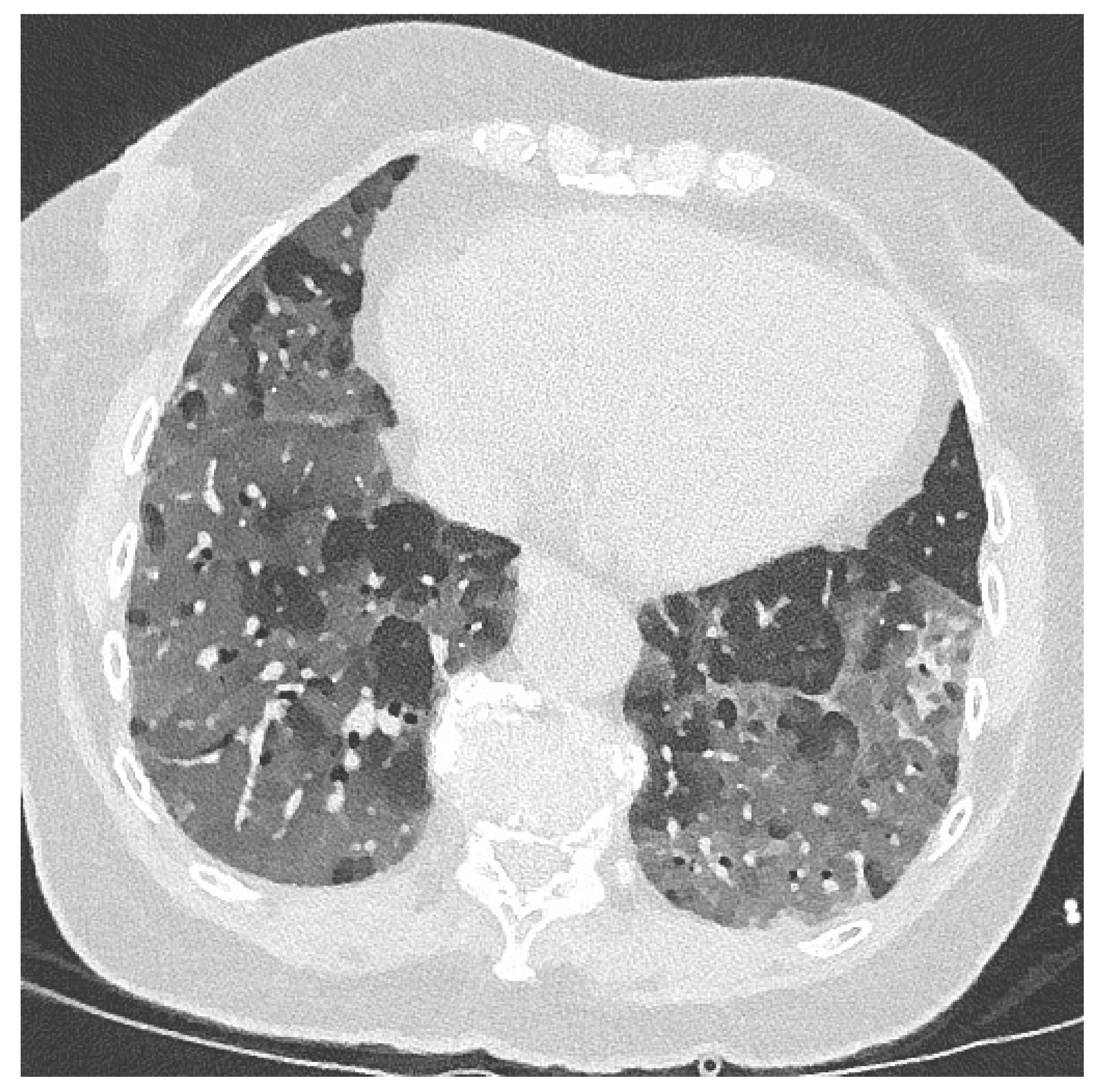
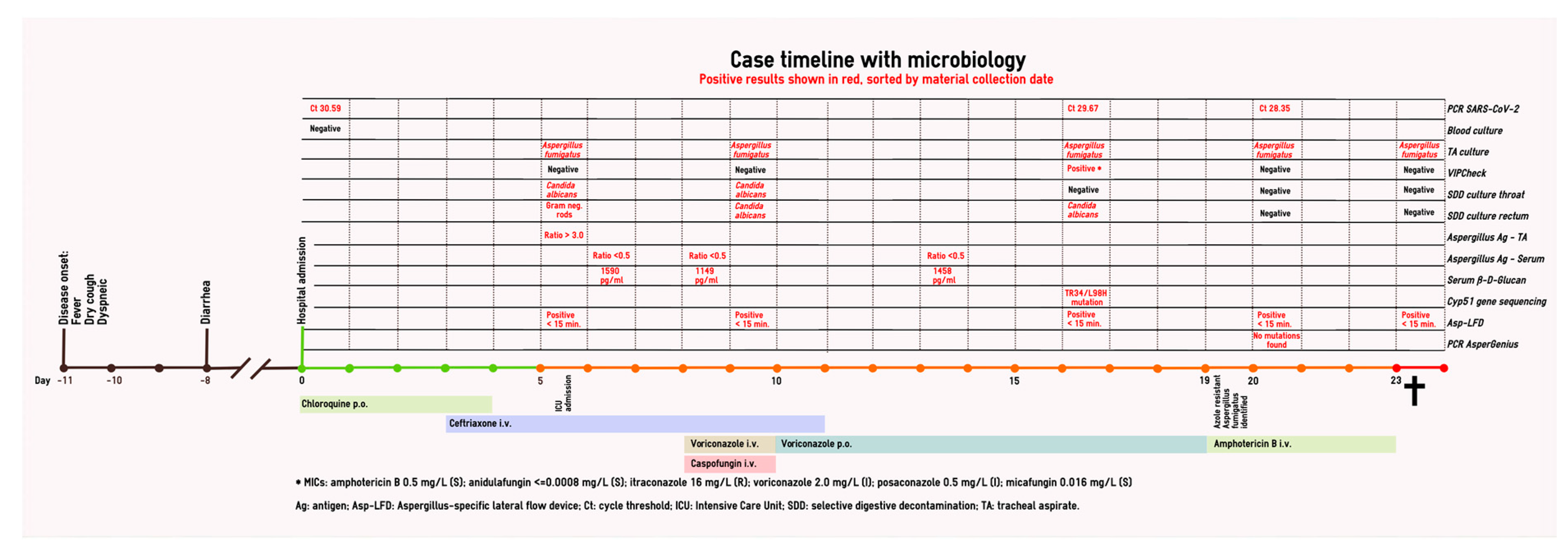
2. Case Report and Results

A 74-year-old patient was admitted because of respiratory insufficiency amid the COVID-19 crisis. Eleven days prior to admission, she had been suffering from fever (38.5 °C) and a dry cough. Three days after symptom onset, she developed diarrhea. Her medical history included complaints of reflux and pain due to arthrosis of the hip and knees, for which she uses a proton-pump inhibitor and a nonsteroidal anti-inflammatory drug pantoprazol and etoricoxib, respectively. She stopped smoking 20 years ago and was healthy and fit otherwise. Patient characteristics can be found in Table 1. This study, “Clinical course and prognostic factors for COVID-19” with project identification code CWZ-nr 027-2020, was approved in March 20202 by the Canisius Wilhelmina Hospital medical ethics committee and patient informed consent was acquired antemortem with opt out possibility.
At presentation to the emergency department, she had been feeling progressively dyspneic for two days. On physical examination, her oxygenation was 82%, with 28 breaths per minute in room air, pulmonary wheezing and an extended expiration. Oxygenation improved to 94% with 5 L O2 via a nasal cannula, but she desaturated during speech. Her BMI was 27.7 (80 kg) and her temperature 37.8 °C. No other aberrant observations on physical examination were made. Her Glasgow Coma Scale was 15 and her ECG was normal. Her C-reactive protein (CRP) was 214 mg/L, and other laboratory findings included slightly elevated leucocytes (12.6 × 10⁹ /L) and neutrophils (8.4 × 10⁹ /L), elevated liver enzymes (alkaline phosphatase 528 U/L; GGT 376 U/L; AST 76 U/L; LD 745 U/L), slightly elevated pro-calcitonin (0.25 µg/L; <0.5 µg/L not suggestive of bacterial infection), increased ferritin (1442 µg/L), and normal electrolyte, glucose and renal function. SARS-CoV-2 nasopharyngeal and throat swabs were taken. A low-dose chest CT demonstrated extensive centralized and peripheral bilateral ground glass opacities with left-sided consolidations and bilateral fibrotic bands without pleural effusions and vascular enlargement (Figure 1). The CO-RADS score was 5 and CT-severity score was 24 out of 25 [11].
Because of the high probability of SARS-CoV-2 infection, chloroquine treatment was started (600 mg and 300 mg on day 1, 300 mg q12h days 2–5), which was national policy at the time. The SARS-CoV-2 PCR of a nasopharyngeal swab was positive (Ct 30.59; E gene [12]). Blood cultures remained negative, as were nasopharynx bacterial cultures taken at admission. The patient was subsequently admitted to our general inpatient respiratory ward. An overview of her hospital course is depicted in Figure 2.
The CRP remained highly stable over the following days at around 200 mg/L with a range of 192–214. However, the patient needed increasing oxygenation with a non-rebreathing mask. Empirical treatment of a suspected bacterial superinfection was started with ceftriaxone i.v. 2000 mg q24h. Five days after admission, the maximum (15 L O2) oxygenation with the non-rebreathing mask became insufficient and the patient was admitted to the ICU for respiratory support and intensive monitoring.
In the ICU, HFNO (high-flow nasal oxygen therapy) and selective digestive decontamination (SDD) were initiated, which includes ceftriaxone i.v. 2000 mg q24h for 4 days and a combined oral non-absorbable suspension of amphotericin B, colistin and tobramycin q6h. In this patient, ceftriaxone was continued de facto for another 4 days. Routine bacterial and fungal (peri-anal, throat and tracheal aspirate) surveillance cultures were done twice weekly in adherence with our local SDD policy [13]. Within a few hours after admission to the ICU, her blood oxygenation became insufficient with HFNO at FiO2 100% and 60 L/min flow. Therefore, she was sedated, intubated and put on a mechanical ventilator. A CT angiography of the chest was performed which demonstrated significant bilateral pulmonary emboli. Anticoagulants (enoxaparine anti-factor Xa) were initiated in therapeutic dosages. Pressure control ventilation was required with the patient in prone position. Because of the need for increasing noradrenaline dosages during circulatory shock, hydrocortisone 100 mg q8h was initiated and continued for five days. Cardiac ultrasound showed a minor tricuspid insufficiency but no major pathology.
Aspergillus fumigatus was recovered from high-volume tracheal aspirate cultures [14] obtained at ICU admission. Aspergillus galactomannan (Platelia Aspergillus; Bio-Rad, Marnes-La-Coquette, France) ratio at this time was >3.0 (positive) in a tracheal aspirate and β-d-glucan (Fungitell assay; Associates of Cape Cod Inc., East Falmouth, MA, USA) in serum was 1590 pg/mL (positive), after which a putative diagnosis of CAPA was made. Serum galactomannan remained negative (<0.5) in three subsequent samples. Voriconazole i.v. 6 mg/kg q12h was started in addition to caspofungin i.v. 70 mg q24h until the VIPcheck (Mediaproducts BV, Groningen, The Netherlands), used to detect azole resistance, was negative. MICs determined with broth microdilution using CLSI methodology of the A. fumigatus isolate were as follows: amphotericin B 0.5 mg/L, micafungin and anidulafungin <0.016 mg/L, itraconazole 1 mg/L, voriconazole 0.25 mg/L, and posaconazole 0.063 mg/L. Voriconazole was switched to oral administration of 200 mg q12h with discontinuation of caspofungin. During SDD, bacterial cultures remained negative throughout her stay in the ICU.
On day 6 after admission (day 2 at the ICU), continuous venovenous hemofiltration was initiated because of rapidly progressive acute renal failure. A. fumigatus was persistently cultured from tracheal aspirate samples during voriconazole treatment and β-d-glucan levels remained positive with 1149 and 1458 pg/µl, at 1 and 6 days (day 8 and 13 after hospital admission) of voriconazole therapy, respectively. Voriconazole serum therapeutic drug monitoring was performed as recommended [15], with therapeutic concentrations of 4.72 mg/L, 2.78 mg/L and 1.43 mg/L at day 13, 15 and 17, respectively.
The respiratory situation improved marginally in the subsequent 7 days but declined steadily thereafter. Pressure support and pressure control ventilation were alternated between days 12 and 19 and attempts to return the patient to a supine position failed several times. After 7 days, A. fumigatus grew on the itraconazole and voriconazole wells of the second VIPcheck on day 19 (tracheal aspirate culture). MICs of this A. fumigatus isolate were as follows: amphotericin B 0.5 mg/L, anidulafungin and micafungin <0.016 mg/L, itraconazole 16 mg/L, voriconazole 2 mg/L and posaconazole 0.5 mg/L. Voriconazole treatment was changed to liposomal amphotericin B 200 mg q24h. Subsequent cyp51A gene sequencing identified a TR34/L98H mutation, probably responsible for the observed azole resistance. On day 22, ventilation and oxygenation of the patient deteriorated further without further treatment options and therapy was discontinued on day 23. An autopsy was not performed.

3. Discussion

We report the first case of azole-resistant CAPA, which occurred in an immunocompetent host during ICU support without a previous history of azole therapy. The A. fumigatus cyp51A gene TR34/L98H mutation found in this patient has been well described as an environmentally acquired mutation [16], which is in line with data from clinical studies where two-thirds of patients with azole-resistant infections had no history of azole pretreatment [10]. This case underscores the importance of early diagnosis and the need for resistance surveillance, comparable to what has been described in influenza patients [9,17], given the emergence of triazole resistance [18,19].
The sensitivity for detection of resistance in primary cultures with the VIPcheck plate depends on the number of A. fumigatus colonies that are tested, as clinical cultures may contain both mixed azole-susceptible and azole-resistant isolates during an infection [20]. We suspect that A. fumigatus isolated in the first tracheal aspirate was already a mixed culture but was missed in initial fungal cultures due to abundance of azole-susceptible A. fumigatus spores. Molecular detection could have given a suggestion to the presence of a mixed culture [21] but PCR could not be performed due to absence of material. The TR34/L98H had a phenotype with high itraconazole MIC (>16 mg/L) and low voriconazole MIC (2 mg/L), similar to strains which have been described only recently in the Netherlands [22].
IPA is known to be problematic to diagnose in the non-neutropenic ICU host [23]. Regardless of the compelling evidence for CAPA in this patient, the EORTC/MSGERC [24] host criteria for invasive fungal disease were not met, nor did the patient meet the AspICU algorithm because we tested tracheal aspirates instead of bronchoalveolar lavage (BAL) fluid [25]. This is in line with findings from other groups, where CAPA patients did not meet the EORTC/MSGERC host criteria either [3,4,5,6]. In addition, the American Association for Bronchology and Interventional Pulmonology (AABIP) has issued a statement advising against routine bronchoscopy in COVID-19 patients, as it poses substantial risk to patients and staff [26]. BAL should only be considered in intubated patients if upper respiratory samples are negative and BAL would significantly change clinical management. Tracheal aspirate cultures, as performed twice weekly in our patient, repeatedly identified A. fumigatus as the only micro-organism present. In the first positive culture, five colonies were tested for resistance with the VIPcheck plate as is recommended to exclude azole resistance [15]. When surveillance cultures of tracheal aspirates were persistently cultured positive with A. fumigatus during voriconazole therapy, we suspected the selection of resistant isolates which were probably already present in the first samples, albeit in undetectable numbers. An autopsy to confirm IPA was not done.
Serum galactomannan testing has been shown to be a fairly sensitive diagnostic tool (70%) in neutropenic patients with pathology-proven invasive aspergillosis [27,28]. However, in patients who are non-neutropenic, serum galactomannan sensitivity of around 25% has been reported [27], which may explain the low number of serum galactomannan positive findings in recently published case reports [6,7] and case series [3,4,5]. The role of β-d-glucan and the Aspergillus-specific lateral flow device (LFD) as an adjunct to the diagnosis of IPA in COVID-19 is not yet clear [2,23]. Serum β-d-glucan was persistently strongly positive in this patient over the course of a week. The specificity for invasive fungal disease of β-d-glucan testing in a mixed ICU population has been shown to be high (86%), with two consecutive positive results [29] compared to those with only fungal colonization and no invasive fungal disease. In addition, multiple other studies report a good sensitivity for the diagnosis of invasive aspergillosis in critically ill patients [30,31,32,33,34]. BAL β-d-glucan in the ICU setting is, however, not recommended, due to its poor specificity and confounders causing false positive results [35].
The LFD is particularly interesting in the ICU due to its short turnaround time. It has demonstrated a higher sensitivity but lower specificity in BAL fluids compared to galactomannan [36] and β-d-glucan [37] in IPA-probable and proven immunocompromised patients. In the ICU setting, however, LFD is suggested to have a lower sensitivity but comparable specificity to galactomannan testing in BAL fluids [35,38]. Noteworthily, a negative predictive value of >96% has been reported in the ICU setting [39]. We used the OLM lateral flow device (AspLFD) on sequential patient tracheal aspirates, yielding positive results on all samples confirming the positive galactomannan result. Although suitable for its negative predictive value or as an additional diagnostic measure, further evaluation of lateral flow technology in critically ill patients is warranted.
Altogether, we describe the clinical course of the first reported patient with azole-resistant CAPA. The contribution of A. fumigatus to this fatal COVID-19 course is highly likely, although autopsy was not performed, as in all previously reported CAPA cases [3,4,5,6,7].

Author Contributions

Conceptualization, J.F.M. and A.S.M.D.; formal analysis, E.F.J.M.; investigation, O.H. and J.B.B.; resources, J.F.M.; data curation, A.S.M.D. and O.H.; writing—original draft preparation, E.M. and J.F.M.; writing—review and editing, A.S.M.D., O.H., J.B.B.; supervision, J.F.M. All authors have read and agreed to the published version of the manuscript.

Funding

This research received no external funding.

Acknowledgments

We thank Paul E Verweij M.D. for helpful discussions.

Conflicts of Interest

The authors declare no conflict of interest.

References

  1. Clancy, C.J.; Nguyen, M.H. COVID-19, superinfections and antimicrobial development: What can we expect? Clin. Infect. Dis. 2020. [Google Scholar] [CrossRef] [PubMed]
  2. Verweij, P.E.; Gangneux, J.-P.; Bassetti, M.; Brüggemann, R.J.M.; Cornely, O.A.; Koehler, P.; Lass-Flörl, C.; van de Veerdonk, F.L.; Chakrabarti, A.; Hoenigl, M. Diagnosing COVID-19-associated pulmonary aspergillosis. Lancet Microbe 2020. [Google Scholar] [CrossRef]
  3. van Arkel, A.L.E.; Rijpstra, T.A.; Belderbos, H.N.A.; van Wijngaarden, P.; Verweij, P.E.; Bentvelsen, R.G. COVID-19 associated pulmonary aspergillosis. Am. J. Respir. Crit. Care Med. 2020. [Google Scholar] [CrossRef] [PubMed]
  4. Alanio, A.; Dellière, S.; Fodil, S.; Bretagne, S.; Mégarbane, B. High prevalence of putative invasive pulmonary aspergillosis in critically Ill COVID-19 patients. Lancet Respir. Med. 2020. [Google Scholar] [CrossRef]
  5. Koehler, P.; Cornely, O.A.; Bottiger, B.W.; Dusse, F.; Eichenauer, D.A.; Fuchs, F.; Hallek, M.; Jung, N.; Klein, F.; Persigehl, T.; et al. COVID-19 associated pulmonary aspergillosis. Mycoses 2020, 63, 528–534. [Google Scholar] [CrossRef]
  6. Marion, B.; Julien, M.; Cécile, N.; Alexandre, L.; Anne-Geneviève, M.; Marc, T.; Renaud, P.; Alexandre, D.; Arnaud, F. Fatal invasive aspergillosis and coronavirus disease in an immunocompetent patient. Emerg. Infect. Dis. 2020, 26. [Google Scholar] [CrossRef]
  7. Prattes, J.; Valentin, T.; Hoenigl, M.; Talakic, E.; Reisinger, A.C.; Eller, P. Invasive pulmonary aspergillosis complicating COVID-19 in the ICU - A case report. Med. Mycol. Case Rep. 2020. [Google Scholar] [CrossRef]
  8. Schauwvlieghe, A.; Rijnders, B.J.A.; Philips, N.; Verwijs, R.; Vanderbeke, L.; Van Tienen, C.; Lagrou, K.; Verweij, P.E.; Van de Veerdonk, F.L.; Gommers, D.; et al. Invasive aspergillosis in patients admitted to the intensive care unit with severe influenza: A retrospective cohort study. Lancet Respir. Med. 2018, 6, 782–792. [Google Scholar] [CrossRef]
  9. Buil, J.B.; Meijer, E.F.J.; Denning, D.W.; Verweij, P.E.; Meis, J.F. Burden of serious fungal infections in the Netherlands. Mycoses 2020, 63, 625–631. [Google Scholar] [CrossRef]
  10. Meis, J.F.; Chowdhary, A.; Rhodes, J.L.; Fisher, M.C.; Verweij, P.E. Clinical implications of globally emerging azole resistance in Aspergillus fumigatus. Philos. Trans. R Soc. Lond. B Biol. Sci. 2016, 371. [Google Scholar] [CrossRef] [Green Version]
  11. Prokop, M.; van Everdingen, W.; van Rees Vellinga, T.; Quarles van Ufford, J.; Stoger, L.; Beenen, L.; Geurts, B.; Gietema, H.; Krdzalic, J.; Schaefer-Prokop, C.; et al. CO-RADS - A categorical CT assessment scheme for patients with suspected COVID-19: Definition and evaluation. Radiology 2020, 201473. [Google Scholar] [CrossRef] [PubMed]
  12. Corman, V.M.; Landt, O.; Kaiser, M.; Molenkamp, R.; Meijer, A.; Chu, D.K.; Bleicker, T.; Brunink, S.; Schneider, J.; Schmidt, M.L.; et al. Detection of 2019 novel coronavirus (2019-nCoV) by real-time RT-PCR. Euro Surveill. 2020, 25. [Google Scholar] [CrossRef] [PubMed] [Green Version]
  13. Oostdijk, E.A.; De Jonge, E.; Kullberg, B.J.; Natsch, S.; De Smet, A.M.; Vandenbroucke-Grauls, C.M.J.E.; van der Vorm, E.; Bonten, M.J.M. SWAB-Richtlijn: Selectieve Decontaminatie bij Patiënten op de Intensive Care. 2018. Available online: https://swab.nl/nl/selectieve-decontaminatie-sdd (accessed on 22 May 2020).
  14. Vergidis, P.; Moore, C.B.; Novak-Frazer, L.; Rautemaa-Richardson, R.; Walker, A.; Denning, D.W.; Richardson, M.D. High-volume culture and quantitative real-time PCR for the detection of Aspergillus in sputum. Clin. Microbiol. Infect. 2019, in press. [Google Scholar] [CrossRef] [PubMed]
  15. Ullmann, A.J.; Aguado, J.M.; Arikan-Akdagli, S.; Denning, D.W.; Groll, A.H.; Lagrou, K.; Lass-Flörl, C.; Lewis, R.E.; Munoz, P.; Verweij, P.E.; et al. Diagnosis and management of Aspergillus diseases: Executive summary of the 2017 ESCMID-ECMM-ERS guideline. Clin. Microbiol. Infect. 2018, 24 (Suppl. 1), e1–e38. [Google Scholar] [CrossRef] [PubMed]
  16. Chowdhary, A.; Kathuria, S.; Xu, J.P.; Meis, J.F. Emergence of azole-resistant Aspergillus fumigatus strains due to agricultural azole use creates an increasing threat to human health. PLoS Pathog. 2013, 9, e1003633. [Google Scholar] [CrossRef]
  17. Talento, A.F.; Dunne, K.; Murphy, N.; O’Connell, B.; Chan, G.; Joyce, E.A.; Hagen, F.; Meis, J.F.; Fahy, R.; Bacon, L.; et al. Post-influenzal triazole-resistant aspergillosis following allogeneic stem cell transplantation. Mycoses 2018, 61, 570–575. [Google Scholar] [CrossRef]
  18. Chowdhary, A.; Sharma, C.; Meis, J.F. Azole-resistant aspergillosis: Epidemiology, molecular mechanisms, and treatment. J. Infect. Dis. 2017, 216, S436–S444. [Google Scholar] [CrossRef] [Green Version]
  19. Lestrade, P.P.; Meis, J.F.; Melchers, W.J.; Verweij, P.E. Triazole resistance in Aspergillus fumigatus: Recent insights and challenges for patient management. Clin. Microbiol. Infect. 2019, 25, 799–806. [Google Scholar] [CrossRef]
  20. Ahmad, S.; Joseph, L.; Hagen, F.; Meis, J.F.; Khan, Z. Concomitant occurrence of itraconazole-resistant and -susceptible strains of Aspergillus fumigatus in routine cultures. J. Antimicrob. Chemother. 2015, 70, 412–415. [Google Scholar] [CrossRef] [Green Version]
  21. Singh, A.; Sharma, B.; Kumar Mahto, K.; Meis, J.F.; Chowdhary, A. High-frequency direct detection of triazole resistance in Aspergillus fumigatus from patients with chronic pulmonary fungal diseases in India. J. Fungi 2020, 6, 67. [Google Scholar] [CrossRef]
  22. Lestrade, P.P.; Buil, J.B.; van der Beek, M.T.; Kuijper, E.J.; van Dijk, K.; Kampinga, G.A.; Rijnders, B.J.A.; Vonk, A.G.; de Greeff, S.C.; Schoffelen, A.F.; et al. Paradoxal trends in azole-resistant Aspergillus fumigatus in a national multicenter surveillance program, 2013–2018. Emerg. Infect. Dis. 2020, 26. [Google Scholar] [CrossRef]
  23. Blot, S.; Rello, J.; Koulenti, D. Diagnosing invasive pulmonary aspergillosis in ICU patients: Putting the puzzle together. Curr. Opin. Crit. Care 2019, 25, 430–437. [Google Scholar] [CrossRef] [PubMed]
  24. Donnelly, J.P.; Chen, S.C.; Kauffman, C.A.; Steinbach, W.J.; Baddley, J.W.; Verweij, P.E.; Clancy, C.J.; Wingard, J.R.; Lockhart, S.R.; Groll, A.H.; et al. Revision and update of the consensus definitions of invasive fungal disease from the European Organization for Research and Treatment of Cancer and the Mycoses Study Group Education and Research Consortium. Clin. Infect. Dis. 2019, in press. [Google Scholar] [CrossRef] [PubMed] [Green Version]
  25. Blot, S.I.; Taccone, F.S.; Van den Abeele, A.M.; Bulpa, P.; Meersseman, W.; Brusselaers, N.; Dimopoulos, G.; Paiva, J.A.; Misset, B.; Rello, J.; et al. A clinical algorithm to diagnose invasive pulmonary aspergillosis in critically ill patients. Am. J. Respir. Crit. Care Med. 2012, 186, 56–64. [Google Scholar] [CrossRef] [Green Version]
  26. Wahidi, M.M.; Lamb, C.; Murgu, S.; Musani, A.; Shojaee, S.; Sachdeva, A.; Maldonado, F.; Mahmood, K.; Kinsey, M.; Sethi, S.; et al. American Association for Bronchology and Interventional Pulmonology (AABIP) statement on the use of bronchoscopy and respiratory specimen collection in patients with suspected or confirmed COVID-19 infection. J. Bronchol. Interv. Pulmonol. 2020. [Google Scholar] [CrossRef] [PubMed]
  27. Meersseman, W.; Lagrou, K.; Maertens, J.; Wilmer, A.; Hermans, G.; Vanderschueren, S.; Spriet, I.; Verbeken, E.; Van Wijngaerden, E. Galactomannan in bronchoalveolar lavage fluid: A tool for diagnosing aspergillosis in intensive care unit patients. Am. J. Respir. Crit. Care Med. 2008, 177, 27–34. [Google Scholar] [CrossRef] [PubMed]
  28. Verweij, P.E.; Weemaes, C.M.; Curfs, J.H.; Bretagne, S.; Meis, J.F. Failure to detect circulating Aspergillus markers in a patient with chronic granulomatous disease and invasive aspergillosis. J. Clin. Microbiol. 2000, 38, 3900–3901. [Google Scholar] [CrossRef] [Green Version]
  29. Talento, A.F.; Dunne, K.; Joyce, E.A.; Palmer, M.; Johnson, E.; White, P.L.; Springer, J.; Loeffler, J.; Ryan, T.; Collins, D.; et al. A prospective study of fungal biomarkers to improve management of invasive fungal diseases in a mixed specialty critical care unit. J. Crit. Care 2017, 40, 119–127. [Google Scholar] [CrossRef]
  30. Acosta, J.; Catalan, M.; del Palacio-Peréz-Medel, A.; Lora, D.; Montejo, J.C.; Cuetara, M.S.; Moragues, M.D.; Ponton, J.; del Palacio, A. A prospective comparison of galactomannan in bronchoalveolar lavage fluid for the diagnosis of pulmonary invasive aspergillosis in medical patients under intensive care: Comparison with the diagnostic performance of galactomannan and of (1→3)-β-d-glucan chromogenic assay in serum samples. Clin. Microbiol. Infect. 2011, 17, 1053–1060. [Google Scholar] [CrossRef] [Green Version]
  31. Acosta, J.; Catalan, M.; Palacio-Pérez-Medel, A.; Montejo, J.; De-La-Cruz-Bértolo, J.; Moragues, M.; Pontón, J.; Finkelman, M.; Palacio, A. Prospective study in critically ill non-neutropenic patients: Diagnostic potential of (1,3)-β-D-glucan assay and circulating galactomannan for the diagnosis of invasive fungal disease. Eur. J. Clin. Microbiol. Infect. Dis. 2011, 31, 721–731. [Google Scholar] [CrossRef]
  32. Boch, T.; Reinwald, M.; Spiess, B.; Liebregts, T.; Schellongowski, P.; Meybohm, P.; Rath, P.M.; Steinmann, J.; Trinkmann, F.; Britsch, S.; et al. Detection of invasive pulmonary aspergillosis in critically ill patients by combined use of conventional culture, galactomannan, 1,3-β-D-glucan and Aspergillus specific nested polymerase chain reaction in a prospective pilot study. J. Crit. Care 2018, 47, 198–203. [Google Scholar] [CrossRef] [PubMed]
  33. De Vlieger, G.; Lagrou, K.; Maertens, J.; Verbeken, E.; Meersseman, W.; Van Wijngaerden, E. Beta-D-glucan detection as a diagnostic test for invasive aspergillosis in immunocompromised critically ill patients with symptoms of respiratory infection: An autopsy-based study. J. Clin. Microbiol. 2011, 49, 3783–3787. [Google Scholar] [CrossRef] [PubMed] [Green Version]
  34. Lahmer, T.; Neuenhahn, M.; Held, J.; Rasch, S.; Schmid, R.M.; Huber, W. Comparison of 1,3-β-d-glucan with galactomannan in serum and bronchoalveolar fluid for the detection of Aspergillus species in immunosuppressed mechanical ventilated critically ill patients. J. Crit. Care 2016, 36, 259–264. [Google Scholar] [CrossRef] [PubMed]
  35. Vanderbeke, L.; Van Wijngaerden, E.; Maertens, J.; Wauters, J.; Lagrou, K. Diagnosis of invasive aspergillosis in intensive care unit patients. Curr. Fungal Infect. Rep. 2020. [Google Scholar] [CrossRef]
  36. Johnson, G.L.; Sarker, S.-J.; Nannini, F.; Ferrini, A.; Taylor, E.; Lass-Flörl, C.; Mutschlechner, W.; Bustin, S.A.; Agrawal, S.G. Aspergillus-specific lateral-flow device and real-time PCR testing of bronchoalveolar lavage fluid: A combination biomarker approach for clinical diagnosis of invasive pulmonary aspergillosis. J. Clin. Microbiol. 2015, 53, 2103–2108. [Google Scholar] [CrossRef] [PubMed] [Green Version]
  37. Hoenigl, M.; Prattes, J.; Spiess, B.; Wagner, J.; Prueller, F.; Raggam, R.B.; Posch, V.; Duettmann, W.; Hoenigl, K.; Wölfler, A.; et al. Performance of galactomannan, 1,3-β-d-glucan, Aspergillus lateral-flow device, conventional culture, and PCR tests with bronchoalveolar lavage fluid for diagnosis of invasive pulmonary aspergillosis. J. Clin. Microbiol. 2014, 52, 2039–2045. [Google Scholar] [CrossRef] [Green Version]
  38. Jenks, J.D.; Mehta, S.R.; Taplitz, R.; Aslam, S.; Reed, S.L.; Hoenigl, M. Point-of-care diagnosis of invasive aspergillosis in non-neutropenic patients: Aspergillus galactomannan lateral flow assay versus Aspergillus-specific lateral flow device test in bronchoalveolar lavage. Mycoses 2019, 62, 230–236. [Google Scholar] [CrossRef]
  39. Eigl, S.; Prattes, J.; Lackner, M.; Willinger, B.; Spiess, B.; Reinwald, M.; Selitsch, B.; Meilinger, M.; Neumeister, P.; Reischies, F.; et al. Multicenter evaluation of a lateral-flow device test for diagnosing invasive pulmonary aspergillosis in ICU patients. Crit. Care 2015, 19, 178. [Google Scholar] [CrossRef] [Green Version]
Figure 1. Low-dose chest CT showing extensive centralized and peripheral bilateral ground glass opacities with left-sided consolidations and bilateral fibrotic bands. No pleural effusion. No vascular enlargement and no specific suggestions of aspergillosis.
Jof 06 00079 g001
Figure 2. Case timeline with microbiology.
Jof 06 00079 g002
Table 1. Patient characteristics
GenderFemale
Age (years)74
Medical historyReflux, polyarthrosis, stopped smoking 20 years ago
MedicationPantoprazol (PPI) and Etoricoxib (NSAID)
Underlying immuno-compromising conditionNone
Initial symptomsFever, dry cough, dyspneic, diarrhea
ARDSProne positioningYes
vvECMONo
Acute renal failureYes, continuous venovenous hemofiltration (CVVH)
IPA definitionEORTC/MSG criteriaN/A
(modified) AspICU algorithmN/A

