A Week of Oral Terbinafine Pulse Regimen Every Three Months to Treat all Dermatophyte Onychomycosis


Abstract
:1. Introduction
2. Materials and Methods
2.1. Patients
2.2. Evaluation of Therapeutic Response
2.3. Statistical Methodology
2.4. Demographic Characteristics
3. Results
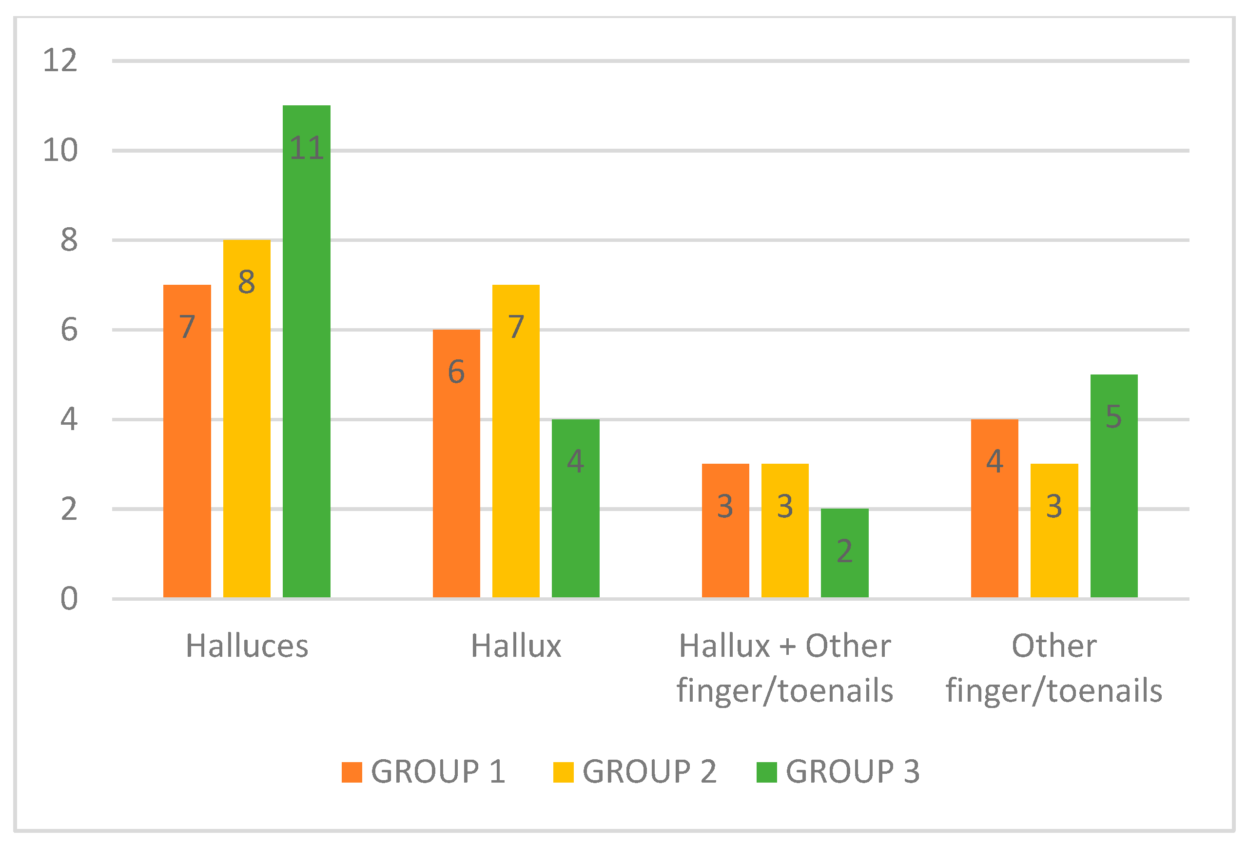
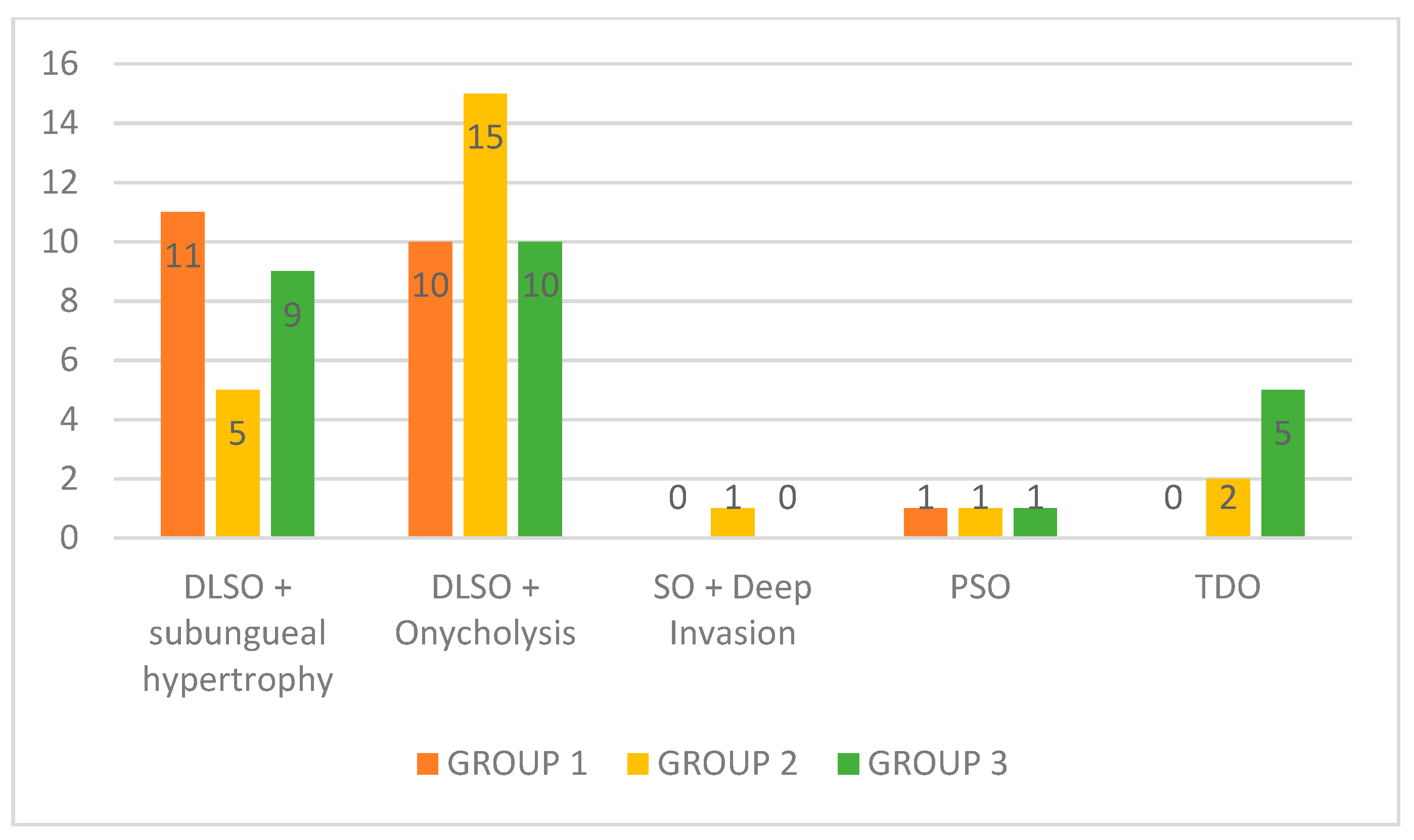
3.1. Relationship between Treatment Results, Affected Nails, Clinical Classification, Presence of Comorbidities, Use of Medications, and Isolated Fungi
3.2. Dropouts and Side Effects
4. Discussion
5. Conclusions
Author Contributions
Funding
Acknowledgments
Conflicts of Interest
Abbreviations
| CI | clinical improvement |
| DLSO | distal lateral subungual onychomycosis |
| MC | mycological cure |
| SO | superficial onychomycosis |
| TC | total cure |
| TDO | total dystrophic onychomycosis |
| TF | therapeutic failure |
| AGNUS | asymmetric gait nail unit syndrome |
References
- Smith, E.B. History of antifungals. J. Am. Acad. Dermatol. 1990, 23, 776–778. [Google Scholar] [CrossRef]
- Neumann, H.A. Oral treatment of onychomycosis of the toe nails; comparison of cost effectiveness of griseofulvin, itraconazole, ketoconazole and terbinafine. Ned. Tijdschr. Geneeskd. 1995, 139, 1350–1351. [Google Scholar]
- Purim, K.S.; Bordignon, G.P.; Queiroz-Telles, F. Fungal infection of the feet in soccer players and non-athlete individuals. Rev. Iberoam. Micol. 2005, 22, 34–38. [Google Scholar] [CrossRef]
- Purim, K.S.; de Freitas, C.F.; Leite, N. Feet dermatophytosis in soccer player. An. Braz. Dermatol. 2009, 84, 550–552. [Google Scholar] [CrossRef]
- Gupta, A.K.; Versteeg, S.G.; Shear, N.H. Onychomycosis in the 21st Century: An Update on Diagnosis, Epidemiology, and Treatment. J. Cutan. Med. Surg. 2017, 21, 525–539. [Google Scholar] [CrossRef]
- Ha, S.J.; Han, K.D.; Song, Y.; Lee, J.H. Weight change and risk of onychomycosis: A nationwide cohort study in Korea. J. Am. Acad. Dermatol. 2018, 78, 613–614. [Google Scholar] [CrossRef]
- Gupta, A.K.; Daigle, D.; Foley, K.A. Network Meta-Analysis of Onychomycosis Treatments. Skin Appendage Disord. 2015, 1, 74–81. [Google Scholar] [CrossRef] [Green Version]
- Garcia-Romero, M.T.; Granados, J.; Vega-Memije, M.E.; Arenas, R. Analysis of genetic polymorphism of the HLA-B and HLA-DR loci in patients with dermatophytic onychomycosis and in their first-degree relatives. Actas Dermosifiliogr. 2012, 103, 59–62. [Google Scholar] [CrossRef]
- Carrillo-Meléndrez, H.; Ortega-Hernández, E.; Granados, J.; Arroyo, S.; Barquera, R.; Arenas, R. Role of HLA-DR Alleles to Increase Genetic Susceptibility to Onychomycosis in Nail Psoriasis. Skin Appendage Disord. 2016, 2, 22–25. [Google Scholar] [CrossRef] [Green Version]
- Abdel-Rahman, S.M. Genetic Predictors of Susceptibility to Dermatophytoses. Mycopathologia 2017, 182, 67–76. [Google Scholar] [CrossRef]
- Gupta, A.K.; Carviel, J.; Shear, N.H. Onychomycosis and Chronic Fungal Disease: Exploiting a Commensal Disguise to Stage a Covert Invasion. J. Cutan. Med. Surg. 2018, 22, 318–322. [Google Scholar] [CrossRef]
- Vlahovic, T.C. Onychomycosis: Evaluation, Treatment Options, Managing Recurrence, and Patient Outcomes. Clin. Podiatr. Med. Surg. 2016, 33, 305–318. [Google Scholar] [CrossRef]
- Lipner, S.R.; Scher, R.K. Part I: Onychomycosis: Clinical Overview and Diagnosis. J. Am. Acad. Dermatol. 2019. [Google Scholar] [CrossRef]
- Zaias, N.; Rebell, G.; Casal, G.; Appel, J. The asymmetric gait toenail unit sign. Skinmed 2012, 10, 213–217. [Google Scholar]
- Zaias, N.; Rebell, G.; Escovar, S. Asymmetric gait nail unit syndrome: The most common worldwide toenail abnormality and onychomycosis. Skinmed 2014, 12, 217–223. [Google Scholar]
- Peres, N.T.; Maranhão, F.C.; Rossi, A.; Martinez-Rossi, N.M. Dermatophytes- Host-pathogen interaction and antifungal resistance. An. Braz. Dermatol. 2010, 85, 657–667. [Google Scholar] [CrossRef]
- Gupta, A.K.; Foley, K.A.; Mays, R.R.; Shear, N.H.; Piguet, V. Monotherapy for toenail onychomycosis: A systematic review and network meta-analysis. Br. J. Dermatol. 2019. [Google Scholar] [CrossRef]
- Gupta, A.K.; Foley, K.A.; Versteeg, S.G. New Antifungal Agents and New Formulations Against Dermatophytes. Mycopathologia 2017, 182, 127–141. [Google Scholar] [CrossRef]
- Gupta, A.K.; Mays, R.R.; Versteeg, S.G.; Shear, N.H.; Friedlander, S.F. Onychomycosis in children: Safety and efficacy of antifungal agents. Pediatr. Dermatol. 2018, 35, 552–559. [Google Scholar] [CrossRef]
- Kreijkamp-Kaspers, S.; Hawke, K.L.; van Driel, M.L. Oral Medications to Treat Toenail Fungal Infection. JAMA 2018, 319, 397–398. [Google Scholar] [CrossRef]
- Dias, M.F.R.G.; Bernardes-Filho, F.; Quaresma-Santos, M.V.P.; Amorim, A.G.D.F.; Schechtman, R.C.; Azulay, D.R. Treatment of superficial mycoses: Review. Part II. An. Braz. Dermatol. 2013, 88, 937–944. [Google Scholar] [CrossRef]
- Salo, H.; Pekurinen, M. Cost effectiveness of oral terbinafine (Lamisil) compared with oral fluconazole (Diflucan) in the treatment of patients with toenail onychomycosis. Pharmacoeconomics 2002, 20, 319–324. [Google Scholar] [CrossRef]
- Jensen, J.C. Clinical pharmacokinetics of terbinafine (Lamisil). Clin. Exp. Dermatol. 1989, 14, 110–113. [Google Scholar] [CrossRef]
- Schatz, F.; Bräutigam, M.; Dobrowolski, E.; Effendy, I.; Haberl, H.; Mensing, H.; Weidinger, G.; Stütz, A. Nail incorporation kinetics of terbinafine in onychomycosis patients. Clin. Exp. Dermatol. 1995, 20, 377–383. [Google Scholar] [CrossRef]
- Leyden, J. Pharmacokinetics and pharmacology of terbinafine and itraconazole. J. Am. Acad. Dermatol. 1998, 38, 42–47. [Google Scholar] [CrossRef]
- Warshaw, E.M.; Fett, D.D.; Bloomfield, H.E.; Grill, J.P.; Nelson, D.B.; Quintero, V.; Carver, S.M.; Zielke, G.R.; Lederle, F.A. Pulse versus continuous terbinafine for onychomycosis: A randomized, double-blind, controlled trial. J. Am. Acad. Dermatol. 2005, 53, 578–584. [Google Scholar] [CrossRef]
- Takahata, Y.; Hiruma, M.; Shiraki, Y.; Tokuhisa, Y.; Sugita, T.; Muto, M. Treatment of dermatophyte onychomycosis with three pulses of terbinafine (500 mg day for a week). Mycoses 2009, 52, 72–76. [Google Scholar] [CrossRef]
- Mishra, M.; Panda, P.; Tripathy, S.; Sengupta, S.; Mishra, K. An open randomized comparative study of oral itraconazole pulse and terbinafine pulse in the treatment of onychomycosis. Indian J. Dermatol. Venereol. Leprol. 2005, 71, 262–266. [Google Scholar] [CrossRef]
- Zaias, N.; Rebell, G. The successful treatment of Trichophyton rubrum nail bed (distal subungual) onychomycosis with intermittent pulse-dosed terbinafine. Arch. Dermatol. 2004, 140, 691–695. [Google Scholar] [CrossRef]
- Guo, S.; Zhong, S.; Zhang, A. Privacy-preserving Kruskal-Wallis test. Comput. Methods Programs Biomed. 2013, 112, 135–145. [Google Scholar] [CrossRef]
- Gupta, A.K.; Stec, N. Recent advances in therapies for onychomycosis and its management. F1000Research 2019, 8. [Google Scholar] [CrossRef]
- Baran, R.; Hay, R.J. New clinical classification for onychomycoses. J. Mycol. Med. 2014, 24, 247–260. [Google Scholar] [CrossRef]
- Villars, V.; Jones, T.C. Clinical efficacy and tolerability of terbinafine (Lamisil)—A new topical and systemic fungicidal drug for treatment of dermatomycoses. Clin. Exp. Dermatol. 1989, 14, 124–127. [Google Scholar] [CrossRef]
- Gupta, A.K.; Paquet, M.; Simpson, F.C. Therapies for the treatment of onychomycosis. Clin. Dermatol. 2013, 31, 544–554. [Google Scholar] [CrossRef]
- Hanna, S.; Andriessen, A.; Beecker, J.; Gilbert, M.; Goldstein, E.; Kalia, S.; King, A.; Kraft, J.; Lynde, C.; Singh, D.; et al. Clinical Insights About Onychomycosis and Its Treatment: A Consensus. J. Drugs Dermatol. 2018, 17, 253–262. [Google Scholar]
- Gupta, A.K.; Mays, R.R.; Versteeg, S.G.; Piraccini, B.M.; Takwale, A.; Shemer, A.; Babaev, M.; Grover, C.; Di Chiacchio, N.G.; Taborda, P.R.O.; et al. Global perspectives for the management of onychomycosis. Int. J. Dermatol. 2018, 1–12. [Google Scholar] [CrossRef]
- Bonsmann, G.; Schiller, M.; Luger, T.A.; Stander, S. Terbinafine-induced subacute cutaneous lupus erythematosus. J. Am. Acad. Dermatol. 2001, 44, 925–931. [Google Scholar] [CrossRef]
- Cerci, F.; Carvalho, M.; Nihi, F. Erythrodermic psoriasis following terbinafine use. J. Am. Acad. Dermatol. 2016, 74. [Google Scholar] [CrossRef]
- Branisteanu, D.E.; Ianosi, S.L.; Dimitriu, A.; Stoleriu, G.; Oanta, A.; Branisteanu, D.C. Drug-induced Rowell syndrome, a rare and difficult to manage disease: A case report. Exp. Ther. Med. 2018, 15, 785–788. [Google Scholar] [CrossRef]
- Ross, C.L.; Shevchenko, A.; Mollanazar, N.K.; Hsu, S.; Motaparthi, K. Acute generalized exanthematous pustulosis due to terbinafine. Dermatol. Ther. 2018, 31, e12617. [Google Scholar] [CrossRef]
- Shapiro, L.E. SNH Drug interactions: Proteins, pumps, and P-450s. J. Am. Acad. Dermatol. 2002, 47, 467–484. [Google Scholar] [CrossRef]
- Nicoletti, P.; Aithal, G.P.; Bjornsson, E.S.; Andrade, R.J.; Sawle, A.; Arrese, M.; Barnhart, H.X.; Bondon-Guitton, E.; Hayashi, P.H.; Bessone, F.; et al. Association of Liver Injury From Specific Drugs, or Groups of Drugs, With Polymorphisms in HLA and Other Genes in a Genome-Wide Association Study. Gastroenterology 2017, 152, 1078–1089. [Google Scholar] [CrossRef] [Green Version]
- Schmitt, J.V.; Bombonatto, G.; Trierweiler, S.M.; Fabri, A.B. General aspects of drug interactions with systemic antifungals in a retrospective study sample. An. Braz. Dermatol. 2013, 88, 476–479. [Google Scholar] [CrossRef]
- Gupta, A.K.; Versteeg, S.G.; Shear, N.H. Common drug-drug interactions in antifungal treatments for superficial fungal infections. Expert Opin. Drug Metab. Toxicol. 2018, 14, 387–398. [Google Scholar] [CrossRef]
- Monod, M.; Mehul, B. Recent Findings in Onychomycosis and Their Application for Appropriate Treatment. J. Fungi 2019, 5, 20. [Google Scholar] [CrossRef]
- Gupta, A.K.; Lynch, L.E.; Kogan, N.; Cooper, E.A. The use of an intermittent terbinafine regimen for the treatment of dermatophyte toenail onychomycosis. J. Eur. Acad. Dermatol. Venereol. 2009, 23, 256–262. [Google Scholar] [CrossRef]
- Christenson, J.; Peterson, G.; Naunton, M.; Bushell, M.; Kosari, S.; Baby, K.; Thomas, J. Challenges and Opportunities in the Management of Onychomycosis. J. Fungi 2018, 4, 87. [Google Scholar] [CrossRef]
- Jansen, R.; Redekop, W.K.; Rutten, F.F. Cost effectiveness of continuous terbinafine compared with intermittent itraconazole in the treatment of dermatophyte toenail onychomycosis: An analysis of based on results from the L.I.ON. study. Lamisil versus Itraconazole in Onychomycosis. Pharmacoeconomics 2001, 19, 401–410. [Google Scholar] [CrossRef]
- Elewski, B.; Pollak, R.; Ashton, S.; Rich, P.; Schlessinger, J.; Tavakkol, A. A randomized, placebo- and active-controlled, parallel-group, multicentre, investigator-blinded study of four treatment regimens of posaconazole in adults with toenail onychomycosis. Br. J. Dermatol. 2012, 166, 389–398. [Google Scholar] [CrossRef]
- Scher, R.K.; Rich, P.; Pariser, D.; Elewski, B. The Epidemiology, Etiology, and Pathophysiology of Onychomycosis. Semin. Cutan. Med. Surg. 2013, 32, S2–S4. [Google Scholar] [CrossRef]
- Hay, R.J.; Baran, R. Onychomycosis: A proposed revision of the clinical classification. J. Am. Acad. Dermatol. 2011, 65, 1219–1227. [Google Scholar] [CrossRef]
- Lipner, S.R.; Scher, R.K. Part II: Onychomycosis: Treatment and Prevention of Recurrence. J. Am. Acad. Dermatol. 2019, 80, 853–867. [Google Scholar] [CrossRef]
- Gupta, A.K.; Foley, K.A. Evidence for biofilms in onychomycosis. Giornale Italiano di Dermatologia e Venereologia 2019, 154, 50–55. [Google Scholar] [CrossRef]
- Finch, J.J.; Warshaw, E.M. Toenail onychomycosis: Current and future treatment options. Dermatol. Ther. 2007, 20, 31–46. [Google Scholar] [CrossRef]
- Zaias, N.E.A. Dermatophyte Onychomycosis: The Active Invasion of a Normal Nail Unit versus the Colonization of an Existing Abnormal Nail Unit by Environmental Fungus. SKINmed 2019, X, 1–5. [Google Scholar]
- World Health Organization. Life Spectancy Situation; World Health Organization: Geneva, Swtizerland, 2019; Available online: https://www.who.int/gho/mortality_burden_disease/life_tables/situation_trends_text/en/ (accessed on 31 July 2019).
- Piraccini, B.; Alessandrini, A. Onychomycosis: A Review. J. Fungi 2015, 1, 30. [Google Scholar] [CrossRef]
- Maddy, A.J.; Tosti, A. Hair and nail diseases in the mature patient. Clin. Dermatol. 2018, 36, 159–166. [Google Scholar] [CrossRef]
- Gupta, A.K.; Versteeg, S.G.; Shear, N.H.; Piguet, V.; Tosti, A.; Piraccini, B.M. A Practical Guide to Curing Onychomycosis: How to Maximize Cure at the Patient, Organism, Treatment, and Environmental Level. Am. J. Clin. Dermatol. 2019, 20, 123–133. [Google Scholar] [CrossRef]
- Geyer, A.S.; Onumah, N.; Uyttendaele, H.; Scher, R.K. Modulation of linear nail growth to treat diseases of the nail. J. Am. Acad. Dermatol. 2004, 50, 229–234. [Google Scholar] [CrossRef]
- Finlay, A.Y.; Lever, L.; Thomas, R.; Dykes, P.J. Nail Matrix Kinetics of Oral Terbinafine in Onychomycosis and Normal Cells. J. Dermatol. Treat. 1990, 1, 51–53. [Google Scholar] [CrossRef]
- Baran, R.; Sigurgeirsson, B.; de Berker, D.; Kaufmann, R.; Lecha, M.; Faergemann, J.; Kerrouche, N.; Sidou, F. A multicentre, randomized, controlled study of the efficacy, safety and cost-effectiveness of a combination therapy with amorolfine nail lacquer and oral terbinafine compared with oral terbinafine alone for the treatment of onychomycosis with matrix involvement. Br. J. Dermatol. 2007, 157, 149–157. [Google Scholar] [CrossRef]
- Yang, E.J.; Lipner, S.R. Pharmacy Costs of Medications for the Treatment of Onychomycosis in the United States. J. Am. Acad. Dermatol. 2019, 81, 276–278. [Google Scholar] [CrossRef]
- Lin, K.; Lipner, S.R. Mobile Phone Reminders for Onychomycosis Medication Adherence. J. Am. Acad. Dermatol. 2018, 80, e105–e107. [Google Scholar] [CrossRef]


| Demographic Characteristics | Group 1 | Group 2 | Group 3 | Total |
|---|---|---|---|---|
| 1. Age | ||||
| Average | 47 | 48 | 48.27 | 47.78 |
| n | 20 | 21 | 22 | 63 |
| Minimum | 26 | 27 | 24 | 24 |
| Maximum | 67 | 70 | 70 | 70 |
| 2. Sex | ||||
| Female (%) | 12 (60.00%) | 12 (57.14%) | 10 (45.45%) | 34 |
| Male (%) | 8 (40.00%) | 9 (42.86%) | 12 (54.55%) | 29 |
| Total | 20 | 21 | 22 | 63 |
| 3. Occupation (%) | ||||
| Possible occupational relationship | 6 (30.00%) | 9 (42.86%) | 5 (22.73%) | 20 |
| No possible occupational relationship | 14 (70.00%) | 12 (57.14%) | 17 (77.72%) | 43 |
| Total | 20 | 21 | 22 | 63 |
| 4. Previous Treatment | ||||
| No (%) | 18 (90.00%) | 19 (90.48%) | 22 (100%) | 59 |
| Yes (%) | 2 (10.00%) | 2 (9.52%) | 0 (0.00%) | 4 |
| Total | 20 | 21 | 22 | 63 |
| 5. Sports Activities (%) | ||||
| None (%) | 9 (45.00%) | 17 (80.95%) | 17 (77.27%) | 43 |
| Effect on the feet (%) | 2 (10.00%) | 2 (9.52%) | 0 (0.00%) | 4 |
| No effect on the feet (%) | 9 (45.00%) | 2 (9.52%) | 5 (22.73%) | 16 |
| Total | 20 | 21 | 22 | 63 |
| 6. Use of Medicines | ||||
| No interaction (%) | 14 (70.00%) | 19 (90.48%) | 18 (81.82%) | 51 |
| Antidepressants (%) | 4 (20.00%) | 1 (4.76%) | 1 (4.55%) | 6 |
| Beta-blockers (%) | 1 (5.00%) | 0 (0.00%) | 1 (4.55%) | 2 |
| Immunosuppressants (%) | 1 (5.00%) | 0 (0.00%) | 0 (0.00%) | 1 |
| >1 Possible interaction (%) | 0 (0.00%) | 1 (4.76%) | 2 (9.09%) | 3 |
| Total | 20 | 21 | 22 | 63 |
| 7. Comorbidities | ||||
| None (%) | 13 (65.00%) | 15 (71.43%) | 16 (72.73%) | 44 |
| Diabetes (%) | 0 (0.00%) | 0 (0.00%) | 1 (4.55%) | 1 |
| Obesity (%) | 1 (5.00%) | 1 (4.76%) | 1 (4.55%) | 3 |
| Hypothyroidism (%) | 2 (10.00%) | 1 (4.76%) | 0 (0.00%) | 3 |
| Depression (%) | 2 (10.00%) | 3 (14.29%) | 1 (4.55%) | 6 |
| Immunodeficiency (%) | 1 (5.00%) | 1 (4.76%) | 1 (4.55%) | 3 |
| >1 Comorbidities (%) | 1 (5.00%) | 0 (0.00%) | 2 (9.09%) | 3 |
| Total | 20 | 21 | 22 | 63 |
| Fungus | Group 1 | Group 2 | Group 3 | Total |
|---|---|---|---|---|
| Trichophyton sp (n, %) | 15 (75.00%) | 13 (61.90%) | 12 (54.55%) | 40 |
| Trichophyton mentagrophytes (n, %) | 2 (10.00%) | 4 (19.05%) | 5 (22.73%) | 11 |
| Trichophyton rubrum (n, %) | 3 (15.00%) | 4 (19.05%) | 4 (18.18%) | 11 |
| Microsporum gypseum (n, %) | 0 (0.00%) | 0 (0.00%) | 1 (4.55%) | 1 |
| Total | 20 | 21 | 22 | 63 |
| Result | Fingernails/Toenails | Clinical Calssification | Comorbidities | Medications | Isolated Fungi |
|---|---|---|---|---|---|
| TC | Right hallux | DLSO + onycholysis | 0 | 0 | Trichophyton sp |
| TC | Right hallux | DLSO + hypertrophy | 0 | 0 | Trichophyton sp |
| TC | Right hallux + 4rth left toenail | DLSO + hypertrophy | 0 | 0 | Trichophyton sp |
| TC | Halluces | DLSO + hypertrophy | depression | antidepressant | Trichophyton sp |
| TC | Right hallux | DLSO + hypertrophy | depression | antidepressant | Trichophyton sp |
| TC | Halluces | DLSO + onycholysis | 0 | 0 | T. rubrum |
| TC | Right hallux | DLSO + hypertrophy | 0 | 0 | T. mentagrophytes |
| TC | Left hallux | DLSO + onycholysis | hypothyroidism | 0 | T. rubrum |
| MC | Halluces | DLSO + onycholysis | obesity + depression | antidepressant | Trichophyton sp |
| MC | Right hallux | DLSO + onycholysis | 0 | 0 | T. mentagrophytes |
| MC | Halluces | DLSO + onycholysis | 0 | 0 | Trichophyton sp |
| CI | Halluces | DLSO + onycholysis | 0 | 0 | Trichophyton sp |
| CI | Halluces | DLSO + onycholysis | 0 | 0 | Trichophyton rubrum |
| TF | Right hallux | DLSO + hypertrophy | hypothyroidism | 0 | T. rubrum |
| TC | Right hallux | DLSO + onycholysis | depression | 0 | T. mentagrophytes |
| TC | Right hallux | DLSO + onycholysis | hypothyroidism | 0 | Trichophyton sp |
| TC | 4rth Right fingernail | DLSO + onycholysis | 0 | 0 | Trichophyton sp |
| TC | Halluces | DLSO + hypertrophy | 0 | 0 | Trichophyton sp |
| TC | Halluces | DLSO + onycholysis | 0 | 0 | T. rubrum |
| TC | Halluces | DLSO + onycholysis | depression | antidepressant | Trichophyton sp |
| TC | Left hallux | DLSO + hypertrophy | 0 | 0 | Trichophyton sp |
| TC | Right hallux | DLSO + onycholysis | HIV | antiretrovirals | Trichophyton sp |
| MC | Left hallux | DLSO + onycholysis | 0 | 0 | Trichophyton sp |
| CI | Halluces | DLSO + onycholysis | 0 | 0 | T. mentagrophytes |
| TF | Right hallux + 2nd left toenail | DLSO + onycholysis | 0 | 0 | T. rubrum |
| TF | Halluces | PSO + SO | 0 | 0 | Trichophyton sp |
| TF | Halluces | DLSO + hypertrophy | 0 | 0 | T. mentagrophytes |
| TF | Halluces | DLSO + hypertrophy + DLSO + onycholysis | 0 | 0 | Trichophyton sp |
| TC | Right hallux + 3rd left toenail | DLSO + hypertrophy | depression | antidepressant | T. mentagrophytes |
| TC | 2nd right + 3rd left toenails | TDO | 0 | 0 | Trichophyton sp |
| TC | Right hallux | TDO | obesity | 0 | Trichophyton sp |
| TC | Right hallux | DLSO + onycholysis | 0 | 0 | Trichophyton sp |
| TC | 2nd right toenail | DLSO + onycholysis + TDO | obesity + depression | antidepressant | T. rubrum |
| TC | Left hallux | DLSO + hypertrophy | 0 | 0 | Trichophyton sp |
| TC | Left hallux | DLSO + onycholysis + TDO | diabetes | o | Trichophyton sp |
| TC | Right hallux | DLSO + onycholysis | 0 | 0 | Trichophyton sp |
| TC | Halluces | DLSO + onycholysis | 0 | 0 | T. rubrum |
| TC | 2nd right + 2nd left toenails | TDO | 0 | 0 | Trichophyton sp |
| TC | 2nd right toenail | DLSO + hypertrophy | 0 | 0 | Trichophyton mentagrophytes |
| CI | Halluces | DLSO + onycholysis | 0 | 0 | Trichophyton sp |
| CI | 3rd right + 3rd left toenail | DLSO + hypertrophy | 0 | 0 | T. mentagrophytes |
| TF | Halluces | DLSO + onycholysis | 0 | 0 | Trichophyton sp |
| TF | Halluces | DLSO + onycholysis | 0 | 0 | T. mentagrophytes |
| Side Effects/Dropout | Group 1 | Group 2 | Group 3 | Total |
|---|---|---|---|---|
| None | 14 (70.00%) | 14 (66.67%) | 15 (68.18%) | 43 |
| Gastralgia | 0 (0.00%) | 3 (14.28%) | 1 (4.55%) | 4 |
| Cutaneous rash | 1 (5.00%) | 0 (0.00%) | 0 (0.00%) | 1 |
| Did not complete (personal reasons) | 5 (25.00%) | 4 (19.05%) | 6 (27.27%) | 15 |
| Total | 20 | 21 | 22 | 63 |
© 2019 by the authors. Licensee MDPI, Basel, Switzerland. This article is an open access article distributed under the terms and conditions of the Creative Commons Attribution (CC BY) license (http://creativecommons.org/licenses/by/4.0/).
Share and Cite
Sprenger, A.B.; Purim, K.S.M.; Sprenger, F.; Queiroz-Telles, F. A Week of Oral Terbinafine Pulse Regimen Every Three Months to Treat all Dermatophyte Onychomycosis. J. Fungi 2019, 5, 82. https://doi.org/10.3390/jof5030082
Sprenger AB, Purim KSM, Sprenger F, Queiroz-Telles F. A Week of Oral Terbinafine Pulse Regimen Every Three Months to Treat all Dermatophyte Onychomycosis. Journal of Fungi. 2019; 5(3):82. https://doi.org/10.3390/jof5030082
Chicago/Turabian StyleSprenger, Anarosa B., Katia Sheylla Malta Purim, Flávia Sprenger, and Flávio Queiroz-Telles. 2019. "A Week of Oral Terbinafine Pulse Regimen Every Three Months to Treat all Dermatophyte Onychomycosis" Journal of Fungi 5, no. 3: 82. https://doi.org/10.3390/jof5030082