Next Issue
Volume 5, December
Previous Issue
Volume 5, June


J. Fungi, Volume 5, Issue 3 (September 2019) – 33 articles

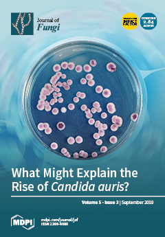
Cover Story (view full-size image): Candida auris is responsible for numerous severe healthcare-associated infections around the world, but its pre-emergence niche and the reasons behind this rapid emergence remain a mystery. This article explores factors that may have influenced its rise, including land use changes (e.g., agriculture and aquaculture), pesticide use, global travel, and changes in the environmental and human microbiome, with the aim of stimulating further research. View this paper.
  • Issues are regarded as officially published after their release is announced to the table of contents alert mailing list.
  • You may sign up for e-mail alerts to receive table of contents of newly released issues.
  • PDF is the official format for papers published in both, html and pdf forms. To view the papers in pdf format, click on the "PDF Full-text" link, and use the free Adobe Reader to open them.
Order results
Result details
Section
Select all
Export citation of selected articles as:
9 pages, 461 KiB  
Communication
Diagnostic Performance of a Novel Multiplex PCR Assay for Candidemia among ICU Patients
by Stefan Fuchs, Cornelia Lass-Flörl and Wilfried Posch
J. Fungi 2019, 5(3), 86; https://doi.org/10.3390/jof5030086 - 17 Sep 2019
Cited by 24 | Viewed by 4060
Abstract
Candidemia poses a major threat to ICU patients and is routinely diagnosed by blood culture, which is known for its low sensitivity and long turnaround times. We compared the performance of a novel, Candida-specific multiplex real-time PCR assay (Fungiplex® Candida IVD [...] Read more.
Candidemia poses a major threat to ICU patients and is routinely diagnosed by blood culture, which is known for its low sensitivity and long turnaround times. We compared the performance of a novel, Candida-specific multiplex real-time PCR assay (Fungiplex® Candida IVD Real-Time PCR Kit) with blood culture and another established diagnostic real-time PCR assay (LightCycler SeptiFast Test) with respect to Candida detection from whole blood samples. Clinical samples from 58 patients were analyzed by standard blood culture (BC) and simultaneously tested with the Fungiplex Candida PCR (FP) and the SeptiFast test (SF) for molecular detection of Candida spp. Compared to BC, the FP test showed high diagnostic power, with a sensitivity of 100% and a specificity of 94.1%. Overall diagnostic accuracy reached 94.6%. Using SF, we found a sensitivity of 60%, a specificity of 96.1%, and an overall diagnostic accuracy of 92.9%. The Fungiplex Candida PCR has shown good sensitivity and specificity on clinical samples of high-risk patients for direct detection of Candida species in whole blood samples. Together with conventional diagnostics (BC and antigen testing), this new multiplex PCR assay may contribute to a rapid and accurate diagnosis of candidiasis. Full article
(This article belongs to the Special Issue Molecular Diagnostics of Fungal Infections)
Show Figures

Figure 1

8 pages, 412 KiB  
Brief Report
Sweet Relief: Determining the Antimicrobial Activity of Medical Grade Honey Against Vaginal Isolates of Candida albicans
by Renée Hermanns, Niels A.J. Cremers, John P. Leeming and Esther T. van der Werf
J. Fungi 2019, 5(3), 85; https://doi.org/10.3390/jof5030085 - 09 Sep 2019
Cited by 24 | Viewed by 4638
Abstract
Recurrent vulvovaginal candidiasis (RVVC) is predicted to increase to almost 158 million cases annually by 2030. Extensive self-diagnosis and easily accessible over-the-counter (OTC) fungistatic drugs contribute to antifungal-resistance, illustrating the need for novel therapies. Honey possesses multiple antimicrobial mechanisms, and there is no [...] Read more.
Recurrent vulvovaginal candidiasis (RVVC) is predicted to increase to almost 158 million cases annually by 2030. Extensive self-diagnosis and easily accessible over-the-counter (OTC) fungistatic drugs contribute to antifungal-resistance, illustrating the need for novel therapies. Honey possesses multiple antimicrobial mechanisms, and there is no antimicrobial resistance towards honey reported. We evaluated the susceptibility of five clinical isolates of Candida albicans and a control strain to regular honey and a medical grade honey (MGH) gel formulation (L-Mesitran, containing 40% honey and vitamins C and E) using an adapted version of the EUCAST protocol at pH 5.2, 4.6, and 4.0. 40% regular honey did not kill or inhibit C. albicans. In contrast, the minimal inhibitory concentration (MIC) of L-Mesitran was 25%–50%, while fungicidal effects occurred at a 50% concentration (MBC) of the MGH formulation, except for one strain which was not killed at pH 4.0. Overall, pH had little effect on antimicrobial activity. MGH formulation L-Mesitran has antimicrobial activity against C. albicans over a relevant pH range. The vitamin supplements or other components of L-Mesitran may enhance the antifungal activity of the honey. This study supports performing clinical trials for conditions, such as RVVC, to find an alternative to available OTC fungistatic drugs. Full article
(This article belongs to the Special Issue Antifungal Drug Development: Rearview and the Horizon)
Show Figures

Figure 1

8 pages, 501 KiB  
Article
Hospital Laboratory Survey for Identification of Candida auris in Belgium
by Klaas Dewaele, Katrien Lagrou, Johan Frans, Marie-Pierre Hayette and Kris Vernelen
J. Fungi 2019, 5(3), 84; https://doi.org/10.3390/jof5030084 - 05 Sep 2019
Cited by 15 | Viewed by 3947
Abstract
Candida auris is a difficult-to-identify, emerging yeast and a cause of sustained nosocomial outbreaks. Presently, not much data exist on laboratory preparedness in Europe. To assess the ability of laboratories in Belgium and Luxembourg to detect this species, a blinded C. auris strain [...] Read more.
Candida auris is a difficult-to-identify, emerging yeast and a cause of sustained nosocomial outbreaks. Presently, not much data exist on laboratory preparedness in Europe. To assess the ability of laboratories in Belgium and Luxembourg to detect this species, a blinded C. auris strain was included in the regular proficiency testing rounds organized by the Belgian public health institute, Sciensano. Laboratories were asked to identify and report the isolate as they would in routine clinical practice, as if grown from a blood culture. Of 142 respondents, 82 (57.7%) obtained a correct identification of C. auris. Of 142 respondents, 27 (19.0%) identified the strain as Candida haemulonii. The remaining labs that did not obtain a correct identification (33/142, 23.2%), reported other yeast species (4/33) or failed to obtain a species identification (29/33). To assess awareness about the infection-control implications of the identification, participants were requested to indicate whether referral of this isolate to a reference laboratory was desirable in a clinical context. Over one-third of all respondents (54/142, 38.0%) stated that they would not send the isolate to a reference laboratory, neither for epidemiological reasons nor for identification confirmation or antifungal susceptibility testing. This comprised 41.5% of the laboratories that submitted an identification of C. auris (34/82). Awareness among Belgian microbiologists, therefore, remains inadequate more than two years after C. auris’ emergence in European clinics. Our data confirm high rates of misidentifications with commonly used identification methods. Programs for raising awareness in European hospitals may be warranted. Full article
(This article belongs to the Special Issue Candida auris)
Show Figures

Figure 1

12 pages, 1890 KiB  
Article
Minimal Inhibitory Concentration (MIC)-Phenomena in Candida albicans and Their Impact on the Diagnosis of Antifungal Resistance
by Ulrike Binder, Maria Aigner, Brigitte Risslegger, Caroline Hörtnagl, Cornelia Lass-Flörl and Michaela Lackner
J. Fungi 2019, 5(3), 83; https://doi.org/10.3390/jof5030083 - 04 Sep 2019
Cited by 11 | Viewed by 3821
Abstract
Antifungal susceptibility testing (AFST) of clinical isolates is a tool in routine diagnostics to facilitate decision making on optimal antifungal therapy. The minimal inhibitory concentration (MIC)-phenomena (trailing and paradoxical effects (PXE)) observed in AFST complicate the unambiguous and reproducible determination of MICs and [...] Read more.
Antifungal susceptibility testing (AFST) of clinical isolates is a tool in routine diagnostics to facilitate decision making on optimal antifungal therapy. The minimal inhibitory concentration (MIC)-phenomena (trailing and paradoxical effects (PXE)) observed in AFST complicate the unambiguous and reproducible determination of MICs and the impact of these phenomena on in vivo outcome are not fully understood. We aimed to link the MIC-phenomena with in vivo treatment response using the alternative infection model Galleria mellonella. We found that Candida albicans strains exhibiting PXE for caspofungin (CAS) had variable treatment outcomes in the Galleria model. In contrast, C. albicans strains showing trailing for voriconazole failed to respond in vivo. Caspofungin- and voriconazole-susceptible C. albicans strains responded to the respective antifungal therapy in vivo. In conclusion, MIC data and subsequent susceptibility interpretation of strains exhibiting PXE and/or trailing should be carried out with caution, as both effects are linked to drug adaptation and treatment response is uncertain to predict. Full article
(This article belongs to the Special Issue Molecular Diagnostics of Fungal Infections)
Show Figures

Figure 1

12 pages, 723 KiB  
Article
A Week of Oral Terbinafine Pulse Regimen Every Three Months to Treat all Dermatophyte Onychomycosis
by Anarosa B. Sprenger, Katia Sheylla Malta Purim, Flávia Sprenger and Flávio Queiroz-Telles
J. Fungi 2019, 5(3), 82; https://doi.org/10.3390/jof5030082 - 04 Sep 2019
Cited by 5 | Viewed by 5993
Abstract
Terbinafine has proved to treat numerous fungal infections, including onychomycosis, successfully. Due to its liver metabolization and dependency on the cytochrome P450 enzyme complex, undesirable drug interaction are highly probable. Additionally to drug interactions, the treatment is long, rising the chances of the [...] Read more.
Terbinafine has proved to treat numerous fungal infections, including onychomycosis, successfully. Due to its liver metabolization and dependency on the cytochrome P450 enzyme complex, undesirable drug interaction are highly probable. Additionally to drug interactions, the treatment is long, rising the chances of the appearance of side effects and abandonment. Pharmacokinetic data suggest that terbinafine maintains a fungicidal effect within the nail up to 30 weeks after its last administration, which has aroused the possibility of a pulse therapy to reduce the side effects while treating onychomycosis. This study’s goal was to evaluate the effectiveness of three different oral terbinafine regimens in treating onychomycosis due to dermatophytes. Sixty-three patients with onychomycosis were sorted by convenience in three different groups. Patients from group 1 received the conventional terbinafine dose (250 mg per day for 3 months). Group 2 received a monthly week-long pulse-therapy dose (500 mg per day for 7 days a month, for 4 months) and group 3 received a 500 mg/day dose for 7 days every 3 months, totaling four treatments. There were no statistical differences regarding the effectiveness or side effects between the groups. Conclusion: A quarterly terbinafine pulse regimen can be a possible alternative for treating onychomycosis caused by dermatophytes. Full article
Show Figures

Figure 1

14 pages, 1066 KiB  
Review
Candida albicans and Staphylococcus aureus Pathogenicity and Polymicrobial Interactions: Lessons beyond Koch’s Postulates
by Olivia A. Todd and Brian M Peters
J. Fungi 2019, 5(3), 81; https://doi.org/10.3390/jof5030081 - 04 Sep 2019
Cited by 45 | Viewed by 6154
Abstract
While Koch’s Postulates have established rules for microbial pathogenesis that have been extremely beneficial for monomicrobial infections, new studies regarding polymicrobial pathogenesis defy these standards. The explosion of phylogenetic sequence data has revolutionized concepts of microbial interactions on and within the host. However, [...] Read more.
While Koch’s Postulates have established rules for microbial pathogenesis that have been extremely beneficial for monomicrobial infections, new studies regarding polymicrobial pathogenesis defy these standards. The explosion of phylogenetic sequence data has revolutionized concepts of microbial interactions on and within the host. However, there remains a paucity of functional follow-up studies to delineate mechanisms driven by such interactions and how they shape health or disease. That said, one particular microbial pairing, the fungal opportunist Candida albicans and the bacterial pathogen Staphylococcus aureus, has received much attention over the last decade. Therefore, the objective of this review is to discuss the multi-faceted mechanisms employed by these two ubiquitous human pathogens during polymicrobial growth, including how they: establish and persist in inter-Kingdom biofilms, tolerate antimicrobial therapy, co-invade host tissue, exacerbate quorum sensing and staphylococcal toxin production, and elicit infectious synergism. Commentary regarding new challenges and remaining questions related to future discovery of this fascinating fungal–bacterial interaction is also provided. Full article
Show Figures

Figure 1

7 pages, 240 KiB  
Communication
Getting Histoplasmosis on the Map of International Recommendations for Patients with Advanced HIV Disease
by Felix Bongomin, Richard Kwizera and David W. Denning
J. Fungi 2019, 5(3), 80; https://doi.org/10.3390/jof5030080 - 02 Sep 2019
Cited by 23 | Viewed by 4079
Abstract
Progressive disseminated histoplasmosis, caused by H. capsulatum, is a life-threatening illness and is an AIDS-defining opportunistic infection. It is neglected, worryingly under-diagnosed, and often misdiagnosed as cancer or tuberculosis with fatal consequences. Globally, over 100,000 cases of disseminated histoplasmosis have been estimated. [...] Read more.
Progressive disseminated histoplasmosis, caused by H. capsulatum, is a life-threatening illness and is an AIDS-defining opportunistic infection. It is neglected, worryingly under-diagnosed, and often misdiagnosed as cancer or tuberculosis with fatal consequences. Globally, over 100,000 cases of disseminated histoplasmosis have been estimated. In 2017, the World Health Organization (WHO) noted that disseminated histoplasmosis is a significant cause of mortality in AIDS patients. Through the rigorous efforts of the Global Action Fund for Fungal Infections (GAFFI) and partners, in 2019, the Histoplasma antigen test was included on the 2nd Edition of the WHO List of Essential Diagnostics. The drugs used in the treatment of histoplasmosis (amphotericin B and itraconazole) are on the WHO Essential Medicine List. The Manaus Declaration on histoplasmosis in the Americas and the Caribbean, where histoplasmosis kills more people with HIV than tuberculosis, advocates for universal access to rapid testing for histoplasmosis and availability of essential drugs for the treatment of histoplasmosis in every country by 2025. Hyperendemic areas are present in the Americas, Caribbean, Southeast Asia, and Latin America. In conclusion, histoplasmosis remains an important clinical and public health problem. To reduce HIV-associated mortality, disseminated histoplasmosis must be addressed through advocacy, increased awareness, and universal access to essential diagnostics and antifungal agents. Full article
(This article belongs to the Special Issue Histoplasma and Histoplasmosis)
8 pages, 470 KiB  
Review
Central Nervous System Infections Due to Aspergillus and Other Hyaline Molds
by Marisa H. Miceli
J. Fungi 2019, 5(3), 79; https://doi.org/10.3390/jof5030079 - 30 Aug 2019
Cited by 25 | Viewed by 4712
Abstract
Central nervous system infections due to Aspergillus spp and other hyaline molds such as Fusarium and Scedosporium spp are rare but fatal conditions. Invasion of the central nervous system (CNS) tends to occur as a result of hematogenous dissemination among immunocompromised patients, and [...] Read more.
Central nervous system infections due to Aspergillus spp and other hyaline molds such as Fusarium and Scedosporium spp are rare but fatal conditions. Invasion of the central nervous system (CNS) tends to occur as a result of hematogenous dissemination among immunocompromised patients, and by local extension or direct inoculation secondary to trauma in immunocompetent hosts. Efforts should be directed to confirm the diagnosis by image-guided stereotactic brain biopsy when feasible. Non-culture methods could be useful to support the diagnosis, but they have not been validated to be performed in cerebral spinal fluid. Treatment of these infections is challenging given the variable susceptibility profile of these pathogens and the penetration of antifungal agents into the brain. Full article
(This article belongs to the Special Issue Fungal Infections of the Central Nervous System)
Show Figures

Figure 1

11 pages, 430 KiB  
Article
Fungal Diseases in Taiwan—National Insurance Data and Estimation
by Yu-Shan Huang, David W. Denning, Shu-Man Shih, Chao A. Hsiung, Un-In Wu, Hsin-Yun Sun, Pao-Yu Chen, Yee-Chun Chen and Shan-Chwen Chang
J. Fungi 2019, 5(3), 78; https://doi.org/10.3390/jof5030078 - 21 Aug 2019
Cited by 7 | Viewed by 3431
Abstract
The burden of fungal diseases based on the real-world national data is limited. This study aimed to estimate the Taiwan incident cases with selected fungal diseases in 2013 using the National Health Insurance Research Database (NHIRD) which covered 99.6% of the 23.4 million [...] Read more.
The burden of fungal diseases based on the real-world national data is limited. This study aimed to estimate the Taiwan incident cases with selected fungal diseases in 2013 using the National Health Insurance Research Database (NHIRD) which covered 99.6% of the 23.4 million population. Over 80,000 incident cases were found and the majority were superficial infections including vulvovaginal candidiasis (477 per 100,000 adult women) and oral candidiasis (90 cases per 100,000 population). Common potentially life-threating fungal diseases were Pneumocystis pneumonia (5.35 cases per 100,000 population), candidemia (3.68), aspergillosis (2.43) and cryptococcal meningitis (1.04). Of the aforementioned cases cancer patients contributed 30.2%, 42.9%, 38.6% and 22.2%, respectively. Of 22,270 HIV-infected persons in NHIRD in 2013, four common diseases were Pneumocystis pneumonia (28.3 cases per 1000 HIV-infected patients), oral candidiasis (17.6), esophageal candidiasis (6.06) and cryptococcal meningitis (2.29). Of pulmonary aspergillosis 32.9% occurred in patients with chronic pulmonary diseases and 26.3% had a prior diagnosis of tuberculosis. There are some notable gaps related to insurance claim data. Cutaneous, urinary tract and eye fungal infections were not captured. Full article
(This article belongs to the Special Issue Fungal Epidemiology)
Show Figures

Figure 1

9 pages, 212 KiB  
Review
Molecular Diagnostics in the Times of Surveillance for Candida auris
by Milena Kordalewska and David S. Perlin
J. Fungi 2019, 5(3), 77; https://doi.org/10.3390/jof5030077 - 20 Aug 2019
Cited by 19 | Viewed by 3795
Abstract
Recently, global health professionals have been significantly challenged by the emergence of Candida auris and its propensity to colonize human skin, persist in the healthcare environment, and cause healthcare-associated outbreaks. Additionally, C. auris isolates are often characterized by elevated minimal inhibitory concentration (MIC) [...] Read more.
Recently, global health professionals have been significantly challenged by the emergence of Candida auris and its propensity to colonize human skin, persist in the healthcare environment, and cause healthcare-associated outbreaks. Additionally, C. auris isolates are often characterized by elevated minimal inhibitory concentration (MIC) values for antifungal drugs. Thus, rapid detection and accurate identification of C. auris together with an assessment of potential antifungal drug resistance has become essential for effective patient management, and infection prevention and control in healthcare facilities. Surprisingly, almost all of the commonly available diagnostic tools rely on recovery (growth) of yeast colonies from collected samples, which delays the diagnostic result by several days or longer. To circumvent these issues, molecular-based DNA amplification assays have been developed to identify C. auris DNA directly from patient samples. Moreover, allele discriminating detection probes can be used to rapidly assess validated mechanisms of echinocandin and azole resistance. Full article
(This article belongs to the Special Issue Candida auris)
13 pages, 4096 KiB  
Article
Diagnosis of Progressive Disseminated Histoplasmosis in Advanced HIV: A Meta-Analysis of Assay Analytical Performance
by Diego H. Caceres, Martha Knuth, Gordana Derado and Mark D. Lindsley
J. Fungi 2019, 5(3), 76; https://doi.org/10.3390/jof5030076 - 18 Aug 2019
Cited by 39 | Viewed by 4891
Abstract
Histoplasmosis is an important cause of mortality in people with advanced HIV, especially in countries with limited access to diagnostic assays. Histoplasmosis can be diagnosed using culture, histopathology, and antibody, antigen, and molecular assays. Several factors may affect the analytical performance of these [...] Read more.
Histoplasmosis is an important cause of mortality in people with advanced HIV, especially in countries with limited access to diagnostic assays. Histoplasmosis can be diagnosed using culture, histopathology, and antibody, antigen, and molecular assays. Several factors may affect the analytical performance of these laboratory assays, including sample type, clinical stage of the disease, and previous use of antifungal treatment, among others. Here we describe the results of a systematic literature review, followed by a meta-analysis of the analytical performances of the diagnostic laboratory assays employed. Our initial search identified 1631 references, of which 1559 references were excluded after title and abstract screening, leaving 72 references identified as studies relevant to the validation of histoplasmosis diagnostic assays. After evaluating the full text, 30 studies were selected for final review, including one paper not identified in the initial search. The meta-analysis for assay analytical performance shows the following results for the overall sensitivity (Sen) and specificity (Spe) of the various methods evaluated: Culture, Sen 77% (no data for specificity calculation); antibody detection assays, Sen 58%/Spe 100%; antigen detection assays, Sen 95%/Spe 97%; and DNA detection assays (molecular), Sen 95%/Spe 99%. Of the 30 studies reviewed, nearly half (n = 13) evaluated Histoplasma antigen assays, which were determined to be the most accurate methodology for diagnosis of progressive disseminated histoplasmosis in advanced HIV (inverse of the negative likelihood ratio was 13.2). Molecular assays appear promising for accurate diagnosis of histoplasmosis, but consensus on exact techniques is needed. Cultures showed variable sensitivity related to sample type and laboratory handling. Finally, antibody assays presented high specificity but low sensitivity. This poor sensitivity is most likely due the highly immunosuppressed state of this patient population. Diagnostic assays are crucial for accurate diagnosis of progressive disseminated histoplasmosis (PDH) with advanced HIV disease. Full article
(This article belongs to the Special Issue Fungal Epidemiology)
Show Figures

Figure 1

15 pages, 2165 KiB  
Article
Estimated Burden of Fungal Infections in Namibia
by Cara M. Dunaiski and David W. Denning
J. Fungi 2019, 5(3), 75; https://doi.org/10.3390/jof5030075 - 16 Aug 2019
Cited by 11 | Viewed by 4012
Abstract
Namibia is a sub-Saharan country with one of the highest HIV infection rates in the world. Although care and support services are available that cater for opportunistic infections related to HIV, the main focus is narrow and predominantly aimed at tuberculosis. We aimed [...] Read more.
Namibia is a sub-Saharan country with one of the highest HIV infection rates in the world. Although care and support services are available that cater for opportunistic infections related to HIV, the main focus is narrow and predominantly aimed at tuberculosis. We aimed to estimate the burden of serious fungal infections in Namibia, currently unknown, based on the size of the population at risk and available epidemiological data. Data were obtained from the World Health Organization (WHO), Joint United Nations Programme on HIV/AIDS (UNAIDS), and published reports. When no data existed, risk populations were used to estimate the frequencies of fungal infections, using the previously described methodology. The population of Namibia in 2011 was estimated at 2,459,000 and 37% were children. Among approximately 516,390 adult women, recurrent vulvovaginal candidiasis (≥4 episodes /year) is estimated to occur in 37,390 (3003/100,000 females). Using a low international average rate of 5/100,000, we estimated 125 cases of candidemia, and 19 patients with intra-abdominal candidiasis. Among survivors of pulmonary tuberculosis (TB) in Namibia 2017, 112 new cases of chronic pulmonary aspergillosis (CPA) are likely, a prevalence of 354 post-TB and a total prevalence estimate of 453 CPA patients in all. Asthma affects 11.2% of adults, 178,483 people, and so allergic bronchopulmonary aspergillosis (ABPA) and severe asthma with fungal sensitization (SAFS) were estimated in approximately 179/100,000 and 237/100,000 people, respectively. Invasive aspergillosis (IA) is estimated to affect 15 patients following leukaemia therapy, and an estimated 0.13% patients admitted to hospital with chronic obstructive pulmonary disease (COPD) (259) and 4% of HIV-related deaths (108) — a total of 383 people. The total HIV-infected population is estimated at 200,000, with 32,371 not on antiretroviral therapy (ART). Among HIV-infected patients, 543 cases of cryptococcal meningitis and 836 cases of Pneumocystis pneumonia are estimated each year. Tinea capitis infections were estimated at 53,784 cases, and mucormycosis at five cases. Data were missing for fungal keratitis and skin neglected fungal tropical diseases such as mycetoma. The present study indicates that approximately 5% of the Namibian population is affected by fungal infections. This study is not an epidemiological study—it illustrates estimates based on assumptions derived from similar studies. The estimates are incomplete and need further epidemiological and diagnostic studies to corroborate, amend them, and improve the diagnosis and management of these diseases. Full article
Show Figures

Figure 1

11 pages, 1146 KiB  
Article
First Analysis of Human Coccidioides Isolates from New Mexico and the Southwest Four Corners Region: Implications for the Distributions of C. posadasii and C. immitis and Human Groups at Risk
by Paris S. Hamm, Miriam I. Hutchison, Pascale Leonard, Sandra Melman and Donald O. Natvig
J. Fungi 2019, 5(3), 74; https://doi.org/10.3390/jof5030074 - 10 Aug 2019
Cited by 9 | Viewed by 4341
Abstract
Coccidioidomycosis (Valley Fever) is a disease caused by species of Coccidioides. The disease is endemic to arid regions of the Southwestern US and while most common in CA and AZ is also present in NM. We present the first genetic analysis of [...] Read more.
Coccidioidomycosis (Valley Fever) is a disease caused by species of Coccidioides. The disease is endemic to arid regions of the Southwestern US and while most common in CA and AZ is also present in NM. We present the first genetic analysis of clinical isolates from NM. Travel and demographic information was available for a number of patients, which included individuals from NM and the Southwestern US Four Corners region. Multi-gene phylogenetic analyses revealed the presence of both C. posadasii and C. immitis. While NM is predicted to be within the endemic range for C. posadasii, our results expand the known range of C. immitis, often considered to be the “California species”. Five of eight infections for which patient ethnicity existed occurred in Native Americans, and two occurred in African Americans. Several isolates came from the northwestern part of NM—outside the predicted “highly-endemic” region. Our study suggests Native Americans represent an unrecognized at-risk group, and it provides a foundation for better defining the geographic distribution of the Coccidioides species and for preventing exposure among populations at risk. In the course of this study, we developed a reliable PCR-based method to distinguish species targeting regions of the mitochondrial genome. Full article
Show Figures

Graphical abstract

10 pages, 630 KiB  
Review
Histoplasmosis and Tuberculosis Co-Occurrence in People with Advanced HIV
by Diego H. Caceres and Audrey Valdes
J. Fungi 2019, 5(3), 73; https://doi.org/10.3390/jof5030073 - 09 Aug 2019
Cited by 29 | Viewed by 5447
Abstract
Distinguishing between histoplasmosis, tuberculosis (TB), and co-occurrence of disease is a frequent dilemma for clinical staff treating people with advanced Human Immunodeficiency Virus (HIV) infection. This problem is most frequently observed in clinical settings in countries where both diseases are endemic. It is [...] Read more.
Distinguishing between histoplasmosis, tuberculosis (TB), and co-occurrence of disease is a frequent dilemma for clinical staff treating people with advanced Human Immunodeficiency Virus (HIV) infection. This problem is most frequently observed in clinical settings in countries where both diseases are endemic. It is also a challenge outside these endemic countries in HIV clinics that take care of patients coming from countries with endemic histoplasmosis and TB. The gold standard for diagnosis of both of these diseases is based on conventional laboratory tests (culture, histopathology and special stains). These tests have several limitations, such as lack of laboratory infrastructure for handling isolates (biosafety level 3), shortage of laboratory staff who have appropriate training and experience, variable analytical performance of tests and long turn-around time. Recently, novel rapid assays for the diagnosis of histoplasmosis and TB became available. However, this technology is not yet widely used. Mortality in immunocompromised patients, such as people with advanced HIV, is directly linked with the ability to rapidly diagnose opportunistic diseases. The aim of this review is to synthesize the main aspects of epidemiology, clinical characteristics, diagnosis and treatment of histoplasmosis/TB co-occurrence in people with advanced HIV. Full article
(This article belongs to the Special Issue Histoplasma and Histoplasmosis)
Show Figures

Figure 1

13 pages, 5652 KiB  
Article
Appressorium: The Breakthrough in Dikarya
by Alexander Demoor, Philippe Silar and Sylvain Brun
J. Fungi 2019, 5(3), 72; https://doi.org/10.3390/jof5030072 - 03 Aug 2019
Cited by 25 | Viewed by 5008
Abstract
Phytopathogenic and mycorrhizal fungi often penetrate living hosts by using appressoria and related structures. The differentiation of similar structures in saprotrophic fungi to penetrate dead plant biomass has seldom been investigated and has been reported only in the model fungus Podospora anserina. [...] Read more.
Phytopathogenic and mycorrhizal fungi often penetrate living hosts by using appressoria and related structures. The differentiation of similar structures in saprotrophic fungi to penetrate dead plant biomass has seldom been investigated and has been reported only in the model fungus Podospora anserina. Here, we report on the ability of many saprotrophs from a large range of taxa to produce appressoria on cellophane. Most Ascomycota and Basidiomycota were able to form appressoria. In contrast, none of the three investigated Mucoromycotina was able to differentiate such structures. The ability of filamentous fungi to differentiate appressoria no longer belongs solely to pathogenic or mutualistic fungi, and this raises the question of the evolutionary origin of the appressorium in Eumycetes. Full article
Show Figures

Graphical abstract

23 pages, 312 KiB  
Review
Central Nervous System Cryptococcal Infections in Non-HIV Infected Patients
by Justin Beardsley, Tania C. Sorrell and Sharon C.-A. Chen
J. Fungi 2019, 5(3), 71; https://doi.org/10.3390/jof5030071 - 02 Aug 2019
Cited by 64 | Viewed by 8072
Abstract
Central nervous system (CNS) cryptococcosis in non-HIV infected patients affects solid organ transplant (SOT) recipients, patients with malignancy, rheumatic disorders, other immunosuppressive conditions and immunocompetent hosts. More recently described risks include the use of newer biologicals and recreational intravenous drug use. Disease is [...] Read more.
Central nervous system (CNS) cryptococcosis in non-HIV infected patients affects solid organ transplant (SOT) recipients, patients with malignancy, rheumatic disorders, other immunosuppressive conditions and immunocompetent hosts. More recently described risks include the use of newer biologicals and recreational intravenous drug use. Disease is caused by Cryptococcus neoformans and Cryptococcus gattii species complex; C. gattii is endemic in several geographic regions and has caused outbreaks in North America. Major virulence determinants are the polysaccharide capsule, melanin and several ‘invasins’. Cryptococcal plb1, laccase and urease are essential for dissemination from lung to CNS and crossing the blood–brain barrier. Meningo-encephalitis is common but intracerebral infection or hydrocephalus also occur, and are relatively frequent in C. gattii infection. Complications include neurologic deficits, raised intracranial pressure (ICP) and disseminated disease. Diagnosis relies on culture, phenotypic identification methods, and cryptococcal antigen detection. Molecular methods can assist. Preferred induction antifungal therapy is a lipid amphotericin B formulation (amphotericin B deoxycholate may be used in non-transplant patients) plus 5-flucytosine for 2–6 weeks depending on host type followed by consolidation/maintenance therapy with fluconazole for 12 months or longer. Control of raised ICP is essential. Clinicians should be vigilant for immune reconstitution inflammatory syndrome. Full article
(This article belongs to the Special Issue Fungal Infections of the Central Nervous System)
10 pages, 385 KiB  
Review
Central Nervous System Infection with Histoplasma capsulatum
by James Riddell IV and L. Joseph Wheat
J. Fungi 2019, 5(3), 70; https://doi.org/10.3390/jof5030070 - 24 Jul 2019
Cited by 17 | Viewed by 7175
Abstract
Histoplasmosis is an endemic fungal infection that may affect both immune compromised and non-immune compromised individuals. It is now recognized that the geographic range of this organism is larger than previously understood, placing more people at risk. Infection with Histoplasma capsulatum may occur [...] Read more.
Histoplasmosis is an endemic fungal infection that may affect both immune compromised and non-immune compromised individuals. It is now recognized that the geographic range of this organism is larger than previously understood, placing more people at risk. Infection with Histoplasma capsulatum may occur after inhalation of conidia that are aerosolized from the filamentous form of the organism in the environment. Clinical syndromes typically associated with histoplasmosis include acute or chronic pneumonia, chronic cavitary pulmonary infection, or mediastinal fibrosis or lymphadenitis. Disseminated infection can also occur, in which multiple organ systems are affected. In up to 10% of cases, infection of the central nervous system (CNS) with histoplasmosis may occur with or without disseminated infection. In this review, we discuss challenges related to the diagnosis of CNS histoplasmosis and appropriate treatment strategies that can lead to successful outcomes. Full article
(This article belongs to the Special Issue Fungal Infections of the Central Nervous System)
Show Figures

Figure 1

25 pages, 1201 KiB  
Review
A Guide to Investigating Suspected Outbreaks of Mucormycosis in Healthcare
by Kathleen P. Hartnett, Brendan R. Jackson, Kiran M. Perkins, Janet Glowicz, Janna L. Kerins, Stephanie R. Black, Shawn R. Lockhart, Bryan E. Christensen and Karlyn D. Beer
J. Fungi 2019, 5(3), 69; https://doi.org/10.3390/jof5030069 - 24 Jul 2019
Cited by 43 | Viewed by 9841
Abstract
This report serves as a guide for investigating mucormycosis infections in healthcare. We describe lessons learned from previous outbreaks and offer methods and tools that can aid in these investigations. We also offer suggestions for conducting environmental assessments, implementing infection control measures, and [...] Read more.
This report serves as a guide for investigating mucormycosis infections in healthcare. We describe lessons learned from previous outbreaks and offer methods and tools that can aid in these investigations. We also offer suggestions for conducting environmental assessments, implementing infection control measures, and initiating surveillance to ensure that interventions were effective. While not all investigations of mucormycosis infections will identify a single source, all can potentially lead to improvements in infection control. Full article
(This article belongs to the Special Issue Fungal Epidemiology)
Show Figures

Figure 1

8 pages, 1714 KiB  
Article
The Burden of Serious Fungal Infections in Tajikistan
by Oktam I. Bobokhojaev, Ali Osmanov, Samariddin P. Aliev, Asliddin S. Radjabzoda, Ziyovuddin T. Avgonov, Safarbek T. Manonov and David W. Denning
J. Fungi 2019, 5(3), 68; https://doi.org/10.3390/jof5030068 - 21 Jul 2019
Cited by 4 | Viewed by 3237
Abstract
Tajikistan is a low-income country in Middle Asia with a population of 8.9 million people. Five percent of the population lives on less than 1.9 USD a day and 54% live on less than 5.5 USD a day. We have estimated the burden [...] Read more.
Tajikistan is a low-income country in Middle Asia with a population of 8.9 million people. Five percent of the population lives on less than 1.9 USD a day and 54% live on less than 5.5 USD a day. We have estimated the burden of serious fungal infections in Tajikistan. It was estimated that 168,834 Tajik women develop recurrent vulvovaginal candidiasis. Among HIV-positive patients, we estimate 490 patients with oesophageal candidiasis and 1260 patients with oral candidiasis, 41 cases of cryptococcal meningitis and 210 cases of Pneumocystis pneumonia annually. According to our estimations there are 774 cases of chronic pulmonary aspergillosis (CPA) as a sequel of tuberculosis; CPA may occur as a consequence of multiple pulmonary conditions and the total prevalence of 4161 cases was estimated. We have estimated 6008 cased of allergic bronchopulmonary aspergillosis (ABPA) and 7930 cases of severe asthma with fungal sensitisation (SAFS), and 137 fungal asthma deaths annually. We have estimated 445 cases of candidemia a year applying a low European rate. There are approximately 283 cases of invasive aspergillosis annually. There are 189,662 (2.1% of the population) people suffering from serious fungal infections in Tajikistan. Hence, improving diagnostics is the first step of understanding a scale of the fungal burden. Full article
Show Figures

Figure 1

19 pages, 2142 KiB  
Review
Emerging Fungal Infections: New Patients, New Patterns, and New Pathogens
by Daniel Z.P. Friedman and Ilan S. Schwartz
J. Fungi 2019, 5(3), 67; https://doi.org/10.3390/jof5030067 - 20 Jul 2019
Cited by 208 | Viewed by 21107
Abstract
The landscape of clinical mycology is constantly changing. New therapies for malignant and autoimmune diseases have led to new risk factors for unusual mycoses. Invasive candidiasis is increasingly caused by non-albicans Candida spp., including C. auris, a multidrug-resistant yeast with the potential [...] Read more.
The landscape of clinical mycology is constantly changing. New therapies for malignant and autoimmune diseases have led to new risk factors for unusual mycoses. Invasive candidiasis is increasingly caused by non-albicans Candida spp., including C. auris, a multidrug-resistant yeast with the potential for nosocomial transmission that has rapidly spread globally. The use of mould-active antifungal prophylaxis in patients with cancer or transplantation has decreased the incidence of invasive fungal disease, but shifted the balance of mould disease in these patients to those from non-fumigatus Aspergillus species, Mucorales, and Scedosporium/Lomentospora spp. The agricultural application of triazole pesticides has driven an emergence of azole-resistant A. fumigatus in environmental and clinical isolates. The widespread use of topical antifungals with corticosteroids in India has resulted in Trichophyton mentagrophytes causing recalcitrant dermatophytosis. New dimorphic fungal pathogens have emerged, including Emergomyces, which cause disseminated mycoses globally, primarily in HIV infected patients, and Blastomyces helicus and B. percursus, causes of atypical blastomycosis in western parts of North America and in Africa, respectively. In North America, regions of geographic risk for coccidioidomycosis, histoplasmosis, and blastomycosis have expanded, possibly related to climate change. In Brazil, zoonotic sporotrichosis caused by Sporothrix brasiliensis has emerged as an important disease of felines and people. Full article
(This article belongs to the Special Issue Fungal Epidemiology)
Show Figures

Figure 1

7 pages, 2016 KiB  
Article
The Burden of Serious Fungal Infections in Kyrgyzstan
by Gulnura K. Turdumambetova, Ali Osmanov and David W. Denning
J. Fungi 2019, 5(3), 66; https://doi.org/10.3390/jof5030066 - 19 Jul 2019
Cited by 4 | Viewed by 3421
Abstract
Kyrgyzstan in Central Asia has a population of 6 million people who have high mortality rates for chronic lung diseases. The mountainous geography, widespread use of biomass fuels for cooking and indoor heating, and high rates of smoking are the major contributing factors. [...] Read more.
Kyrgyzstan in Central Asia has a population of 6 million people who have high mortality rates for chronic lung diseases. The mountainous geography, widespread use of biomass fuels for cooking and indoor heating, and high rates of smoking are the major contributing factors. We have estimated the number of serious fungal infections in order to define the burden of these diseases in Kyrgyzstan. We estimated 774 cases of chronic pulmonary aspergillosis (CPA) as a sequel of tuberculosis (TB); CPA occurs as a sequel of multiple conditions, so a total prevalence of 3097 cases was estimated, which is among the highest rates in the world. An estimated 2205 patients have allergic bronchopulmonary aspergillosis (ABPA) and 2911 have severe asthma with fungal sensitization (SAFS), which may be an underestimate. There are approximately 292 cases of invasive aspergillosis annually. The number of adult women who get recurrent vulvovaginal candidiasis is 175,949. We approximated 787 cases of oral and 294 cases of esophageal candidiasis, 25 cases of cryptococcal meningitis, and 101 cases of Pneumocystis pneumonia annually in HIV-positive patients. The incidence of candidemia was estimated at 300. We have estimated that a total of 185,961 people (3% of the population) have serious fungal infection in Kyrgyzstan. Given this burden, diagnostic improvements are necessary. Full article
Show Figures

Figure 1

17 pages, 2673 KiB  
Review
Diagnosis and Management of Central Nervous System Cryptococcal Infections in HIV-Infected Adults
by Caleb Skipper, Mahsa Abassi and David R Boulware
J. Fungi 2019, 5(3), 65; https://doi.org/10.3390/jof5030065 - 19 Jul 2019
Cited by 22 | Viewed by 5988
Abstract
Cryptococcal meningitis persists as a significant source of morbidity and mortality in persons with HIV/AIDS, particularly in sub-Saharan Africa. Despite increasing access to antiretrovirals, persons presenting with advanced HIV disease remains common, and Cryptococcus remains the most frequent etiology of adult meningitis. We [...] Read more.
Cryptococcal meningitis persists as a significant source of morbidity and mortality in persons with HIV/AIDS, particularly in sub-Saharan Africa. Despite increasing access to antiretrovirals, persons presenting with advanced HIV disease remains common, and Cryptococcus remains the most frequent etiology of adult meningitis. We performed a literature review and herein present the most up-to-date information on the diagnosis and management of cryptococcosis. Recent advances have dramatically improved the accessibility of timely and affordable diagnostics. The optimal initial antifungal management has been newly updated after the completion of a landmark clinical trial. Beyond antifungals, the control of intracranial pressure and mitigation of toxicities remain hallmarks of effective treatment. Cryptococcal meningitis continues to present challenging complications and continued research is needed. Full article
(This article belongs to the Special Issue Fungal Infections of the Central Nervous System)
Show Figures

Figure 1

15 pages, 252 KiB  
Review
Central Nervous System Infection with Other Endemic Mycoses: Rare Manifestation of Blastomycosis, Paracoccidioidomycosis, Talaromycosis, and Sporotrichosis
by Carol A. Kauffman
J. Fungi 2019, 5(3), 64; https://doi.org/10.3390/jof5030064 - 18 Jul 2019
Cited by 22 | Viewed by 4564
Abstract
The central nervous system (CNS) is not a major organ involved with infections caused by the endemic mycoses, with the possible exception of meningitis caused by Coccidioides species. When CNS infection does occur, the manifestations vary among the different endemic mycoses; mass-like lesions [...] Read more.
The central nervous system (CNS) is not a major organ involved with infections caused by the endemic mycoses, with the possible exception of meningitis caused by Coccidioides species. When CNS infection does occur, the manifestations vary among the different endemic mycoses; mass-like lesions or diffuse meningeal involvement can occur, and isolated chronic meningitis, as well as widely disseminated acute infection that includes the CNS, are described. This review includes CNS infection caused by Blastomyces dermatitidis, Paracoccidioides brasiliensis, Talaromyces marneffei, and the Sporothrix species complex. The latter is not geographically restricted, in contrast to the classic endemic mycoses, but it is similar in that it is a dimorphic fungus. CNS infection with B. dermatitidis can present as isolated chronic meningitis or a space-occupying lesion usually in immunocompetent hosts, or as one manifestation of widespread disseminated infection in patients who are immunosuppressed. P. brasiliensis more frequently causes mass-like intracerebral lesions than meningitis, and most often CNS disease is part of disseminated infection found primarily in older patients with the chronic form of paracoccidioidomycosis. T. marneffei is the least likely of the endemic mycoses to cause CNS infection. Almost all reported cases have been in patients with advanced HIV infection and almost all have had widespread disseminated infection. Sporotrichosis is known to cause isolated chronic meningitis, primarily in immunocompetent individuals who do not have Sporothrix involvement of other organs. In contrast, CNS infection in patients with advanced HIV infection occurs as part of widespread disseminated infection. Full article
(This article belongs to the Special Issue Fungal Infections of the Central Nervous System)
14 pages, 2056 KiB  
Article
Longitudinal Changes in CD4+, CD8+ T Cell Phenotype and Activation Marker Expression Following Antiretroviral Therapy Initiation among Patients with Cryptococcal Meningitis
by Alice Bayiyana, Samuel Okurut, Rose Nabatanzi, Godfrey Zziwa, David R. Boulware, Fredrick Lutwama and David Meya
J. Fungi 2019, 5(3), 63; https://doi.org/10.3390/jof5030063 - 17 Jul 2019
Cited by 3 | Viewed by 4297
Abstract
Despite improvement in the prognosis of HIV/AIDS (human immunodeficiency virus/acquired immune deficiency syndrome) patients on antiretroviral therapy (ART), cryptococcal meningitis (CM) still causes 10–15% mortality among HIV-infected patients. The immunological impact of ART on the CD4+ and CD8+ T cell repertoire [...] Read more.
Despite improvement in the prognosis of HIV/AIDS (human immunodeficiency virus/acquired immune deficiency syndrome) patients on antiretroviral therapy (ART), cryptococcal meningitis (CM) still causes 10–15% mortality among HIV-infected patients. The immunological impact of ART on the CD4+ and CD8+ T cell repertoire during cryptococcal co-infection is unclear. We determined longitudinal phenotypic changes in T cell subsets among patients with CM after they initiated ART. We hypothesized that ART alters the clonotypic phenotype and structural composition of CD4+ and CD8+ T cells during CM co-infection. For this substudy, peripheral blood mononuclear cells (PBMC) were isolated at four time points from CM patients following ART initiation during the parent study (ClinicalTrials.gov number, NCT01075152). Phenotypic characterization of CD4+ and CD8+ T cells was done using T cell surface marker monoclonal antibodies by flow cytometry. There was variation in the expression of immunophenotypic markers defining central memory (CD27+CD45R0+), effector memory (CD45R0+CD27), immune activation (CD38+ and Human Leucocyte Antigen DR (HLA-DR+), and exhaustion (Programmed cell death protein one (PD-1) in the CD4+ T cell subset. In comparison to the CD4+ T cell population, the CD8+ central memory subset declined gradually with minimal increase in the effector memory subset. Both CD4+ and CD8+ T cell immune exhaustion and activation markers remained elevated over 12 weeks. The relative surge and decline in the expression of T cell surface markers outlines a variation in the differentiation of CD4+ T cells during ART treatment during CM co-infection. Full article
Show Figures

Figure 1

13 pages, 2224 KiB  
Article
Structural Differences Influence Biological Properties of Glucosylceramides from Clinical and Environmental Isolates of Scedosporium aurantiacum and Pseudallescheria minutispora
by Adriana Caneppa, Jardel Vieira de Meirelles, Rodrigo Rollin-Pinheiro, Mariana Ingrid Dutra da Silva Xisto, Livia Cristina Liporagi-Lopes, Lauro de Souza, Maria Teresa Villela Romanos and Eliana Barreto-Bergter
J. Fungi 2019, 5(3), 62; https://doi.org/10.3390/jof5030062 - 15 Jul 2019
Cited by 7 | Viewed by 2817
Abstract
Scedosporium/Lomentospora complex is composed of filamentous fungi, including some clinically relevant species, such as Pseudallescheria boydii, Scedosporium aurantiacum, and Scedosporium apiospermum. Glucosylceramide (GlcCer), a conserved neutral glycosphingolipid, has been described as an important cell surface molecule playing a role in fungal [...] Read more.
Scedosporium/Lomentospora complex is composed of filamentous fungi, including some clinically relevant species, such as Pseudallescheria boydii, Scedosporium aurantiacum, and Scedosporium apiospermum. Glucosylceramide (GlcCer), a conserved neutral glycosphingolipid, has been described as an important cell surface molecule playing a role in fungal morphological transition and pathogenesis. The present work aimed at the evaluation of GlcCer structures in S. aurantiacum and Pseudallescheria minutispora, a clinical and an environmental isolate, respectively, in order to determine their participation in fungal growth and host-pathogen interactions. Structural analysis by positive ion-mode ESI-MS (electrospray ionization mass spectrometer) revealed the presence of different ceramide moieties in GlcCer in these species. Monoclonal antibodies against Aspergillus fumigatus GlcCer could recognize S. aurantiacum and P. minutispora conidia, suggesting a conserved epitope in fungal GlcCer. In addition, these antibodies reduced fungal viability, enhanced conidia phagocytosis by macrophages, and decreased fungal survival inside phagocytic cells. Purified GlcCer from both species led to macrophage activation, increasing cell viability as well as nitric oxide and superoxide production in different proportions between the two species. These results evidenced some important properties of GlcCer from species of the Scedosporium/Lomentospora complex, as well as the effects of monoclonal anti-GlcCer antibodies on fungal cells and host-pathogen interaction. The differences between the two species regarding the observed biological properties suggest that variation in GlcCer structures and strain origin could interfere in the role of GlcCer in host-pathogen interaction. Full article
(This article belongs to the Special Issue Lipids and Fungal Infectious Diseases)
Show Figures

Figure 1

5 pages, 964 KiB  
Communication
Hypoxia Decreases Diagnostic Biomarkers for Aspergillosis In Vitro
by Elisabeth Maurer, Maria Aigner, Cornelia Lass-Flörl and Ulrike Binder
J. Fungi 2019, 5(3), 61; https://doi.org/10.3390/jof5030061 - 11 Jul 2019
Cited by 1 | Viewed by 2785
Abstract
The aim of the study was to evaluate the influence of hypoxia on galactomannan and (1,3)-β-d-glucan release of clinically relevant Aspergilli in vitro. Hypoxia decreased biomass and consequently led to lower biomarker release. However, when normalized to biomass, hypoxia led to [...] Read more.
The aim of the study was to evaluate the influence of hypoxia on galactomannan and (1,3)-β-d-glucan release of clinically relevant Aspergilli in vitro. Hypoxia decreased biomass and consequently led to lower biomarker release. However, when normalized to biomass, hypoxia led to increased levels of biomarkers at early growth stages (24 h). Antifungals (amphotericin B and voriconazole) decreased the galactomannan amount of A. fumigatus, even more prominently in hypoxia. Full article
(This article belongs to the Special Issue Molecular Diagnostics of Fungal Infections)
Show Figures

Figure 1

11 pages, 238 KiB  
Review
CNS Infections Caused by Brown-Black Fungi
by Jon Velasco and Sanjay Revankar
J. Fungi 2019, 5(3), 60; https://doi.org/10.3390/jof5030060 - 10 Jul 2019
Cited by 27 | Viewed by 3907
Abstract
Central nervous system (CNS) infections caused by brown-black or dematiaceous fungi are distinctly rare and represent a small proportion of infections termed phaeohyphomycoses. However, these are becoming more commonly reported. Though many fungi have been implicated in disease, most cases are caused by [...] Read more.
Central nervous system (CNS) infections caused by brown-black or dematiaceous fungi are distinctly rare and represent a small proportion of infections termed phaeohyphomycoses. However, these are becoming more commonly reported. Though many fungi have been implicated in disease, most cases are caused by only a few species, Cladophialophora bantiana being the most common. Most of the fungi described are molds, and often cause infection in immunocompetent individuals, in contrast to infection with other more common molds such as Aspergillus, which is usually seen in highly immunocompromised patients. Diagnosis is challenging, as there are no specific tests for this group of fungi. In addition, these infections are often refractory to standard drug therapies, requiring an aggressive combined surgical and medical approach to improve outcomes, yet mortality remains high. There are no standardized treatments due to a lack of randomized clinical trials, though guidelines have been published based on available data and expert opinion. Full article
(This article belongs to the Special Issue Fungal Infections of the Central Nervous System)
20 pages, 545 KiB  
Review
Mucormycosis of the Central Nervous System
by Amanda Chikley, Ronen Ben-Ami and Dimitrios P Kontoyiannis
J. Fungi 2019, 5(3), 59; https://doi.org/10.3390/jof5030059 - 08 Jul 2019
Cited by 64 | Viewed by 5727
Abstract
Mucormycosis involves the central nervous system by direct extension from infected paranasal sinuses or hematogenous dissemination from the lungs. Incidence rates of this rare disease seem to be rising, with a shift from the rhino-orbital-cerebral syndrome typical of patients with diabetes mellitus and [...] Read more.
Mucormycosis involves the central nervous system by direct extension from infected paranasal sinuses or hematogenous dissemination from the lungs. Incidence rates of this rare disease seem to be rising, with a shift from the rhino-orbital-cerebral syndrome typical of patients with diabetes mellitus and ketoacidosis, to disseminated disease in patients with hematological malignancies. We present our current understanding of the pathobiology, clinical features, and diagnostic and treatment strategies of cerebral mucormycosis. Despite advances in imaging and the availability of novel drugs, cerebral mucormycosis continues to be associated with high rates of death and disability. Emerging molecular diagnostics, advances in experimental systems and the establishment of large patient registries are key components of ongoing efforts to provide a timely diagnosis and effective treatment to patients with cerebral mucormycosis. Full article
(This article belongs to the Special Issue Fungal Infections of the Central Nervous System)
Show Figures

Figure 1

17 pages, 672 KiB  
Perspective
On the Origins of a Species: What Might Explain the Rise of Candida auris?
by Brendan R. Jackson, Nancy Chow, Kaitlin Forsberg, Anastasia P. Litvintseva, Shawn R. Lockhart, Rory Welsh, Snigdha Vallabhaneni and Tom Chiller
J. Fungi 2019, 5(3), 58; https://doi.org/10.3390/jof5030058 - 06 Jul 2019
Cited by 96 | Viewed by 23176
Abstract
Candida auris is an emerging multidrug-resistant yeast first described in 2009 that has since caused healthcare-associated outbreaks of severe human infections around the world. In some hospitals, it has become a leading cause of invasive candidiasis. C. auris is markedly different from most [...] Read more.
Candida auris is an emerging multidrug-resistant yeast first described in 2009 that has since caused healthcare-associated outbreaks of severe human infections around the world. In some hospitals, it has become a leading cause of invasive candidiasis. C. auris is markedly different from most other pathogenic Candida species in its genetics, antifungal resistance, and ability to spread between patients. The reasons why this fungus began spreading widely in the last decade remain a mystery. We examine available data on C. auris and related species, including genomic epidemiology, phenotypic characteristics, and sites of detection, to put forth hypotheses on its possible origins. C. auris has not been detected in the natural environment; related species have been detected in in plants, insects, and aquatic environments, as well as from human body sites. It can tolerate hypersaline environments and higher temperatures than most Candida species. We explore hypotheses about the pre-emergence niche of C. auris, whether in the environmental or human microbiome, and speculate on factors that might have led to its spread, including the possible roles of healthcare, antifungal use, and environmental changes, including human activities that might have expanded its presence in the environment or caused increased human contact. Full article
(This article belongs to the Special Issue Candida auris)
Show Figures

Figure 1

11 pages, 686 KiB  
Review
Necrotizing Mucormycosis of Wounds Following Combat Injuries, Natural Disasters, Burns, and Other Trauma
by Thomas J. Walsh, Duane R. Hospenthal, Vidmantas Petraitis and Dimitrios P. Kontoyiannis
J. Fungi 2019, 5(3), 57; https://doi.org/10.3390/jof5030057 - 04 Jul 2019
Cited by 34 | Viewed by 5373
Abstract
Necrotizing mucormycosis is a devastating complication of wounds incurred in the setting of military (combat) injuries, natural disasters, burns, or other civilian trauma. Apophysomyces species, Saksenaea species and Lichtheimia (formerly Absidia) species, although uncommon as causes of sinopulmonary mucormycosis, are relatively frequent agents [...] Read more.
Necrotizing mucormycosis is a devastating complication of wounds incurred in the setting of military (combat) injuries, natural disasters, burns, or other civilian trauma. Apophysomyces species, Saksenaea species and Lichtheimia (formerly Absidia) species, although uncommon as causes of sinopulmonary mucormycosis, are relatively frequent agents of trauma-related mucormycosis. The pathogenesis of these infections likely involves a complex interaction among organism, impaired innate host defenses, and biofilms related to traumatically implanted foreign materials. Effective management depends upon timely diagnosis, thorough surgical debridement, and early initiation of antifungal therapy. Full article
(This article belongs to the Special Issue Mucorales and Mucormycosis)
Show Figures

Figure 1

Previous Issue
Next Issue
Back to TopTop