


J. Fungi, Volume 3, Issue 2 (June 2017) – 14 articles
Cover Story (view full-size image):
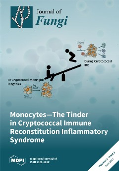
A third of adults with AIDS and cryptococcal meningitis (CM) develop immune reconstitution inflammatory syndrome (IRIS) after the initiation of antiretroviral therapy (ART). We demonstrate that monocyte subset phenotype and cytokine responses prior to ART are associated with, and may be predictive of, CM–IRIS. Larger studies to further delineate innate immunological responses and the efficacy of immunomodulatory therapies during cryptococcal IRIS are warranted. View this paper
- Issues are regarded as officially published after their release is announced to the table of contents alert mailing list.
- You may sign up for e-mail alerts to receive table of contents of newly released issues.
- PDF is the official format for papers published in both, html and pdf forms. To view the papers in pdf format, click on the "PDF Full-text" link, and use the free Adobe Reader to open them.
Previous Issue
Next Issue