
Thoracic Tumor Associated with a Unilateral Empyema in a Beef Cow: A Case Report
 , and
, and 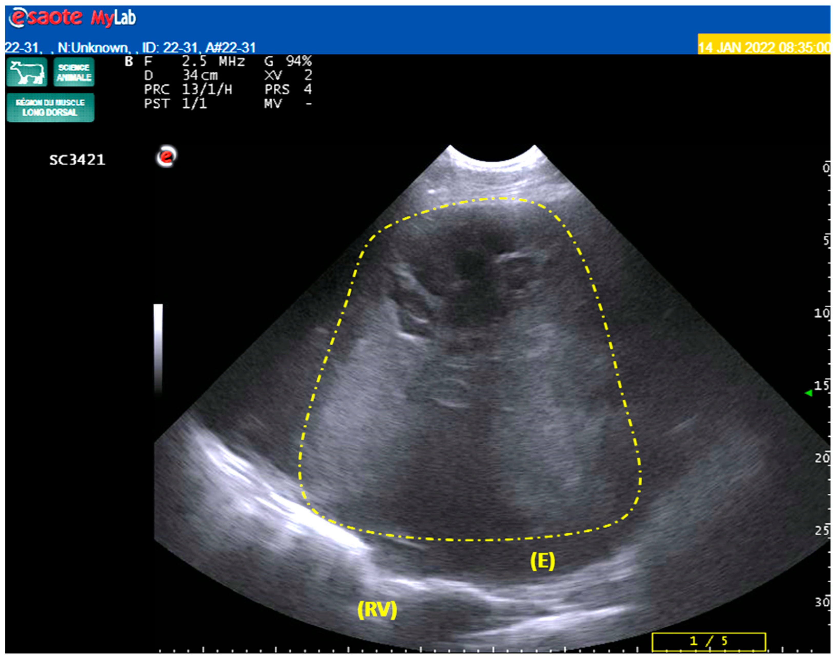
Abstract
:Simple Summary
Abstract
1. Introduction
2. Detailed Case Description
2.1. Case Presentation and Gross Findings
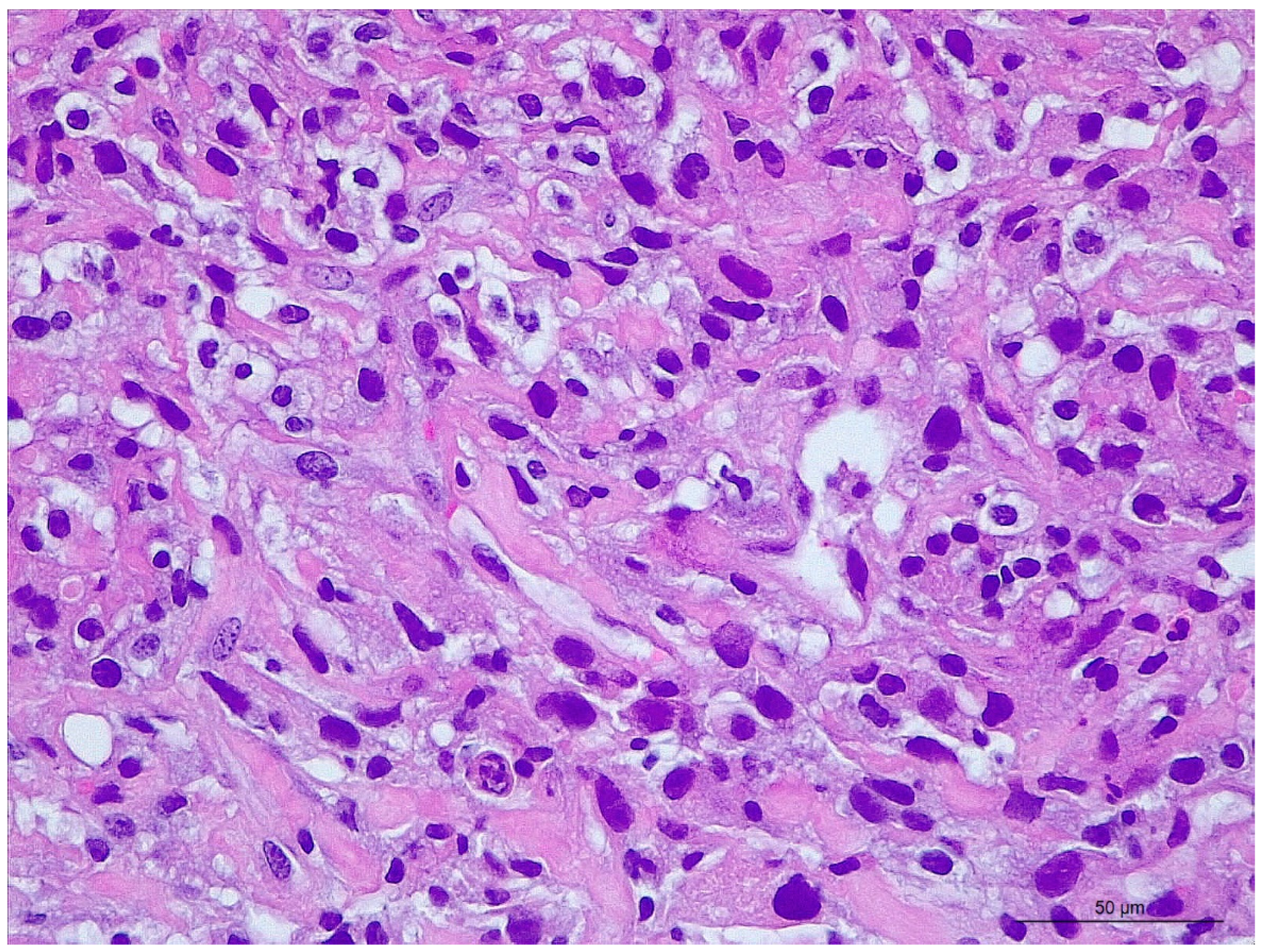
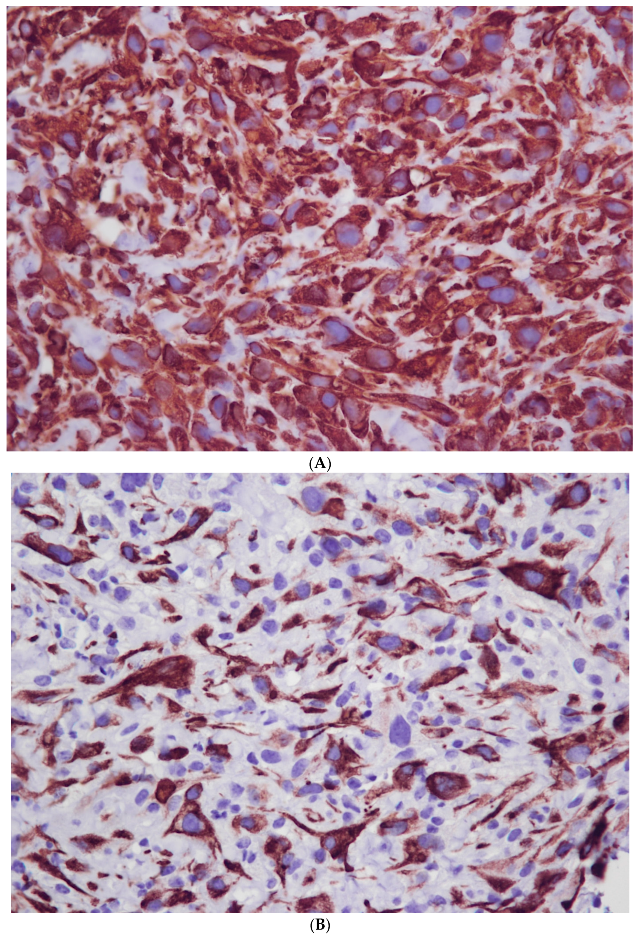
2.2. Final Diagnosis
2.3. Treatment and Outcome
3. Discussion
4. Conclusions
Supplementary Materials
Author Contributions
Funding
Institutional Review Board Statement
Informed Consent Statement
Data Availability Statement
Acknowledgments
Conflicts of Interest
Abbreviations
References
- Maxie, M.G. Jubb, Kennedy & Palmer’s Pathology of Domestic Animals, 6th ed.; Elsevier: Philadelphia, PA, USA, 2016; Volume 3, 957p. [Google Scholar]
- Vidal, E.; Tolosa, E.; Espinar, S.; de Val, B.P.; Nofrarías, M.; Alba, A.; Allepuz, A.; Grau-Roma, L.; López-Soria, S.; Martínez, J.; et al. Six-Year Follow-up of Slaughterhouse Surveillance (2008–2013): The Catalan Slaughterhouse Support Network (SESC). Vet. Pathol. 2016, 53, 532–544. [Google Scholar] [CrossRef] [PubMed]
- Babkine, M.; Blond, L. Ultrasonography of the bovine respiratory system and its practical application. Vet. Clin. N. Am. Food Anim. Pract. 2009, 25, 633–649. [Google Scholar] [CrossRef] [PubMed]
- Misdorp, W. Tumours in Calves: Comparative Aspects. J. Comp. Pathol. 2002, 127, 96–105. [Google Scholar] [CrossRef]
- Cappelleri, A.; Minoli, L.; Pigoli, C.; Costa, A.; Zaghini, L.; Bassanini, L.; Sinelli, M.; Perri, M.; Luini, M.V.; Tagliabue, G.; et al. Retrospective study of tumors from cattle slaughtered in Lombardy (Italy): Preliminary evaluation on the establishment of a bovine cancer registry. Vet. Ital. 2022, 58, 67–75. [Google Scholar] [PubMed]
- Sousa, D.Z.; Luis Rivera, L.C.; Didier Quevedo, D.C.; Gorino, A.C.; Biagio, S.; Laufer, R. Pulmonary adenocarcinoma in cattle. Rev. MVZ Córdoba 2014, 19, 4358–4363. [Google Scholar] [CrossRef]
- Gorig, A. Sarkomatose bei einem drei Wochen alten kalbe. Dtsch. Tierarztl. Wochenschr. 1893, 1, 321. [Google Scholar]
- Jacinto, J.G.P.; Bolcato, M.; Gentile, A.; Benazzi, C.; Muscatello, L.V. Congenital Suborbital Undifferentiated Sarcoma in a Crossbred Calf. Animals 2021, 11, 534. [Google Scholar] [CrossRef] [PubMed]
- Schwartz, I.; Levy, D. Pathobiology of bovine leukemia virus. Vet. Res. 1994, 25, 521–536. [Google Scholar] [PubMed]
- Seluanov, A.; Gladyshev, V.N.; Vijg, J.; Gorbunova, V. Mechanisms of cancer resistance in long-lived mammals. Nat. Rev. Cancer 2018, 18, 433–441. [Google Scholar] [CrossRef] [PubMed]
- Bertone, A.L. Neoplasms of the bovine gastrointestinal tract. Vet. Clin. N. Am. Food Anim. Pract. 1990, 6, 515–524. [Google Scholar] [CrossRef]
- Lucena, R.B.; Rissi, D.R.; Kommers, G.D.; Pierezan, F.; Oliveira-Filho, J.C.; Macedo, J.T.S.A.; Flores, M.M.; Barros, C.S.L. A Retrospective Study of 586 Tumours in Brazilian Cattle. J. Comp. Pathol. 2011, 145, 20–24. [Google Scholar] [CrossRef]
- Jourquin, S.; Lowie, T.; Debruyne, F.; Chantillon, L.; Vereecke, N.; Boyen, F.; Boone, R.; Bokma, J.; Pardon, B. Dynamics of subclinical pneumonia in male dairy calves in relation to antimicrobial therapy and production outcomes. J. Dairy Sci. 2023, 106, 676–689. [Google Scholar] [CrossRef]
- Rhodes, V.; Ryan, E.G.; Hayes, C.J.; Mcaloon, C.; O’Grady, L.; Hoey, S.; Mee, J.F.; Pardon, B.; Earley, B.; Mcaloon, C.G. Diagnosis of respiratory disease in preweaned dairy calves using sequential thoracic ultrasonography and clinical respiratory scoring: Temporal transitions and association with growth rates. J. Dairy Sci. 2021, 104, 11165–11175. [Google Scholar] [CrossRef]
- Cramer, M.C.; Ollivett, T.L. Growth of preweaned, group-housed dairy calves diagnosed with respiratory disease using clinical respiratory scoring and thoracic ultrasound—A cohort study. J. Dairy Sci. 2019, 102, 4322–4331. [Google Scholar] [CrossRef] [PubMed]
- Andrade Neto, A.Q.; Souto, R.J.C.; de Paula Cajueiro, J.F.; De Mendonça, C.L.; Driemeier, D.; De Almeida Souza, J.C.; de Souza Mendonça, F.; Bastos Afonso, J.A. Primary Pulmonary Adenocarcinoma in a Cow. Acta Sci. Vet. 2019, 47, 6. [Google Scholar] [CrossRef]
- Barley, J.P. Pulmonary papillary adenocarcinoma in an adult cow. Vet. Irel. J. 2011, 1, 674–675. [Google Scholar]
- Carminato, A.; Bozzato, E.; Trevisan, L.; Vascellari, M.; Catania, S.; De Palma, D.; Mutinelli, F. Blastoma polmonare del bovino—Bovine pulmonary blastoma. Large Anim. Rev. 2008, 14, 3–5. [Google Scholar]
- Roth, L.; Bradley, G.A. Pulmonary hamartoma in a calf. J. Comp. Pathol. 1991, 105, 471–474. [Google Scholar] [CrossRef] [PubMed]
- Üzum, N.; Özcay, N.; Ataoglu, Ö. Benign multicystic peritoneal mesothelioma. Turk. J. Gastroenterol. 2009, 20, 138–141. [Google Scholar]
- Hollington, P. Benign multicystic mesothelioma. ANZ J. Surg. 2010, 80, 186–187. [Google Scholar] [CrossRef]
- Magnusson, R.A.; Veit, H.P. Mesothelioma in a calf. J. Am. Vet. Med. Assoc. 1987, 191, 233–234. [Google Scholar] [PubMed]
- Wolfe, D.F.; Carson, R.L.; Hudson, R.S.; Boosinger, T.R.; Mysinger, P.W.; Powe, T.A., Jr.; Claxton, M.S.; Angel, K.L. Mesothelioma in cattle: Eight cases (1970–1988). J. Am. Vet. Med. Assoc. 1991, 199, 486–491. [Google Scholar]
- Pizarro, M.; Brandau, C.; Sanchez, M.A.; Flores, J.M. Immunocytochemical identification of a bovine peritoneal mesothelioma. Zentralbl. Veterinarmed. A 1992, 39, 476–480. [Google Scholar] [CrossRef] [PubMed]
- Girard, C.A.; Cecyre, A. Diffuse abdominal epithelioid mesothelioma in a cow. Can. Vet. J. 1995, 36, 440–441. [Google Scholar]
- Braun, U.; Rutten, M.; Bleul, U.; Previtali, M.; Kruger, S.; Gerspach, C.; Geiger, S.; Sydler, T. Biphasisches Mesotheliom bei einer Braunviehkuh: Klinische, histomorphologische, immunhistochemische und elektronenmikroskopische Befunde [Biphasic mesothelioma in a Swiss Braunvieh cow: Clinical, histological, immunohistochemical and electron microscopical findings]. Schweiz. Arch. Tierheilkd. 2012, 154, 33–38. (In German) [Google Scholar] [CrossRef]
- Gerbaudo, V.H.; Sugarbaker, D.J.; Britz-Cunningham, S.; Di Carli, M.F.; Mauceri, C.; Treves, S.T. Assessment of malignant pleural mesothelioma with (18)F-FDG dual-head gamma-camera coincidence imaging: Comparison with histopathology. J. Nucl. Med. 2002, 43, 1144–1149. [Google Scholar] [PubMed]
- Takasu, M.; Shirota, K.; Uchida, N.; Iguchi, T.; Nishii, N.; Ohba, Y.; Maeda, S.; Miyazawa, K.; Murase, T.; Kitagawa, H. Pericardial mesothelioma in a neonatal calf. J. Vet. Med. Sci. 2006, 68, 519–521. [Google Scholar] [CrossRef]
- Thomson, R.G.; McGavin, M.D.; Carlton, W.; Zachary, J.F. Thomson’s Special Veterinary Pathology, 3rd ed.; Mosby: Boston, MA, USA, 2000; 755p. [Google Scholar]
- Peli, A.; Bolcato, M.; Roccaro, M.; Gentile, A.; Militerno, G. Mesothelioma in cattle: Two case reports. Large Anim. Rev. 2018, 24, 89–92. [Google Scholar]
- Kawashima, Y.; Fujimoto, A.; Saito, M.; Mikami, O.; Ishikawa, Y.; Kadota, K. Histological comparison of malignant epithelioid mesothelioma in young and adult cattle. J. Vet. Med. Sci. 2021, 83, 968–972. [Google Scholar] [CrossRef]
- Bengston, J.S. Primary reticulum cell sarcoma of the lymph nodes of a cow with widespread metastases. Am. J. Pathol. 1938, 14, 365–376.5. [Google Scholar]
- Devbhandari, M.P.; Meraj, S.; Jones, M.T.; Kadir, I.; Bridgewater, B. Primary cardiac sarcoma: Reports of two cases and a review of current literature. J. Cardiothorac. Surg. 2007, 2, 34. [Google Scholar] [CrossRef] [PubMed]
- Marlowe, K.W.; Robat, C.S.; Clarke, D.M.; Taylor, A.; Touret, M.; Husbands, B.D.; Vail, D.M. Primary pulmonary histiocytic sarcoma in dogs: A retrospective analysis of 37 cases (2000–2015). Vet. Comp. Oncol. 2018, 16, 658–663. [Google Scholar] [CrossRef]
- Armbrust, L.J.; Biller, D.S.; Bamford, A.; Chun, R.; Garrett, L.D.; Sanderson, M.W. Comparison of three-view thoracic radiography and computed tomography for detection of pulmonary nodules in dogs with neoplasia. J. Am. Vet. Med. Assoc. 2012, 240, 1088–1094. [Google Scholar] [CrossRef] [PubMed]
- Merritt, R.M.; Williams, M.F.; James, T.H.; Porubsky, E.S. Detection of cervical metastasis. A meta-analysis comparing computed tomography with physical examination. Arch. Otolaryngol. Head Neck Surg. 1997, 123, 149–152. [Google Scholar] [CrossRef]
- Dernell, W.S.; Withrow, S.J.; Kuntz, C.A.; Powers, B.E. Principles of treatment for soft tissue sarcoma. Clin. Tech. Small Anim. Pract. 1998, 13, 59–64. [Google Scholar] [CrossRef]
- Macewan, E.G.; Powers, B.E.; Macy, D. Soft tissue sarcomas. In Small Animal Clinical Oncology, 3rd ed.; Withrow, S.J., MacEwen, E.G., Eds.; WB Saunders: Philadelphia, PA, USA, 2001; pp. 283–304. [Google Scholar]
- Ehrhart, N. Soft-tissue sarcomas in dogs: A review. J. Am. Anim. Hosp. Assoc. 2005, 41, 241–246. [Google Scholar] [CrossRef]
- Guardado, F.J.; Gabadage, K.; Allen, A.L. Presumptive metastatic leiomyosarcoma in a feedlot steer. Can. Vet. J. 2022, 63, 819–824. [Google Scholar] [PubMed]
- Ikeda, N.; Yoshida, T.; Seki, A.; Nakamura, M.; Tanaka, T.; Ichikawa, R.; Nakahara, R.; Orihara, K.; Kobayashi, M.; Yamashita, R.; et al. Extraskeletal chondrosarcoma in the abdominal cavity of a cow. J. Vet. Med. Sci. 2019, 81, 1749–1752. [Google Scholar] [CrossRef]
- Fujimoto, A.; Wada, Y.; Kanada, T.; Ishikawa, Y.; Kadota, K. Mast Cell Sarcoma with Megakaryocytic Differentiation in a Calf. J. Vet. Med. Sci. 2012, 74, 1643–1646. [Google Scholar] [CrossRef]


Disclaimer/Publisher’s Note: The statements, opinions and data contained in all publications are solely those of the individual author(s) and contributor(s) and not of MDPI and/or the editor(s). MDPI and/or the editor(s) disclaim responsibility for any injury to people or property resulting from any ideas, methods, instructions or products referred to in the content. |
© 2023 by the authors. Licensee MDPI, Basel, Switzerland. This article is an open access article distributed under the terms and conditions of the Creative Commons Attribution (CC BY) license (https://creativecommons.org/licenses/by/4.0/).
Share and Cite
Robcis, R.; De Campos, C.; Garapin, B.; Lucas, M.-N.; Poujade, A.; Gaide, N.; Delverdier, M.; Maillard, R. Thoracic Tumor Associated with a Unilateral Empyema in a Beef Cow: A Case Report. Vet. Sci. 2023, 10, 376. https://doi.org/10.3390/vetsci10060376
Robcis R, De Campos C, Garapin B, Lucas M-N, Poujade A, Gaide N, Delverdier M, Maillard R. Thoracic Tumor Associated with a Unilateral Empyema in a Beef Cow: A Case Report. Veterinary Sciences. 2023; 10(6):376. https://doi.org/10.3390/vetsci10060376
Chicago/Turabian StyleRobcis, Rodolphe, Charly De Campos, Bénédicte Garapin, Marie-Noëlle Lucas, Agnès Poujade, Nicolas Gaide, Maxence Delverdier, and Renaud Maillard. 2023. "Thoracic Tumor Associated with a Unilateral Empyema in a Beef Cow: A Case Report" Veterinary Sciences 10, no. 6: 376. https://doi.org/10.3390/vetsci10060376