
Seroprevalence and Risk Factors for Toxoplasma gondii Infection in Horses
 , ,
, ,  , ,
, ,  and
and
Abstract
:Simple Summary
Abstract
1. Introduction
2. Material and Methods
2.1. Ethical Statement
2.2. Study Area
2.3. Sample Collection and Preparation
2.4. Serological Examination
2.5. Statistical Analysis
3. Results
4. Discussion
5. Conclusions
Author Contributions
Funding
Institutional Review Board Statement
Informed Consent Statement
Data Availability Statement
Acknowledgments
Conflicts of Interest
References
- Dubey, J.P. Toxoplasmosis of Animals and Humans; CRC Press: Boca Raton, FL, USA, 2016. [Google Scholar]
- Innes, E. A brief history and overview of Toxoplasma gondii. Zoonoses Public Health 2010, 57, 1–7. [Google Scholar] [CrossRef] [PubMed]
- Selim, A.; Marawan, M.A.; Abdelhady, A.; Wakid, M.H. Seroprevalence and Potential Risk Factors of Toxoplasma gondii in Dromedary Camels. Agriculture 2023, 13, 129. [Google Scholar] [CrossRef]
- Dabritz, H.; Conrad, P.A. Cats and Toxoplasma: Implications for public health. Zoonoses Public Health 2010, 57, 34–52. [Google Scholar] [CrossRef] [PubMed]
- Schlüter, D.; Däubener, W.; Schares, G.; Groß, U.; Pleyer, U.; Lüder, C. Animals are key to human toxoplasmosis. Int. J. Med. Microbiol. 2014, 304, 917–929. [Google Scholar] [CrossRef] [PubMed]
- Tong, W.H.; Pavey, C.; O’Handley, R.; Vyas, A. Behavioral biology of Toxoplasma gondii infection. Parasites Vectors 2021, 14, 1–6. [Google Scholar] [CrossRef] [PubMed]
- Yang, S.; Parmley, S.F. Toxoplasma gondii expresses two distinct lactate dehydrogenase homologous genes during its life cycle in intermediate hosts. Gene 1997, 184, 1–12. [Google Scholar] [CrossRef]
- Hill, D.; Dubey, J. Toxoplasma gondii: Transmission, diagnosis and prevention. Clin. Microbiol. Infect. 2002, 8, 634–640. [Google Scholar] [CrossRef] [Green Version]
- Tassi, P. Toxoplasma gondii infection in horses. A review. Parassitologia 2007, 49, 7–15. [Google Scholar]
- Masatani, T.; Takashima, Y.; Takasu, M.; Matsuu, A.; Amaya, T. Prevalence of anti-Toxoplasma gondii antibody in domestic horses in Japan. Parasitol. Int. 2016, 65, 146–150. [Google Scholar] [CrossRef]
- Stelzer, S.; Basso, W.; Silván, J.B.; Ortega-Mora, L.M.; Maksimov, P.; Gethmann, J.; Conraths, F.; Schares, G. Toxoplasma gondii infection and toxoplasmosis in farm animals: Risk factors and economic impact. Food Waterborne Parasitol. 2019, 15, e00037. [Google Scholar] [CrossRef]
- Dubey, J.P.; Murata, F.; Cerqueira-Cézar, C.; Kwok, O. Public health and economic importance of Toxoplasma gondii infections in goats: The last decade. Res. Vet. Sci. 2020, 132, 292–307. [Google Scholar] [CrossRef]
- Al Hamada, A.; Habib, I.; Barnes, A.; Robertson, I. Risk factors associated with seropositivity to Toxoplasma among sheep and goats in Northern Iraq. Vet. Parasitol. Reg. Stud. Rep. 2019, 15, 100264. [Google Scholar] [CrossRef] [PubMed]
- Souza, I.B.d.; Fernandes, P.R.; Silva, T.R.M.; Santos, C.V.B.; Silva, N.M.M.d.; Ubirajara Filho, C.R.C.; Carvalho, G.A.d.; Alves, L.C.; Mota, R.A.; Ramos, R.A.N. Seroprevalence of Neospora caninum and Toxoplasma gondii in dogs from an urban area of North-eastern Brazil: A spatial approach. Rev. Da Soc. Bras. De Med. Trop. 2019, 52–55. [Google Scholar] [CrossRef] [Green Version]
- Olsen, A.; Berg, R.; Tagel, M.; Must, K.; Deksne, G.; Enemark, H.L.; Alban, L.; Johansen, M.V.; Nielsen, H.V.; Sandberg, M. Seroprevalence of Toxoplasma gondii in domestic pigs, sheep, cattle, wild boars, and moose in the Nordic-Baltic region: A systematic review and meta-analysis. Parasite Epidemiol. Control. 2019, 5, e00100. [Google Scholar] [CrossRef] [PubMed]
- Guo, M.; Dubey, J.P.; Hill, D.; Buchanan, R.L.; Gamble, H.R.; Jones, J.L.; Pradhan, A.K. Prevalence and risk factors for Toxoplasma gondii infection in meat animals and meat products destined for human consumption. J. Food Prot. 2015, 78, 457–476. [Google Scholar] [CrossRef]
- Ibrahim, H.M.; Huang, P.; Salem, T.A.; Talaat, R.M.; Nasr, M.I.; Xuan, X.; Nishikawa, Y. Prevalence of Neospora caninum and Toxoplasma gondii antibodies in northern Egypt. Am. J. Trop. Med. Hyg. 2009, 80, 263–267. [Google Scholar] [CrossRef] [Green Version]
- Shaapan, R.; El-Nawawi, F.; Tawfik, M. Sensitivity and specificity of various serological tests for the detection of Toxoplasma gondii infection in naturally infected sheep. Vet. Parasitol. 2008, 153, 359–362. [Google Scholar] [CrossRef] [PubMed]
- El-Ghaysh, A. Seroprevalence of Toxoplasma gondii in Egyptian donkeys using ELISA. Vet. Parasitol. 1998, 80, 71–73. [Google Scholar] [CrossRef]
- Ghazy, A.; Shaapan, R.; Abdel-Rahman, E.H. Comparative serological diagnosis of toxoplasmosis in horses using locally isolated Toxoplasma gondii. Vet. Parasitol. 2007, 145, 31–36. [Google Scholar] [CrossRef]
- Dubey, J.; Murata, F.; Cerqueira-Cézar, C.; Kwok, O.; Su, C. Economic and public health importance of Toxoplasma gondii infections in sheep: 2009–2020. Vet. Parasitol. 2020, 286, 109195. [Google Scholar] [CrossRef]
- Kakakhel, M.A.; Wu, F.; Anwar, Z.; Saif, I.; ul Akbar, N.; Gul, N.; Ali, I.; Feng, H.; Wang, W. The presence of Toxoplasma gondii in soil, their transmission, and their influence on the small ruminants and human population: A review. Microbial. Pathogenesis 2021, 158, 104850. [Google Scholar] [CrossRef]
- García-Bocanegra, I.; Cabezón, O.; Arenas-Montes, A.; Carbonero, A.; Dubey, J.; Perea, A.; Almería, S. Seroprevalence of Toxoplasma gondii in equids from Southern Spain. Parasitol. Int. 2012, 61, 421–424. [Google Scholar] [CrossRef] [PubMed]
- Gazyağci, S.; Macun, H.; Babür, C. Investigation of seroprevalance of toxoplasmosis in mares and stallions in Ankara province, Turkey. Iran. J. Vet. Res. 2011, 12, 354–356. [Google Scholar]
- Cazarotto, C.J.; Balzan, A.; Grosskopf, R.K.; Boito, J.P.; Portella, L.P.; Vogel, F.F.; Fávero, J.F.; Cucco, D.d.C.; Biazus, A.H.; Machado, G. Horses seropositive for Toxoplasma gondii, Sarcocystis spp. and Neospora spp.: Possible risk factors for infection in Brazil. Microb. Pathog. 2016, 99, 30–35. [Google Scholar] [CrossRef] [PubMed]
- Aroussi, A.; Vignoles, P.; Dalmay, F.; Wimel, L.; Dardé, M.-L.; Mercier, A.; Ajzenberg, D. Detection of Toxoplasma gondii DNA in horse meat from supermarkets in France and performance evaluation of two serological tests. Parasite 2015, 22, 14–22. [Google Scholar] [CrossRef] [Green Version]
- Alvarado-Esquivel, C.; Alvarado-Esquivel, D.; Dubey, J.P. Prevalence of Toxoplasma gondii antibodies in domestic donkeys (Equus asinus) in Durango, Mexico slaughtered for human consumption. BMC Vet. Res. 2015, 11, 1–4. [Google Scholar] [CrossRef] [Green Version]
- Dixon, B.; Fayer, R.; Santín, M.; Hill, D.; Dubey, J. Protozoan parasites: Cryptosporidium, Giardia, Cyclospora, and Toxoplasma. Rapid Detect. Charact. Enumer. Foodborne Pathog. 2011, 349–370. [Google Scholar]
- Shwab, E.K.; Saraf, P.; Zhu, X.-Q.; Zhou, D.-H.; McFerrin, B.M.; Ajzenberg, D.; Schares, G.; Hammond-Aryee, K.; van Helden, P.; Higgins, S.A. Human impact on the diversity and virulence of the ubiquitous zoonotic parasite Toxoplasma gondii. Proc. Natl. Acad. Sci. USA 2018, 115, E6956–E6963. [Google Scholar] [CrossRef] [Green Version]
- El-Geddawi, O.A.; El-Sayad, M.H.; Sadek, N.A.; Hussien, N.A.; Ahmed, M.A. Detection of T. gondii infection in blood donors in Alexandria, Egypt, using serological and molecular strategies. Parasitol. United J. 2016, 9, 24. [Google Scholar] [CrossRef]
- El-Sayed, N.M.; Abdel-Wahab, M.M.; Kishik, S.M.; Alhusseini, N.F. Do we need to screen Egyptian voluntary blood donors for toxoplasmosis? Asian Pac. J. Trop. Dis. 2016, 6, 260–264. [Google Scholar] [CrossRef]
- Matsuo, K.; Kamai, R.; Uetsu, H.; Goto, H.; Takashima, Y.; Nagamune, K. Seroprevalence of Toxoplasma gondii infection in cattle, horses, pigs and chickens in Japan. Parasitol. Int. 2014, 63, 638–639. [Google Scholar] [CrossRef] [PubMed] [Green Version]
- Terkawi, M.A.; Kameyama, K.; Rasul, N.H.; Xuan, X.; Nishikawa, Y. Development of an immunochromatographic assay based on dense granule protein 7 for serological detection of Toxoplasma gondii infection. Clin. Vaccine Immunol. 2013, 20, 596–601. [Google Scholar] [CrossRef] [PubMed] [Green Version]
- Gu, Y.; Wang, Z.; Cai, Y.; Li, X.; Wei, F.; Shang, L.; Li, J.; Liu, Q. A comparative study of Toxoplasma gondii seroprevalence in mink using a modified agglutination test, a Western blot, and enzyme-linked immunosorbent assays. J. Vet. Diagn. Investig. 2015, 27, 616–620. [Google Scholar] [CrossRef] [PubMed] [Green Version]
- Fereig, R.M.; Mahmoud, H.Y.; Mohamed, S.G.; AbouLaila, M.R.; Abdel-Wahab, A.; Osman, S.A.; Zidan, S.A.; El-Khodary, S.A.; Mohamed, A.E.A.; Nishikawa, Y. Seroprevalence and epidemiology of Toxoplasma gondii in farm animals in different regions of Egypt. Vet. Parasitol. Reg. Stud. Rep. 2016, 3, 1–6. [Google Scholar] [CrossRef]
- Shaapan, R.; Ghazy, A. Isolation of Toxoplasma gondii from horse meat in Egypt. Pak. J. Biol. Sci. PJBS 2007, 10, 174–177. [Google Scholar] [CrossRef] [Green Version]
- Daniel, W.W.; Cross, C.L. Biostatistics: A Foundation for Analysis in the Health Sciences; Wiley: New York, NY, USA, 2018. [Google Scholar]
- Haridy, F.M.; Shoukry, N.M.; Hassan, A.A.; Morsy, T.A. ELISA-seroprevalence of Toxoplasma gondii in draught horses in Greater Cairo, Egypt. J. Egypt. Soc. Parasitol. 2009, 39, 821–826. [Google Scholar]
- Zhang, X.-X.; Ren, W.-X.; Hou, G.; Liu, Q.; Yu, T.-Q.; Zhao, Q.; Ni, H.-B. Seroprevalence and risk factors of Toxoplasma gondii infection in horses in Jilin Province and Inner Mongolia Autonomous Region, Northern China. Acta Tropica 2018, 187, 119–123. [Google Scholar] [CrossRef]
- Almeida, J.C.; Vidotto, O.; Ferreira, E.P.; Ribeiro, L.P.; Mongruel, A.C.; Vieira, T.S.; Freire, R.L.; Mota, R.A.; Vieira, R.F. Serosurvey of anti-Toxoplasma gondii antibodies in sport horses from Paraiba state, Northeastern Brazil. Acta Parasitol. 2017, 62, 225–227. [Google Scholar] [CrossRef]
- Saqib, M.; Hussain, M.; Sajid, M.; Mansoor, M.; Asi, M.; Fadya, A.; Zohaib, A.; Sial, A.; Muhammad, G.; Ullah, I. Sero-epidemiology of equine toxoplasmosis using a latex agglutination test in the three metropolises of Punjab, Pakistan. Trop. Biomed. 2015, 32, 276–285. [Google Scholar]
- Bártová, E.; Sedlák, K.; Kobédová, K.; Budíková, M.; Atuman, Y.J.; Kamani, J. Seroprevalence and risk factors of Neospora spp. and Toxoplasma gondii infections among horses and donkeys in Nigeria, West Africa. Acta Parasitol. 2017, 62, 606–609. [Google Scholar] [CrossRef]
- Razmi, G.R.; Abedi, V.; Yaghfoori, S. Serological study of Toxoplasma gondii infection in Turkoman horses in the North Khorasan Province, Iran. J. Parasit. Dis. 2016, 40, 515–519. [Google Scholar] [CrossRef] [Green Version]
- Alanazi, A.D.; Alyousif, M.S. Prevalence of antibodies to Toxoplasma gondii in horses in Riyadh Province, Saudi Arabia. J. Parasitol. 2011, 97, 943–945. [Google Scholar] [CrossRef] [PubMed]
- Aharonson-Raz, K.; Baneth, G.; Lopes, A.P.; Brancal, H.; Schallig, H.; Cardoso, L.; Steinman, A. Low seroprevalence of Leishmania infantum and Toxoplasma gondii in the horse population in Israel. Vector-Borne Zoonotic Dis. 2015, 15, 726–731. [Google Scholar] [CrossRef] [PubMed]
- Lopes, A.P.; Sousa, S.; Dubey, J.; Ribeiro, A.J.; Silvestre, R.; Cotovio, M.; Schallig, H.D.; Cardoso, L.; Cordeiro-da-Silva, A. Prevalence of antibodies to Leishmania infantum and Toxoplasma gondii in horses from the north of Portugal. Parasites Vectors 2013, 6, 1–4. [Google Scholar] [CrossRef] [PubMed] [Green Version]
- Boughattas, S.; Bergaoui, R.; Essid, R.; Aoun, K.; Bouratbine, A. Seroprevalence of Toxoplasma gondii infection among horses in Tunisia. Parasites Vectors 2011, 4, 1–3. [Google Scholar] [CrossRef] [PubMed] [Green Version]
- Karatepe, B.; Babür, C.; Karatepe, M.; Kılıç, S. Seroprevalence of toxoplasmosis in horses in Niğde Province of Turkey. Trop. Anim. Health Prod. 2010, 42, 385–389. [Google Scholar] [CrossRef]
- Uggla, A.; Mattson, S.; Juntti, N. Prevalence of antibodies to Toxoplasma gondii in cats, dogs and horses in Sweden. Acta Vet. Scand. 1990, 31, 219–222. [Google Scholar] [CrossRef]
- Kouam, M.K.; Diakou, A.; Kanzoura, V.; Papadopoulos, E.; Gajadhar, A.A.; Theodoropoulos, G. A seroepidemiological study of exposure to Toxoplasma, Leishmania, Echinococcus and Trichinella in equids in Greece and analysis of risk factors. Vet. Parasitol. 2010, 170, 170–175. [Google Scholar] [CrossRef] [PubMed]
- Rinaldi, L.; Scala, A. Toxoplasmosis in livestock in Italy: An epidemiological update. Parassitologia 2008, 50, 59–61. [Google Scholar]
- AKCA, A.; Babur, C.; ARSLAN, M.; Gicik, Y.; Kara, M.; KILIC, S. Prevalence of antibodies to Toxoplasma gondii in horses in the province of Kars, Turkey. Vet. Med. 2004, 49, 9–13. [Google Scholar] [CrossRef] [Green Version]
- Güçlü, Z.; Karaer, Z.; Babür, C.; Kiliç, S. Investigation of Toxoplasma gondii antibodies in sport horses bred in Ankara province. Positivity 2007, 16, 256. [Google Scholar]
- Bártová, E.; Sedlák, K.; Syrová, M.; Literák, I. Neospora spp. and Toxoplasma gondii antibodies in horses in the Czech Republic. Parasitol. Res. 2010, 107, 783–785. [Google Scholar] [CrossRef] [PubMed]
- Meng, Q.-F.; Li, D.; Yao, G.-Z.; Zou, Y.; Cong, W.; Shan, X.-F. Seroprevalence of Toxoplasma gondii infection and variables associated with seropositivity in donkeys in eastern China. Parasite 2018, 25, 66. [Google Scholar] [CrossRef] [Green Version]
- Selim, A.; Abdelhady, A. The first detection of anti-West Nile virus antibody in domestic ruminants in Egypt. Trop. Anim. Health Prod. 2020, 52, 3147–3151. [Google Scholar] [CrossRef] [PubMed]
- Selim, A.; Ali, A.-F.; Ramadan, E. Prevalence and molecular epidemiology of Johne’s disease in Egyptian cattle. Acta tropica 2019, 195, 1–5. [Google Scholar] [CrossRef] [PubMed]
- Selim, A.; Attia, K.; Ramadan, E.; Hafez, Y.M.; Salman, A. Seroprevalence and molecular characterization of Brucella species in naturally infected cattle and sheep. Prev. Vet. Med. 2019, 171, 104756. [Google Scholar] [CrossRef] [PubMed]
- Selim, A.; Khater, H.; Almohammed, H.I. A recent update about seroprevalence of ovine neosporosis in Northern Egypt and its associated risk factors. Sci. Rep. 2021, 11, 14043. [Google Scholar] [CrossRef]
- Prestrud, K.W.; Åsbakk, K.; Fuglei, E.; Mørk, T.; Stien, A.; Ropstad, E.; Tryland, M.; Gabrielsen, G.W.; Lydersen, C.; Kovacs, K.M. Serosurvey for Toxoplasma gondii in arctic foxes and possible sources of infection in the high Arctic of Svalbard. Vet. Parasitol. 2007, 150, 6–12. [Google Scholar] [CrossRef]
- Dhimal, M.; Ahrens, B.; Kuch, U. Species composition, seasonal occurrence, habitat preference and altitudinal distribution of malaria and other disease vectors in eastern Nepal. Parasites Vectors 2014, 7, 1–11. [Google Scholar] [CrossRef] [Green Version]
- Selim, A.; Ali, A.-F.; Moustafa, S.M.; Ramadan, E. Molecular and serological data supporting the role of Q fever in abortions of sheep and goats in northern Egypt. Microb. Pathog. 2018, 125, 272–275. [Google Scholar] [CrossRef]
- Selim, A.; Manaa, E.; Khater, H. Seroprevalence and risk factors for lumpy skin disease in cattle in Northern Egypt. Trop. Anim. Health Prod. 2021, 53, 350. [Google Scholar] [CrossRef] [PubMed]
- Selim, A.; Manaa, E.A.; Alanazi, A.D.; Alyousif, M.S. Seroprevalence, risk factors and molecular identification of bovine leukemia virus in Egyptian cattle. Animals 2021, 11, 319. [Google Scholar] [CrossRef] [PubMed]
- Selim, A.; Manaa, E.A.; Waheed, R.M.; Alanazi, A.D. Seroprevalence, associated risk factors analysis and first molecular characterization of chlamydia abortus among Egyptian sheep. Comp. Immunol. Microbiol. Infect. Dis. 2021, 74, 101600. [Google Scholar] [CrossRef] [PubMed]
- Dubey, J. Persistence of encysted Toxoplasma gondii in tissues of equids fed oocysts. Am. J. Vet. Res. 1985, 46, 1753–1754. [Google Scholar]
- Selim, A.; Megahed, A.; Kandeel, S.; Alouffi, A.; Almutairi, M.M. West Nile virus seroprevalence and associated risk factors among horses in Egypt. Sci. Rep. 2021, 11, 20932. [Google Scholar] [CrossRef]
- Selim, A.; Megahed, A.A.; Kandeel, S.; Abdelhady, A. Risk factor analysis of bovine leukemia virus infection in dairy cattle in Egypt. Comp. Immunol. Microbiol. Infect. Dis. 2020, 72, 101517. [Google Scholar] [CrossRef]
- Selim, A.; Yang, E.; Rousset, E.; Thiéry, R.; Sidi-Boumedine, K. Characterization of Coxiella burnetii strains from ruminants in a Galleria mellonella host-based model. New Microbes New Infect. 2018, 24, 8–13. [Google Scholar] [CrossRef]
- Selim, A.M.; Elhaig, M.M.; Gaede, W. Development of multiplex real-time PCR assay for the detection of Brucella spp., Leptospira spp. and Campylobacter foetus. Vet. Ital. 2014, 50, 75. [Google Scholar]
- Selim, A.M.; Elhaig, M.M.; Moawed, S.A.; El-Nahas, E. Modeling the potential risk factors of bovine viral diarrhea prevalence in Egypt using univariable and multivariable logistic regression analyses. Vet. World 2018, 11, 259. [Google Scholar] [CrossRef] [Green Version]
- Ling, C.; Wan, P. A report of investigations of antibodies to Toxoplasma gondii in the horse and mule in Sichuan Province. Zhongguo Shouyi Ke-Ji 1984, 4, 32–34. [Google Scholar]
- Cherif, M.; Ait Oudhia, K.; Khelef, D. Detection of anti-Toxoplasma gondiiantibodies among horses (Equus caballus) and donkeys (Equus asinus) in Tiaret province, northwestern Algeria. Revue Méd. Vét. 2015, 166, 271–274. [Google Scholar]
- Wang, J.-L.; Zhou, D.-H.; Chen, J.; Liu, G.-X.; Pu, W.-B.; Liu, T.-Y.; Qin, S.-Y.; Yin, M.-Y.; Zhu, X.-Q. The prevalence of antibodies to Toxoplasma gondii in horses in Changji Hui Autonomous Prefecture, Xinjiang, northwestern China. Rev. Bras. De Parasitol. Veterinária 2015, 24, 298–302. [Google Scholar] [CrossRef] [PubMed] [Green Version]
- Li, X.; Ni, H.-B.; Ren, W.-X.; Jiang, J.; Gong, Q.-L.; Zhang, X.-X. Seroprevalence of Toxoplasma gondii in horses: A global systematic review and meta-analysis. Acta Trop. 2020, 201, 105222. [Google Scholar] [CrossRef] [PubMed]
- Tavalla, M.; Sabaghan, M.; Abdizadeh, R.; Khademvatan, S.; Rafiei, A.; Piranshahi, A.R. Seroprevalence of Toxoplasma gondii and Neospora spp. infections in Arab horses, southwest of Iran. Jundishapur J. Microbiol. 2015, 8, e14939. [Google Scholar] [CrossRef] [Green Version]
- Ouslimani, S.F.; Tennah, S.; Azzag, N.; Derdour, S.Y.; China, B.; Ghalmi, F. Seroepidemiological study of the exposure to Toxoplasma gondii among horses in Algeria and analysis of risk factors. Vet. World 2019, 12, 2007–2016. [Google Scholar] [CrossRef] [Green Version]
- Alvarado-Esquivel, C.; García-Machado, C.; Alvarado-Esquivel, D.; Vitela-Corrales, J.; Villena, I.; Dubey, J. Seroprevalence of Toxoplasma gondii infection in domestic sheep in Durango State, Mexico. J. Parasitol. 2012, 98, 271–273. [Google Scholar] [CrossRef]
- Lee, S.-H.; Lee, S.-E.; Seo, M.-G.; Goo, Y.-K.; Cho, K.-H.; Cho, G.-J.; Kwon, O.-D.; Kwak, D.; Lee, W.-J. Evidence of Toxoplasma gondii exposure among horses in Korea. J. Vet. Med. Sci. 2014, 76, 1663–1665. [Google Scholar] [CrossRef] [Green Version]
- Bártová, E.; Machaèová, T.; Sedlák, K.; Budíková, M.; Mariani, U.; Veneziano, V. Seroprevalence of antibodies of Neospora spp. and Toxoplasma gondii in horses from southern Italy. Folia Parasitol. 2015, 62, 1. [Google Scholar] [CrossRef]
- Millán, J.; Candela, M.G.; Palomares, F.; Cubero, M.J.; Rodríguez, A.; Barral, M.; de la Fuente, J.; Almería, S.; León-Vizcaíno, L. Disease threats to the endangered Iberian lynx (Lynx pardinus). Vet. J. 2009, 182, 114–124. [Google Scholar] [CrossRef]
- García-Bocanegra, I.; Dubey, J.; Martínez, F.; Vargas, A.; Cabezón, O.; Zorrilla, I.; Arenas, A.; Almería, S. Factors affecting seroprevalence of Toxoplasma gondii in the endangered Iberian lynx (Lynx pardinus). Vet. Parasitol. 2010, 167, 36–42. [Google Scholar] [CrossRef]
- Mainar, R.; De La Cruz, C.; Asensio, A.; Domínguez, L.; Vázquez-Boland, J. Prevalence of agglutinating antibodies to Toxoplasma gondii in small ruminants of the Madrid region, Spain, and identification of factors influencing seropositivity by multivariate analysis. Vet. Res. Commun. 1996, 20, 153–159. [Google Scholar] [CrossRef] [PubMed]


| Variable | Category | No. of Horses | Distribution |
|---|---|---|---|
| Locality | Giza | 110 | 26.2% |
| Kafr El Sheikh | 110 | 26.2% | |
| Qalyubia | 100 | 23.8% | |
| Gharbia | 100 | 23.8% | |
| Breed | Arabian | 60 | 14.3% |
| Thoroughbred | 200 | 47.6% | |
| Mixed | 160 | 38.1% | |
| Sex | Male | 190 | 45.2% |
| Female | 230 | 54.7% | |
| Age | <5 year | 130 | 31% |
| 5–10 | 180 | 42.8% | |
| >10 | 110 | 26.2% | |
| Presence of cats | Yes | 180 | 42.9% |
| No | 240 | 57.1% | |
| Presence of domestic ruminants | Yes | 120 | 28.6% |
| No | 300 | 71.4% |
| Factor | No. of Examined Horses | No. of Positive | % of Positive | 95% CI | p-Value |
|---|---|---|---|---|---|
| Locality | |||||
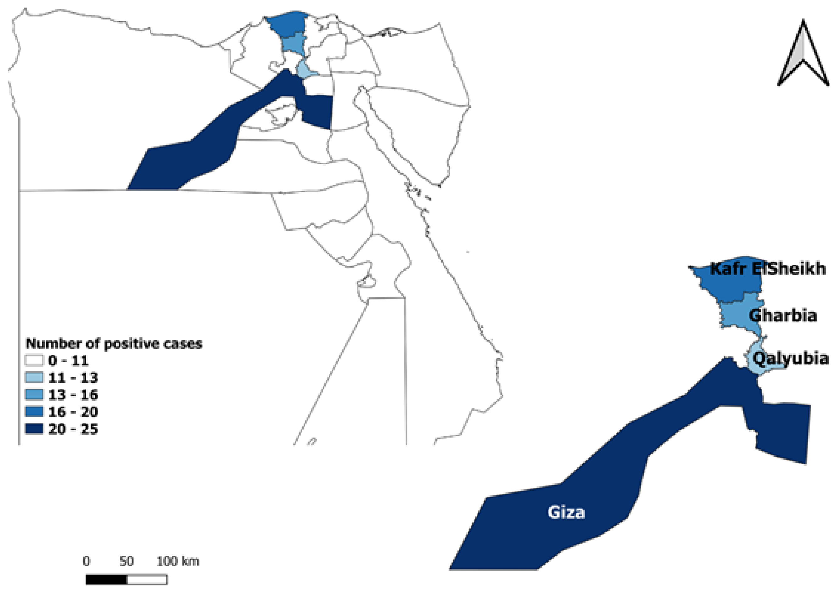
| Giza | 110 | 23 | 20.9 | 14.36–29.43 | |
| Kafr El Sheikh | 110 | 19 | 17.3 | 11.34–25.41 | |
| Qalyubia | 100 | 12 | 12.0 | 7–19.81 | 0.31 |
| Gharbia | 100 | 14 | 14.0 | 8.53–22.14 |
| Factor | No. of Examined Horses | No. of Positive | % of Positive | 95% CI | Statistics |
|---|---|---|---|---|---|
| Breed | |||||
| Arabian | 60 | 5 | 8.3 | 3.61–18.06 | χ2 = 6.167 df = 2 p = 0.04 * |
| Thoroughbred | 200 | 29 | 14.5 | 10.29–20.05 | |
| Mixed | 160 | 34 | 21.3 | 15.62–28.22 | |
| Sex | |||||
| Male | 190 | 20 | 10.5 | 6.92–15.7 | χ2 = 8.203 df = 1 p = 0.004 * |
| Female | 230 | 48 | 20.9 | 16.12–26.58 | |
| Age | |||||
| <5 years | 130 | 13 | 10.0 | 5.94–16.36 | χ2 = 7.165 df = 2 p = 0.02 * |
| 5–10 years | 180 | 30 | 16.7 | 11.93–22.8 | |
| >10 years | 110 | 25 | 22.7 | 15.9–31.4 | |
| Presence of cats | |||||
| Yes | 180 | 38 | 21.1 | 15.78–27.64 | χ2 = 5.621 df = 1 p = 0.02 * |
| No | 240 | 30 | 12.5 | 8.9–17.28 | |
| Presence of domestic ruminants | |||||
| Yes | 120 | 28 | 23.3 | 16.66–31.65 | χ2 = 6.317 df = 1 p = 0.01 * |
| No | 300 | 40 | 13.3 | 9.94–17.64 | |
| Total | 420 | 68 | 16.2 | 12.98–20.02 |
| Variable | B | S.E. | OR | 95% CI for OR | p-Value | |
|---|---|---|---|---|---|---|
| Lower | Upper | |||||
| Breed | ||||||
| Thoroughbred | 0.574 | 0.519 | 1.78 | 0.64 | 4.90 | 0.268 |
| Mixed | 0.965 | 0.519 | 2.63 | 0.95 | 7.26 | 0.063 |
| Sex | ||||||
| Female | 0.852 | 0.296 | 2.35 | 1.31 | 4.19 | 0.004 |
| Age | ||||||
| 5–10 years | 0.803 | 0.380 | 2.23 | 1.06 | 4.70 | 0.034 |
| >10 years | 1.023 | 0.388 | 2.78 | 1.30 | 5.95 | 0.008 |
| Presence of cats | ||||||
| Yes | 0.679 | 0.283 | 1.97 | 1.13 | 3.44 | 0.017 |
| Presence of domestic ruminants | ||||||
| Yes | 0.769 | 0.297 | 2.16 | 1.21 | 3.86 | 0.010 |
Disclaimer/Publisher’s Note: The statements, opinions and data contained in all publications are solely those of the individual author(s) and contributor(s) and not of MDPI and/or the editor(s). MDPI and/or the editor(s) disclaim responsibility for any injury to people or property resulting from any ideas, methods, instructions or products referred to in the content. |
© 2023 by the authors. Licensee MDPI, Basel, Switzerland. This article is an open access article distributed under the terms and conditions of the Creative Commons Attribution (CC BY) license (https://creativecommons.org/licenses/by/4.0/).
Share and Cite
Marzok, M.; AL-Jabr, O.A.; Salem, M.; Alkashif, K.; Sayed-Ahmed, M.; Wakid, M.H.; Kandeel, M.; Selim, A. Seroprevalence and Risk Factors for Toxoplasma gondii Infection in Horses. Vet. Sci. 2023, 10, 237. https://doi.org/10.3390/vetsci10030237
Marzok M, AL-Jabr OA, Salem M, Alkashif K, Sayed-Ahmed M, Wakid MH, Kandeel M, Selim A. Seroprevalence and Risk Factors for Toxoplasma gondii Infection in Horses. Veterinary Sciences. 2023; 10(3):237. https://doi.org/10.3390/vetsci10030237
Chicago/Turabian StyleMarzok, Mohamed, Omar A. AL-Jabr, Mohamed Salem, Khalid Alkashif, Mohamed Sayed-Ahmed, Majed H. Wakid, Mahmoud Kandeel, and Abdelfattah Selim. 2023. "Seroprevalence and Risk Factors for Toxoplasma gondii Infection in Horses" Veterinary Sciences 10, no. 3: 237. https://doi.org/10.3390/vetsci10030237