Review

Modern Imaging Techniques in the Study and Disease Diagnosis of the Mammary Glands of Animals

by
Mariana S. Barbagianni
* and
Pagona G. Gouletsou
Faculty of Veterinary Science, School of Health Sciences, University of Thessaly, 43100 Karditsa, Greece
*
Author to whom correspondence should be addressed.
Vet. Sci. 2023, 10(2), 83; https://doi.org/10.3390/vetsci10020083
Submission received: 7 December 2022 / Revised: 12 January 2023 / Accepted: 20 January 2023 / Published: 22 January 2023
(This article belongs to the Special Issue Comparative Oncology and Veterinary Cancer Surveillance)

Abstract

:

Simple Summary

Various imaging techniques may be applied in research or clinical practice to evaluate pathological conditions of mammary glands and contribute to a diagnosis. This review describes imaging techniques that are used on farms and companion animals’ mammary glands. Specifically, computed tomography, positron emission tomography, magnetic resonance imaging, ultrasonographic techniques such as Doppler, contrast-enhanced and three-dimensional examination, and also elastography are presented. Furthermore, the relative bibliography is reviewed and discussed.

Abstract

The study of the structure and function of the animals’ mammary glands is of key importance, as it reveals pathological processes at their onset, thus contributing to their immediate treatment. The most frequently studied mammary diseases are mastitis in cows and ewes and mammary tumours in dogs and cats. Various imaging techniques such as computed tomography, positron emission tomography, magnetic resonance imaging, and ultrasonographic techniques (Doppler, contrast-enchanced, three-dimensional and elastography) are available and can be applied in research or clinical practice in order to evaluate possible abnormalities in mammary glands, as well as to assist in the differential diagnosis. In this review, the above imaging technologies are described, and the perspectives of each method are highlighted. It is inferred that ultrasonographic modalities are the most frequently used imaging techniques for the diagnosis of clinical or subclinical mastitis and treatment guidance on a farm. In companion animals, a combination of imaging techniques should be applied for a more accurate diagnosis of mammary tumours. In any case, the confirmation of the diagnosis is provided by laboratory techniques.

1. Introduction

Various imaging techniques are available and can be applied in research or clinical practice, in order to evaluate possible abnormalities of mammary glands of domestic animals, as well as to assist in the differential diagnosis of masses within these organs [1,2]. Some of these techniques use radiation and rely on its spatial and temporal interaction with tissues in order to educe useful information from observations [3] whilst others use magnetic field and radio or high-frequency sound waves [4,5].
Anatomical features, structures or physiological processes can be assessed by employing different imaging methods [6,7]. The main methods used in the imaging of mammary glands of animals include computed tomography, positron emission tomography, magnetic resonance imaging, and ultrasonographic techniques (e.g., B-mode ultrasonography, Doppler ultrasonography, contrast-enchased ultrasonography, three-dimensional ultrasonography, and elastography) [2,4,5,8].
Computed tomography and magnetic resonance imaging provide information regarding the structure of the organs whilst positron emission tomography mainly provides information about their function [3]. The functional information derived from positron emission tomography images can be combined with anatomical information obtained from computed tomography images, thus in combination constitute the positron emission tomography/computed tomography scanning technique [3]. Doppler ultrasonography, contrast-enhanced ultrasonography, three-dimensional ultrasonography, and elastography are easy, fast, non-invasive diagnostic techniques, enabling immediate results for investigation of mammary gland abnormalities, although there are differences in the diagnostic efficacy between these modalities [5].
Although in human medicine clear diagnostic imaging guidelines have been proposed to investigate diseases of the mammary glands [9], in veterinary work, similar protocols have not been developed thus far. At the moment, only an algorithm has been proposed for the evaluation of sentinel lymph nodes in dogs with mammary carcinoma [10].
The objective of this review is to describe the process of modern imaging techniques that apply in the study and disease diagnosis of the mammary glands of animals and to highlight the perspectives of each method.

2. Computed Tomography

Computed tomography (CT) is a useful technique for the evaluation of anatomical structures, which employs high spatial resolution and moderate differentiation of tissue contrast [6]. For computed tomography imaging, an X-ray tube and X-ray detectors are necessary, which are positioned on opposite sides of a rotating ring. The X-ray source is rotated 360° around the animal, and data are obtained at each degree of rotation in order to calculate the degree of attenuation in small squares of the cross-section [11]. To construct a three-dimensional image, abundant two-dimensional X-ray images are taken around the axis of rotation [6]. X-rays pass through different tissues of the body of the animals and are attenuated at different rates, which depend on the density of different tissues [3]. In general, the equipment and scanning protocols that are being used for humans can be used, with some modifications, for animals [1]. Specifically, the doses of ionizing radiation required for the examination in animals are smaller than those used in humans, but not negligible [3]. However, in animals, the examination is always performed with the animal under general anaesthesia [12].
In farm animals, CT imaging has been reported only in cows. This technique has been used for the estimation of mammary parenchyma of heifers, showing a significant accuracy [13], as it excluded extra-parenchymal tissue (fat) more reliably than did physical dissection [11]. Moreover, CT has been employed for assessing mammary development as a means to predict future milk production [14]. However, there is no report yet on the use of CT for the diagnosis of pathological conditions of the mammary gland in farm animals.
In companion animals, CT has been used for mammary tumour diagnosis [15]. Indeed, the method has been suggested to be performed in all female dogs with malignant mammary tumours [2]. The CT is the examination of choice in cases of mammary tumours because it can provide thorough anatomical details, accurate depiction of calcification [15], visualization of soft tissue components of tumours, and high sensitivity in the detection of potential metastases [16]. In a study of sentinel lymph node metastasis in canine mammary tumours, contrast-enhanced CT imaging showed high sensitivity and specificity (87.5% and 89.3%, respectively) [17].
The advantages of CT imaging are the high spatial resolution (1 mm) and the accuracy of the images provided [6]. The disadvantages of the technique include the low soft tissue contrast, the little information regarding the function of the organs, the use of ionizing radiation [3], the reduced availability of equipment, and the high cost [14].

3. Positron Emission Tomography

Positron emission tomography (PET) is a relatively new imaging technique that is primarily applied in companion animals and can provide information about functional processes in three dimensions [8]. Intravenous administration of radiolabeled tracers is needed for this examination while specific positron emission tomography tracers are currently used for the evaluation of specific metabolic processes, e.g., glucose metabolism, oxygen utilization, or blood volume [7]. The tracer that is mainly used in veterinary work is the fluorine-18 fluorodeoxyglucose, which is a glucose analogue; in this, the hydroxyl group on the two-carbon of a glucose molecule is replaced by a radioactive fluoride isotope (18F) [8]. This tracer is also preferred in PET/CT scans in human oncology in order to discriminate between malignant and benign lesions [18], given that cancer cells have higher glucose metabolism and resort to the use of anaerobic glycolysis (‘Warburg phenomenon’) [19,20,21]. In fact, in human medicine, it has been found that PET/CT shows low sensitivity (52.2%,) but high specificity (91.6%) for axillary lymph node metastasis in patients with breast cancer [22]. During positron emission tomography imaging, blood samples should be collected repeatedly to evaluate radioactivity in the blood during the course of the study [23] while the time course of radioactivity within the target tissue (i.e., the mammary glands) is recorded by the scanner. The radioactive half-life period of isotopes used in this examination is short, which limits the availability of positron emission tomography imaging [3]. Although the technique has a lower spatial resolution compared to other imaging techniques, the use of combined PET/CT techniques can assist the investigator to overcome this factor [3]. In such a case, CT images must be taken immediately before PET scanning [8].
For performing the examination, animals should be fasted for 12 h [24], and a catheter should be placed intravenously for inducing anaesthesia and for injecting the tracers [3]. It is recommended to assess the animals continuously throughout the imaging procedure, by monitoring vital parametres (heart rate, respiration rate, body temperature, reflexes), as well as performing an electrocardiographic examination and pulse goniometry [24,25]. Occasionally, blood pressure, blood glucose concentration, and blood gases concentration should also be monitored [25].
Based on the current bibliography, in farm animals, the method has not been applied diagnostically so far. On the other hand, in companion animals, PET has been employed for the detection of mammary tumours [8,26]. Sanchez et al. [8] standardized the maximum uptake value of glucose (mean value: 1.1) and correlated it with tumour size and benign vs. malignant lesions. They found that the minimum tumour size needed to distinguish malignant lesions according to the maximum uptake value of glucose was 1.5 cm and a glucose uptake value >2 was 100% sensitive for malignancy, although no association between the maximum uptake value of glucose and histologic subtype or grade had been found.
The advantage of PET imaging is the collection of information through true functional imaging [8]. The disadvantages of this technique include the little anatomical information, the low sensitivity of the costly equipment, the reduced availability of equipment [24], and the necessity for using radioactive tracers. [3].

4. Magnetic Resonance Imaging

Magnetic resonance imaging (MRI) provides detailed images with better soft tissue contrast compared to CT [3]. The principles of the technology refer to using a powerful magnetic field in order to align the nuclear magnetization of atoms of hydrogen present in the water within the body of the animal examined [27]. Radiofrequency fields are used, with the aim to modify the alignment of this magnetization, which causes the hydrogen nuclei to produce a magnetic field that rotates and thus can be detected by the scanner. This signal can be modulated by further magnetic fields, which aim to produce adequate information for reconstructing an image of the animal under examination [3]. As magnetic resonance imaging explores only the morphological features of the organ under examination, the use of contrast-enhanced MRI in human medicine has been found to improve the specificity and sensitivity of diagnosis achieved with MRI alone [28,29].
MRI can be useful and provides important information for diagnosis in humans, in which mammography, ultrasonographic examination, or mammary biopsy could not lead to a diagnosis because it may show evidence of mammary tumours that had not been detected hitherto (i.e., by means of previous diagnostic techniques) [30].
During the performance of the technique, the animals examined must be under anaesthesia to allow easier manipulations and a speedier process. The mammary glands should be milked out (especially in ruminants) in order to decongest and visualize the mammary parenchyma more accurately with no compression [31]. In companion animals, the technique is performed in accordance with the principles of human mammography [4].
In farm animals, MRI has been used for the assessment of mammary glands. Specifically, Fowler et al. [31] and Stelwagen et al. [32], by this modality, estimated the volume of the mammary parenchyma in goats and heifers, respectively. Until now, MRI has not been used for the diagnosis of pathological conditions of the mammary gland in farm animals.
In companion animals, conventional static MRI was applied for the examination of the morphological characteristics of mammary tumours in dogs, such as their size, shape, and their tissue structure [4], while dynamic contrast-enhanced MRI providing information about the physiological properties of tumours [4]. Garamvolgyi et al. [4] used contrast-enhanced MRI for imaging the physiology of microcirculation, a technique that improved the diagnostic approach of canine mammary tumours. Later, Jaramillo-Chaustre et al. [33] reported the use of low-field equipment of MRI with administration of contrast agent to differentiate signal intensities of lymph nodes that collect lymph from tumour regions regarding healthy lymph nodes in dogs.
The advantages of magnetic resonance imaging include the high spatial (1 mm) resolution achieved during the examination, the excellent soft tissue contrast, and the lack of requirement to use X-rays or radioactive tracers [3]. Additionally, in human medicine, it has been found that MRI provides high sensitivity (94.6%) and specificity (74.2%) for breast cancer diagnosis, compared with ultrasound, or mammography. The sensitivity increases more with the combination of imaging techniques (ultrasonography, mammography, and MRI) [34]. The disadvantages of this technique include the need for high-cost equipment [3] and the reduced availability of equipment.

5. Ultrasonography

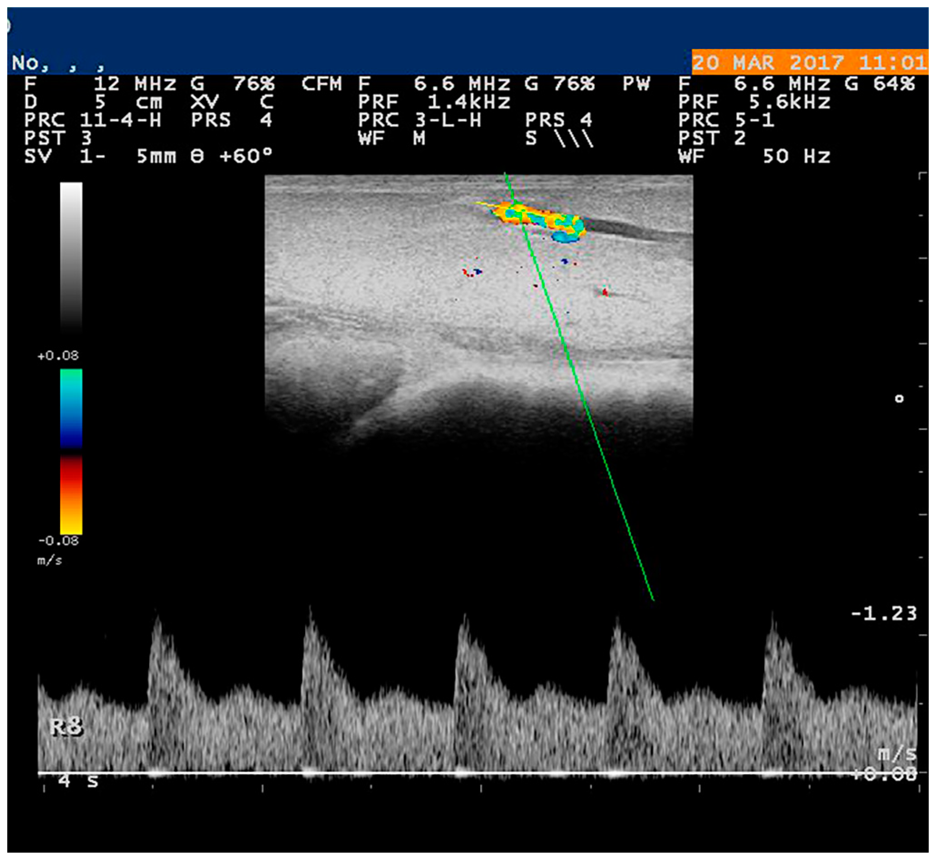
5.1. Doppler Examination

Doppler ultrasonography can be used for the estimation of blood flow into the mammary glands. Power Doppler imaging uses the amplitude of a Doppler shift to detect blood movement, but without information regarding its direction [35]. Colour Doppler images are generally combined with grayscale (B-mode) images to display duplex ultrasonography images while colour Doppler flow is the presentation of the velocity by colour scale and provides information regarding its localization with the identification of the type of vessel [5].
Usually, a B-mode ultrasonographic examination precedes the Doppler examination. The B-mode ultrasonographic examination provides information about the dimensions of the mammary glands, as well as about the echotexture (homogeneous or heterogeneous) and the echogenicity (hypo, hyperechoic, or mixed) of the parenchyma [5]. In a study of canine mastitis, it has been found that, during inflammation, the tissue had reduced echogenicity and that the distinct layers, characteristic of the normal mammary tissue, were lost [36]. In cattle mastitis, the sonographic image depends on the degree of structural changes that occur in the tissue [37]. A non-homogeneous hypoechoic pattern was imaged in the case of acute mastitis [38] while a hyperechoic pattern was imaged in chronic mastitis as a result of fibrosis [37]. However, criteria allowing the differentiation between inflammation and neoplasia by ultrasonographic examination are not provided by current literature. Inflammation is very common in mammary neoplasia, especially during the prolonged process of tumour, and also, in clinical practice, mastitis may be indistinguishable from a mammary tumour with present inflammation [39], as has been reported in human medicine [40].
During the evaluation of vascularization by means of spectral Doppler, the settings of the equipment are of particular importance in reliable measurement. In particular, the angle between the Doppler beam and the vessel’s long axis (‘Doppler angle’) should not exceed 60°, colour gain must be adjusted in a way that colour visualizes only inside the vessel, and the Pulse Repetition Frequency should be set appropriately to avoid the ‘aliasing phenomenon’ [41]. The Doppler gate, which is dependent on the diametre of the vessel, should be regulated at 2 to 4 mm and positioned at the center of the vessel under examination (Figure 1). A minimum of three continuous and consecutive waves are needed for an accurate evaluation. The parametres measured in most studies are the following: (a) peak systolic velocity (PSV), (b) end diastolic velocity (EDV), (c) resistance index [RI = (PSV − EDV)/PSV], and (d) pulsatility index [PI = (PSV − EDV)/TAMV] (TAMV: time-averaged maximum velocity); in some cases, important information can also be extracted by (e) systolic: diastolic velocity ratio [SV/DV = (ASF/ADF)] (ASF: average diastolic flow and ADF: average systolic flow), (f) time-averaged maximum velocity (TAMV), (g) systolic acceleration (A), and (h) total blood volume [Q = (TAVmean × S)] (TAVmean: time-averaged mean velocity, S: cross-sectional surface of the vessel) [41,42]. Finally, the characterization of blood flow pattern has also been reported, as the laminar or turbulent flow and the high or low resistivity are observations that may contribute to the diagnosis [43]. Potentially, the assessment of the blood flow pattern can be used to assess the efficacy of anti-vascular therapy [44,45].
During the examination, the farm animals should be in a standing position, restrained by an assistant [41] whilst the companion animals should be in lateral recumbency and no sedation during examination is needed [4]. The hair of the specific region should be fully clipped [41].
In farm animals, the technique has been applied in healthy animals for monitoring and evaluation of mammary blood flow in lactating [46] or dry [47] cows, in heifers [48], in ewes [49,50] and goats [51,52]. Regarding mastitis, many studies have employed the Doppler ultrasonographic examination for the investigation of the infection in cattle [53], buffaloes [54], and sheep [50], thus nowadays the Doppler examination is considered a precise method for the diagnosis of this disease. Santos et al. [55] have indicated that increased PSV and RI in cases of mastitis in goats are probably related to more severe infections. Furthermore, the examination of the supramammary lymph nodes may also provide useful information regarding infection in cows [42].
In companion animals, by using colour flow imaging, a mainly peripheral vascular pattern has been observed in benign tumours in dogs whilst in mammary malignant lesions, a mixed pattern has been usually reported [43,56]. Moreover, by using the spectral mode, increased blood flow has been observed in malignant lesions, specifically a significant increase in PSV [43] whilst no differences were detected in PI or RI [57]. The use of the modality in mastitis in dogs has been reported by Balaci et al. [58] and Trasch et al. [36], who used Doppler examination for evaluating the efficacy of treatment protocols against mastitis. It seems that Doppler ultrasound evaluations may assist in the prediction of the malignancy of canine mammary masses but with moderate sensitivity and specificity [5].
The advantages of the technique are the real-time information about vascularization and haemodynamic aspects of blood vessel flow that it can provide [59] and the affordable cost of the equipment [41]. The disadvantage may be the inability to detect small lesions or microcalcifications that may lead to false negative results in companion animal tumours [60].

5.2. Contrast-Enhanced Examination

Contrast-enhanced ultrasonographic examination (CEUS) is a novel imaging technique that, in humans, is employed for the evaluation of mammary tumours [61]. Recently, CEUS has been found useful for the detection of lesions in mammary glands of animals [62]. For this examination, the ultrasound equipment must be fitted with specific software to perform the secondary harmonic imaging and inverted pulse whilst the use of a contrast agent is also necessary [62]. The microbubbles used as contrast agent must possess certain characteristics: stability with resistance to external pressures to prevent their dissolution and reduced gas diffusion into the blood [63]. Nowadays, the microbubbles used have a small size (1–10 mm in diametre); include a protein, lipid, or polymer shell capsule; and contain an inert and relatively insoluble gas [56,64,65]. Great attention must be paid to parametres like depth, gain, and focal zones, which must be maintained constant throughout the examination [5]. Video clips are taken immediately and for a short period after the injection of the contrast agent and recorded. Microbubble perfusion and the dynamic enhancement of the image of each lesion are thus analyzed, considering the presence or absence of the contrast agent in the tissue and the times of tissue perfusion from wash-in to enhancement peak to washout [5] (Figure 2).
During the performance of the technique, the contrast agent is administrated intravenously through the cephalic (dogs, cats) or the jugular (ruminants) vein [62,66].
In human medicine, as found in mouse animal model, the tumour growth time can be identified by using CEUS examination [61]. CEUS is applied for predicting the nature of mammary lesions, in that way reducing the number of biopsies performed in patients [67]. The increased correlation of lesion vascularization in mammary tumours found by using CEUS with the results of MRI, further highlights the significant accuracy of the method [68]. A recent variation of the classical CEUS is the intradermal and subcutaneous injection of contrast agent in the region of the mammary areola, which may be of clinical application in breast cancer patients [69]. Although there are many studies certifying the diagnostic efficacy of CEUS examination for the characterization of mammary lesions in humans [70,71], in animal health the technique has received limited attention. In farm animals, there is only one study [62]. This study indicated that the reduced perfusion of a contrast agent into the mammary parenchyma of ewes correlated with the reduced amount of functional mammary tissue in long-standing mammary lesions [62].
In companion animals, the method has been described in mammary glands and inguinal lymph nodes in healthy dogs at the various stages of the oestrous cycle [66] and also in dogs with mammary tumour [10]. Moreover, a transcutaneous injection of contrast agent has been applied in dogs’ mammary glands, which was used as an animal model [72,73]. Recently, Stan et al. [10] proposed the combination of B-mode, Doppler, and CEUS examinations along with real-time elastography for the assessment of sentinel lymph nodes in dogs with mammary carcinoma. They indicated that elastography had the highest accuracy in identifying metastases in sentinel lymph nodes whilst CEUS and Doppler examinations had similar accuracy. However, in another study, the use of CEUS in mammary lesions of dogs did not conclude to differentiation of neoplastic and non-neoplastic tissues [74].
The advantages of CEUS examination include the imaging of the progression of vessel formation and evaluation of blood flow in functional vessels [75], which is important information in the investigation of mammary tumours. The disadvantages may be the inability to detect small lesions, as in a study in human hepatocellular carcinoma where the smallest detectable lesions ranged from 3 to 5 mm in diameter [76], and the low specificity (16.7%) of the method [5].

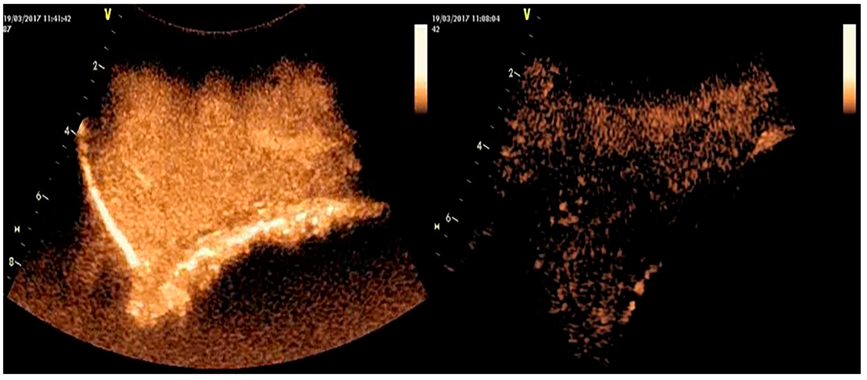
5.3. Three-Dimensional Examination

Three-dimensional (3-D) ultrasonography is a relatively new imaging method that has received a limited application in mammary imaging in animals [77,78] despite its extensive use and rapid development in humans [79,80]. The three-dimensional ultrasonographic examination of the breast is a useful tool for various procedures, e.g., breast biopsy or anatomic guidance during breast neoplasia surgery [81,82].
For this technique, a conventional transducer with or without an electromagnetic position sensor can be used. However, in the latter case, defects may appear on the display, as, during movement of the transducer across the region of interest in order to capture multiple two-dimensional images, some areas may easily be under- or over-scanned and the slice spacing may vary; hence, the data corresponding to the volume of structures of interest would not be accurate if an electromagnetic position sensor was not used [83]. Therefore, nowadays, it is preferable to use the ‘volume transducer’, which is a modern transducer with a positioning system and scanning mechanics (a built-in mechanical drive to carry out a fully automatic sweep). The transducer is held and not moved whilst the transducer elements automatically sweep through the volume box, which is the operator-selected region of interest [77].
In a two-dimensional ultrasonographic examination, the target tissue is scanned in different planes and the operator must reconstruct a multitude of images in his mind in order to receive a virtual three-dimensional image [84]. In contrast, in the three-dimensional examination, image reconstruction is performed directly by the software of the equipment, and the processed signals are presented to the monitor as a real three-dimensional image [84,85].
In farm animals, there are only two bibliographic reports. In these, Fasulkov et al. [78] and Franz et al. [77] applied the technique for the visualization and characterization of structures of lactating mammary glands in healthy cattle and obtained clear imaging of the teat, the ductal system, and the mammary parenchyma. The same procedures in animals as in the Doppler examination are required for the performance of the 3-D examination [78].
In companion animals, the application of this technique has not been reported yet.
The advantage of this technique is the better sonographic discrimination of malignant from benign masses than 2D, as 3-D provides more information on vascular morphology [86]. The disadvantages could be the high cost of the necessary equipment and the need for specialized knowledge for applying the technique [87].

5.4. Elastography

Elastography is an ultrasonographic technique employed to measure the elasticity and stiffness of tissues, in order to increase diagnostic sensitivity [88]. The tissue elasticity is assessed by the evaluation of the pressure force applied on the tissue and the resulting deformation. The tissue deformation can be estimated by high-frequency echo signals, whilst a value of tension cannot be evaluated on the basis of the tissue measurement. In this respect, the procedure of exerting force on the tissues must be performed under strictly controlled conditions [89,90].
The elasticity of a tissue (even within the same organ) may vary according to the physiological state (e.g., lactating or dry mammary gland) or the pathological process (e.g., inflammation, neoplasia) in there. Hence, by using the appropriate software in the ultrasonographic equipment, this elasticity can be visualized [91]. During the examination, the equipment initially receives digitized radiofrequency echo signs from the tissue as it is (i.e., without compression); then, the compression applied to the tissue by the transducer along the radiation axis results in some displacement of the tissue; finally, the equipment receives a second, post-compression digitized radiofrequency echo sign from the same tissue [92]. The data received from these two echo lines (i.e., before and after compression) undergo processing, which results in the production of the ‘elastogram’ (i.e., the elastographic image).
Several types of elastographic techniques can be applied, which depend upon the force that is applied onto the tissue; these include real-time elastography, acoustic radiation force impulse imaging, transient elastography, point shear wave elastography, and shear wave elastography [93]. The determinant for the success of the technique is the force exerted by the ultrasound transducer on the tissue under examination [90]. Shear wave elastography is an advanced technique that allows objective measurement of the elasticity of a tissue [94,95]. In real-time elastography, the stiffness of the tissue is transposed in colour mode, where each colour signifies a level of stiffness: images with blue areas correspond to soft tissue areas, i.e., more elastic, whilst reddened areas correspond to more rigid tissue areas, i.e., with lower elasticity [5]. Then, the evaluation of shades of colours into scores of elasticities has been made possible [96,97].
In farm animals, the technique was described by Skerl et al. [94], who used samples of mammary glands of cattle to monitor the pressure applied during manually performed elastography.
In companion animals, acoustic radiation force impulse imaging (ARFI) has been used in dogs [5,98] or cats [99] with mammary tumours. Feliciano et al. [98] compared acoustic radiation force impulse results with carcinoma types and grades, as they were estimated by histopathological classification. They found that the method could identify complex carcinoma types with moderate accuracy due to the shorter periods of contrast wash-in and peak enhancement times observed in this type of tumour. They also found that the increase in perfusion times (wash-in, peak enhancement, and washout times) enables the detection of grade II and III carcinomas with moderate accuracy. Although the diagnostic technique is not sensitive enough for the differentiation of special and complex carcinoma types [98], it is highly recommended in veterinary oncology for malignancy prediction of canine mammary masses [5].
Additionally, shear wave elastography has been used for the differential diagnosis of benign from malignant lesions in canine mammary tumours [95,100]. Glińska-Suchocka et al. [95] performed shear wave elastographic examination in neoplastic mammary glands of dogs and then fine-needle aspiration biopsy to determine the nature and degree of malignancy of the lesions. They found that benign neoplasms of the mammary gland showed low stiffness, whereas malignant neoplasms were characterized by high stiffness. On the other hand, Pieczewska et al. [100] evaluated by shear wave elastography the density of the canine mammary tumour before and after the treatment with aglepristone and found no influence on the density of the tumour’s tissue. Among the various elastography techniques, shear wave elastography has been noted as the most promising tool, as it allows fast and non-invasive diagnosis of malignant mammary tumours in dogs [100].
Based on the above studies, it is inferred that elastography is a useful tool for predicting malignancy with high sensitivity (94.7%) and specificity (97.2%) [5], but, in many cases, the biopsy cannot be avoided [101].

6. Discussion

A variety of techniques can be used to visualize mammary lesions in the various available imaging techniques. In CT, x-ray radiation is used to receive a cross-sectional image of the body in MRI a magnetic field forces the hydrogen cellular nuclei to align in different positions, and in PET, the radiation emitted from the animal after injection of radioactive tracers is visualized [3]. In ultrasonographic examination, high-frequency sound waves are used with different modalities in order to receive tissue-related information [5]. Although imaging techniques vary, their use in animals depends on animal species and pathological condition. The mammary diseases that have been mainly studied in animals are mastitis and mammary tumours. Mastitis occurs frequently in cows and ewes and has a significant impact on animal’s welfare and a huge economical effect on the dairy industry [102,103], so its early diagnosis is imperative. On the other hand, mammary tumours have been studied thoroughly in dogs and cats. Mammary tumours are the most common neoplasm in intact female dogs [104] and the third most common neoplasm in domestic cats [105]. Therefore, the imaging of mammary tumours as well as the diagnostic accuracy of each method has concerned many researchers.
In human medicine, mammography and ultrasonography have an important role in the diagnosis of mammary diseases, but still, a biopsy guided with ultrasound examination is necessary for the histopathological confirmation of the findings [9]. MRI can provide additional information [106]. In addition, CEUS studies can reveal the presence of nodular-type lesions [9] while the combination of PET/CT may be useful in cases of mammary infections [9] and can be used to confirm mammary neoplasia [107,108,109]. Furthermore, Doppler examination can detect increased vascularization within the mammary gland [110] and also dilated lactiferous ducts with echoic content or with thickened walls, a finding that MRI can also reveal [106]. In regard to the mammary neoplasia, Doppler examination has been used for the quantitative evaluation of the perfusion of the mammary parenchyma and the vascularization of neoplastic tissues [111,112], as well as for the differentiation between malignant and benign mammary lesions [113]. It seems that sometimes, imaging features cannot provide differentiation between mammary neoplasia or infection; for example, ultrasonographic examination would show irregular hypoechoic masses with increased vascularity while MRI would show irregular, enhancing masses or non-mass enhancement with micro-abscesses [114]. Although in human medicine diagnostic imaging guidelines have been proposed to investigate diseases of the mammary glands [9], in veterinary work, similar protocols have not been developed thus far and only an algorithm has been proposed for the evaluation of sentinel lymph nodes in dogs with mammary carcinoma [10].
In farm animals, the ultrasonographic examination of the mammary glands has been studied extensively [37,41,50] while the rest of the imaging methods were studied less, mainly for the estimation of the normal parenchyma [24,31] or for highlighting the benefits of the methods in human medicine when animal model studies were performed [115]. The easy transfer of the ultrasound machine to the farm, the fast real-time imaging, and its application without the need for anesthetizing the animal [41] are reasons for its wide use (Table 1). Ultrasonography has been proposed as a diagnostic technique for the diagnosis of subclinical mastitis in sheep [116] and as a tool indicating the proper time to start treatment [117] and also monitoring the progress of mastitis in cows [118]. After the ultrasonographic examination, microbiological examination and California mastitis test (CMT) of milk samples are employed to confirm the disease [119].
Nowadays, in clinical practice, mammary neoplasia in dogs and cats is usually detected by radiography, ultrasound, or computed tomography and, subsequently, is confirmed with histopathological examination [2,120,121]. For the differentiation of mammary malignant and benign tumours, Doppler examination (colour flow and spectral mode) is useful [10] whilst contrast-enhanced ultrasonography and acoustic radiation force impulse imaging elastography can be applied for the identification of some of the characteristics of mammary carcinoma in dogs [98]. Feliciano et al. [5] recommended the use of acoustic radiation imaging elastography in veterinary oncology, as it allows the fast, non-invasive, and complication-free prediction of malignant tumours in dogs. On the other hand, Gasser et al. [74] attempted to reveal differences between different types of benign mammary lesions in dogs by using Doppler, acoustic radiation imaging elastography, and contrast-enhanced techniques, but no conclusive findings could be obtained. Additionally, in animals with mammary tumours, the examination of lymph nodes and detection of potential metastases is particularly important, as well as the detection of lung metastasis, where the CT has been found to have higher sensitivity than chest radiographies [2]. In cases of metastatic lymph nodes, acoustic radiation imaging-elastography is the ideal method that may differentiate free and reactive tumour metastatic lymph nodes [122]. Contrast-enhanced ultrasound has improved the diagnostic possibilities regarding mammary abnormalities, as contributes to malignancy prediction [123]. Last but not least, MRI and PET are promising methodologies that have been applied to companion animals [4,8]; however, high equipment costs limit their application in clinical practice [3,4].
In conclusion, concerning mastitis in farm animals, ultrasonographic examination (Doppler, CEUS, elastography) could be performed as a first examination for the diagnosis of clinical or subclinical mastitis and treatment guidance, but a bacteriological examination and a California mastitis test (CMT) of milk samples should follow for the confirmation of the disease [116]. Regarding mammary tumours in companion animals, a combination of imaging techniques should be applied for accurate diagnosis. Initially, a first assessment of the extent of the tumour, its vascularity, and its characterization could be carried out using Doppler ultrasonography [124], and then, depending on the available equipment, CT, MRI, or PET can be applied, taking into account that CT and MRI provide information about anatomy while PET about the functionality of the tissue. Whichever imaging modality is used, biopsy or ultrasound-guided biopsy in cases of small sized not palpated lesions for histopathological examination is essential for an accurate diagnosis [125].

Author Contributions

Conceptualization, M.S.B., P.G.G.; visualization, M.S.B.; writing—original draft preparation, M.S.B.; writing—review and editing, M.S.B., P.G.G. All authors have read and agreed to the published version of the manuscript.

Funding

This work received no external funding.

Institutional Review Board Statement

Not applicable.

Informed Consent Statement

Not applicable.

Data Availability Statement

Not applicable.

Conflicts of Interest

The authors declare no conflict of interest.

References

  1. Olsen, A.K.; Zeidler, D.; Pedersen, K.; Sørensen, M.; Jensen, S.B.; Munk, O.L. Imaging techniques: CT, MRI, and PET scanning. In Swine in the Laboratory. Surgery, Anesthesia, Imaging, and Experimental Techniques, 3rd ed.; Swindle, M.M., Smith, A.C., Eds.; CRC Press: Boca Raton, FL, USA, 2007; pp. 387–395. [Google Scholar]
  2. Otoni, C.C.; Rahal, S.C.; Vulcano, L.C.; Ribeiro, S.M.; Hette, K.; Giordano, T.; Doiche, D.P.; Amorim, R.L. Survey radiography and computerized tomography imaging of the thorax in female dogs with mammary tumors. Acta Vet. Scand. 2010, 52, 20. [Google Scholar] [CrossRef] [PubMed] [Green Version]
  3. Alstrup, A.K.O.; Winterdahl, M. Imaging Techniques in Large Animals. Scand. J. Lab. Anim. Sci. 2009, 36, 55–66. [Google Scholar] [CrossRef]
  4. Garamvoglyi, R.; Petrasi, Z.S.; Hevesi, A.; Jakab, C.S.; Vajda, Z.S.; Bogner, P.; Repa, I. Magnetic resonance imaging technique for the examination of canine mammary tumours. Acta Vet. Hung. 2006, 54, 143–159. [Google Scholar] [CrossRef] [Green Version]
  5. Feliciano, M.A.R.; Uscategui, R.A.R.; Maronezi, M.C.; Simões, A.P.R.; Silva, P.; Gasser, B.; Pavan, L.; Carvalho, C.F.; Canola, J.C.; Vicente, W.R.R. Ultrasonography methods for predicting malignancy in canine mammary tumors. PLoS ONE 2017, 12, e0178143. [Google Scholar] [CrossRef] [PubMed] [Green Version]
  6. Prokop, M. Principles of CT, Spiral CT, and Multislice CT. In Spiral and Multislice Computed Tomography of the Body, 1st ed.; Prokop, M., Galanski, M., Eds.; Thieme: Stuttgart, Germany, 2001; pp. 2–7. [Google Scholar]
  7. Maisey, M.N. Positron Emission Tomography in Clinical Medicine. In Positron Emission Tomography: Basic Sciences, 1st ed.; Bailey, D.L., Townsend, D.W., Valk, P.E., Maisey, M.N., Eds.; Springer: London, UK, 2005; pp. 1–12. [Google Scholar]
  8. Sánchez, D.; Romero, L.; López, S.; Campuzano, M.; Ortega, R.; Morales, A.; Guadarrama, M.; Cesarman-Maus, G.; García-Pérez, O.; Lizano, M. 18F-FDG—PET/CT in Canine Mammary Gland Tumors. Front. Vet. Sci. 2019, 6, 280. [Google Scholar] [CrossRef]
  9. Grover, H.; Grover, S.B.; Goyal, P.; Hedge, R.; Gupta, S.; Malhotra, S.; Li, S.; Gupta, N. Clinical and imaging features of idiopathic granulomatous mastitis—The diagnostic challenges and a brief review. Clin. Imaging 2021, 69, 126–132. [Google Scholar] [CrossRef]
  10. Stan, F.; Gudea, A.; Damian, A.; Gal, A.F.; Papuc, I.; Pop, A.R.; Martonos, C. Ultrasonographic Algorithm for the Assessment of Sentinel Lymph Nodes That Drain the Mammary Carcinomas in Female Dogs. Animals 2020, 10, 2366. [Google Scholar] [CrossRef]
  11. Sørensen, M.T.; Sejrsen, K.; Foldager, J. Estimation of Pubertal Mammary Development in Heifers by Computed Tomography. J. Dairy Sci. 1987, 70, 265–270. [Google Scholar] [CrossRef]
  12. Keane, M.; Paul, E.; Sturrock, C.J.; Rauch, C.; Rutland, C.S. Computed Tomography in Veterinary Medicine: Currently Published and Tomorrow’s Vision. In Computed Tomography—Advanced Applications, 1st ed.; Halefoglu, A.M., Ed.; IntechOpen: London, UK, 2017; pp. 271–283. [Google Scholar]
  13. Sejrsen, K.; Foldager, J.; Sørensen, M.T.; Akers, R.M.; Bauman, D.E. Effect of exogenous bovine somatotropin on pubertal mammary development in heifers. J. Dairy Sci. 1986, 69, 1528–1535. [Google Scholar] [CrossRef]
  14. Akers, M.R. A 100-Year Review: Mammary development and lactation. J. Dairy Sci. 2017, 100, 10332–10352. [Google Scholar] [CrossRef]
  15. Kim, S.; Kwon, K.; Choi, H.; Lee, Y. Evaluation of Mammary Gland Calcification in Dogs; Radiography and Computed Tomography. J. Emb. Trans. 2017, 32, 183–192. [Google Scholar] [CrossRef]
  16. Boone, J.M.; Kwan, A.L.; Yang, K.; Burkett, G.W.; Lindfors, K.K.; Nelson, T.R. Computed tomography for imaging the breast. J. Mammary Gland Biol. Neoplasia 2006, 11, 103–111. [Google Scholar] [CrossRef] [PubMed]
  17. Soultani, C.; Patsikas, M.N.; Karayannopoulou, M.; Jakovljevic, S.; Chryssogonidis, I.; Papazoglou, L.; Papaioannou, N.; Papadopoulou, P.; Pavlidou, K.; Ilia, G.M.; et al. Assessment of sentinel lymph node metastasis in canine mammary gland tumors using computed tomographic indirect lymphography. Vet. Radiol. Ultrasound. 2017, 58, 186–196. [Google Scholar] [CrossRef] [PubMed]
  18. Agrawal, A.; Rangarajan, V. Appropriateness criteria of FDG PET/CT in oncology. Indian J. Radiol. Imaging 2015, 25, 88–101. [Google Scholar] [CrossRef] [PubMed]
  19. Ganapathy, V.; Thangaraju, M.; Prasad, P.D. Nutrient transporters in cancer: Relevance to Warburg hypothesis and beyond. Pharmacol. Ther. 2009, 121, 29–40. [Google Scholar] [CrossRef]
  20. Vander Heiden, M.G.; Cantley, L.C.; Thompson, C.B. Understanding the Warburg effect: The metabolic requirements of cell proliferation. Science 2009, 324, 1029–1033. [Google Scholar] [CrossRef] [PubMed] [Green Version]
  21. Shen, B.; Huang, T.; Sun, Y.; Jin, Z.; Li, X.F. Revisit 18F-fluorodeoxyglucose oncology positron emission tomography: “Systems molecular imaging” of glucose metabolism. Oncotarget 2017, 8, 43536–43542. [Google Scholar] [CrossRef] [PubMed] [Green Version]
  22. Kasem, J.; Wazir, U.; Mokbel, K. Sensitivity, Specificity and the Diagnostic Accuracy of PET/CT for Axillary Staging in Patients with Stage I-III Cancer: A Systematic Review of the Literature. In Vivo 2021, 35, 23–30. [Google Scholar] [CrossRef] [PubMed]
  23. Meyer, P.T.; Circiumaru, V.; Cardi, C.A.; Thomas, D.H.; Bal, H.; Acton, P.D. Simplified quantification of small animal [18F] FDG PET studies using a standard arterial input function. Eur. J. Nucl. Med. Mol. Imaging 2006, 33, 948–954. [Google Scholar] [CrossRef]
  24. Danielsen, E.; Smith, D.F.; Poulsen, P.H.; Østergaard, L.; Gee, A.; Ishizu, K.; Venkatachalam, T.K.; Benders, D.; Hansen, S.; Gjedde, A.; et al. Positron emission tomography of living brain in minipigs and domestic pigs. Scand J. Lab. Anim. Sci. 1998, 25, 127–135. [Google Scholar]
  25. Olsen, A.K.; Keiding, S.; Munk, O.L. Effect of hypercapnia on cerebral blood flow and blood volume in pigs studied by positron emission tomography. Comp. Med. 2006, 56, 416–420. [Google Scholar] [PubMed]
  26. Black, N.F.; McJames, S.; Rust, T.C.; Kardmas, D.J. Evaluation of rapid dual-tracer 62Cu-PTSM + 62Cu-ATSM PET in dogs with spontaneously occurring tumors. Phys. Med. Biol. 2008, 53, 217–232. [Google Scholar] [CrossRef] [PubMed] [Green Version]
  27. Brown, R.W.; Cheng, Y.-C.N.; Haacke, E.M.; Thompson, M.R.; Venkatesan, R. Classical Response of a Single Nucleus to a Magnetic Field. In Magnetic Resonance Imaging: Physical Principles and Sequence Design, 2nd ed.; Brown, R.W., Cheng, Y.-C.N., Haacke, E.M., Thompson, M.R., Venkatesan, R., Eds.; Wiley-Blackwell: New York, NY, USA, 2014; pp. 19–36. [Google Scholar]
  28. Müller-Schimpfle, M.; Stoll, P.; Stern, W.; Kurz, S.; Dammann, F.; Claussen, C.D. Do mammography, sonography and MR mammography have a diagnostic benefit compared with mammography and sonography? AJR Am. J. Roentgenol. 1997, 168, 1323–1329. [Google Scholar] [CrossRef] [PubMed]
  29. Friedrich, M. MRI of the breast: State of the art. Eur. Radiol. 1998, 8, 707–725. [Google Scholar] [CrossRef]
  30. Lepori, D. Inflammatory breast disease: The radiologist’s role. Diagn. Interv. Imaging 2015, 96, 1045–1064. [Google Scholar] [CrossRef] [Green Version]
  31. Fowler, P.A.; Knight, C.H.; Cameron, G.G.; Foster, M.A. Use of magnetic resonance imaging in the study of goat mammary glands in vivo. J. Reprod. Fert. 1990, 89, 359–366. [Google Scholar] [CrossRef]
  32. Stelwagen, K.; McBride, B.W.; Grieve, D.G.; Towner, R.A. Nuclear magnetic resonance imaging and proton spectroscopy used as a technique to assess mammary gland composition in Holstein heifers. Can. J. Anim. Sci. 1990, 70, 1151–1154. [Google Scholar] [CrossRef]
  33. Jaramillo-Chaustre, X.L.; Bustamante-Cano, J.J.; Gómez-Parra, F. Lymphography by MRI for animal model canine. Iteckne 2017, 14, 46–61. [Google Scholar] [CrossRef]
  34. Aristokli, N.; Polycarpou, I.; Themistocleous, S.C.; Sophocleous, D.; Mamais, I. Comparison of the diagnostic performance of Magnetic Resonance Imaging (MRI), ultrasound and mammography for detection of breast cancer based on tumor type, breast density and patient’s history: A review. Radiography 2022, 28, 848–856. [Google Scholar] [CrossRef]
  35. Hamper, U.M.; DeJong, M.R.; Caskey, C.I.; Sheth, S. Power Doppler imaging: Clinical experience and correlations with color Doppler US and other imaging modalities. RadioGraphics 1997, 17, 499–513. [Google Scholar] [CrossRef] [Green Version]
  36. Trasch, K.; Wehrend, A.; Bostedt, H. Ultrasonographic description of canine mastitis. Vet. Radiol. Ultrasound 2007, 48, 580–584. [Google Scholar] [CrossRef] [PubMed]
  37. Flöck, M.; Winter, P. Diagnostic ultrasonography in cattle with diseases of the mammary gland. Vet. J. 2006, 171, 314–321. [Google Scholar] [CrossRef] [PubMed]
  38. Banting, A. Ultrasonographic examination of the mammary gland in cows with induced S. aureus mastitis: A criteria for prognosis and evaluation of therapy. Cattle Pract. 1998, 6, 121–124. [Google Scholar]
  39. Kaszak, I.; Ruszczak, A.; Kanafa, S.; Witkowska-Piłaszewicz, O.; Sacharczuk, M.; Jurka, P. New insights of canine mastitis—A review. Anim. Sci. Pap. Rep. 2018, 36, 33–44. [Google Scholar]
  40. Guirguis, M.S.; Adrada, B.; Santiago, L.; Candelaria, R.; Arribas, E. Mimickers of breast malignancy: Imaging findings, pathologic concordance and clinical management. Insights Imaging 2021, 12, 53. [Google Scholar] [CrossRef] [PubMed]
  41. Petridis, I.G.; Barbagianni, M.S.; Ioannidi, K.S.; Samaras, E.; Fthenakis, G.C.; Vloumidi, E.I. Doppler ultrasonographic examination in sheep. Small Rumin. Res. 2017, 152, 22–32. [Google Scholar] [CrossRef]
  42. Risvanli, A.; Dogan, H.; Safak, T.; Kilic, M.A.; Seker, I. The relationship between mastitis and the B-mode, colour Doppler ultrasonography measurements of supramammary lymph nodes in cows. J. Dairy Res. 2019, 86, 315–318. [Google Scholar] [CrossRef]
  43. Feliciano, M.A.; Vicente, W.R.; Silva, M.A. Conventional and Doppler ultrasound for the differentiation of benign and malignant canine mammary tumours. J. Small Anim. Pract. 2012, 53, 332–337. [Google Scholar] [CrossRef]
  44. McDonald, D.M.; Choyke, P.L. Imaging of angiogenesis: From microscope to clinic. Nat. Med. 2003, 9, 713–725. [Google Scholar] [CrossRef]
  45. Abma, E.; Stock, E.; De Spiegelaere, W.; Brantegem, L.V.; Vanderperren, K.; Ni, Y.; Vynck, M.; Daminet, S.; Clercq, K.D.; de Rooster, H. Power Doppler ultrasound and contrast-enhanced ultrasound demonstrate non-invasive tumour vascular response to anti-vascular therapy in canine cancer patients. Sci. Rep. 2019, 9, 9262. [Google Scholar] [CrossRef] [Green Version]
  46. Piccione, G.; Arcigli, A.; Fazio, F.; Giudice, E.; Caola, G. Pulsed wave-Doppler ultrasonographic evaluation of mammary blood flow speed in cows during different productive periods. Acta Sci. Vet. 2004, 32, 171–175. [Google Scholar] [CrossRef]
  47. Braun, U.; Forster, E. B-mode and colour Doppler sonographic examination of the milk vein and musculophrenic vein in dry cows and cows with a milk yield of 10 and 20 kg. Acta Vet. Scand. 2012, 54, 15. [Google Scholar] [CrossRef] [PubMed] [Green Version]
  48. Esselburn, K.M.; Hill, T.M.; Bateman, H.G., 2nd; Fluharty, F.L.; Moeller, S.J.; O’Diam, K.M.; Daniels, K.M. Examination of weekly parenchymal area by ultrasound, mammary mass, and composition in Holstein heifers reared on 1 or 3 diets from birth to 2 months of age. J. Dairy Sci. 2015, 98, 5280–5293. [Google Scholar] [CrossRef] [PubMed]
  49. Piccione, G.; Arcigli, A.; Assenza, A.; Percipalle, M.; Caola, G. Pulsed wave-Doppler ultrasonographic evaluation of the mammary blood flow in the ewe. Acta Vet. Brno 2004, 73, 23–27. [Google Scholar] [CrossRef]
  50. Barbagianni, M.S.; Mavrogianni, V.S.; Vasileiou, N.G.C.; Fthenakis, G.C.; Petridis, I.G. Ultrasonographic examination of the udder in sheep. Small Rumin. Res. 2017, 152, 86–99. [Google Scholar] [CrossRef]
  51. Nielsen, M.O.; Jakobsen, K.; Jørgensen, N.J. Changes in mammary blood flow during the lactation period in goats measured by the ultrasound Doppler principle. Comp. Biochem. Physiol. A Comp. Physiol. 1990, 97, 519–524. [Google Scholar] [CrossRef]
  52. Christensen, K.; Nielsen, M.O.; Bauer, R.; Hilden, K. Evaluation of mammary blood flow measurements in lactation goats using the ultrasound Doppler principle. Comp. Biochem. Physiol. A Comp. Physiol. 1989, 92, 385–392. [Google Scholar] [CrossRef]
  53. Porcionato, M.A.; Soares, W.V.; Reis, C.B.; Cortinhas, C.S.; Mestieri, L.; Santos, M.V. Milk flow, teat morphology and subclinical mastitis prevalence in Gir cows. Pesqui. Agropecu. Bras. 2010, 45, 1507–1512. [Google Scholar] [CrossRef] [Green Version]
  54. Rambabu, K.; Sreenu, M.; Kumar, R.V.S.; Rao, T.S.C. Ultrasonography of the udder and teat in buffaloes. Buffalo Bull. 2009, 28, 5–10. Available online: http://ibic.lib.ku.ac.th/.../2009-5.htm (accessed on 12 May 2022).
  55. Santos, V.J.C.; Simplício, K.M.M.G.; Sanchez, D.C.C.; Almeida, V.T.; Teixeira, P.P.M.; Coutinho, L.N.; Rodrigues, L.F.S.; Oliveira, M.E.F.; Feliciano, M.A.R.; Vicente, W.R.R. Conventional and Doppler ultrasonography on a goat with gangrenous mastitis. Vicente. Arq. Bras. Med. Vet. Zootec. 2014, 66, 1931–1935. [Google Scholar] [CrossRef] [Green Version]
  56. Mantziaras, G.; Luvoni, G.C. Advanced ultrasound techniques in small animal reproduction imaging. Reprod. Domest. Anim. 2020, 2, 17–25. [Google Scholar] [CrossRef] [PubMed]
  57. Soler, M.; Dominguez, E.; Lucas, X.; Novellas, R.; Gomes-Coelho, K.V.; Espada, Y.; Agut, A. Comparison between ultrasonographic findings of benign and malignant canine mammary gland tumours using B-mode, colour Doppler, power Doppler and spectral Doppler. Res. Vet. Sci. 2016, 107, 141–146. [Google Scholar] [CrossRef] [PubMed]
  58. Balaci, I.; Ciupe, S.; Pop, A.; Parlapan, L.; Arion, A.; Vasiu, I.; Purdoiu, R.; Papuc, I.; Groza, I. Ultrasonographic Findings of Mastitic and Normal Mammary Gland in Bitches. Vet. Med. 2015, 72, 110–116. [Google Scholar] [CrossRef] [PubMed] [Green Version]
  59. Carvalho, C.F.; Chammas, M.C.; Cerri, G.G. Physical principles of Doppler ultrasonography. Cienc. Rural 2008, 38, 872–879. [Google Scholar] [CrossRef]
  60. Lana, S.E.; Rutteman, G.R.; Withrow, S.J. Tumors of the Mammary Gland. In Withrow and MacEwen’s Small Animal Clinical Oncology, 4th ed.; Withrow, S.J., Vail, D.M., Eds.; Saunders Elsevier: St. Louis, MO, USA, 2007; pp. 619–638. [Google Scholar] [CrossRef]
  61. Wang, M.; Feng, H.L.; Liu, Y.Q.; Liu, H.; Jiang, Y.X.; Zhu, Q.L.; Dai, Q.; Li, J.C. Angiogenesis Research in Mouse Mammary Cancer Based on Contrast-enhanced Ultrasonography: Exploratory Study. Acad. Radiol. 2018, 25, 889–897. [Google Scholar] [CrossRef] [PubMed]
  62. Mantziaras, G.; Vasileiou, N.G.; Ioannidi, K.S.; Mavrogianni, V.S.; Gougoulis, D.A.; Fthenakis, G.C.; Petridis, I.G.; Barbagianni, M.S. Use of contrast-enhanced ultrasonographic examination to evaluate health status of mammary glands of ewes at the end of a lactation period. J. Dairy Res. 2018, 85, 39–43. [Google Scholar] [CrossRef]
  63. Bouakaz, A.; De Jong, N.; Cachard, C. Standard properties of ultrasound contrast agents. Ultrasound Med. Biol. 1998, 24, 469–472. [Google Scholar] [CrossRef]
  64. Cosgrove, D.; Harvey, C. Clinical uses of microbubbles in diagnosis and treatment. Med. Biol. Eng. Comput. 2009, 47, 813–826. [Google Scholar] [CrossRef]
  65. Appis, A.W.; Tracy, M.J.; Feinstein, S.B. Update on the safety and efficacy of commercial ultrasound contrast agents in cardiac applications. Echo Res. Pract. 2015, 2, 55–62. [Google Scholar] [CrossRef] [Green Version]
  66. Vanderperren, K.; Saunders, J.H.; Van der Vekens, E.; Wydooghe, E.; de Rooster, H.; Duchateau, L.; Stock, E. B-mode and contrast-enhanced ultrasonography of the mammary gland during the estrous cycle of dogs. Anim. Reprod. Sci. 2018, 199, 15–23. [Google Scholar] [CrossRef]
  67. Lee, S.C.; Tchelepi, H.; Grant, E.; Desai, B.; Luo, C.; Groshen, S.; Hovanessian-Larsen, L. Contrast-Enhanced Ultrasound Imaging of Breast Masses: Adjunct Tool to Decrease the Number of False-Positive Biopsy Results. J. Ultrasound Med. 2019, 38, 2259–2273. [Google Scholar] [CrossRef] [PubMed]
  68. Caproni, N.; Marchisio, F.; Pecchi, A.; Canossi, B.; Battista, R.; D’Alimonte, P.; Torricelli, P. Contrast-enhanced ultrasound in the characterisation of breast masses: Utility of quantitative analysis in comparison with MRI. Eur. Radiol. 2010, 20, 1384–1395. [Google Scholar] [CrossRef] [PubMed]
  69. Luo, J.; Feng, L.; Zhou, Q.; Chen, Q.; Liu, J.; Wu, C.; Luo, J.; Chen, J.; Wu, H.; Deng, W. The value of contrast-enhanced ultrasound in determining the location of sentinel lymph nodes in breast cancer. Cancer Imaging 2021, 21, 28. [Google Scholar] [CrossRef]
  70. Liu, H.; Jiang, Y.X.; Liu, J.B.; Zhu, Q.L.; Sun, Q. Evaluation of breast lesions with contrast-enhanced ultrasound using the microvascular imaging technique: Initial observations. Breast 2008, 17, 532–539. [Google Scholar] [CrossRef]
  71. Wan, C.; Du, J.; Fang, H.; Li, F.; Wang, L. Evaluation of breast lesions by contrast enhanced ultrasound: Qualitative and quantitative analysis. Eur. J. Radiol. 2012, 81, e444–e450. [Google Scholar] [CrossRef]
  72. Gelb, H.R.; Freeman, L.J.; Rohleder, J.J.; Snyder, P.W. Feasibility of contrast-enhanced ultrasound-guided biopsy of sentinel lymph nodes in dogs. Vet. Radiol. Ultrasound. 2010, 6, 628–633. [Google Scholar] [CrossRef] [PubMed]
  73. Favril, S.; Stock, E.; Hernot, S.; Hesta, M.; Polis, I.; Vanderperren, K.; de Rooster, H. Sentinel lymph node mapping by near-infrared fluorescence imaging and contrast-enhanced ultrasound in healthy dogs. Vet. Comp. Oncol. 2019, 17, 89–98. [Google Scholar] [CrossRef] [Green Version]
  74. Gasser, B.; Rodriguez, M.G.K.; Uscategui, R.A.R.; Silva, P.A.; Maronezi, M.C.; Pavan, L.; Feliciano, M.A.R.; Vicente, W.R.R. Ultrasonographic characteristics of benign mammary lesions in bitches. Vet. Med. 2018, 63, 216–224. [Google Scholar] [CrossRef] [Green Version]
  75. Lamuraglia, M.; Bridal, S.L.; Santin, M.; Izzi, G.; Rixe, O.; Paradiso, A.; Lucidarme, O. Clinical relevance of contrast-enhanced ultrasound in monitoring anti-angiogenic therapy of cancer: Current status and perspectives. Crit. Rev. Oncol. Hematol. 2010, 73, 202–212. [Google Scholar] [CrossRef]
  76. Leoni, S.; Piscaglia, F.; Golfieri, R.; Camaggi, V.; Vidili, G.; Pini, P.; Bolondi, L. The impact of vascular and nonvascular findings on the noninvasive diagnosis of small hepatocellular carcinoma based on the EASL and AASLD criteria. Am. J. Gastroenterol. 2010, 105, 599–609. [Google Scholar] [CrossRef]
  77. Franz, S.; Hofmann-Parisot, M.M.; Baumgartner, W. Evaluation of three-dimensional ultrasonography of the bovine mammary gland. Am. J. Vet. Res. 2004, 65, 1159–1163. [Google Scholar] [CrossRef] [PubMed]
  78. Fasulkov, I.; Karadaev, M.; Vasilev, N.; Nikolov, M.; Nonov, T. Three-dimensional ultrasonography of the mammary gland in lactating cows. Tradit. Mod. Vet. Med. 2018, 3, 109–113. [Google Scholar] [CrossRef]
  79. Kotsianos, D.; Wirth, S.; Fischer, T.; Hiltawsky, K.; Sittek, H.; Reiser, M. 3D-Ultraschall (3D-US) in der Diagnostik von Mammaherdbefunden [3D ultrasound (3D US) in the diagnosis of focal breast lesions]. Radiologe 2005, 45, 237–244. [Google Scholar] [CrossRef] [PubMed]
  80. Weismann, C.; Mayr, C.; Egger, H.; Auer, A. Breast Sonography—2D, 3D, 4D Ultrasound or Elastography? Breast Care 2011, 6, 98–103. [Google Scholar] [CrossRef] [Green Version]
  81. Pretorius, D.H.; Nelson, T.R. Three-dimensional ultrasound. Ultrasound Obstet. Gynecol. 1995, 5, 219–221. [Google Scholar] [CrossRef]
  82. Riccabona, M.; Nelson, T.R.; Pretorius, D.H. Three-dimensional ultrasound: Accuracy of distance and volume measurements. Ultrasound Obstet Gynecol. 1996, 7, 429–434. [Google Scholar] [CrossRef]
  83. Nelson, T.R.; Pretorius, D.H.; Hull, A.; Riccabona, M.; Sklansky, M.S.; James, G. Sources and impact of artifacts on clinical three-dimensional ultrasound imaging. Ultrasound Obstet Gynecol. 2000, 16, 374–383. [Google Scholar] [CrossRef]
  84. Dimitrova, V.; Markov, D.; Dimitrov, R. 3D and 4D ultrasonography in obstetrics. Akush. Ginekol. (Sofiia) 2007, 46, 31–40. (In Bulgarian) [Google Scholar]
  85. Pretorius, D.H.; Nelson, T.R.; James, G. Three-dimensional ultrasound in obstetrics. In Diagnostic Imaging of Fetal Anomalies; Nyberg, D.A., McGahan, J.P., Pretorius, D.H., Pilu, G., Eds.; Lippincott Williams & Wilkins: Philadelphia, PA, USA, 2003; pp. 969–988. ISBN 0-7817-3211-5. [Google Scholar]
  86. Chen, D.R.; Lai, H.W. Three-dimensional ultrasonography for breast malignancy detection. Expert Opin. Med. Diagn. 2011, 5, 253–261. [Google Scholar] [CrossRef]
  87. Hildebrandt, T.B.; Drews, B.; Kurz, J.; Hermes, R.; Yang, S.; Göritz, F. Pregnancy monitoring in dogs and cats using 3D and 4D ultrasonography. Reprod. Domest. Anim. 2009, 44, 125–128. [Google Scholar] [CrossRef]
  88. Ophir, J.; Céspedes, I.; Ponnekanti, H.; Yazdi, Y.; Li, X. Elastography: A quantitative method for imaging the elasticity of biological tissues. Ultrason. Imaging 1991, 13, 111–134. [Google Scholar] [CrossRef]
  89. Dobruch-Sobczak, K.; Sudoł-Szopińska, I. The usefulness of sonoelastography in the differential diagnosis of solid breast lesions. Ultrasound Med. Biol. 2011, 37, 100. [Google Scholar] [CrossRef]
  90. Li, Y.; Snedeker, J.G. Elastography: Modality-specific approaches, clinical applications, and research horizons. Skeletal Radiol. 2011, 40, 389–397. [Google Scholar] [CrossRef] [Green Version]
  91. Alam, F.; Naito, K.; Horiguchi, J.; Fukuda, H.; Tachikake, T.; Ito, K. Accuracy of sonographic elastography in the differential diagnosis of enlarged cervical lymph nodes: Comparison with conventional B-mode sonography. AJR Am. J. Roentgenol. 2008, 191, 604–610. [Google Scholar] [CrossRef] [PubMed]
  92. Ophir, J.; Garra, B.; Kallel, F.; Konofagou, E.; Krouskop, T.; Righetti, R.; Varghese, T. Elastographic imaging. Ultrasound Med. Biol. 2000, 26, 23–29. [Google Scholar] [CrossRef] [PubMed]
  93. Bamber, J.; Cosgrove, D.; Dietrich, C.F.; Fromageau, J.; Bojunga, J.; Calliada, F.; Cantisani, V.; Correas, J.M.; D’Onofrio, M.; Drakonaki, E.E.; et al. EFSUMB guidelines and recommendations on the clinical use of ultrasound elastography. Part 1: Basic principles and technology. Ultraschall Med. 2013, 34, 169–184. [Google Scholar] [CrossRef] [Green Version]
  94. Skerl, K.; Eichhorn, B.; Poltorjanoks, R.; Cochran, S.; Evans, A. Introduction of a Measurement Setup to Monitor the Pressure Applied During Handheld Ultrasound Elastography. Ultrasound Med. Biol. 2020, 46, 2556–2559. [Google Scholar] [CrossRef]
  95. Glińska-Suchocka, K.; Jankowski, M.; Kubiak, K.; Spuzak, J.; Dzimira, S.; Nicpon, J. Application of shear wave elastography in the diagnosis of mammary gland neoplasm in dogs. Pol. J. Vet. Sci. 2013, 16, 477–482. [Google Scholar] [CrossRef] [Green Version]
  96. Dudea, S.M.; Lenghel, M.; Botar-Jid, C.; Vasilescu, D.; Duma, M. Ultrasonography of superficial lymph nodes: Benign vs. malignant. Med. Ultrason. 2012, 14, 294–306. [Google Scholar]
  97. Lenghel, L.M.; Bolboaca, S.D.; Botar-Jid, C.; Baciut, G.; Dudea, S.M. The value of a new score for sonoelastographic differentiation between benign and malignant cervical lymph nodes. Med. Ultrason. 2012, 14, 271–277. [Google Scholar]
  98. Feliciano, M.A.R.; Ramirez, R.A.U.; Maronezi, M.C.; Maciel, G.S.; Avante, M.L.; Senhorello, I.L.S.; Mucédola, T.; Gasser, B.; Carvalho, C.F.; Vicente, W.R.R. Accuracy of four ultrasonography techniques in predicting histopathological classification of canine mammary carcinomas. Vet. Radiol. Ultrasound 2018, 59, 444–452. [Google Scholar] [CrossRef] [PubMed]
  99. Feliciano, M.A.R.; Maronezi, M.C.; Brito, M.B.S.; Simões, A.P.R.; Maciel, G.S.; Castanheira, T.L.L.; Garrido, E.; Uscategui, R.R.; Miceli, N.G.; Vicente, W.R.R. Doppler and Elastography as complementary diagnostic methods for mammary neoplasms in female cats. Arq. Bras. Med. Vet. Zootec. 2015, 67, 935–939. [Google Scholar] [CrossRef] [Green Version]
  100. Pieczewska, B.; Glińska-Suchocka, K.; Niżański, W.; Dzięcioł, M. Decreased Size of Mammary Tumors Caused by Preoperative Treatment with Aglepristone in Female Domestic Dogs (Canis familiaris) Do Not Influence the Density of the Benign Neoplastic Tissue Measured Using Shear Wave Elastography Technique. Animals 2021, 11, 527. [Google Scholar] [CrossRef] [PubMed]
  101. Balleyguier, C.; Ciolovan, L.; Ammari, S.; Canale, S.; Sethom, S.; Al Rouhbane, R.; Vielh, P.; Dromain, C. Breast elastography: The technical process and its applications. Diagn. Interv. Imaging 2013, 94, 503–513. [Google Scholar] [CrossRef] [Green Version]
  102. Sharun, K.; Dhama, K.; Tiwari, R.; Gugjoo, M.B.; Iqbal Yatoo, M.; Patel, S.K.; Pathak, M.; Karthik, K.; Khurana, S.K.; Singh, R.; et al. Advances in therapeutic and managemental approaches of bovine mastitis: A comprehensive review. Vet. Q. 2021, 41, 107–136. [Google Scholar] [CrossRef]
  103. Mørk, T.; Waage, S.; Tollersrud, T.; Kvitle, B.; Sviland, S. Clinical mastitis in ewes; bacteriology, epidemiology and clinical features. Acta Vet. Scand. 2007, 49, 23. [Google Scholar] [CrossRef]
  104. Valdivia, G.; Alonso-Diez, Á.; Pérez-Alenza, D.; Peña, L. From Conventional to Precision Therapy in Canine Mammary Cancer: A Comprehensive Review. Front. Vet. Sci. 2021, 8, 623800. [Google Scholar] [CrossRef]
  105. Giménez, F.; Hecht, S.; Craig, L.E.; Legendre, A.M. Early detection, aggressive therapy: Optimizing the management of feline mammary masses. J. Feline Med. Surg. 2010, 12, 214–224. [Google Scholar] [CrossRef]
  106. Pluguez-Turull, C.W.; Nanyes, J.E.; Quintero, C.J.; Alizai, H.; Mais, D.D.; Kist, K.A.; Dornbluth, N.C. Idiopathic Granulomatous Mastitis: Manifestations at Multimodality Imaging and Pitfalls. Radiographics 2018, 38, 330–356. [Google Scholar] [CrossRef]
  107. Bazelaire, C.; Groheux, D.; Chapellier, M.; Sabatier, F.; Scémama, A.; Pluvinage, A.; Albiter, M.; de Kerviler, E. Breast inflammation: Indications for MRI and PET-CT. Diagn. Interv. Imaging 2012, 93, 104–115. [Google Scholar] [CrossRef] [Green Version]
  108. Dong, A.; Wang, Y.; Lu, J.; Zuo, C. Spectrum of the Breast Lesions with Increased 18F-FDG Uptake on PET/CT. Clin. Nucl. Med. 2016, 41, 543–557. [Google Scholar] [CrossRef] [Green Version]
  109. Kitajima, K.; Miyoshi, Y. Present and future role of FDG-PET/CT imaging in the management of breast cancer. Jpn. J. Radiol. 2016, 34, 167–180. [Google Scholar] [CrossRef]
  110. Jesinger, R.A.; Lattin, G.E.; Ballard, E.A.; Zelasko, S.M.; Glassman, L.M. Vascular abnormalities of the breast: Arterial and venous disorders, vascular masses, and mimic lesions with radiologic-pathologic correlation. Radiographics 2011, 31, 117–136. [Google Scholar] [CrossRef] [Green Version]
  111. Dzięcioł, M.; Scholbach, T.; Stańczyk, E.; Ostrowska, J.; Kinda, W.; Woźniak, M.; Atamaniuk, W.; Skrzypczak, P.; Niżański, W.; Wieczorek, A.; et al. Dynamic tissue perfusion measurement in the reproductive organs of the female and male dogs. Bull. Vet. Inst. Pulawy 2014, 58, 149–155. [Google Scholar] [CrossRef] [Green Version]
  112. Park, A.H.; Seo, B.K. Up-to-date Doppler techniques for breast tumor vascularity: Superb microvascular imaging and contrast-enhanced ultrasound. Ultrasonography 2018, 37, 98–106. [Google Scholar] [CrossRef] [PubMed] [Green Version]
  113. Lee, S.W.; Choi, H.Y.; Baek, S.Y.; Lim, S.M. Role of color and power Doppler imaging in differentiating between malignant and benign solid breast masses. J. Clin. Ultrasound 2002, 30, 459–464. [Google Scholar] [CrossRef]
  114. Fazzio, R.T.; Shah, S.S.; Sandhu, N.P.; Glazebrook, K.N. Idiopathic granulomatous mastitis: Imaging update and review. Insights Imaging 2016, 7, 531–539. [Google Scholar] [CrossRef] [PubMed] [Green Version]
  115. Schroeder, T.; Melo, M.F.V.; Musch, G.; Harris, R.S.; Venegas, J.G.; Winkler, T. Image-derived input function for assessment of 18F-FDG uptake by the inflamed lung. J. Nucl. Med. 2007, 48, 1889–1896. [Google Scholar] [CrossRef] [PubMed] [Green Version]
  116. Hussein, A.H.; EL-Khabaz Khaled, A.S.; Malek, S.S. Is udder ultrasonography a diagnostic tool for subclinical mastitis in sheep? Small Rumin. Res. 2015, 129, 121–128. [Google Scholar] [CrossRef]
  117. Suzuki, N.; Kurose, T.; Kaneko, S.; Haraguchi, A.; Isobe, N. Outcome prediction from the first examination in clinical mastitis using ultrasonography in dairy cows. Anim. Sci. J. 2020, 91, e13452. [Google Scholar] [CrossRef]
  118. Narváez-Semanate, J.L.; Daza-Bolaños, C.A.; Valencia-Hoyos, C.E.; Hurtado-Garzón, D.T.; Acosta-Jurado, D.C. Diagnostic methods of subclinical mastitis in bovine milk: An overview. Rev. Fac. Nac. Agron. 2022, 75, 10077–10088. [Google Scholar] [CrossRef]
  119. Ntemka, A.; Tsakmakidis, I.; Boscos, C.; Theodoridis, A.; Kiossis, E. The Role of Ewes’ Udder Health on Echotexture and Blood Flow Changes during the Dry and Lactation Periods. Animals 2022, 12, 2230. [Google Scholar] [CrossRef]
  120. Souza, P.M.; Mamprim, M.J.; Lopes, M.D. Mode B-Ultrasound and Doppler in Malignant Tumors of the Mammary Gland of Female Dogs. Ultrasound Med. Biol. 2013, 39, 78. [Google Scholar] [CrossRef]
  121. Petrucci, G.; Henriques, J.; Gregório, H.; Vicente, G.; Prada, J.; Pires, I.; Lobo, L.; Medeiros, R.; Queiroga, F. Metastatic feline mammary cancer: Prognostic factors, outcome and comparison of different treatment modalities—A retrospective multicentre study. J. Feline Med. Surg. 2021, 23, 549–556. [Google Scholar] [CrossRef] [PubMed]
  122. Silva, P.; Uscategui, R.A.R.; Maronezi, M.C.; Gasser, B.; Pavan, L.; Gatto, I.R.H.; de Almeida, V.T.; Vicente, W.R.R.; Feliciano, M.A.R. Ultrasonography for lymph nodes metastasis identification in bitches with mammary neoplasms. Sci. Rep. 2018, 8, 17708. [Google Scholar] [CrossRef] [PubMed] [Green Version]
  123. Hillaert, A.; Stock, E.; Duchateau, L.; de Rooster, H.; Devriendt, N.; Vanderperren, K. B-Mode and Contrast-Enhanced Ultrasonography Aspects of Benign and Malignant Superficial Neoplasms in Dogs: A Preliminary Study. Animals 2022, 12, 2765. [Google Scholar] [CrossRef]
  124. Marquardt, C.; Burkhardt, E.; Failing, K.; Wehrend, A. Sonographic examination of mammary tumors in bitches. Part 1: Individual criteria detectable by sonography and their correlation with tumor dignity. Tierärztl. Prax. 2003, 31, 275–283. [Google Scholar] [CrossRef]
  125. Nyman, H.T.; Nielsen, O.L.; McEvoy, F.J.; Lee, M.H.; Martinussen, T.; Hellmén, E.; Kristensen, A.T. Comparison of B-mode and Doppler ultrasonographic findings with histologic features of benign and malignant mammary tumors in dogs. Am. J. Vet. Res. 2006, 67, 985–991. [Google Scholar] [CrossRef]
Figure 1. Spectral waveform of the external pudendal artery in the mammary gland of ewe. Image taken and processed on a MyLab® 30 ultrasonography system with linear transducer 6.6 MHz and scanning depth 50 mm.
Vetsci 10 00083 g001
Figure 2. Contrast-enhanced ultrasonographic presentation of mammary parenchyma at the 6th month of the lactation period. Along the long axis of the udder, left: imaging of mammary gland of a healthy ewe, with peak enhancement of mammary parenchyma in 24 s—right: imaging of mammary gland of a ewe with history of mastitis, with reduced enhancement of mammary parenchyma in 46 s (images taken and processed on a Vivid-I ultrasonography system (General Electric) with convex transducer, imaging frequency: 2.0/4.0 MHz—mechanical index: 0.09—power: 22 dB—scanning depth: 60 mm—contrast agent: 20 µL sulphur hexafluoride in microbubbles) [62].
Vetsci 10 00083 g002
Table 1. Diagnostic imaging methods applied to animals.
MethodMain TargetPreparationEquipmentCostSpecies
Computed Tomography [12,14,15,17]Mammary tumours
and metastatic lymph nodes
Under general anaesthesia
Optional administration of contrast agent
Not portable++Companion animals
Positron Emission Tomography [3,8,24]Mammary tumours
and metastatic lymph nodes
Under general anaesthesia
Administration of radioactive isotopes
Not portable++++Companion animals
Magnetic Resonance Imaging [3,4,31]Mammary tumoursUnder general anaesthesia
Optional administration of contrast agent
Not portable+++Companion animals
Doppler Examination
[41,43,53]
Mammary tumours
Mastitis
The hair of the region fully clippedPortable+Companion animals (tumours)
Farm animals (mastitis)
Contrast-enhanced ultrasonographic examination (CEUS)
[62,66,10]
Mammary tumours
Mastitis
The hair of the region fully clipped
Administration of contrast agent
Portable++Companion animals (tumours)
Farm animals (mastitis)
Three-Dimensional
[77,78,87]
No report in mammary diseasesThe hair of the region fully clippedPortable+++Farm animals (healthy)
Elastography
[5,99,100]
Mammary tumoursThe hair of the region fully clippedPortable++Companion animals
(tumours)
Disclaimer/Publisher’s Note: The statements, opinions and data contained in all publications are solely those of the individual author(s) and contributor(s) and not of MDPI and/or the editor(s). MDPI and/or the editor(s) disclaim responsibility for any injury to people or property resulting from any ideas, methods, instructions or products referred to in the content.

