Next Issue
Volume 8, September
Previous Issue
Volume 8, July


Bioengineering, Volume 8, Issue 8 (August 2021) – 16 articles

Cover Story (view full-size image): 
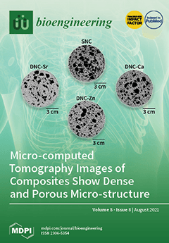
A new class of porous 3D dual network composite hydrogel scaffolds with osteogenic ions: Ca2+, Zn2+, and Sr2+ was developed by sequential formation of the networks with differences in the ion-alginate affinity, accounting for the differences in properties. These composites resembled the morphology of trabecular bone with high interconnectivity and isotropy, allowing cells to thrive and to evenly dissipate energy from multidirectional loads. The simple fabrication process and low costs of these scaffolds with osteoinductive properties thus have translational potential to replace autografting in bone defects, especially for oral and maxillofacial surgery. View this paper.
  • Issues are regarded as officially published after their release is announced to the table of contents alert mailing list.
  • You may sign up for e-mail alerts to receive table of contents of newly released issues.
  • PDF is the official format for papers published in both, html and pdf forms. To view the papers in pdf format, click on the "PDF Full-text" link, and use the free Adobe Reader to open them.
Order results
Result details
Section
Select all
Export citation of selected articles as:
18 pages, 5011 KiB  
Article
Modulation of Synthetic Tracheal Grafts with Extracellular Matrix Coatings
by Lumei Liu, Sayali Dharmadhikari, Robert A. Pouliot, Michael M. Li, Peter M. Minneci, Zhenghong Tan, Kimberly Shontz, Jed Johnson, Susan D. Reynolds, Christopher K. Breuer, Daniel J. Weiss and Tendy Chiang
Bioengineering 2021, 8(8), 116; https://doi.org/10.3390/bioengineering8080116 - 20 Aug 2021
Cited by 4 | Viewed by 2484
Abstract
Synthetic scaffolds for the repair of long-segment tracheal defects are hindered by insufficient biocompatibility and poor graft epithelialization. In this study, we determined if extracellular matrix (ECM) coatings improved the biocompatibility and epithelialization of synthetic tracheal grafts (syn-TG). Porcine and human ECM substrates [...] Read more.
Synthetic scaffolds for the repair of long-segment tracheal defects are hindered by insufficient biocompatibility and poor graft epithelialization. In this study, we determined if extracellular matrix (ECM) coatings improved the biocompatibility and epithelialization of synthetic tracheal grafts (syn-TG). Porcine and human ECM substrates (pECM and hECM) were created through the decellularization and lyophilization of lung tissue. Four concentrations of pECM and hECM coatings on syn-TG were characterized for their effects on scaffold morphologies and on in vitro cell viability and growth. Uncoated and ECM-coated syn-TG were subsequently evaluated in vivo through the orthotopic implantation of segmental grafts or patches. These studies demonstrated that ECM coatings were not cytotoxic and, enhanced the in vitro cell viability and growth on syn-TG in a dose-dependent manner. Mass spectrometry demonstrated that fibrillin, collagen, laminin, and nephronectin were the predominant ECM components transferred onto scaffolds. The in vivo results exhibited similar robust epithelialization of uncoated and coated syn-TG patches; however, the epithelialization remained poor with either uncoated or coated scaffolds in the segmental replacement models. Overall, these findings demonstrated that ECM coatings improve the seeded cell biocompatibility of synthetic scaffolds in vitro; however, they do not improve graft epithelialization in vivo. Full article
(This article belongs to the Special Issue Design and Fabrication of Artificial Stem Cell Microenvironments)
Show Figures

Figure 1

19 pages, 706 KiB  
Review
Biosurfactants: Properties and Applications in Drug Delivery, Biotechnology and Ecotoxicology
by Thiago R. Bjerk, Patricia Severino, Sona Jain, Conrado Marques, Amélia M. Silva, Tatiana Pashirova and Eliana B. Souto
Bioengineering 2021, 8(8), 115; https://doi.org/10.3390/bioengineering8080115 - 13 Aug 2021
Cited by 61 | Viewed by 7712
Abstract
Surfactants are amphiphilic compounds having hydrophilic and hydrophobic moieties in their structure. They can be of synthetic or of microbial origin, obtained respectively from chemical synthesis or from microorganisms’ activity. A new generation of ecofriendly surfactant molecules or biobased surfactants is increasingly growing, [...] Read more.
Surfactants are amphiphilic compounds having hydrophilic and hydrophobic moieties in their structure. They can be of synthetic or of microbial origin, obtained respectively from chemical synthesis or from microorganisms’ activity. A new generation of ecofriendly surfactant molecules or biobased surfactants is increasingly growing, attributed to their versatility of applications. Surfactants can be used as drug delivery systems for a range of molecules given their capacity to create micelles which can promote the encapsulation of bioactives of pharmaceutical interest; besides, these assemblies can also show antimicrobial properties. The advantages of biosurfactants include their high biodegradability profile, low risk of toxicity, production from renewable sources, functionality under extreme pH and temperature conditions, and long-term physicochemical stability. The application potential of these types of polymers is related to their properties enabling them to be processed by emulsification, separation, solubilization, surface (interfacial) tension, and adsorption for the production of a range of drug delivery systems. Biosurfactants have been employed as a drug delivery system to improve the bioavailability of a good number of drugs that exhibit low aqueous solubility. The great potential of these molecules is related to their auto assembly and emulsification capacity. Biosurfactants produced from bacteria are of particular interest due to their antibacterial, antifungal, and antiviral properties with therapeutic and biomedical potential. In this review, we discuss recent advances and perspectives of biosurfactants with antimicrobial properties and how they can be used as structures to develop semisolid hydrogels for drug delivery, in environmental bioremediation, in biotechnology for the reduction of production costs and also their ecotoxicological impact as pesticide alternative. Full article
(This article belongs to the Special Issue Hydrogels Used for Biomanufacturing)
Show Figures

Figure 1

18 pages, 1462 KiB  
Article
WinBEST-KIT: Biochemical Reaction Simulator for Analyzing Multi-Layered Metabolic Pathways
by Tatsuya Sekiguchi, Hiroyuki Hamada and Masahiro Okamoto
Bioengineering 2021, 8(8), 114; https://doi.org/10.3390/bioengineering8080114 - 11 Aug 2021
Viewed by 2719
Abstract
We previously developed the biochemical reaction simulator WinBEST-KIT. In recent years, research interest has shifted from analysis of individual biochemical reactions to analysis of metabolic pathways as systems. These large-scale and complicated metabolic pathways can be considered as characteristic multi-layered structures, which, for [...] Read more.
We previously developed the biochemical reaction simulator WinBEST-KIT. In recent years, research interest has shifted from analysis of individual biochemical reactions to analysis of metabolic pathways as systems. These large-scale and complicated metabolic pathways can be considered as characteristic multi-layered structures, which, for convenience, are separated from whole biological systems according to their specific roles. These pathways include reactants having the same name but with unique stoichiometric coefficients arranged across many different places and connected between arbitrary layers. Accordingly, in this study, we have developed a new version of WinBEST-KIT that allows users (1) to utilize shortcut symbols that can be arranged with multiple reactants having the same name but with unique stoichiometric coefficients, thereby providing a layout that is similar to metabolic pathways depicted in biochemical textbooks; (2) to create layers that divide large-scale and complicated metabolic pathways according to their specific roles; (3) to connect the layers by using shortcut symbols; and (4) to analyze the interactions between these layers. These new and existing features allow users to create and analyze such multi-layered metabolic pathways efficiently. Furthermore, WinBEST-KIT supports SBML, making it possible for users to utilize these new and existing features to create and publish SBML models. Full article
(This article belongs to the Special Issue New Bioinformatics Tools)
Show Figures

Figure 1

31 pages, 9086 KiB  
Review
A Review of Biomaterials and Scaffold Fabrication for Organ-on-a-Chip (OOAC) Systems
by Luana A. Osório, Elisabete Silva and Ruth E. Mackay
Bioengineering 2021, 8(8), 113; https://doi.org/10.3390/bioengineering8080113 - 06 Aug 2021
Cited by 32 | Viewed by 8432
Abstract
Drug and chemical development along with safety tests rely on the use of numerous clinical models. This is a lengthy process where animal testing is used as a standard for pre-clinical trials. However, these models often fail to represent human physiopathology. This may [...] Read more.
Drug and chemical development along with safety tests rely on the use of numerous clinical models. This is a lengthy process where animal testing is used as a standard for pre-clinical trials. However, these models often fail to represent human physiopathology. This may lead to poor correlation with results from later human clinical trials. Organ-on-a-Chip (OOAC) systems are engineered microfluidic systems, which recapitulate the physiochemical environment of a specific organ by emulating the perfusion and shear stress cellular tissue undergoes in vivo and could replace current animal models. The success of culturing cells and cell-derived tissues within these systems is dependent on the scaffold chosen; hence, scaffolds are critical for the success of OOACs in research. A literature review was conducted looking at current OOAC systems to assess the advantages and disadvantages of different materials and manufacturing techniques used for scaffold production; and the alternatives that could be tailored from the macro tissue engineering research field. Full article
(This article belongs to the Special Issue Organs-on-Chips, Volume 2)
Show Figures

Figure 1

16 pages, 4487 KiB  
Article
Self-Organogenesis from 2D Micropatterns to 3D Biomimetic Biliary Trees
by Emilie Gontran, Lorena Loarca, Cyrille El Kassis, Latifa Bouzhir, Dmitry Ayollo, Elsa Mazari-Arrighi, Alexandra Fuchs and Pascale Dupuis-Williams
Bioengineering 2021, 8(8), 112; https://doi.org/10.3390/bioengineering8080112 - 05 Aug 2021
Cited by 2 | Viewed by 3751
Abstract
Background and Aims: Globally, liver diseases account for 2 million deaths per year. For those with advanced liver disease the only curative approach is liver transplantation. However, less than 10% of those in need get a liver transplant due to limited organ availability. [...] Read more.
Background and Aims: Globally, liver diseases account for 2 million deaths per year. For those with advanced liver disease the only curative approach is liver transplantation. However, less than 10% of those in need get a liver transplant due to limited organ availability. To circumvent this challenge, there has been a great focus in generating a bioengineered liver. Despite its essential role in liver functions, a functional biliary system has not yet been developed. In this framework, exploration of epithelial cell self-organogenesis and microengineering-driven geometrical cell confinement allow to envision the bioengineering of a functional biomimetic intrahepatic biliary tract. Approach: three-dimensional (3D) bile ducts were built in vitro by restricting cell adhesion to two-dimensional (2D) patterns to guide cell self-organization. Tree shapes mimicking the configuration of the human biliary system were micropatterned on glass slides, restricting cell attachment to these areas. Different tree geometries and culture conditions were explored to stimulate self-organogenesis of normal rat cholangiocytes (NRCs) used as a biliary cell model, either alone or in co-culture with human umbilical endothelial cells (HUVECs). Results: Pre-seeding the micropatterns with HUVECs promoted luminogenesis with higher efficiency to yield functional branched biliary tubes. Lumen formation, apico-basal polarity, and preservation of the cholangiocyte phenotype were confirmed. Moreover, intact and functional biliary structures were detached from the micropatterns for further manipulation. Conclusion: This study presents physiologically relevant 3D biliary duct networks built in vitro from 2D micropatterns. This opens opportunities for investigating bile duct organogenesis, physiopathology, and drug testing. Full article
Show Figures

Figure 1

22 pages, 6072 KiB  
Article
Simulation-Based Clarification of Appropriate Factors for Presenting Phosphene in Two Directions Avoiding Electrical Interference
by Manami Kanamaru, Phan Xuan Tan and Eiji Kamioka
Bioengineering 2021, 8(8), 111; https://doi.org/10.3390/bioengineering8080111 - 05 Aug 2021
Cited by 2 | Viewed by 2381
Abstract
Walking support systems are essential for blind people. In this study, the presentation of phosphene position is focused on as a method to detect obstacles for blind people. When the phosphene is used in a walking support system, it is necessary to accurately [...] Read more.
Walking support systems are essential for blind people. In this study, the presentation of phosphene position is focused on as a method to detect obstacles for blind people. When the phosphene is used in a walking support system, it is necessary to accurately present the phosphene in at least three directions of the visual field. Controlling the presentation of phosphene position has been reported in several previous studies. However, methodologies to present phosphene in multiple directions without any electric interference have not as yet been investigated. In this study, therefore, appropriate stimulation factors are clarified by the simulation of electric field on the eyeball surface which is strongly related to the presentation of phosphene position in the visual field. As a result of the simulation, it was revealed that the distance of each electrode does not give a significant effect to the eyeball surface. However, the phase of alternating current significantly changed the electric field on the eyeball surface. From investigation of the simulation results, it was clarified that the transition of the electric field on the eyeball surface can be controlled using anti-phase stimulation. In addition, the methodology to present the phosphene at least in two directions was verified. Full article
Show Figures

Figure 1

15 pages, 769 KiB  
Review
How Do Mechanics Guide Fibroblast Activity? Complex Disruptions during Emphysema Shape Cellular Responses and Limit Research
by Mathew N. Leslie, Joshua Chou, Paul M. Young, Daniela Traini, Peta Bradbury and Hui Xin Ong
Bioengineering 2021, 8(8), 110; https://doi.org/10.3390/bioengineering8080110 - 05 Aug 2021
Cited by 5 | Viewed by 3913
Abstract
The emphysema death toll has steadily risen over recent decades, causing the disease to become the third most common cause of death worldwide in 2019. Emphysema is currently incurable and could be due to a genetic condition (Alpha-1 antitrypsin deficiency) or exposure to [...] Read more.
The emphysema death toll has steadily risen over recent decades, causing the disease to become the third most common cause of death worldwide in 2019. Emphysema is currently incurable and could be due to a genetic condition (Alpha-1 antitrypsin deficiency) or exposure to pollutants/irritants, such as cigarette smoke or poorly ventilated cooking fires. Despite the growing burden of emphysema, the mechanisms behind emphysematous pathogenesis and progression are not fully understood by the scientific literature. A key aspect of emphysematous progression is the destruction of the lung parenchyma extracellular matrix (ECM), causing a drastic shift in the mechanical properties of the lung (known as mechanobiology). The mechanical properties of the lung such as the stiffness of the parenchyma (measured as the elastic modulus) and the stretch forces required for inhalation and exhalation are both reduced in emphysema. Fibroblasts function to maintain the structural and mechanical integrity of the lung parenchyma, yet, in the context of emphysema, these fibroblasts appear incapable of repairing the ECM, allowing emphysema to progress. This relationship between the disturbances in the mechanical cues experienced by an emphysematous lung and fibroblast behaviour is constantly overlooked and consequently understudied, thus warranting further research. Interestingly, the failure of current research models to integrate the altered mechanical environment of an emphysematous lung may be limiting our understanding of emphysematous pathogenesis and progression, potentially disrupting the development of novel treatments. This review will focus on the significance of emphysematous lung mechanobiology to fibroblast activity and current research limitations by examining: (1) the impact of mechanical cues on fibroblast activity and the cell cycle, (2) the potential role of mechanical cues in the diminished activity of emphysematous fibroblasts and, finally, (3) the limitations of current emphysematous lung research models and treatments as a result of the overlooked emphysematous mechanical environment. Full article
Show Figures

Figure 1

14 pages, 1970 KiB  
Review
Combined Strategies to Prompt the Biological Reduction of Chlorinated Aliphatic Hydrocarbons: New Sustainable Options for Bioremediation Application
by Marta M. Rossi, Edoardo Dell’Armi, Laura Lorini, Neda Amanat, Marco Zeppilli, Marianna Villano and Marco Petrangeli Papini
Bioengineering 2021, 8(8), 109; https://doi.org/10.3390/bioengineering8080109 - 03 Aug 2021
Cited by 14 | Viewed by 3968
Abstract
Groundwater remediation is one of the main objectives to minimize environmental impacts and health risks. Chlorinated aliphatic hydrocarbons contamination is prevalent and presents particularly challenging scenarios to manage with a single strategy. Different technologies can manage contamination sources and plumes, although they are [...] Read more.
Groundwater remediation is one of the main objectives to minimize environmental impacts and health risks. Chlorinated aliphatic hydrocarbons contamination is prevalent and presents particularly challenging scenarios to manage with a single strategy. Different technologies can manage contamination sources and plumes, although they are usually energy-intensive processes. Interesting alternatives involve in-situ bioremediation strategies, which allow the chlorinated contaminant to be converted into non-toxic compounds by indigenous microbial activity. Despite several advantages offered by the bioremediation approaches, some limitations, like the relatively low reaction rates and the difficulty in the management and control of the microbial activity, can affect the effectiveness of a bioremediation approach. However, those issues can be addressed through coupling different strategies to increase the efficiency of the bioremediation strategy. This mini review describes different strategies to induce the reduction dechlorination reaction by the utilization of innovative strategies, which include the increase or the reduction of contaminant mobility as well as the use of innovative strategies of the reductive power supply. Subsequently, three future approaches for a greener and more sustainable intervention are proposed. In particular, two bio-based materials from renewable resources are intended as alternative, long-lasting electron-donor sources (e.g., polyhydroxyalkanoates from mixed microbial cultures) and a low-cost adsorbent (e.g., biochar from bio-waste). Finally, attention is drawn to novel bio-electrochemical systems that use electric current to stimulate biological reactions. Full article
(This article belongs to the Special Issue Bioengineering in Remediation of Polluted Environments)
Show Figures

Figure 1

29 pages, 7773 KiB  
Review
Stem Cell Niche Microenvironment: Review
by Mohamed Abdul-Al, George Kumi Kyeremeh, Morvarid Saeinasab, Saeed Heidari Keshel and Farshid Sefat
Bioengineering 2021, 8(8), 108; https://doi.org/10.3390/bioengineering8080108 - 28 Jul 2021
Cited by 17 | Viewed by 4186
Abstract
The cornea comprises a pool of self-regenerating epithelial cells that are crucial to preserving clarity and visibility. Limbal epithelial stem cells (LESCs), which live in a specialized stem cell niche (SCN), are crucial for the survival of the human corneal epithelium. They live [...] Read more.
The cornea comprises a pool of self-regenerating epithelial cells that are crucial to preserving clarity and visibility. Limbal epithelial stem cells (LESCs), which live in a specialized stem cell niche (SCN), are crucial for the survival of the human corneal epithelium. They live at the bottom of the limbal crypts, in a physically enclosed microenvironment with a number of neighboring niche cells. Scientists also simplified features of these diverse microenvironments for more analysis in situ by designing and recreating features of different SCNs. Recent methods for regenerating the corneal epithelium after serious trauma, including burns and allergic assaults, focus mainly on regenerating the LESCs. Mesenchymal stem cells, which can transform into self-renewing and skeletal tissues, hold immense interest for tissue engineering and innovative medicinal exploration. This review summarizes all types of LESCs, identity and location of the human epithelial stem cells (HESCs), reconstruction of LSCN and artificial stem cells for self-renewal. Full article
(This article belongs to the Special Issue Design and Fabrication of Artificial Stem Cell Microenvironments)
Show Figures

Figure 1

22 pages, 8146 KiB  
Article
Dual Network Composites of Poly(vinyl alcohol)-Calcium Metaphosphate/Alginate with Osteogenic Ions for Bone Tissue Engineering in Oral and Maxillofacial Surgery
by Lilis Iskandar, Lucy DiSilvio, Jonathan Acheson and Sanjukta Deb
Bioengineering 2021, 8(8), 107; https://doi.org/10.3390/bioengineering8080107 - 28 Jul 2021
Cited by 2 | Viewed by 3388
Abstract
Despite considerable advances in biomaterials-based bone tissue engineering technologies, autografts remain the gold standard for rehabilitating critical-sized bone defects in the oral and maxillofacial (OMF) region. A majority of advanced synthetic bone substitutes (SBS’s) have not transcended the pre-clinical stage due to inferior [...] Read more.
Despite considerable advances in biomaterials-based bone tissue engineering technologies, autografts remain the gold standard for rehabilitating critical-sized bone defects in the oral and maxillofacial (OMF) region. A majority of advanced synthetic bone substitutes (SBS’s) have not transcended the pre-clinical stage due to inferior clinical performance and translational barriers, which include low scalability, high cost, regulatory restrictions, limited advanced facilities and human resources. The aim of this study is to develop clinically viable alternatives to address the challenges of bone tissue regeneration in the OMF region by developing ‘dual network composites’ (DNC’s) of calcium metaphosphate (CMP)—poly(vinyl alcohol) (PVA)/alginate with osteogenic ions: calcium, zinc and strontium. To fabricate DNC’s, single network composites of PVA/CMP with 10% (w/v) gelatine particles as porogen were developed using two freeze–thawing cycles and subsequently interpenetrated by guluronate-dominant sodium alginate and chelated with calcium, zinc or strontium ions. Physicochemical, compressive, water uptake, thermal, morphological and in vitro biological properties of DNC’s were characterised. The results demonstrated elastic 3D porous scaffolds resembling a ‘spongy bone’ with fluid absorbing capacity, easily sculptable to fit anatomically complex bone defects, biocompatible and osteoconductive in vitro, thus yielding potentially clinically viable for SBS alternatives in OMF surgery. Full article
(This article belongs to the Special Issue Multifunctional Scaffolds for Musculoskeletal Regeneration)
Show Figures

Figure 1

12 pages, 3543 KiB  
Article
Engineering and Monitoring 3D Cell Constructs with Time-Evolving Viscoelasticity for the Study of Liver Fibrosis In Vitro
by Ludovica Cacopardo and Arti Ahluwalia
Bioengineering 2021, 8(8), 106; https://doi.org/10.3390/bioengineering8080106 - 27 Jul 2021
Cited by 9 | Viewed by 2991
Abstract
Liver fibrosis is generally associated with an over-production and crosslinking of extracellular matrix proteins, causing a progressive increase in both the elastic and viscous properties of the hepatic tissue. We describe a strategy for mimicking and monitoring the mechano-dynamics of the 3D microenvironment [...] Read more.
Liver fibrosis is generally associated with an over-production and crosslinking of extracellular matrix proteins, causing a progressive increase in both the elastic and viscous properties of the hepatic tissue. We describe a strategy for mimicking and monitoring the mechano-dynamics of the 3D microenvironment associated with liver fibrosis. Cell-laden gelatin hydrogels were crosslinked with microbial transglutaminase using a purpose-designed cytocompatible two-step protocol, which allows for the exposure of cells to a mechanically changing environment during culturing. A bioreactor was re-engineered to monitor the mechanical properties of cell constructs over time. The results showed a shift towards a more elastic (i.e., solid-like) behaviour, which is likely related to an increase in cell stress. The method effectively mimics the time-evolving mechanical microenvironment associated with liver fibrosis and could provide novel insights into pathophysiological processes in which both elastic and viscous properties of tissues change over time. Full article
Show Figures

Figure 1

19 pages, 6210 KiB  
Article
Delivery of Bioactive Compounds to Improve Skin Cell Responses on Microfabricated Electrospun Microenvironments
by David H. Ramos-Rodriguez, Sheila MacNeil, Frederik Claeyssens and Ilida Ortega Asencio
Bioengineering 2021, 8(8), 105; https://doi.org/10.3390/bioengineering8080105 - 27 Jul 2021
Cited by 10 | Viewed by 2865
Abstract
The introduction of microtopographies within biomaterial devices is a promising approach that allows one to replicate to a degree the complex native environment in which human cells reside. Previously, our group showed that by combining electrospun fibers and additive manufacturing it is possible [...] Read more.
The introduction of microtopographies within biomaterial devices is a promising approach that allows one to replicate to a degree the complex native environment in which human cells reside. Previously, our group showed that by combining electrospun fibers and additive manufacturing it is possible to replicate to an extent the stem cell microenvironment (rete ridges) located between the epidermal and dermal layers. Our group has also explored the use of novel proangiogenic compounds to improve the vascularization of skin constructs. Here, we combine our previous approaches to fabricate innovative polycaprolactone fibrous microtopographical scaffolds loaded with bioactive compounds (2-deoxy-D-ribose, 17β-estradiol, and aloe vera). Metabolic activity assay showed that microstructured scaffolds can be used to deliver bioactive agents and that the chemical relation between the working compound and the electrospinning solution is critical to replicate as much as possible the targeted morphologies. We also reported that human skin cell lines have a dose-dependent response to the bioactive compounds and that their inclusion has the potential to improve cell activity, induce blood vessel formation and alter the expression of relevant epithelial markers (collagen IV and integrin β1). In summary, we have developed fibrous matrixes containing synthetic rete-ridge-like structures that can deliver key bioactive compounds that can enhance skin regeneration and ultimately aid in the development of a complex wound healing device. Full article
(This article belongs to the Special Issue Design and Fabrication of Artificial Stem Cell Microenvironments)
Show Figures

Figure 1

20 pages, 3459 KiB  
Article
Kinetic Analysis of Lidocaine Elimination by Pig Liver Cells Cultured in 3D Multi-Compartment Hollow Fiber Membrane Network Perfusion Bioreactors
by Gerardo Catapano, Juliane K. Unger, Elisabetta M. Zanetti, Gionata Fragomeni and Jörg C. Gerlach
Bioengineering 2021, 8(8), 104; https://doi.org/10.3390/bioengineering8080104 - 23 Jul 2021
Cited by 1 | Viewed by 2869
Abstract
Liver cells cultured in 3D bioreactors is an interesting option for temporary extracorporeal liver support in the treatment of acute liver failure and for animal models for preclinical drug screening. Bioreactor capacity to eliminate drugs is generally used for assessing cell metabolic competence [...] Read more.
Liver cells cultured in 3D bioreactors is an interesting option for temporary extracorporeal liver support in the treatment of acute liver failure and for animal models for preclinical drug screening. Bioreactor capacity to eliminate drugs is generally used for assessing cell metabolic competence in different bioreactors or to scale-up bioreactor design and performance for clinical or preclinical applications. However, drug adsorption and physical transport often disguise the intrinsic drug biotransformation kinetics and cell metabolic state. In this study, we characterized the intrinsic kinetics of lidocaine elimination and adsorption by porcine liver cells cultured in 3D four-compartment hollow fiber membrane network perfusion bioreactors. Models of lidocaine transport and biotransformation were used to extract intrinsic kinetic information from response to lidocaine bolus of bioreactor versus adhesion cultures. Different from 2D adhesion cultures, cells in the bioreactors are organized in liver-like aggregates. Adsorption on bioreactor constituents significantly affected lidocaine elimination and was effectively accounted for in kinetic analysis. Lidocaine elimination and cellular monoethylglicinexylidide biotransformation featured first-order kinetics with near-to-in vivo cell-specific capacity that was retained for times suitable for clinical assist and drug screening. Different from 2D cultures, cells in the 3D bioreactors challenged with lidocaine were exposed to close-to-physiological lidocaine and monoethylglicinexylidide concentration profiles. Kinetic analysis suggests bioreactor technology feasibility for preclinical drug screening and patient assist and that drug adsorption should be accounted for to assess cell state in different cultures and when laboratory bioreactor design and performance is scaled-up to clinical use or toxicological drug screening. Full article
Show Figures

Graphical abstract

16 pages, 6997 KiB  
Article
TCA Cycle and Its Relationship with Clavulanic Acid Production: A Further Interpretation by Using a Reduced Genome-Scale Metabolic Model of Streptomyces clavuligerus
by Howard Ramirez-Malule, Víctor A. López-Agudelo, David Gómez-Ríos, Silvia Ochoa, Rigoberto Ríos-Estepa, Stefan Junne and Peter Neubauer
Bioengineering 2021, 8(8), 103; https://doi.org/10.3390/bioengineering8080103 - 22 Jul 2021
Cited by 5 | Viewed by 2753
Abstract
Streptomyces clavuligerus (S. clavuligerus) has been widely studied for its ability to produce clavulanic acid (CA), a potent inhibitor of β-lactamase enzymes. In this study, S. clavuligerus cultivated in 2D rocking bioreactor in fed-batch operation produced CA at comparable rates to [...] Read more.
Streptomyces clavuligerus (S. clavuligerus) has been widely studied for its ability to produce clavulanic acid (CA), a potent inhibitor of β-lactamase enzymes. In this study, S. clavuligerus cultivated in 2D rocking bioreactor in fed-batch operation produced CA at comparable rates to those observed in stirred tank bioreactors. A reduced model of S. clavuligerus metabolism was constructed by using a bottom-up approach and validated using experimental data. The reduced model was implemented for in silico studies of the metabolic scenarios arisen during the cultivations. Constraint-based analysis confirmed the interrelations between succinate, oxaloacetate, malate, pyruvate, and acetate accumulations at high CA synthesis rates in submerged cultures of S. clavuligerus. Further analysis using shadow prices provided a first view of the metabolites positive and negatively associated with the scenarios of low and high CA production. Full article
(This article belongs to the Special Issue Strategies for the Efficient Development of Microbial Bioprocesses)
Show Figures

Figure 1

3 pages, 177 KiB  
Editorial
Biocomposite Inks for 3D Printing
by Gary Chinga-Carrasco
Bioengineering 2021, 8(8), 102; https://doi.org/10.3390/bioengineering8080102 - 22 Jul 2021
Viewed by 2226
Abstract
Three-dimensional (3D) printing has evolved massively during the last years and is demonstrating its potential in tissue engineering, wound dressings, cell culture models for drug testing, and prosthesis, to name a few [...] Full article
(This article belongs to the Special Issue Biocomposite Inks for 3D Printing)
14 pages, 4299 KiB  
Article
Numerical Models Can Assist Choice of an Aortic Phantom for In Vitro Testing
by Giulia Comunale, Luigi Di Micco, Daniela Paola Boso, Francesca Maria Susin and Paolo Peruzzo
Bioengineering 2021, 8(8), 101; https://doi.org/10.3390/bioengineering8080101 - 21 Jul 2021
Cited by 5 | Viewed by 2473
Abstract
(1) Background: The realization of appropriate aortic replicas for in vitro experiments requires a suitable choice of both the material and geometry. The matching between the grade of details of the geometry and the mechanical response of the materials is an open issue [...] Read more.
(1) Background: The realization of appropriate aortic replicas for in vitro experiments requires a suitable choice of both the material and geometry. The matching between the grade of details of the geometry and the mechanical response of the materials is an open issue that deserves attention. (2) Methods: To explore this issue, we performed a series of Fluid–Structure Interaction simulations, which compared the dynamics of three aortic models. Specifically, we reproduced a patient-specific geometry with a wall of biological tissue or silicone, and a parametric geometry based on in vivo data made in silicone. The biological tissue and the silicone were modeled with a fiber-oriented anisotropic and isotropic hyperelastic model, respectively. (3) Results: Clearly, both the aorta’s geometry and its constitutive material contribute to the determination of the aortic arch deformation; specifically, the parametric aorta exhibits a strain field similar to the patient-specific model with biological tissue. On the contrary, the local geometry affects the flow velocity distribution quite a lot, although it plays a minor role in the helicity along the arch. (4) Conclusions: The use of a patient-specific prototype in silicone does not a priori ensure a satisfactory reproducibility of the real aorta dynamics. Furthermore, the present simulations suggest that the realization of a simplified replica with the same compliance of the real aorta is able to mimic the overall behavior of the vessel. Full article
Show Figures

Figure 1

Previous Issue
Next Issue
Back to TopTop