Next Issue
Volume 7, December
Previous Issue
Volume 7, June


Bioengineering, Volume 7, Issue 3 (September 2020) – 54 articles

Cover Story (view full-size image): 
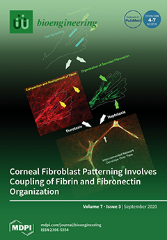
Using a 3D fibrin matrix model, corneal fibroblasts have been shown to secrete, bind, and organize fibronectin into tracks that facilitate cell spreading and migration. Here, we demonstrate that during fibroblast spreading, fibronectin is organized in coordination with the compaction and realignment of fibrin fibers by cellular traction forces. Over time, this leads to the formation of an interconnected network of cells, fibronectin, and compacted fibrin tracks. Interestingly, interconnected networks of fibroblasts are often observed during in vivo corneal wound healing, and it is hypothesized that these networks may facilitate more efficient wound repopulation and closure. View this paper
  • Issues are regarded as officially published after their release is announced to the table of contents alert mailing list.
  • You may sign up for e-mail alerts to receive table of contents of newly released issues.
  • PDF is the official format for papers published in both, html and pdf forms. To view the papers in pdf format, click on the "PDF Full-text" link, and use the free Adobe Reader to open them.
Order results
Result details
Section
Select all
Export citation of selected articles as:
12 pages, 1437 KiB  
Article
A Small Study of Bacterial Contamination of Anaerobic Digestion Materials and Survival in Different Feed Stocks
by Lauren Russell, Paul Whyte, Annetta Zintl, Stephen Gordon, Bryan Markey, Theo de Waal, Enda Cummins, Stephen Nolan, Vincent O’Flaherty, Florence Abram, Karl Richards, Owen Fenton and Declan Bolton
Bioengineering 2020, 7(3), 116; https://doi.org/10.3390/bioengineering7030116 - 22 Sep 2020
Cited by 6 | Viewed by 3360
Abstract
If pathogens are present in feedstock materials and survive in anaerobic digestion (AD) formulations at 37 °C, they may also survive the AD process to be disseminated in digestate spread on farmland as a fertilizer. The aim of this study was to investigate [...] Read more.
If pathogens are present in feedstock materials and survive in anaerobic digestion (AD) formulations at 37 °C, they may also survive the AD process to be disseminated in digestate spread on farmland as a fertilizer. The aim of this study was to investigate the prevalence of Salmonella spp., Escherichia coli O157, Listeria monocytogenes, Enterococcus faecalis and Clostridium spp. in AD feed and output materials and survival/growth in four formulations based on food waste, bovine slurry and/or grease-trap waste using International Organization for Standardization (ISO) or equivalent methods. The latter was undertaken in 100 mL Ramboldi tubes, incubated at 37 °C for 10 d with surviving cells enumerated periodically and the T90 values (time to achieve a 1 log reduction) calculated. The prevalence rates for Salmonella spp., Escherichia coli O157, Listeria monocytogenes, Enterococcus faecalis and Clostridium spp. were 3, 0, 5, 11 and 10/13 in food waste, 0, 0, 2, 3 and 2/3 in bovine slurry, 1, 0, 8, 7 and 8/8 in the mixing tank, 5, 1, 17, 18 and 17 /19 in raw digestate and 0, 0, 0, 2 and 2/2 in dried digestate, respectively. Depending on the formulation, T90 values ranged from 1.5 to 2.8 d, 1.6 to 2.8 d, 3.1 to 23.5 d, 2.2 to 6.6 d and 2.4 to 9.1 d for Salmonella Newport, Escherichia coli O157, Listeria monocytogenes, Enterococcus faecalis and Clostridium sporogenes, respectively. It was concluded that AD feed materials may be contaminated with a range of bacterial pathogens and L. monocytogenes may survive for extended periods in the test formulations incubated at 37 °C. Full article
(This article belongs to the Special Issue Current Advances in Anaerobic Digestion Technology)
Show Figures

Figure 1

40 pages, 2988 KiB  
Review
Innovative Human Three-Dimensional Tissue-Engineered Models as an Alternative to Animal Testing
by Patrick Bédard, Sara Gauvin, Karel Ferland, Christophe Caneparo, Ève Pellerin, Stéphane Chabaud and Stéphane Bolduc
Bioengineering 2020, 7(3), 115; https://doi.org/10.3390/bioengineering7030115 - 17 Sep 2020
Cited by 70 | Viewed by 17858
Abstract
Animal testing has long been used in science to study complex biological phenomena that cannot be investigated using two-dimensional cell cultures in plastic dishes. With time, it appeared that more differences could exist between animal models and even more when translated to human [...] Read more.
Animal testing has long been used in science to study complex biological phenomena that cannot be investigated using two-dimensional cell cultures in plastic dishes. With time, it appeared that more differences could exist between animal models and even more when translated to human patients. Innovative models became essential to develop more accurate knowledge. Tissue engineering provides some of those models, but it mostly relies on the use of prefabricated scaffolds on which cells are seeded. The self-assembly protocol has recently produced organ-specific human-derived three-dimensional models without the need for exogenous material. This strategy will help to achieve the 3R principles. Full article
Show Figures

Figure 1

18 pages, 4331 KiB  
Article
Optimization of Co-Culture Conditions for a Human Vascularized Adipose Tissue Model
by Feipeng Yang, Ronald N. Cohen and Eric M. Brey
Bioengineering 2020, 7(3), 114; https://doi.org/10.3390/bioengineering7030114 - 17 Sep 2020
Cited by 25 | Viewed by 5633
Abstract
In vitro adipose tissue models can be used to provide insight into fundamental aspects of adipose physiology. These systems may serve as replacements for animal models, which are often poor predictors of obesity and metabolic diseases in humans. Adipose tissue consists of a [...] Read more.
In vitro adipose tissue models can be used to provide insight into fundamental aspects of adipose physiology. These systems may serve as replacements for animal models, which are often poor predictors of obesity and metabolic diseases in humans. Adipose tissue consists of a rich vasculature that is essential to its function. However, the study of endothelial cell–adipocyte interactions has been challenging due to differences in culture conditions required for the survival and function of each cell type. To address this issue, we performed an extensive evaluation of the cell culture media composition to identify the conditions optimal for the co-culture of endothelial cells and adipocytes. The effects of individual media factors on cell survival, proliferation, and differentiation were systematically explored. Several media factors were determined to disrupt the co-culture system. Optimized culture conditions were identified and used to generate a vascularized human adipose microtissue. An interconnected vascular network was established within an adipose micro-tissue, and the networks were anastomosed with perfused channels to form a functional network. In conclusion, media conditions were identified that enabled endothelial cell–adipocyte co-culture and were used to support the formation of a vascularized adipose tissue within a microfluidic device. Full article
Show Figures

Figure 1

20 pages, 2746 KiB  
Article
Biomimetic 3D Models for Investigating the Role of Monocytes and Macrophages in Atherosclerosis
by Anna Garcia-Sabaté, Walaa Kamal E. Mohamed, Jiranuwat Sapudom, Aseel Alatoom, Layla Al Safadi and Jeremy C. M. Teo
Bioengineering 2020, 7(3), 113; https://doi.org/10.3390/bioengineering7030113 - 16 Sep 2020
Cited by 24 | Viewed by 12174
Abstract
Atherosclerosis, the inflammation of artery walls due to the accumulation of lipids, is the most common underlying cause for cardiovascular diseases. Monocytes and macrophages are major cells that contribute to the initiation and progression of atherosclerotic plaques. During this process, an accumulation of [...] Read more.
Atherosclerosis, the inflammation of artery walls due to the accumulation of lipids, is the most common underlying cause for cardiovascular diseases. Monocytes and macrophages are major cells that contribute to the initiation and progression of atherosclerotic plaques. During this process, an accumulation of LDL-laden macrophages (foam cells) and an alteration in the extracellular matrix (ECM) organization leads to a local vessel stiffening. Current in vitro models are carried out onto two-dimensional tissue culture plastic and cannot replicate the relevant microenvironments. To bridge the gap between in vitro and in vivo conditions, we utilized three-dimensional (3D) collagen matrices that allowed us to mimic the ECM stiffening during atherosclerosis by increasing collagen density. First, human monocytic THP-1 cells were embedded into 3D collagen matrices reconstituted at low and high density. Cells were subsequently differentiated into uncommitted macrophages (M0) and further activated into pro- (M1) and anti-inflammatory (M2) phenotypes. In order to mimic atherosclerotic conditions, cells were cultured in the presence of oxidized LDL (oxLDL) and analyzed in terms of oxLDL uptake capability and relevant receptors along with their cytokine secretomes. Although oxLDL uptake and larger lipid size could be observed in macrophages in a matrix dependent manner, monocytes showed higher numbers of oxLDL uptake cells. By analyzing major oxLDL uptake receptors, both monocytes and macrophages expressed lectin-like oxidized low-density lipoprotein receptor-1 (LOX1), while enhanced expression of scavenger receptor CD36 could be observed only in M2. Notably, by analyzing the secretome of macrophages exposed to oxLDL, we demonstrated that the cells could, in fact, secrete adipokines and growth factors in distinct patterns. Besides, oxLDL appeared to up-regulate MHCII expression in all cells, while an up-regulation of CD68, a pan-macrophage marker, was found only in monocytes, suggesting a possible differentiation of monocytes into a pro-inflammatory macrophage. Overall, our work demonstrated that collagen density in the plaque could be one of the major factors driving atherosclerotic progression via modulation of monocyte and macrophages behaviors. Full article
Show Figures

Graphical abstract

27 pages, 3676 KiB  
Review
Translational Roadmap for the Organs-on-a-Chip Industry toward Broad Adoption
by Vanessa Allwardt, Alexander J. Ainscough, Priyalakshmi Viswanathan, Stacy D. Sherrod, John A. McLean, Malcolm Haddrick and Virginia Pensabene
Bioengineering 2020, 7(3), 112; https://doi.org/10.3390/bioengineering7030112 - 16 Sep 2020
Cited by 51 | Viewed by 10751
Abstract
Organs-on-a-Chip (OOAC) is a disruptive technology with widely recognized potential to change the efficiency, effectiveness, and costs of the drug discovery process; to advance insights into human biology; to enable clinical research where human trials are not feasible. However, further development is needed [...] Read more.
Organs-on-a-Chip (OOAC) is a disruptive technology with widely recognized potential to change the efficiency, effectiveness, and costs of the drug discovery process; to advance insights into human biology; to enable clinical research where human trials are not feasible. However, further development is needed for the successful adoption and acceptance of this technology. Areas for improvement include technological maturity, more robust validation of translational and predictive in vivo-like biology, and requirements of tighter quality standards for commercial viability. In this review, we reported on the consensus around existing challenges and necessary performance benchmarks that are required toward the broader adoption of OOACs in the next five years, and we defined a potential roadmap for future translational development of OOAC technology. We provided a clear snapshot of the current developmental stage of OOAC commercialization, including existing platforms, ancillary technologies, and tools required for the use of OOAC devices, and analyze their technology readiness levels. Using data gathered from OOAC developers and end-users, we identified prevalent challenges faced by the community, strategic trends and requirements driving OOAC technology development, and existing technological bottlenecks that could be outsourced or leveraged by active collaborations with academia. Full article
(This article belongs to the Special Issue Organs-on-Chips, Volume 2)
Show Figures

Graphical abstract

16 pages, 29376 KiB  
Review
Biomimetic Nanocarriers for Cancer Target Therapy
by Clara Guido, Gabriele Maiorano, Barbara Cortese, Stefania D’Amone and Ilaria Elena Palamà
Bioengineering 2020, 7(3), 111; https://doi.org/10.3390/bioengineering7030111 - 14 Sep 2020
Cited by 35 | Viewed by 5997
Abstract
Nanotechnology offers innovative tools for the design of biomimetic nanocarriers for targeted cancer therapy. These nano-systems present several advantages such as cargo’s protection and modulation of its release, inclusion of stimuli-responsive elements, and enhanced tumoral accumulation. All together, these nano-systems suffer low therapeutic [...] Read more.
Nanotechnology offers innovative tools for the design of biomimetic nanocarriers for targeted cancer therapy. These nano-systems present several advantages such as cargo’s protection and modulation of its release, inclusion of stimuli-responsive elements, and enhanced tumoral accumulation. All together, these nano-systems suffer low therapeutic efficacy in vivo because organisms can recognize and remove foreign nanomaterials. To overcome this important issue, different modifications on nanoparticle surfaces were exploited in order to reach the desired therapeutic efficacy eliciting, also, the response of immune system against cancer cells. For this reason, more recently, a new strategy involving cell membrane-covered nanoparticles for biomedical application has been attracting increasing attention. Membranes from red blood cells, platelets, leukocytes, tumor, and stem cells, have been exploited as biomimetic coatings of nanoparticles for evading clearance or stimulated immune system by maintaining in the same way their targeting capability. In this review, the use of different cell sources as coating of biomimetic nanocarriers for cancer therapy is discussed. Full article
(This article belongs to the Collection Nanoparticles for Therapeutic and Diagnostic Applications)
Show Figures

Figure 1

12 pages, 2264 KiB  
Article
Elastin-Collagen Based Hydrogels as Model Scaffolds to Induce Three-Dimensional Adipocyte Culture from Adipose Derived Stem Cells
by Kristen Newman, Kendra Clark, Bhuvaneswari Gurumurthy, Pallabi Pal and Amol V. Janorkar
Bioengineering 2020, 7(3), 110; https://doi.org/10.3390/bioengineering7030110 - 12 Sep 2020
Cited by 15 | Viewed by 4020
Abstract
This study aimed to probe the effect of formulation of scaffolds prepared using collagen and elastin-like polypeptide (ELP) and their resulting physico-chemical and mechanical properties on the adipogenic differentiation of human adipose derived stem cells (hASCs). Six different ELP-collagen scaffolds were prepared by [...] Read more.
This study aimed to probe the effect of formulation of scaffolds prepared using collagen and elastin-like polypeptide (ELP) and their resulting physico-chemical and mechanical properties on the adipogenic differentiation of human adipose derived stem cells (hASCs). Six different ELP-collagen scaffolds were prepared by varying the collagen concentration (2 and 6 mg/mL), ELP addition (6 mg/mL), or crosslinking of the scaffolds. FTIR spectroscopy indicated secondary bonding interactions between collagen and ELP, while scanning electron microscopy revealed a porous structure for all scaffolds. Increased collagen concentration, ELP addition, and presence of crosslinking decreased swelling ratio and increased elastic modulus and compressive strength of the scaffolds. The scaffold characteristics influenced cell morphology, wherein the hASCs seeded in the softer, non-crosslinked scaffolds displayed a spread morphology. We determined that stiffer and/or crosslinked elastin-collagen based scaffolds constricted the spreading of hASCs, leading to a spheroid morphology and yielded an enhanced adipogenic differentiation as indicated by Oil Red O staining. Overall, this study underscored the importance of spheroid morphology in adipogenic differentiation, which will allow researchers to create more physiologically-relevant three-dimensional, in vitro culture models. Full article
(This article belongs to the Special Issue Cell–Biomaterial Interactions)
Show Figures

Figure 1

10 pages, 1083 KiB  
Article
SlicerArduino: A Bridge between Medical Imaging Platform and Microcontroller
by Paolo Zaffino, Alessio Merola, Domenico Leuzzi, Virgilio Sabatino, Carlo Cosentino and Maria Francesca Spadea
Bioengineering 2020, 7(3), 109; https://doi.org/10.3390/bioengineering7030109 - 11 Sep 2020
Cited by 2 | Viewed by 3469
Abstract
Interaction between medical image platform and external environment is a desirable feature in several clinical, research, and educational scenarios. In this work, the integration between 3D Slicer package and Arduino board is introduced, enabling a simple and useful communication between the two software/hardware [...] Read more.
Interaction between medical image platform and external environment is a desirable feature in several clinical, research, and educational scenarios. In this work, the integration between 3D Slicer package and Arduino board is introduced, enabling a simple and useful communication between the two software/hardware platforms. The open source extension, programmed in Python language, manages the connection process and offers a communication layer accessible from any point of the medical image suite infrastructure. Deep integration with 3D Slicer code environment is provided and a basic input–output mechanism accessible via GUI is also made available. To test the proposed extension, two exemplary use cases were implemented: (1) INPUT data to 3D Slicer, to navigate on basis of data detected by a distance sensor connected to the board, and (2) OUTPUT data from 3D Slicer, to control a servomotor on the basis of data computed through image process procedures. Both goals were achieved and quasi-real-time control was obtained without any lag or freeze, thus boosting the integration between 3D Slicer and Arduino. This integration can be easily obtained through the execution of few lines of Python code. In conclusion, SlicerArduino proved to be suitable for fast prototyping, basic input–output interaction, and educational purposes. The extension is not intended for mission-critical clinical tasks. Full article
Show Figures

Graphical abstract

12 pages, 2100 KiB  
Article
Formulation and Characterization of Alginate Dialdehyde, Gelatin, and Platelet-Rich Plasma-Based Bioink for Bioprinting Applications
by Lakshmi T. Somasekharan, Naresh Kasoju, Riya Raju and Anugya Bhatt
Bioengineering 2020, 7(3), 108; https://doi.org/10.3390/bioengineering7030108 - 09 Sep 2020
Cited by 28 | Viewed by 6742
Abstract
Layer-by-layer additive manufacturing process has evolved into three-dimensional (3D) “bio-printing” as a means of constructing cell-laden functional tissue equivalents. The process typically involves the mixing of cells of interest with an appropriate hydrogel, termed as “bioink”, followed by printing and tissue maturation. An [...] Read more.
Layer-by-layer additive manufacturing process has evolved into three-dimensional (3D) “bio-printing” as a means of constructing cell-laden functional tissue equivalents. The process typically involves the mixing of cells of interest with an appropriate hydrogel, termed as “bioink”, followed by printing and tissue maturation. An ideal bioink should have adequate mechanical, rheological, and biological features of the target tissues. However, native extracellular matrix (ECM) is made of an intricate milieu of soluble and non-soluble extracellular factors, and mimicking such a composition is challenging. To this end, here we report the formulation of a multi-component bioink composed of gelatin and alginate -based scaffolding material, as well as a platelet-rich plasma (PRP) suspension, which mimics the insoluble and soluble factors of native ECM respectively. Briefly, sodium alginate was subjected to controlled oxidation to yield alginate dialdehyde (ADA), and was mixed with gelatin and PRP in various volume ratios in the presence of borax. The formulation was systematically characterized for its gelation time, swelling, and water uptake, as well as its morphological, chemical, and rheological properties; furthermore, blood- and cytocompatibility were assessed as per ISO 10993 (International Organization for Standardization). Printability, shape fidelity, and cell-laden printing was evaluated using the RegenHU 3D Discovery bioprinter. The results indicated the successful development of ADA–gelatin–PRP based bioink for 3D bioprinting and biofabrication applications. Full article
(This article belongs to the Special Issue Biocomposite Inks for 3D Printing)
Show Figures

Figure 1

16 pages, 3064 KiB  
Review
Sonodelivery in Skeletal Muscle: Current Approaches and Future Potential
by Richard E. Decker, Zachary E. Lamantia, Todd S. Emrick and Marxa L. Figueiredo
Bioengineering 2020, 7(3), 107; https://doi.org/10.3390/bioengineering7030107 - 09 Sep 2020
Cited by 5 | Viewed by 3670
Abstract
There are currently multiple approaches to facilitate gene therapy via intramuscular gene delivery, such as electroporation, viral delivery, or direct DNA injection with or without polymeric carriers. Each of these methods has benefits, but each method also has shortcomings preventing it from being [...] Read more.
There are currently multiple approaches to facilitate gene therapy via intramuscular gene delivery, such as electroporation, viral delivery, or direct DNA injection with or without polymeric carriers. Each of these methods has benefits, but each method also has shortcomings preventing it from being established as the ideal technique. A promising method, ultrasound-mediated gene delivery (or sonodelivery) is inexpensive, widely available, reusable, minimally invasive, and safe. Hurdles to utilizing sonodelivery include choosing from a large variety of conditions, which are often dependent on the equipment and/or research group, and moderate transfection efficiencies when compared to some other gene delivery methods. In this review, we provide a comprehensive look at the breadth of sonodelivery techniques for intramuscular gene delivery and suggest future directions for this continuously evolving field. Full article
(This article belongs to the Special Issue Advances in Skeletal Muscle Tissue Engineering)
Show Figures

Figure 1

16 pages, 1926 KiB  
Article
Material Characterization and Substrate Suitability Assessment of Chicken Manure for Dry Batch Anaerobic Digestion Processes
by Harald Wedwitschka, Daniela Gallegos Ibanez, Franziska Schäfer, Earl Jenson and Michael Nelles
Bioengineering 2020, 7(3), 106; https://doi.org/10.3390/bioengineering7030106 - 07 Sep 2020
Cited by 16 | Viewed by 3870
Abstract
Chicken manure is an agricultural residue material with a high biomass potential. The energetical utilization of this feedstock via anaerobic digestion is an interesting waste treatment option. One waste treatment technology most appropriate for the treatment of stackable (non-free-flowing) dry organic waste materials [...] Read more.
Chicken manure is an agricultural residue material with a high biomass potential. The energetical utilization of this feedstock via anaerobic digestion is an interesting waste treatment option. One waste treatment technology most appropriate for the treatment of stackable (non-free-flowing) dry organic waste materials is the dry batch anaerobic digestion process. The aim of this study was to evaluate the substrate suitability of chicken manure from various sources as feedstock for percolation processes. Chicken manure samples from different housing forms were investigated for their chemical and physical material properties, such as feedstock composition, permeability under compaction and material compressibility. The permeability under compaction of chicken manure ranged from impermeable to sufficiently permeable depending on the type of chicken housing, manure age and bedding material used. Porous materials, such as straw and woodchips, were successfully tested as substrate additives with the ability to enhance material mixture properties to yield superior permeability and allow sufficient percolation. In dry anaerobic batch digestion trials at lab scale, the biogas generation of chicken manure with and without any structure material addition was investigated. Digestion trials were carried out without solid inoculum addition and secondary methanization of volatile components. The specific methane yield of dry chicken manure was measured and found to be 120 to 145 mL/g volatile solids (VS) and 70 to 75 mL/g fresh matter (FM), which represents approximately 70% of the methane potential based on fresh mass of common energy crops, such as corn silage. Full article
(This article belongs to the Special Issue Current Advances in Anaerobic Digestion Technology)
Show Figures

Figure 1

22 pages, 662 KiB  
Review
Electrospun Scaffolds and Induced Pluripotent Stem Cell-Derived Cardiomyocytes for Cardiac Tissue Engineering Applications
by Taylor Cook Suh, Alaowei Y. Amanah and Jessica M. Gluck
Bioengineering 2020, 7(3), 105; https://doi.org/10.3390/bioengineering7030105 - 06 Sep 2020
Cited by 23 | Viewed by 6073
Abstract
Tissue engineering (TE) combines cells, scaffolds, and growth factors to assemble functional tissues for repair or replacement of tissues and organs. Cardiac TE is focused on developing cardiac cells, tissues, and structures—most notably the heart. This review presents the requirements, challenges, and research [...] Read more.
Tissue engineering (TE) combines cells, scaffolds, and growth factors to assemble functional tissues for repair or replacement of tissues and organs. Cardiac TE is focused on developing cardiac cells, tissues, and structures—most notably the heart. This review presents the requirements, challenges, and research surrounding electrospun scaffolds and induced pluripotent stem cell (iPSC)-derived cardiomyocytes (CMs) towards applications to TE hearts. Electrospinning is an attractive fabrication method for cardiac TE scaffolds because it produces fibers that demonstrate the optimal potential for mimicking the complex structure of the cardiac extracellular matrix (ECM). iPSCs theoretically offer the capacity to generate limitless numbers of CMs for use in TE hearts, however these iPSC-CMs are electrophysiologically, morphologically, mechanically, and metabolically immature compared to adult CMs. This presents a functional limitation to their use in cardiac TE, and research aiming to address this limitation is presented in this review. Full article
(This article belongs to the Special Issue Cardiac Regeneration)
Show Figures

Figure 1

35 pages, 4428 KiB  
Review
Repositioning Natural Antioxidants for Therapeutic Applications in Tissue Engineering
by Pasquale Marrazzo and Cian O’Leary
Bioengineering 2020, 7(3), 104; https://doi.org/10.3390/bioengineering7030104 - 02 Sep 2020
Cited by 35 | Viewed by 6475
Abstract
Although a large panel of natural antioxidants demonstrate a protective effect in preventing cellular oxidative stress, their low bioavailability limits therapeutic activity at the targeted injury site. The importance to deliver drug or cells into oxidative microenvironments can be realized with the development [...] Read more.
Although a large panel of natural antioxidants demonstrate a protective effect in preventing cellular oxidative stress, their low bioavailability limits therapeutic activity at the targeted injury site. The importance to deliver drug or cells into oxidative microenvironments can be realized with the development of biocompatible redox-modulating materials. The incorporation of antioxidant compounds within implanted biomaterials should be able to retain the antioxidant activity, while also allowing graft survival and tissue recovery. This review summarizes the recent literature reporting the combined role of natural antioxidants with biomaterials. Our review highlights how such functionalization is a promising strategy in tissue engineering to improve the engraftment and promote tissue healing or regeneration. Full article
Show Figures

Graphical abstract

18 pages, 6803 KiB  
Article
Additive Manufacturing of Prostheses Using Forest-Based Composites
by Erik Stenvall, Göran Flodberg, Henrik Pettersson, Kennet Hellberg, Liselotte Hermansson, Martin Wallin and Li Yang
Bioengineering 2020, 7(3), 103; https://doi.org/10.3390/bioengineering7030103 - 01 Sep 2020
Cited by 21 | Viewed by 5013
Abstract
A custom-made prosthetic product is unique for each patient. Fossil-based thermoplastics are the dominant raw materials in both prosthetic and industrial applications; there is a general demand for reducing their use and replacing them with renewable, biobased materials. A transtibial prosthesis sets strict [...] Read more.
A custom-made prosthetic product is unique for each patient. Fossil-based thermoplastics are the dominant raw materials in both prosthetic and industrial applications; there is a general demand for reducing their use and replacing them with renewable, biobased materials. A transtibial prosthesis sets strict demands on mechanical strength, durability, reliability, etc., which depend on the biocomposite used and also the additive manufacturing (AM) process. The aim of this project was to develop systematic solutions for prosthetic products and services by combining biocomposites using forestry-based derivatives with AM techniques. Composite materials made of polypropylene (PP) reinforced with microfibrillated cellulose (MFC) were developed. The MFC contents (20, 30 and 40 wt%) were uniformly dispersed in the polymer PP matrix, and the MFC addition significantly enhanced the mechanical performance of the materials. With 30 wt% MFC, the tensile strength and Young´s modulus was about twice that of the PP when injection molding was performed. The composite material was successfully applied with an AM process, i.e., fused deposition modeling (FDM), and a transtibial prosthesis was created based on the end-user’s data. A clinical trial of the prosthesis was conducted with successful outcomes in terms of wearing experience, appearance (color), and acceptance towards the materials and the technique. Given the layer-by-layer nature of AM processes, structural and process optimizations are needed to maximize the reinforcement effects of MFC to eliminate variations in the binding area between adjacent layers and to improve the adhesion between layers. Full article
(This article belongs to the Special Issue Biocomposite Inks for 3D Printing)
Show Figures

Figure 1

14 pages, 5748 KiB  
Article
Micropatterning Decellularized ECM as a Bioactive Surface to Guide Cell Alignment, Proliferation, and Migration
by Emily Cady, Jacob A. Orkwis, Rachel Weaver, Lia Conlin, Nicolas N. Madigan and Greg M. Harris
Bioengineering 2020, 7(3), 102; https://doi.org/10.3390/bioengineering7030102 - 31 Aug 2020
Cited by 11 | Viewed by 4218
Abstract
Bioactive surfaces and materials have displayed great potential in a variety of tissue engineering applications but often struggle to completely emulate complex bodily systems. The extracellular matrix (ECM) is a crucial, bioactive component in all tissues and has recently been identified as a [...] Read more.
Bioactive surfaces and materials have displayed great potential in a variety of tissue engineering applications but often struggle to completely emulate complex bodily systems. The extracellular matrix (ECM) is a crucial, bioactive component in all tissues and has recently been identified as a potential solution to be utilized in combination with biomaterials. In tissue engineering, the ECM can be utilized in a variety of applications by employing the biochemical and biomechanical cues that are crucial to regenerative processes. However, viable solutions for maintaining the dimensionality, spatial orientation, and protein composition of a naturally cell-secreted ECM remain challenging in tissue engineering. Therefore, this work used soft lithography to create micropatterned polydimethylsiloxane (PDMS) substrates of a three-dimensional nature to control cell adhesion and alignment. Cells aligned on the micropatterned PDMS, secreted and assembled an ECM, and were decellularized to produce an aligned matrix biomaterial. The cells seeded onto the decellularized, patterned ECM showed a high degree of alignment and migration along the patterns compared to controls. This work begins to lay the groundwork for elucidating the immense potential of a natural, cell-secreted ECM for directing cell function and offers further guidance for the incorporation of natural, bioactive components for emerging tissue engineering technologies. Full article
(This article belongs to the Special Issue Extracellular Matrix in Wound Healing)
Show Figures

Figure 1

10 pages, 2279 KiB  
Article
Utilization of an Industry Byproduct, Corymbia maculata Leaves, by Aspergillus terreus to Produce Lovastatin
by Mishal Subhan, Rani Faryal and Ian Macreadie
Bioengineering 2020, 7(3), 101; https://doi.org/10.3390/bioengineering7030101 - 30 Aug 2020
Cited by 3 | Viewed by 3157
Abstract
Due to its ability to lower cholesterol levels, simvastatin is a leading drug for the prevention of strokes and heart disease: it also lowers the incidence of neurodegenerative diseases. Simvastatin is made from lovastatin, a precursor produced by the industrial fungus, Aspergillus terreus [...] Read more.
Due to its ability to lower cholesterol levels, simvastatin is a leading drug for the prevention of strokes and heart disease: it also lowers the incidence of neurodegenerative diseases. Simvastatin is made from lovastatin, a precursor produced by the industrial fungus, Aspergillus terreus. In this study, Corymbia maculata leaves were tested as a novel substrate for the growth of a new isolate of A. terreus and a lovastatin-resistant strain of A. terreus to produce lovastatin. Corymbia maculata (spotted gum) is well utilized by forest industries as a source of timber because of its high strength, durability and smooth texture. However, the leaves are a major waste product. Growth of A. terreus on Corymbia maculata leaves, in solid-state fermentation resulted in the production of lovastatin. Fermentation of media using fresh leaves of Corymbia maculata produced more lovastatin (4.9 mg g−1), than the sun-dried leaves (3.2 mg g−1). Levels of lovastatin were further increased by the lovastatin-resistant strain of A. terreus (Lvs-r), which produced twice the amount of the parental strain. The production of lovastatin was confirmed by HPLC and LC–MS/MS studies. The study suggests that the utilization of a cheap substrate for the production of lovastatin can have a potential economic benefit. Full article
Show Figures

Figure 1

14 pages, 1432 KiB  
Review
Advanced Techniques for Skeletal Muscle Tissue Engineering and Regeneration
by Moon Sung Kang, Seok Hyun Lee, Won Jung Park, Ji Eun Lee, Bongju Kim and Dong-Wook Han
Bioengineering 2020, 7(3), 99; https://doi.org/10.3390/bioengineering7030099 - 26 Aug 2020
Cited by 30 | Viewed by 6774
Abstract
Tissue engineering has recently emerged as a novel strategy for the regeneration of damaged skeletal muscle tissues due to its ability to regenerate tissue. However, tissue engineering is challenging due to the need for state-of-the-art interdisciplinary studies involving material science, biochemistry, and mechanical [...] Read more.
Tissue engineering has recently emerged as a novel strategy for the regeneration of damaged skeletal muscle tissues due to its ability to regenerate tissue. However, tissue engineering is challenging due to the need for state-of-the-art interdisciplinary studies involving material science, biochemistry, and mechanical engineering. For this reason, electrospinning and three-dimensional (3D) printing methods have been widely studied because they can insert embedded muscle cells into an extracellular-matrix-mimicking microenvironment, which helps the growth of seeded or laden cells and cell signals by modulating cell–cell interaction and cell–matrix interaction. In this mini review, the recent research trends in scaffold fabrication for skeletal muscle tissue regeneration using advanced techniques, such as electrospinning and 3D bioprinting, are summarized. In conclusion, the further development of skeletal muscle tissue engineering techniques may provide innovative results with clinical potential for skeletal muscle regeneration. Full article
(This article belongs to the Special Issue Advances in Skeletal Muscle Tissue Engineering)
Show Figures

Graphical abstract

13 pages, 8735 KiB  
Article
Investigating Curcumin/Intestinal Epithelium Interaction in a Millifluidic Bioreactor
by Joana Costa, Vanessa Almonti, Ludovica Cacopardo, Daniele Poli, Simona Rapposelli and Arti Ahluwalia
Bioengineering 2020, 7(3), 100; https://doi.org/10.3390/bioengineering7030100 - 26 Aug 2020
Cited by 8 | Viewed by 4088
Abstract
Multidrug resistance is still an obstacle for chemotherapeutic treatments. One of the proteins involved in this phenomenon is the P-glycoprotein, P-gp, which is known to be responsible for the efflux of therapeutic substances from the cell cytoplasm. To date, the identification of a [...] Read more.
Multidrug resistance is still an obstacle for chemotherapeutic treatments. One of the proteins involved in this phenomenon is the P-glycoprotein, P-gp, which is known to be responsible for the efflux of therapeutic substances from the cell cytoplasm. To date, the identification of a drug that can efficiently inhibit P-gp activity remains a challenge, nevertheless some studies have identified natural compounds suitable for that purpose. Amongst them, curcumin has shown an inhibitory effect on the protein in in vitro studies using Caco-2 cells. To understand if flow can modulate the influence of curcumin on the protein’s activity, we studied the uptake of a P-gp substrate under static and dynamic conditions. Caco-2 cells were cultured in bioreactors and in Transwells and the basolateral transport of rhodamine-123 was assessed in the two systems as a function of the P-gp activity. Experiments were performed with and without pre-treatment of the cells with an extract of curcumin or an arylmethyloxy-phenyl derivative to evaluate the inhibitory effect of the natural substance with respect to a synthetic compound. The results indicated that the P-gp activity of the cells cultured in the bioreactors was intrinsically lower, and that the effect of both natural and synthetic inhibitors was up modulated by the presence of flow. Our study underlies the fact that the use of more sophisticated and physiologically relevant in vitro models can bring new insights on the therapeutic effects of natural substances such as curcumin. Full article
Show Figures

Figure 1

14 pages, 611 KiB  
Review
Pre-Clinical Cell Therapeutic Approaches for Repair of Volumetric Muscle Loss
by Mahdis Shayan and Ngan F. Huang
Bioengineering 2020, 7(3), 97; https://doi.org/10.3390/bioengineering7030097 - 20 Aug 2020
Cited by 19 | Viewed by 5310
Abstract
Extensive damage to skeletal muscle tissue due to volumetric muscle loss (VML) is beyond the inherent regenerative capacity of the body, and results in permanent functional debilitation. Current clinical treatments fail to fully restore native muscle function. Recently, cell-based therapies have emerged as [...] Read more.
Extensive damage to skeletal muscle tissue due to volumetric muscle loss (VML) is beyond the inherent regenerative capacity of the body, and results in permanent functional debilitation. Current clinical treatments fail to fully restore native muscle function. Recently, cell-based therapies have emerged as a promising approach to promote skeletal muscle regeneration following injury and/or disease. Stem cell populations, such as muscle stem cells, mesenchymal stem cells and induced pluripotent stem cells (iPSCs), have shown a promising capacity for muscle differentiation. Support cells, such as endothelial cells, nerve cells or immune cells, play a pivotal role in providing paracrine signaling cues for myogenesis, along with modulating the processes of inflammation, angiogenesis and innervation. The efficacy of cell therapies relies on the provision of instructive microenvironmental cues and appropriate intercellular interactions. This review describes the recent developments of cell-based therapies for the treatment of VML, with a focus on preclinical testing and future trends in the field. Full article
(This article belongs to the Special Issue Advances in Skeletal Muscle Tissue Engineering)
Show Figures

Figure 1

14 pages, 2182 KiB  
Article
Application of Fibre Bragg Grating Sensors in Strain Monitoring and Fracture Recovery of Human Femur Bone
by Ali Najafzadeh, Dinusha Serandi Gunawardena, Zhengyong Liu, Ton Tran, Hwa-Yaw Tam, Jing Fu and Bernard K. Chen
Bioengineering 2020, 7(3), 98; https://doi.org/10.3390/bioengineering7030098 - 19 Aug 2020
Cited by 10 | Viewed by 4156
Abstract
Fibre Bragg Grating (FBG) sensors are gaining popularity in biomedical engineering. However, specific standards for in vivo testing for their use are absolutely limited. In this study, in vitro experimental tests were performed to investigate the behaviors and applications of gratings attached to [...] Read more.
Fibre Bragg Grating (FBG) sensors are gaining popularity in biomedical engineering. However, specific standards for in vivo testing for their use are absolutely limited. In this study, in vitro experimental tests were performed to investigate the behaviors and applications of gratings attached to intact and fractured thighbone for a range of compression loading (<300 N) based around some usual daily activities. The wavelength shifts and the corresponding strain sensitivities of the FBG sensors were measured to determine their effectiveness in monitoring the femoral fracture healing process. Four different arrangements of FBG sensors were selected to measure strains at different critical locations on the femoral sawbones surface. Data obtained for intact and plated sawbones were compared using both embedded longitudinal and coiled FBG arrays. Strains were measured close to the fracture, posterior linea aspera and popliteal surface areas, as well as at the proximal and distal ends of the synthetic femur; their responses are discussed herein. The gratings on the longitudinally secured FBG arrays were found to provide high levels of sensitivity and precise measurements, even for relatively small loads (<100 N). Nevertheless, embedding angled FBG sensors is essential to measure the strain generated by applied torque on the femur bone. The maximum recorded strain of the plated femur was 503.97 µε for longitudinal and −274.97 µε for coiled FBG arrays, respectively. These project results are important to configure effective arrangements and orientations of FBG sensors with respect to fracture position and fixation implant for future in vivo experiments. Full article
Show Figures

Figure 1

19 pages, 5665 KiB  
Article
Mechanical Enhancement of Cytocompatible 3D Scaffolds, Consisting of Hydroxyapatite Nanocrystals and Natural Biomolecules, Through Physical Cross-Linking
by Despoina Brasinika, Elias P. Koumoulos, Kyriaki Kyriakidou, Eleni Gkartzou, Maria Kritikou, Ioannis K. Karoussis and Costas A. Charitidis
Bioengineering 2020, 7(3), 96; https://doi.org/10.3390/bioengineering7030096 - 19 Aug 2020
Cited by 3 | Viewed by 3709
Abstract
Bioinspired scaffolds mimicking natural bone-tissue properties holds great promise in tissue engineering applications towards bone regeneration. Within this work, a way to reinforce mechanical behavior of bioinspired bone scaffolds was examined by applying a physical crosslinking method. Scaffolds consisted of hydroxyapatite nanocrystals, biomimetically [...] Read more.
Bioinspired scaffolds mimicking natural bone-tissue properties holds great promise in tissue engineering applications towards bone regeneration. Within this work, a way to reinforce mechanical behavior of bioinspired bone scaffolds was examined by applying a physical crosslinking method. Scaffolds consisted of hydroxyapatite nanocrystals, biomimetically synthesized in the presence of collagen and l-arginine. Scaffolds were characterized by X-ray diffraction, Fourier transform infrared spectroscopy, scanning electron microscopy (SEM), microcomputed tomography, and nanoindentation. Results revealed scaffolds with bone-like nanostructure and composition, thus an inherent enhanced cytocompatibility. Evaluation of porosity proved the development of interconnected porous network with bimodal pore size distribution. Mechanical reinforcement was achieved through physical crosslinking with riboflavin irradiation, and nanoindentation tests indicated that within the experimental conditions of 45% humidity and 37 °C, photo-crosslinking led to an increase in the scaffold’s mechanical properties. Elastic modulus and hardness were augmented, and specifically elastic modulus values were doubled, approaching equivalent values of trabecular bone. Cytocompatibility of the scaffolds was assessed using MG63 human osteosarcoma cells. Cell viability was evaluated by double staining and MTT assay, while attachment and morphology were investigated by SEM. The results suggested that scaffolds provided a cell friendly environment with high levels of viability, thus supporting cell attachment, spreading and proliferation. Full article
(This article belongs to the Special Issue Biomaterials for Bone Tissue Engineering)
Show Figures

Figure 1

6 pages, 192 KiB  
Editorial
Introduction to the Special Issue on Advances in Biological Tissue Biomechanics
by Chung-Hao Lee and Jun Liao
Bioengineering 2020, 7(3), 95; https://doi.org/10.3390/bioengineering7030095 - 17 Aug 2020
Cited by 1 | Viewed by 3060
(This article belongs to the Special Issue Advances in Biological Tissue Biomechanics)
18 pages, 2765 KiB  
Review
Recent Advances of Polyaniline-Based Biomaterials for Phototherapeutic Treatments of Tumors and Bacterial Infections
by Chiranjeevi Korupalli, Poliraju Kalluru, Karthik Nuthalapati, Naresh Kuthala, Suresh Thangudu and Raviraj Vankayala
Bioengineering 2020, 7(3), 94; https://doi.org/10.3390/bioengineering7030094 - 13 Aug 2020
Cited by 26 | Viewed by 5341
Abstract
Conventional treatments fail to completely eradicate tumor or bacterial infections due to their inherent shortcomings. In recent years, photothermal therapy (PTT) has emerged as an attractive treatment modality that relies on the absorption of photothermal agents (PTAs) at a specific wavelength, thereby transforming [...] Read more.
Conventional treatments fail to completely eradicate tumor or bacterial infections due to their inherent shortcomings. In recent years, photothermal therapy (PTT) has emerged as an attractive treatment modality that relies on the absorption of photothermal agents (PTAs) at a specific wavelength, thereby transforming the excitation light energy into heat. The advantages of PTT are its high efficacy, specificity, and minimal damage to normal tissues. To this end, various inorganic nanomaterials such as gold nanostructures, carbon nanostructures, and transition metal dichalcogenides have been extensively explored for PTT applications. Subsequently, the focus has shifted to the development of polymeric PTAs, owing to their unique properties such as biodegradability, biocompatibility, non-immunogenicity, and low toxicity when compared to inorganic PTAs. Among various organic PTAs, polyaniline (PANI) is one of the best-known and earliest-reported organic PTAs. Hence, in this review, we cover the recent advances and progress of PANI-based biomaterials for PTT application in tumors and bacterial infections. The future prospects in this exciting area are also addressed. Full article
Show Figures

Figure 1

13 pages, 1174 KiB  
Article
Effects of Lasers and Their Delivery Characteristics on Machined and Micro-Roughened Titanium Dental Implant Surfaces
by Thomas Fenelon, Mahmoud M. Bakr, Laurence J. Walsh and Roy George
Bioengineering 2020, 7(3), 93; https://doi.org/10.3390/bioengineering7030093 - 11 Aug 2020
Cited by 5 | Viewed by 2913
Abstract
The aim of the study was to investigate the effects of neodymium: yttrium aluminium garnet (Nd:YAG) (1064 nm) and erbium: yttrium aluminium garnet (Er:YAG) (2940 nm) laser energy on titanium when delivered with conventional optics (focusing handpieces or plain ended optical fibres) or [...] Read more.
The aim of the study was to investigate the effects of neodymium: yttrium aluminium garnet (Nd:YAG) (1064 nm) and erbium: yttrium aluminium garnet (Er:YAG) (2940 nm) laser energy on titanium when delivered with conventional optics (focusing handpieces or plain ended optical fibres) or with a conical tip. Machined and micro-roughened implant discs were subjected to laser irradiation under a variety of energy settings either dry (without water) or wet (with water). Samples were scanned using a 3D non-contact laser profilometer and analysed for surface roughness, volume of peaks and the maximum diameter of the ablated area. Conical tip designs when used with both lasers showed no surface effect at any power setting on both machined and micro-roughened implant surfaces, regardless of the irrigation condition. When used with conventional delivery systems, laser effects on titanium were dose related, and were more profound with the Nd:YAG than with the Er:YAG laser. High laser pulse energies caused surface fusion which reduced the roughness of micro-roughened titanium surfaces. Likewise, repeated pulses and higher power densities also caused greater surface modifications. The presence of water reduced the influence of laser irradiation on titanium. It may be concluded that conical fibres can reduce unwanted surface modification, and this may be relevant to clinical protocols for debridement or disinfection of titanium dental implants. Full article
(This article belongs to the Special Issue Future Use of Technology and Materials for Dental Restoration)
Show Figures

Figure 1

36 pages, 3775 KiB  
Review
From Human Pluripotent Stem Cells to 3D Cardiac Microtissues: Progress, Applications and Challenges
by Mariana A. Branco, Joaquim M.S. Cabral and Maria Margarida Diogo
Bioengineering 2020, 7(3), 92; https://doi.org/10.3390/bioengineering7030092 - 10 Aug 2020
Cited by 8 | Viewed by 5174
Abstract
The knowledge acquired throughout the years concerning the in vivo regulation of cardiac development has promoted the establishment of directed differentiation protocols to obtain cardiomyocytes (CMs) and other cardiac cells from human pluripotent stem cells (hPSCs), which play a crucial role in the [...] Read more.
The knowledge acquired throughout the years concerning the in vivo regulation of cardiac development has promoted the establishment of directed differentiation protocols to obtain cardiomyocytes (CMs) and other cardiac cells from human pluripotent stem cells (hPSCs), which play a crucial role in the function and homeostasis of the heart. Among other developments in the field, the transition from homogeneous cultures of CMs to more complex multicellular cardiac microtissues (MTs) has increased the potential of these models for studying cardiac disorders in vitro and for clinically relevant applications such as drug screening and cardiotoxicity tests. This review addresses the state of the art of the generation of different cardiac cells from hPSCs and the impact of transitioning CM differentiation from 2D culture to a 3D environment. Additionally, current methods that may be employed to generate 3D cardiac MTs are reviewed and, finally, the adoption of these models for in vitro applications and their adaptation to medium- to high-throughput screening settings are also highlighted. Full article
Show Figures

Figure 1

40 pages, 7678 KiB  
Review
Progress in Delivery of siRNA-Based Therapeutics Employing Nano-Vehicles for Treatment of Prostate Cancer
by Milad Ashrafizadeh, Kiavash Hushmandi, Ebrahim Rahmani Moghadam, Vahideh Zarrin, Sharareh Hosseinzadeh Kashani, Saied Bokaie, Masoud Najafi, Shima Tavakol, Reza Mohammadinejad, Noushin Nabavi, Chia-Ling Hsieh, Atefeh Zarepour, Ehsan Nazarzadeh Zare, Ali Zarrabi and Pooyan Makvandi
Bioengineering 2020, 7(3), 91; https://doi.org/10.3390/bioengineering7030091 - 10 Aug 2020
Cited by 70 | Viewed by 7768
Abstract
Prostate cancer (PCa) accounts for a high number of deaths in males with no available curative treatments. Patients with PCa are commonly diagnosed in advanced stages due to the lack of symptoms in the early stages. Recently, the research focus was directed toward [...] Read more.
Prostate cancer (PCa) accounts for a high number of deaths in males with no available curative treatments. Patients with PCa are commonly diagnosed in advanced stages due to the lack of symptoms in the early stages. Recently, the research focus was directed toward gene editing in cancer therapy. Small interfering RNA (siRNA) intervention is considered as a powerful tool for gene silencing (knockdown), enabling the suppression of oncogene factors in cancer. This strategy is applied to the treatment of various cancers including PCa. The siRNA can inhibit proliferation and invasion of PCa cells and is able to promote the anti-tumor activity of chemotherapeutic agents. However, the off-target effects of siRNA therapy remarkably reduce its efficacy in PCa therapy. To date, various carriers were designed to improve the delivery of siRNA and, among them, nanoparticles are of importance. Nanoparticles enable the targeted delivery of siRNAs and enhance their potential in the downregulation of target genes of interest. Additionally, nanoparticles can provide a platform for the co-delivery of siRNAs and anti-tumor drugs, resulting in decreased growth and migration of PCa cells. The efficacy, specificity, and delivery of siRNAs are comprehensively discussed in this review to direct further studies toward using siRNAs and their nanoscale-delivery systems in PCa therapy and perhaps other cancer types. Full article
(This article belongs to the Collection Nanoparticles for Therapeutic and Diagnostic Applications)
Show Figures

Graphical abstract

15 pages, 3768 KiB  
Article
Hemodynamic Performance of Dysfunctional Prosthetic Heart Valve with the Concomitant Presence of Subaortic Stenosis: In Silico Study
by Othman Smadi, Anas Abdelkarim, Samer Awad and Thakir D. Almomani
Bioengineering 2020, 7(3), 90; https://doi.org/10.3390/bioengineering7030090 - 07 Aug 2020
Cited by 4 | Viewed by 3520
Abstract
The prosthetic heart valve is vulnerable to dysfunction after surgery, thus a frequent assessment is required. Doppler electrocardiography and its quantitative parameters are commonly used to assess the performance of the prosthetic heart valves and provide detailed information on the interaction between the [...] Read more.
The prosthetic heart valve is vulnerable to dysfunction after surgery, thus a frequent assessment is required. Doppler electrocardiography and its quantitative parameters are commonly used to assess the performance of the prosthetic heart valves and provide detailed information on the interaction between the heart chambers and related prosthetic valves, allowing early detection of complications. However, in the case of the presence of subaortic stenosis, the accuracy of Doppler has not been fully investigated in previous studies and guidelines. Therefore, it is important to evaluate the accuracy of the parameters in such cases to get early detection, and a proper treatment plan for the patient, at the right time. In the current study, a CFD simulation was performed for the blood flow through a Bileaflet Mechanical Heart Valve (BMHV) with concomitant obstruction in the Left Ventricle Outflow Tract (LVOT). The current study explores the impact of the presence of the subaortic on flow patterns. It also investigates the accuracy of (BMHV) evaluation using Doppler parameters, as proposed in the American Society of Echocardiography (ASE) guidelines. Full article
(This article belongs to the Special Issue The Next Generation of Prosthetic Heart Valves)
Show Figures

Figure 1

18 pages, 3069 KiB  
Article
Coupling of Fibrin Reorganization and Fibronectin Patterning by Corneal Fibroblasts in Response to PDGF BB and TGFβ1
by Miguel Miron-Mendoza, Dalia Vazquez, Nerea García-Rámila, Hikaru R. Ikebe and W. Matthew Petroll
Bioengineering 2020, 7(3), 89; https://doi.org/10.3390/bioengineering7030089 - 07 Aug 2020
Cited by 6 | Viewed by 3560
Abstract
We previously reported that corneal fibroblasts within 3D fibrin matrices secrete, bind, and organize fibronectin into tracks that facilitate cell spreading and migration. Other cells use these fibronectin tracks as conduits, which leads to the development of an interconnected cell/fibronectin network. In this [...] Read more.
We previously reported that corneal fibroblasts within 3D fibrin matrices secrete, bind, and organize fibronectin into tracks that facilitate cell spreading and migration. Other cells use these fibronectin tracks as conduits, which leads to the development of an interconnected cell/fibronectin network. In this study, we investigate how cell-induced reorganization of fibrin correlates with fibronectin track formation in response to two growth factors present during wound healing: PDGF BB, which stimulates cell spreading and migration; and TGFβ1, which stimulates cellular contraction and myofibroblast transformation. Both PDGF BB and TGFβ1 stimulated global fibrin matrix contraction (p < 0.005); however, the cell and matrix patterning were different. We found that, during PDGF BB-induced cell spreading, fibronectin was organized simultaneously with the generation of tractional forces at the leading edge of pseudopodia. Over time this led to the formation of an interconnected network consisting of cells, fibronectin and compacted fibrin tracks. Following culture in TGFβ1, cells were less motile, produced significant local fibrin reorganization, and formed fewer cellular connections as compared to PDGF BB (p < 0.005). Although bands of compacted fibrin tracks developed in between neighboring cells, fibronectin labeling was not generally present along these tracks, and the correlation between fibrin and fibronectin labeling was significantly less than that observed in PDGF BB (p < 0.001). Taken together, our results show that cell-induced extracellular matrix (ECM) reorganization can occur independently from fibronectin patterning. Nonetheless, both events seem to be coordinated, as corneal fibroblasts in PDGF BB secrete and organize fibronectin as they preferentially spread along compacted fibrin tracks between cells, producing an interconnected network in which cells, fibronectin and compacted fibrin tracks are highly correlated. This mechanism of patterning could contribute to the formation of organized cellular networks that have been observed following corneal injury and refractive surgery. Full article
(This article belongs to the Special Issue Extracellular Matrix in Wound Healing)
Show Figures

Figure 1

23 pages, 3740 KiB  
Review
Mechanisms of the Osteogenic Switch of Smooth Muscle Cells in Vascular Calcification: WNT Signaling, BMPs, Mechanotransduction, and EndMT
by John Tyson, Kaylee Bundy, Cameron Roach, Hannah Douglas, Valerie Ventura, Mary Frances Segars, Olivia Schwartz and C. LaShan Simpson
Bioengineering 2020, 7(3), 88; https://doi.org/10.3390/bioengineering7030088 - 06 Aug 2020
Cited by 27 | Viewed by 8640
Abstract
Characterized by the hardening of arteries, vascular calcification is the deposition of hydroxyapatite crystals in the arterial tissue. Calcification is now understood to be a cell-regulated process involving the phenotypic transition of vascular smooth muscle cells into osteoblast-like cells. There are various pathways [...] Read more.
Characterized by the hardening of arteries, vascular calcification is the deposition of hydroxyapatite crystals in the arterial tissue. Calcification is now understood to be a cell-regulated process involving the phenotypic transition of vascular smooth muscle cells into osteoblast-like cells. There are various pathways of initiation and mechanisms behind vascular calcification, but this literature review highlights the wingless-related integration site (WNT) pathway, along with bone morphogenic proteins (BMPs) and mechanical strain. The process mirrors that of bone formation and remodeling, as an increase in mechanical stress causes osteogenesis. Observing the similarities between the two may aid in the development of a deeper understanding of calcification. Both are thought to be regulated by the WNT signaling cascade and bone morphogenetic protein signaling and can also be activated in response to stress. In a pro-calcific environment, integrins and cadherins of vascular smooth muscle cells respond to a mechanical stimulus, activating cellular signaling pathways, ultimately resulting in gene regulation that promotes calcification of the vascular extracellular matrix (ECM). The endothelium is also thought to contribute to vascular calcification via endothelial to mesenchymal transition, creating greater cell plasticity. Each of these factors contributes to calcification, leading to increased cardiovascular mortality in patients, especially those suffering from other conditions, such as diabetes and kidney failure. Developing a better understanding of the mechanisms behind calcification may lead to the development of a potential treatment in the future. Full article
(This article belongs to the Special Issue Cell–Biomaterial Interactions)
Show Figures

Figure 1

19 pages, 5539 KiB  
Article
Development of a Novel Medical Device for Mucositis and Peri-Implantitis Treatment
by Gloria Cosoli, Lorenzo Scalise, Alfredo De Leo, Paola Russo, Gerardo Tricarico, Enrico Primo Tomasini and Graziano Cerri
Bioengineering 2020, 7(3), 87; https://doi.org/10.3390/bioengineering7030087 - 05 Aug 2020
Cited by 3 | Viewed by 3865
Abstract
In spite of all the developments in dental implantology techniques, peri-implant diseases are frequent (prevalence up to 80% and 56% of subjects for mucositis and peri-implantitis, respectively) and there is an urgency for an effective treatment strategy. This paper presents an innovative electromedical [...] Read more.
In spite of all the developments in dental implantology techniques, peri-implant diseases are frequent (prevalence up to 80% and 56% of subjects for mucositis and peri-implantitis, respectively) and there is an urgency for an effective treatment strategy. This paper presents an innovative electromedical device for the electromagnetic treatment of mucositis and peri-implantitis diseases. This device is also equipped with a measurement part for bioimpedance, which reflects the health conditions of a tissue, thus allowing clinicians to objectively detect impaired areas and to monitor the severity of the disease, evaluate the treatment efficacy, and adjust it accordingly. The design of the device was realized considering literature data, clinical evidence, numerical simulation results, and electromagnetic compatibility (EMC) pre-compliance tests, involving both clinicians and engineers, to better understand all the needs and translate them into design requirements. The reported system is being tested in more than 50 dental offices since 2019, providing efficient treatments for mucositis and peri-implantitis, with success rates of approximately 98% and 80%, respectively. Full article
(This article belongs to the Special Issue Future Use of Technology and Materials for Dental Restoration)
Show Figures

Figure 1

Previous Issue
Next Issue
Back to TopTop