
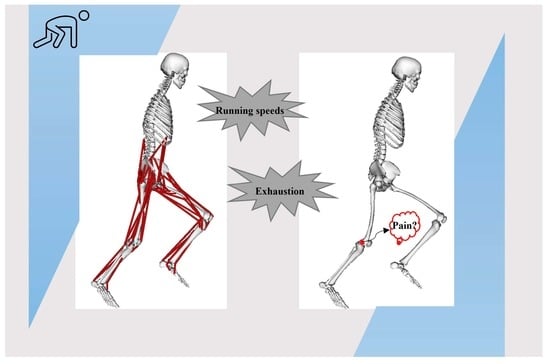
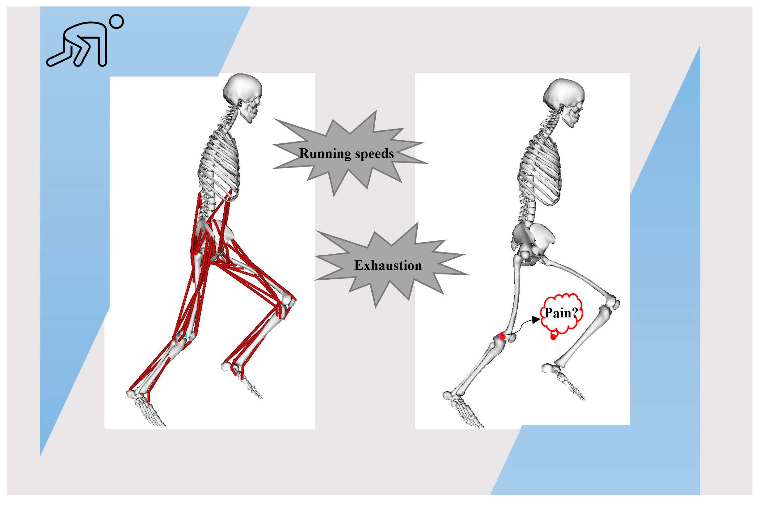
Effects of Running Speeds and Exhaustion on Iliotibial Band Strain during Running


Abstract
:
1. Introduction
2. Methodology
2.1. Participants
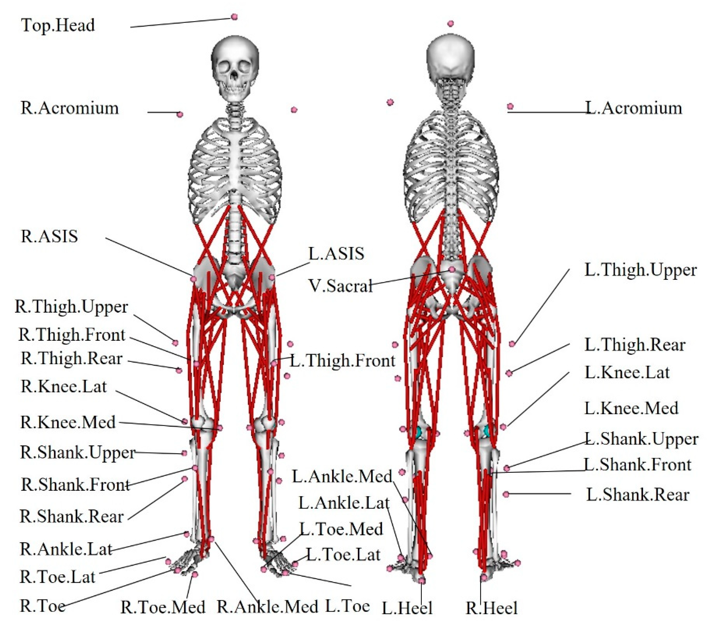
2.2. Experiments
2.3. MSK Model
2.4. Data Analysis
3. Results
4. Discussion
5. Conclusions
Author Contributions
Funding
Institutional Review Board Statement
Informed Consent Statement
Data Availability Statement
Conflicts of Interest
References
- Aderem, J.; Louw, Q.A. Biomechanical risk factors associated with iliotibial band syndrome in runners: A systematic review. BMC Musculoskelet. Disord. 2015, 16, 356. [Google Scholar] [CrossRef] [Green Version]
- Fairclough, J.; Hayashi, K.; Toumi, H.; Lyons, K.; Bydder, G.; Phillips, N.; Best, T.M.; Benjamin, M. The functional anatomy of the iliotibial band during flexion and extension of the knee: Implications for understanding iliotibial band syndrome. J. Anat. 2006, 208, 309–316. [Google Scholar] [CrossRef]
- Eng, C.M.; Arnold, A.S.; Lieberman, D.E.; Biewener, A.A. The capacity of the human iliotibial band to store elastic energy during running. J. Biomech. 2015, 48, 3341–3348. [Google Scholar] [CrossRef] [Green Version]
- Taunton, J.E.; Ryan, M.B.; Clement, D.B.; McKenzie, D.C.; Lloyd-Smith, D.R.; Zumbo, B.D. A retrospective case-control analysis of 2002 running injuries. Br. J. Sport. Med. 2002, 36, 95–101. [Google Scholar] [CrossRef] [Green Version]
- Devan, M.R.; Pescatello, L.S.; Faghri, P.; Anderson, J. A prospective study of overuse knee injuries among female athletes with muscle imbalances and structural abnormalities. J. Athl. Train. 2004, 39, 263. [Google Scholar] [PubMed]
- Fairclough, J.; Hayashi, K.; Toumi, H.; Lyons, K.; Bydder, G.; Phillips, N.; Best, T.M.; Benjamin, M. Is iliotibial band syndrome really a friction syndrome? J. Sci. Med. Sport 2007, 10, 74–76; discussion 77–78. [Google Scholar] [CrossRef]
- Hamill, J.; Miller, R.; Noehren, B.; Davis, I. A prospective study of iliotibial band strain in runners. Clin. Biomech. 2008, 23, 1018–1025. [Google Scholar] [CrossRef] [PubMed]
- Ferber, R.; Noehren, B.; Hamill, J.; Davis, I.S. Competitive female runners with a history of iliotibial band syndrome demonstrate atypical hip and knee kinematics. J. Orthop. Sport. Phys. 2010, 40, 52–58. [Google Scholar] [CrossRef] [Green Version]
- Sinclair, J.; Ingram, J.; Butters, B.; Brooks, D.; Stainton, P.; Taylor, P.J. A three-experiment examination of iliotibial band strain characteristics during different conditions using musculoskeletal simulation. Sport Sci. Health 2020, 16, 727–736. [Google Scholar] [CrossRef]
- Meardon, S.A.; Campbell, S.; Derrick, T.R. Step width alters iliotibial band strain during running. Sport. Biomech. 2012, 11, 464–472. [Google Scholar] [CrossRef]
- Day, E.M.; Gillette, J.C. Acute Effects of Wedge Orthoses and Sex on Iliotibial Band Strain During Overground Running in Nonfatiguing Conditions. J. Orthop. Sport. Phys. 2019, 49, 743–750. [Google Scholar] [CrossRef]
- Schache, A.G.; Dorn, T.W.; Williams, G.P.; Brown, N.A.; Pandy, M.G. Lower-limb muscular strategies for increasing running speed. J. Orthop. Sport. Phys. 2014, 44, 813–824. [Google Scholar] [CrossRef] [PubMed] [Green Version]
- Hafer, J.F.; Brown, A.M.; deMille, P.; Hillstrom, H.J.; Garber, C.E. The effect of a cadence retraining protocol on running biomechanics and efficiency: A pilot study. J. Sport. Sci. 2015, 33, 724–731. [Google Scholar] [CrossRef]
- Petersen, J.; Sorensen, H.; Nielsen, R.O. Cumulative loads increase at the knee joint with slow-speed running compared to faster running: A biomechanical study. J. Orthop. Sport. Phys. 2015, 45, 316–322. [Google Scholar] [CrossRef] [PubMed]
- Mei, Q.; Gu, Y.; Xiang, L.; Baker, J.S.; Fernandez, J. Foot Pronation Contributes to Altered Lower Extremity Loading After Long Distance Running. Front. Physiol. 2019, 10, 573. [Google Scholar] [CrossRef]
- Jewell, C.; Hamill, J.; von Tscharner, V.; Boyer, K.A. Altered multi-muscle coordination patterns in habitual forefoot runners during a prolonged, exhaustive run. Eur. J. Sport Sci. 2019, 19, 1062–1071. [Google Scholar] [CrossRef] [PubMed]
- Chen, T.L.; Wong, D.W.; Wang, Y.; Tan, Q.; Lam, W.K.; Zhang, M. Changes in segment coordination variability and the impacts of the lower limb across running mileages in half marathons: Implications for running injuries. J. Sport Health Sci. 2022, 11, 67–74. [Google Scholar] [CrossRef]
- Miller, R.H.; Lowry, J.L.; Meardon, S.A.; Gillette, J.C. Lower extremity mechanics of iliotibial band syndrome during an exhaustive run. Gait Posture 2007, 26, 407–413. [Google Scholar] [CrossRef]
- Sinclair, J.; Taylor, P.J.; Liles, N.B. Effects of running with minimal and conventional footwear in habitual and non-habitual users: A musculoskeletal simulation and statistical parametric mapping based approach. Footwear Sci. 2019, 12, 25–38. [Google Scholar] [CrossRef]
- Meardon, S.A.; Willson, J.D.; Gries, S.R.; Kernozek, T.W.; Derrick, T.R. Bone stress in runners with tibial stress fracture. Clin. Biomech. 2015, 30, 895–902. [Google Scholar] [CrossRef]
- Hutchinson, L.A.; Lichtwark, G.A.; Willy, R.W.; Kelly, L.A. The Iliotibial Band: A Complex Structure with Versatile Functions. Sport. Med. 2022, 52, 995–1008. [Google Scholar] [CrossRef] [PubMed]
- Tomiyama, S.; Usui, S.; Tamura, Y. Iliotibial band strain during two running speed conditions focused on the hip joint angle. In Proceedings of the 33 International Conference of Biomechanics in Sports (2015), Poitiers, France, 29 June–3 July 2015. [Google Scholar]
- Baker, R.L.; Souza, R.B.; Rauh, M.J.; Fredericson, M.; Rosenthal, M.D. Differences in Knee and Hip Adduction and Hip Muscle Activation in Runners with and without Iliotibial Band Syndrome. PM&R 2018, 10, 1032–1039. [Google Scholar] [CrossRef]
- Radzak, K.N.; Stickley, C.D. Fatigue-Induced Hip-Abductor Weakness and Changes in Biomechanical Risk Factors for Running-Related Injuries. J. Athl. Train. 2020, 55, 1270–1276. [Google Scholar] [CrossRef]
- Noehren, B.; Davis, I.; Hamill, J. ASB clinical biomechanics award winner 2006 prospective study of the biomechanical factors associated with iliotibial band syndrome. Clin. Biomech. 2007, 22, 951–956. [Google Scholar] [CrossRef] [PubMed]
- Fredericson, M.; Cookingham, C.L.; Chaudhari, A.M.; Dowdell, B.C.; Oestreicher, N.; Sahrmann, S.A. Hip abductor weakness in distance runners with iliotibial band syndrome. Clin. J. Sport Med. 2000, 10, 169–175. [Google Scholar] [CrossRef]
- Brindle, R.A.; Ebaugh, D.D.; Willson, J.D.; Finley, M.A.; Shewokis, P.A.; Milner, C.E. Relationships of hip abductor strength, neuromuscular control, and hip width to femoral length ratio with peak hip adduction angle in healthy female runners. J. Sport. Sci. 2020, 38, 2291–2297. [Google Scholar] [CrossRef]
- Brown, A.M.; Zifchock, R.A.; Hillstrom, H.J.; Song, J.; Tucker, C.A. The effects of fatigue on lower extremity kinematics, kinetics and joint coupling in symptomatic female runners with iliotibial band syndrome. Clin. Biomech. 2016, 39, 84–90. [Google Scholar] [CrossRef]
- Willwacher, S.; Kurz, M.; Robbin, J.; Thelen, M.; Hamill, J.; Kelly, L.; Mai, P. Running-Related Biomechanical Risk Factors for Overuse Injuries in Distance Runners: A Systematic Review Considering Injury Specificity and the Potentials for Future Research. Sport. Med. 2022, 52, 1863–1877. [Google Scholar] [CrossRef]
- Monti, R.J.; Roy, R.R.; Zhong, H.; Edgerton, V.R. Mechanical properties of rat soleus aponeurosis and tendon during variable recruitment in situ. J. Exp. Biol. 2003, 206, 3437–3445. [Google Scholar] [CrossRef] [Green Version]
- Birnbaum, K.; Siebert, C.H.; Pandorf, T.; Schopphoff, E.; Prescher, A.; Niethard, F.U. Anatomical and biomechanical investigations of the iliotibial tract. Surg. Radiol. Anat. 2004, 26, 433–446. [Google Scholar] [CrossRef] [PubMed]


| Participant Characteristics | Value | |
|---|---|---|
| Mass/kg | 64.73 (±11.65) | |
| Height/cm | 170.90 (±8.38) | |
| Pre-30 min run | Normal speed/(m/s) | 3.33 (±0.36) |
| Fast speed/(m/s) | 3.90 (±0.41) | |
| Post-30 min run | Normal speed/(m/s) | 3.32 (±0.36) |
| Fast speed/(m/s) | 3.93 (±0.38) |
| Joint Angles/° | Before Exhaustion (Mean ± SD) | After Exhaustion (Mean ± SD) | ||
|---|---|---|---|---|
| Normal Speed | Fast Speed | Normal Speed | Fast Speed | |
| Hip flexion | 29.70 (±4.00) | 34.96 (±5.19) b | 32.40 (±4.85) a | 37.86 (±4.55) a,b |
| Hip adduction | 13.89 (±2.85) | 14.22 (±3.21) | 15.15 (±2.99) | 15.48 (±2.82) a |
| Hip internal rotation | 7.90 (±6.56) | 6.97 (±5.92) | 8.01 (±5.82) | 5.63 (±6.22) b |
| Knee flexion | 44.59 (±3.55) | 46.81 (±3.86) b | 46.94 (±3.69) a | 49.09 (±3.43) a,b |
| Knee adduction | 5.92 (±3.28) | 6.44 (±3.46) | 6.26 (±3.91) | 6.71 (±4.11) |
| Knee internal rotation | 7.67 (±5.29) | 7.99 (±5.84) | 8.92 (±6.22) | 8.722 (±6.70) |
| Moment (Nm/kg) | Before Exhaustion (Mean ± SD) | After Exhaustion (Mean ± SD) | ||
|---|---|---|---|---|
| Normal Speed | Fast Speed | Normal Speed | Fast Speed | |
| Hip extension | 2.33 (±0.62) | 2.77 (±0.51) b | 2.42 (±0.47) | 2.87 (±0.60) b |
| Hip abduction | 1.18 (±0.39) | 1.42 (±0.72) | 1.32 (±0.42) | 1.47 (±0.47) |
| Hip internal rotation | 0.26 (±0.11) | 0.32 (±0.14) | 0.31 (±0.10) | 0.36 (±0.13) b |
| Knee extension | 1.73 (±0.32) | 1.75 (±0.30) | 1.87 (±0.36) a | 1.86 (±0.32) |
| Knee adduction | 0.66 (±0.30) | 0.76 (±0.48) | 0.73 (±0.31) | 0.79 (±0.38) |
| Knee internal rotation | 0.25 (±0.15) | 0.31 (±0.26) | 0.28 (±0.19) | 0.32 (±0.20) |
| Variables | Before Exhaustion (Mean ± SD) | After Exhaustion (Mean ± SD) | ||
|---|---|---|---|---|
| Normal Speed | Fast Speed | Normal Speed | Fast Speed | |
| ITB strain/% | 9.54 (±0.59) | 9.56 (±0.66) | 9.63 (±0.70) | 9.53 (±0.80) |
| ITB strain rate/(%/s) | 35.05 (±7.79) | 44.76 (±8.86) b | 38.57 (±9.04) a | 48.44 (±10.90) a,b |
Disclaimer/Publisher’s Note: The statements, opinions and data contained in all publications are solely those of the individual author(s) and contributor(s) and not of MDPI and/or the editor(s). MDPI and/or the editor(s) disclaim responsibility for any injury to people or property resulting from any ideas, methods, instructions or products referred to in the content. |
© 2023 by the authors. Licensee MDPI, Basel, Switzerland. This article is an open access article distributed under the terms and conditions of the Creative Commons Attribution (CC BY) license (https://creativecommons.org/licenses/by/4.0/).
Share and Cite
Chen, S.; Wang, Y.; Bing, F.; Zhang, M. Effects of Running Speeds and Exhaustion on Iliotibial Band Strain during Running. Bioengineering 2023, 10, 417. https://doi.org/10.3390/bioengineering10040417
Chen S, Wang Y, Bing F, Zhang M. Effects of Running Speeds and Exhaustion on Iliotibial Band Strain during Running. Bioengineering. 2023; 10(4):417. https://doi.org/10.3390/bioengineering10040417
Chicago/Turabian StyleChen, Shanefei, Yan Wang, Fangbo Bing, and Ming Zhang. 2023. "Effects of Running Speeds and Exhaustion on Iliotibial Band Strain during Running" Bioengineering 10, no. 4: 417. https://doi.org/10.3390/bioengineering10040417