
In Vitro Antibody Quantification with Hyperspectral Imaging in a Large Field of View for Clinical Applications
 , , , ,
  , and
, and 
Abstract
:1. Introduction
2. Materials and Methods
2.1. Sampling
2.2. Hyperspectral Imaging
2.3. Antibody
2.4. Statistics
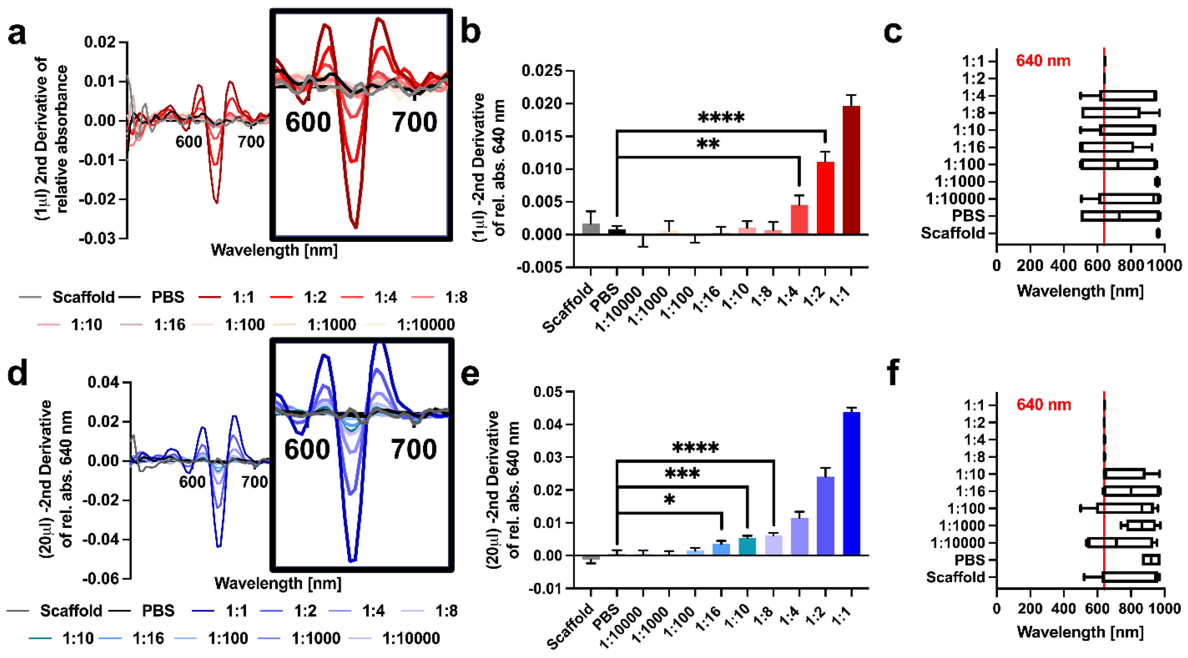
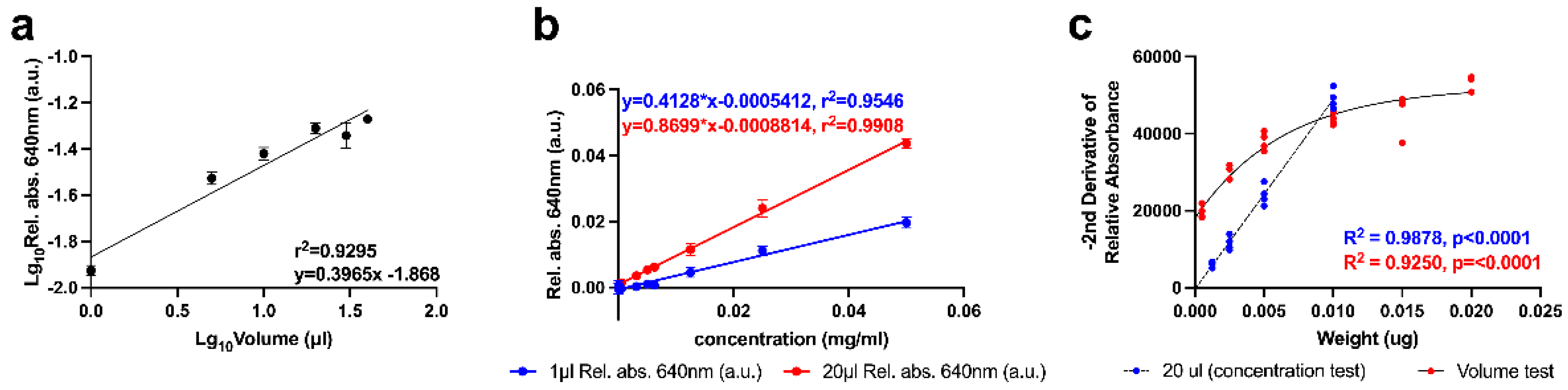
3. Results
4. Discussion
5. Conclusions
Supplementary Materials
Author Contributions
Funding
Institutional Review Board Statement
Informed Consent Statement
Data Availability Statement
Conflicts of Interest
References
- Calin, M.A.; Parasca, S.V.; Savastru, D.; Manea, D. Hyperspectral Imaging in the Medical Field: Present and Future. Appl. Spectrosc. Rev. 2014, 49, 435–447. [Google Scholar] [CrossRef]
- Li, Q.; He, X.; Wang, Y.; Liu, H.; Xu, D.; Guo, F. Review of spectral imaging technology in biomedical engineering: Achievements and challenges. J. Biomed. Opt. 2013, 18, 100901. [Google Scholar] [CrossRef] [Green Version]
- Sun, L.; Zhou, M.; Li, Q.; Hu, M.; Wen, Y.; Zhang, J.; Lu, Y.; Chu, J. Diagnosis of cholangiocarcinoma from microscopic hyperspectral pathological dataset by deep convolution neural networks. Methods 2022, 202, 22–30. [Google Scholar] [CrossRef] [PubMed]
- Wang, Q.; Li, Q.; Zhou, M.; Sun, Z.; Liu, H.; Wang, Y. A hyperspectral vessel image registration method for blood oxygenation mapping. PLoS ONE 2017, 12, e0178499. [Google Scholar] [CrossRef] [Green Version]
- Lu, G.; Fei, B. Medical hyperspectral imaging: A review. J. Biomed. Opt. 2014, 19, 10901. [Google Scholar] [CrossRef]
- Akalin, A.; Mu, X.; Kon, M.A.; Ergin, A.; Remiszewski, S.H.; Thompson, C.M.; Raz, D.J.; Diem, M. Classification of malignant and benign tumors of the lung by infrared spectral histopathology (SHP). Lab. Investig. 2015, 95, 406–421. [Google Scholar] [CrossRef] [Green Version]
- Cinelli, L.; Felli, E.; Baratelli, L.; Ségaud, S.; Baiocchini, A.; Okamoto, N.; Rodríguez-Luna, M.R.; Elmore, U.; Rosati, R.; Partelli, S.; et al. Single Snapshot Imaging of Optical Properties (SSOP) for Perfusion Assessment during Gastric Conduit Creation for Esophagectomy: An Experimental Study on Pigs. Cancers 2021, 13, 6079. [Google Scholar] [CrossRef] [PubMed]
- Rodríguez-Luna, M.R.; Okamoto, N.; Cinelli, L.; Baratelli, L.; Ségaud, S.; Rodríguez-Gómez, A.; Keller, D.S.; Zonoobi, E.; Bannone, E.; Marescaux, J.; et al. Quantification of bowel ischaemia using real-time multispectral Single Snapshot Imaging of Optical Properties (SSOP). Surg. Endosc. 2022, 37, 2395–2403. [Google Scholar] [CrossRef]
- Sun, J.; Wu, Z.; Wang, L.; Yao, Q.; Li, M.; Yao, G. Adaptive denoising hyperspectral data for visualization enhancement of intraoperative tissue. J. Biophotonics 2022, 15, e202200083. [Google Scholar] [CrossRef]
- De Landro, M.; García-Molina, I.E.; Barberio, M.; Felli, E.; Agnus, V.; Pizzicannella, M.; Diana, M.; Zappa, E.; Saccomandi, P. Hyperspectral Imagery for Assessing Laser-Induced Thermal State Change in Liver. Sensors 2021, 21, 643. [Google Scholar] [CrossRef]
- De Landro, M.; Felli, E.; Collins, T.; Nkusi, R.; Baiocchini, A.; Barberio, M.; Orrico, A.; Pizzicannella, M.; Hostettler, A.; Diana, M.; et al. Prediction of In Vivo Laser-Induced Thermal Damage with Hyperspectral Imaging Using Deep Learning. Sensors 2021, 21, 6934. [Google Scholar] [CrossRef] [PubMed]
- Felli, E.; Cinelli, L.; Bannone, E.; Giannone, F.; Muttillo, E.M.; Barberio, M.; Keller, D.S.; Rodríguez-Luna, M.R.; Okamoto, N.; Collins, T.; et al. Hyperspectral Imaging in Major Hepatectomies: Preliminary Results from the Ex-Machyna Trial. Cancers 2022, 14, 5591. [Google Scholar] [CrossRef] [PubMed]
- Okamoto, N.; Rodríguez-Luna, M.R.; Bencteux, V.; Al-Taher, M.; Cinelli, L.; Felli, E.; Urade, T.; Nkusi, R.; Mutter, D.; Marescaux, J.; et al. Computer-Assisted Differentiation between Colon-Mesocolon and Retroperitoneum Using Hyperspectral Imaging (HSI) Technology. Diagnostics 2022, 12, 2225. [Google Scholar] [CrossRef]
- Rodríguez-Luna, M.R.; Okamoto, N.; Al-Taher, M.; Keller, D.S.; Cinelli, L.; Ashoka, A.H.; Klymchenko, A.S.; Marescaux, J.; Diana, M. In Vivo Imaging Evaluation of Fluorescence Intensity at Tail Emission of Near-Infrared-I (NIR-I) Fluorophores in a Porcine Model. Life 2022, 12, 1123. [Google Scholar] [CrossRef] [PubMed]
- Halicek, M.; Fabelo, H.; Ortega, S.; Callico, G.M.; Fei, B. In-Vivo and Ex-Vivo Tissue Analysis through Hyperspectral Imaging Techniques: Revealing the Invisible Features of Cancer. Cancers 2019, 11, 756. [Google Scholar] [CrossRef] [PubMed] [Green Version]
- Khouj, Y.; Dawson, J.; Coad, J.; Vona-Davis, L. Hyperspectral Imaging and K-Means Classification for Histologic Evaluation of Ductal Carcinoma In Situ. Front. Oncol. 2018, 8, 17. [Google Scholar] [CrossRef] [PubMed] [Green Version]
- Manni, F.; van der Sommen, F.; Fabelo, H.; Zinger, S.; Shan, C.; Edstrom, E.; Elmi-Terander, A.; Ortega, S.; Callico, G.M.; de With, P.H.N. Hyperspectral Imaging for Glioblastoma Surgery: Improving Tumor Identification Using a Deep Spectral-Spatial Approach. Sensors 2020, 20, 6955. [Google Scholar] [CrossRef]
- Marinovich, M.L.; Azizi, L.; Macaskill, P.; Irwig, L.; Morrow, M.; Solin, L.J.; Houssami, N. The Association of Surgical Margins and Local Recurrence in Women with Ductal Carcinoma In Situ Treated with Breast-Conserving Therapy: A Meta-Analysis. Ann. Surg. Oncol. 2016, 23, 3811–3821. [Google Scholar] [CrossRef] [Green Version]
- Arbyn, M.; Redman, C.W.E.; Verdoodt, F.; Kyrgiou, M.; Tzafetas, M.; Ghaem-Maghami, S.; Petry, K.U.; Leeson, S.; Bergeron, C.; Nieminen, P.; et al. Incomplete excision of cervical precancer as a predictor of treatment failure: A systematic review and meta-analysis. Lancet Oncol. 2017, 18, 1665–1679. [Google Scholar] [CrossRef] [Green Version]
- He, F.; Zhang, W.; Shen, Y.; Yu, P.; Bao, Q.; Wen, J.; Hu, C.; Qiu, S. Effects of resection margins on local recurrence of osteosarcoma in extremity and pelvis: Systematic review and meta-analysis. Int. J. Surg. 2016, 36, 283–292. [Google Scholar] [CrossRef] [PubMed]
- Tummers, W.S.; Groen, J.V.; Mulder, B.G.S.; Farina-Sarasqueta, A.; Morreau, J.; Putter, H.; van de Velde, C.J.; Vahrmeijer, A.L.; Bonsing, B.A.; Mieog, J.S.; et al. Impact of resection margin status on recurrence and survival in pancreatic cancer surgery. Br. J. Surg. 2019, 106, 1055–1065. [Google Scholar] [CrossRef] [PubMed] [Green Version]
- Bickenbach, K.A.; Gonen, M.; Strong, V.; Brennan, M.F.; Coit, D.G. Association of positive transection margins with gastric cancer survival and local recurrence. Ann. Surg. Oncol. 2013, 20, 2663–2668. [Google Scholar] [CrossRef] [PubMed]
- Zeng, W.G.; Liu, M.J.; Zhou, Z.X.; Wang, Z.J. A Distal Resection Margin of ≤1 mm and Rectal Cancer Recurrence After Sphincter-Preserving Surgery: The Role of a Positive Distal Margin in Rectal Cancer Surgery. Dis. Colon Rectum 2017, 60, 1175–1183. [Google Scholar] [CrossRef] [PubMed]
- Demir, B.; Incaz, S.; Uckuyulu, E.I.; Oysu, C. Accuracy of Frozen Section Examination in Oral Cavity Cancers. Ear Nose Throat J. 2022, 101, NP354–NP357. [Google Scholar] [CrossRef]
- Liu, Y.J.; Smith-Chakmakova, F.; Rassaei, N.; Han, B.; Enomoto, L.M.; Crist, H.; Hollenbeak, C.S.; Karamchandani, D.M. Frozen Section Interpretation of Pancreatic Margins: Subspecialized Gastrointestinal Pathologists Versus General Pathologists. Int. J. Surg. Pathol. 2016, 24, 108–115. [Google Scholar] [CrossRef]
- Keereweer, S.; Kerrebijn, J.D.; van Driel, P.B.; Xie, B.; Kaijzel, E.L.; Snoeks, T.J.; Que, I.; Hutteman, M.; van der Vorst, J.R.; Mieog, J.S.; et al. Optical image-guided surgery--where do we stand? Mol. Imaging Biol. 2011, 13, 199–207. [Google Scholar] [CrossRef] [Green Version]
- Wang, S.; Zhang, Y. HMGB1 in inflammation and cancer. J. Hematol. Oncol. 2020, 13, 116. [Google Scholar] [CrossRef]
- Rapoport, B.L.; Steel, H.C.; Theron, A.J.; Heyman, L.; Smit, T.; Ramdas, Y.; Anderson, R. High Mobility Group Box 1 in Human Cancer. Cells 2020, 9, 1664. [Google Scholar] [CrossRef]
- Holmer, A.; Marotz, J.; Wahl, P.; Dau, M.; Kammerer, P.W. Hyperspectral imaging in perfusion and wound diagnostics—methods and algorithms for the determination of tissue parameters. Biomed. Tech. 2018, 63, 547–556. [Google Scholar] [CrossRef]
- Sano, K.; Mitsunaga, M.; Nakajima, T.; Choyke, P.L.; Kobayashi, H. In vivo breast cancer characterization imaging using two monoclonal antibodies activatably labeled with near infrared fluorophores. Breast Cancer Res. 2012, 14, R61. [Google Scholar] [CrossRef] [Green Version]
- Iqbal, N.; Iqbal, N. Human Epidermal Growth Factor Receptor 2 (HER2) in Cancers: Overexpression and Therapeutic Implications. Mol. Biol. Int. 2014, 2014, 852748. [Google Scholar] [CrossRef] [PubMed]
- Stummer, W.; Pichlmeier, U.; Meinel, T.; Wiestler, O.D.; Zanella, F.; Reulen, H.J. Fluorescence-guided surgery with 5-aminolevulinic acid for resection of malignant glioma: A randomised controlled multicentre phase III trial. Lancet Oncol. 2006, 7, 392–401. [Google Scholar] [CrossRef] [PubMed]
- Nagengast, W.B.; Hartmans, E.; Garcia-Allende, P.B.; Peters, F.T.M.; Linssen, M.D.; Koch, M.; Koller, M.; Tjalma, J.J.J.; Karrenbeld, A.; Jorritsma-Smit, A.; et al. Near-infrared fluorescence molecular endoscopy detects dysplastic oesophageal lesions using topical and systemic tracer of vascular endothelial growth factor A. Gut 2019, 68, 7–10. [Google Scholar] [CrossRef] [PubMed] [Green Version]
- Eggert, D.; Bengs, M.; Westermann, S.; Gessert, N.; Gerstner, A.O.H.; Mueller, N.A.; Bewarder, J.; Schlaefer, A.; Betz, C.; Laffers, W. In vivo detection of head and neck tumors by hyperspectral imaging combined with deep learning methods. J. Biophotonics 2021, 15, e202100167. [Google Scholar] [CrossRef]
- Barberio, M.; Benedicenti, S.; Pizzicannella, M.; Felli, E.; Collins, T.; Jansen-Winkeln, B.; Marescaux, J.; Viola, M.G.; Diana, M. Intraoperative Guidance Using Hyperspectral Imaging: A Review for Surgeons. Diagnostics 2021, 11, 2066. [Google Scholar] [CrossRef]
- Yao, W.; Wang, J.; Zhu, L.; Jia, X.; Xu, L.; Tian, X.; Hu, S.; Wu, S.; Wei, L. Epigenetic Regulator KDM4D Restricts Tumorigenesis via Modulating SYVN1/HMGB1 Ubiquitination Axis in Esophageal Squamous Cell Carcinoma. Front. Oncol. 2021, 11, 761346. [Google Scholar] [CrossRef] [PubMed]
- Porter, R.J.; Murray, G.I.; Brice, D.P.; Petty, R.D.; McLean, M.H. Novel biomarkers for risk stratification of Barrett’s oesophagus associated neoplastic progression-epithelial HMGB1 expression and stromal lymphocytic phenotype. Br. J. Cancer 2020, 122, 545–554. [Google Scholar] [CrossRef] [PubMed]
- Barberio, M.; Longo, F.; Fiorillo, C.; Seeliger, B.; Mascagni, P.; Agnus, V.; Lindner, V.; Geny, B.; Charles, A.L.; Gockel, I.; et al. HYPerspectral Enhanced Reality (HYPER): A physiology-based surgical guidance tool. Surg. Endosc. 2020, 34, 1736–1744. [Google Scholar] [CrossRef]
- Urade, T.; Felli, E.; Barberio, M.; Al-Taher, M.; Felli, E.; Goffin, L.; Agnus, V.; Ettorre, G.M.; Marescaux, J.; Mutter, D.; et al. Hyperspectral enhanced reality (HYPER) for anatomical liver resection. Surg. Endosc. 2021, 35, 1844–1850. [Google Scholar] [CrossRef]


Disclaimer/Publisher’s Note: The statements, opinions and data contained in all publications are solely those of the individual author(s) and contributor(s) and not of MDPI and/or the editor(s). MDPI and/or the editor(s) disclaim responsibility for any injury to people or property resulting from any ideas, methods, instructions or products referred to in the content. |
© 2023 by the authors. Licensee MDPI, Basel, Switzerland. This article is an open access article distributed under the terms and conditions of the Creative Commons Attribution (CC BY) license (https://creativecommons.org/licenses/by/4.0/).
Share and Cite
De Landro, M.; Cinelli, L.; Marchese, N.; Spano, G.; Barberio, M.; Vincent, C.; Marescaux, J.; Mutter, D.; De Mathelin, M.; Gioux, S.; et al. In Vitro Antibody Quantification with Hyperspectral Imaging in a Large Field of View for Clinical Applications. Bioengineering 2023, 10, 370. https://doi.org/10.3390/bioengineering10030370
De Landro M, Cinelli L, Marchese N, Spano G, Barberio M, Vincent C, Marescaux J, Mutter D, De Mathelin M, Gioux S, et al. In Vitro Antibody Quantification with Hyperspectral Imaging in a Large Field of View for Clinical Applications. Bioengineering. 2023; 10(3):370. https://doi.org/10.3390/bioengineering10030370
Chicago/Turabian StyleDe Landro, Martina, Lorenzo Cinelli, Nicola Marchese, Giulia Spano, Manuel Barberio, Cindy Vincent, Jacques Marescaux, Didier Mutter, Michel De Mathelin, Sylvain Gioux, and et al. 2023. "In Vitro Antibody Quantification with Hyperspectral Imaging in a Large Field of View for Clinical Applications" Bioengineering 10, no. 3: 370. https://doi.org/10.3390/bioengineering10030370