

De Novo Design of Imidazopyridine-Tethered Pyrazolines That Target Phosphorylation of STAT3 in Human Breast Cancer Cells
 , , , , , , , ,
Abstract
:1. Introduction
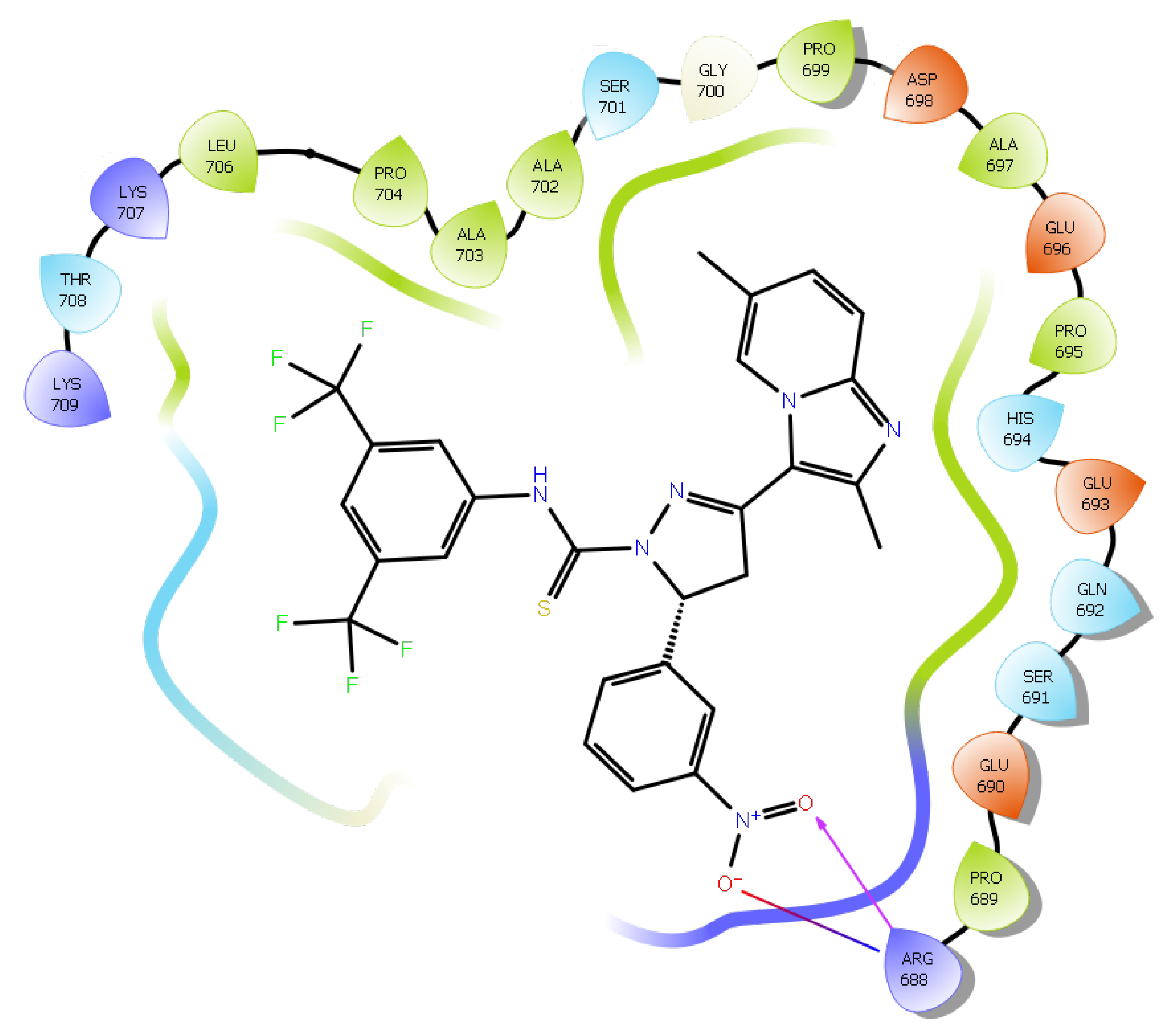
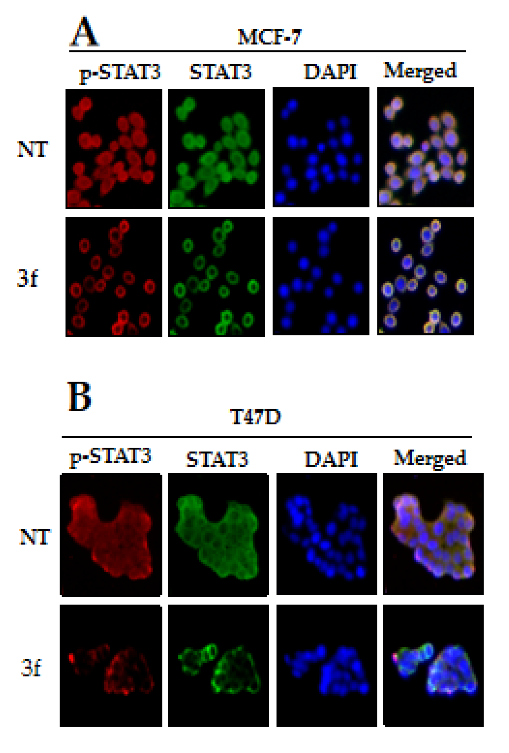
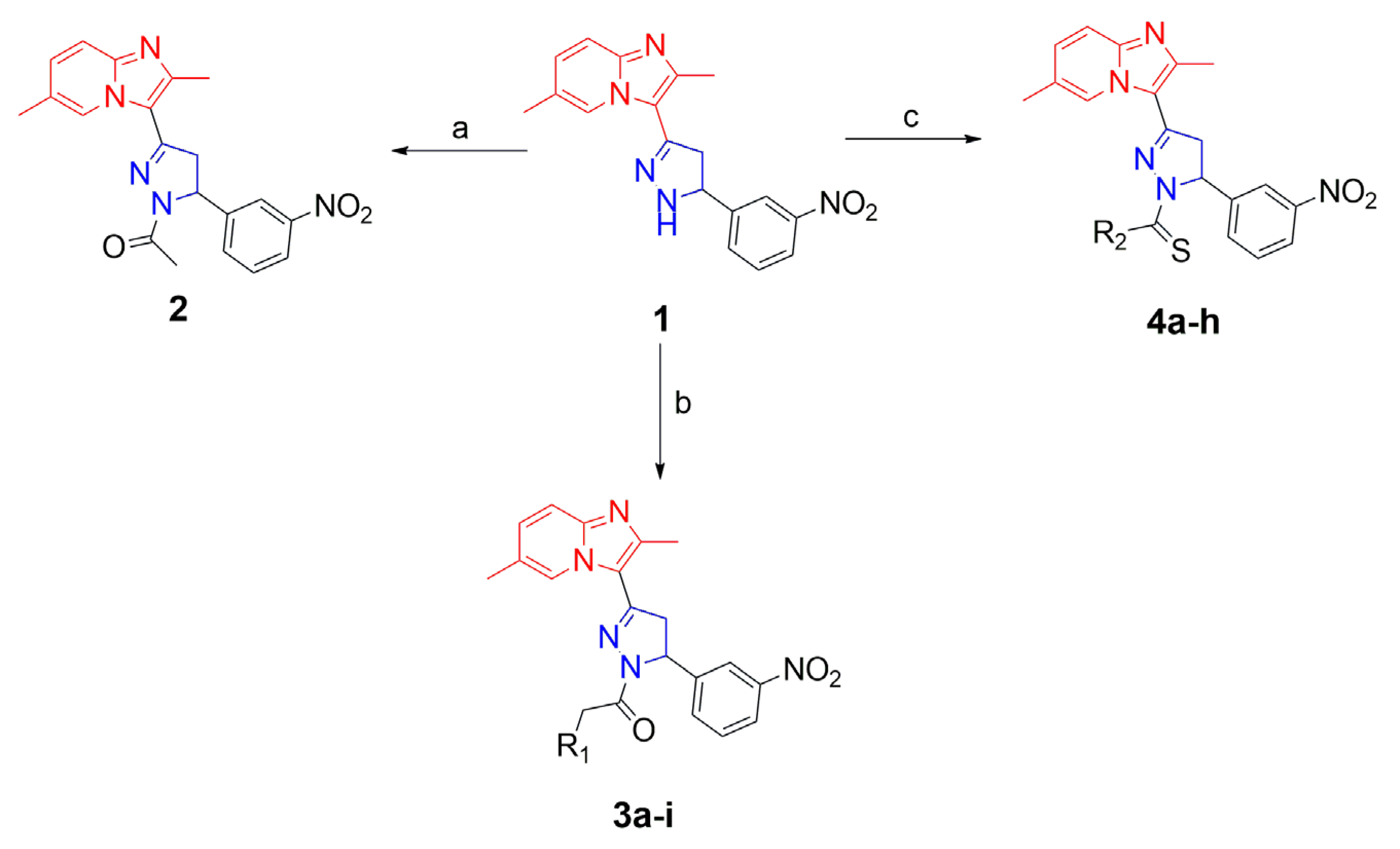
2. Results and Discussion
3. Materials and Methods
3.1. General Procedure for the Synthesis of Imidazole-Pyridine Substituted Pyrazoline Derivatives
3.2. Synthesis of 2-Pyrazoline Derivatives (3a–i) from 2,6-Dimethyl-3-(5-(3-nitrophenyl)-4,5-dihydro-1H-pyrazol-3-yl)imidazo[1,2-a]pyridine
3.3. Synthesis of 2-Pyrazoline Derivatives (4a–h) from 2,6-Dimethyl-3-(5-(3-nitrophenyl)-4,5-dihydro-1H-pyrazol-3-yl)imidazo[1,2-a]pyridine
3.4. 1-(3-(2,6-Dimethylimidazo[1,2-a]pyridin-3-yl)-5-(3-nitrophenyl)-4,5-dihydro-1H-pyrazol-1-yl)ethanone (2)
3.5. 1-(2-(3-(2,6-Dimethylimidazo[1,2-a]pyridin-3-yl)-5-(3-nitrophenyl)-4,5-dihydro-1H-pyrazol-1-yl)-2-oxoethyl)piperidin-4-one (3a)
3.6. Tert-butyl-4-(2-(3-(2,6-dimethylimidazo[1,2-a]pyridin-3-yl)-5-(3-nitrophenyl)-4,5-dihydro-1H-pyrazol-1-yl)-2-oxoethyl)piperazine-1-carboxylate (3b)
3.7. 1-(3-(2,6-Dimethylimidazo[1,2-a]pyridin-3-yl)-5-(3-nitrophenyl)-4,5-dihydro-1H-pyrazol-1-yl)-2-(piperazin-1-yl)ethanone (3c)
3.8. 2-(4-Acetylpiperazin-1-yl)-1-(3-(2,6-dimethylimidazo[1,2-a]pyridin-3-yl)-5-(3-nitrophenyl)-4,5-dihydro-1H-pyrazol-1-yl)ethanone (3d)
3.9. 2-(4-(4-Chlorophenyl)piperazin-1-yl)-1-(3-(2,6-dimethylimidazo[1,2-a]pyridin-3-yl)-5-(3-nitrophenyl)-4,5-dihydro-1H-pyrazol-1-yl)ethanone (3e)
3.10. 2-(4-(2,3-Dichlorophenyl)piperazin-1-yl)-1-(3-(2,6-dimethylimidazo[1,2-a]pyridin-3-yl)-5-(3-nitrophenyl)-4,5-dihydro-1H-pyrazol-1-yl)ethanone (3f)
3.11. 2-(4-(4-Chloro-2-fluorophenyl)piperazin-1-yl)-1-(3-(2,6-dimethylimidazo[1,2-a]pyridin-3-yl)-5-(3-nitrophenyl)-4,5-dihydro-1H-pyrazol-1-yl)ethanone (3g)
3.12. 2-(4-(3,4-Difluorophenyl)piperazin-1-yl)-1-(3-(2,6-dimethylimidazo[1,2-a]pyridin-3-yl)-5-(3-nitrophenyl)-4,5-dihydro-1H-pyrazol-1-yl)ethanone (3h)
3.13. 1-(3-(2,6-Dimethylimidazo[1,2-a]pyridin-3-yl)-5-(3-nitrophenyl)-4,5-dihydro-1H-pyrazol-1-yl)-2-(4-(2-fluorophenyl)piperazin-1-yl)ethanone (3i)
3.14. 3-(2,6-Dimethylimidazo[1,2-a]pyridin-3-yl)-5-(3-nitrophenyl)-N-phenyl-4,5-dihydro-1H-pyrazoline-1-carbothioamide (4a)
3.15. 3-(2,6-Dimethylimidazo[1,2-a]pyridin-3-yl)-5-(3-nitrophenyl)-N-(4-nitrophenyl)-4,5-dihydro-1H-pyrazole-1-carbothioamide (4b)
3.16. N-Cyclohexyl-3-(2,6-dimethylimidazo[1,2-a]pyridin-3-yl)-5-(3-nitrophenyl)-4,5-dihydro-1H-pyrazoline-1-carbothioamide (4c)
3.17. N-(4-Chlorophenyl)-3-(2,6-dimethylimidazo[1,2-a]pyridin-3-yl)-5-(3-nitrophenyl)-4,5-dihydro-1H-pyrazoline-1-carbothioamide (4d)
3.18. N-(3-Chlorophenyl)-3-(2,6-dimethylimidazo[1,2-a]pyridin-3-yl)-5-(3-nitrophenyl)-4,5-dihydro-1H-pyrazoline-1-carbothioamide (4e)
3.19. 3-(2,6-Dimethylimidazo[1,2-a]pyridin-3-yl)-5-(3-nitrophenyl)-N-(p-tolyl)-4,5-dihydro-1H-pyrazoline-1-carbothioamide (4f)
3.20. N-(3,5-Bis(trifluoromethyl)phenyl)-3-(2,6-dimethylimidazo[1,2-a]pyridin-3-yl)-5-(3-nitrophenyl)-4,5-dihydro-1H-pyrazoline-1-carbothioamide (4g)
3.21. N-Butyl-3-(2,6-dimethylimidazo[1,2-a]pyridin-3-yl)-5-(3-nitrophenyl)-4,5-dihydro-1H-pyrazoline-1-carbothioamide (4h)
3.22. Cell Viability Assay
3.23. Preparation of Whole Cell Lysates
3.24. Western Blot Analysis
3.25. In Silico DFT Calculations
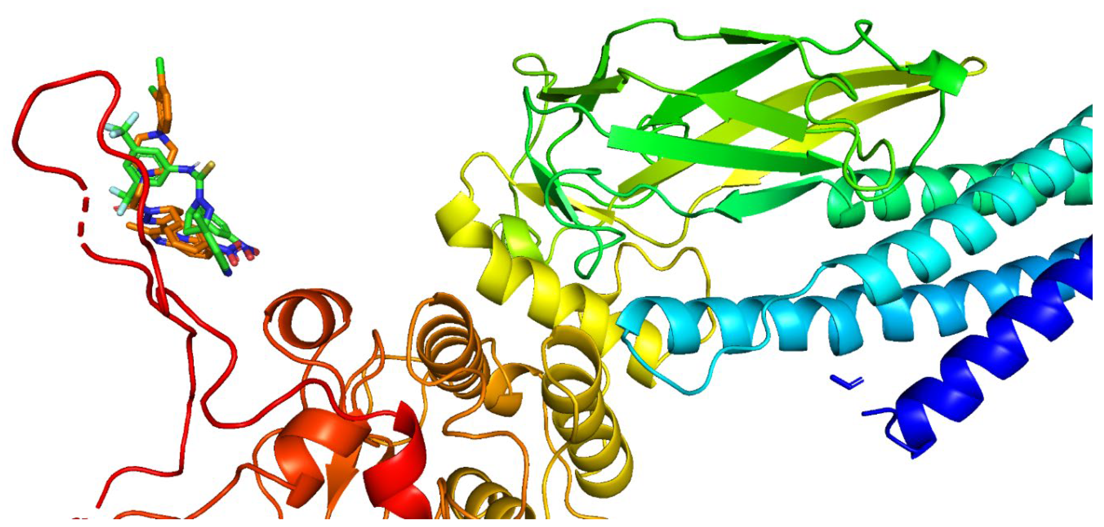
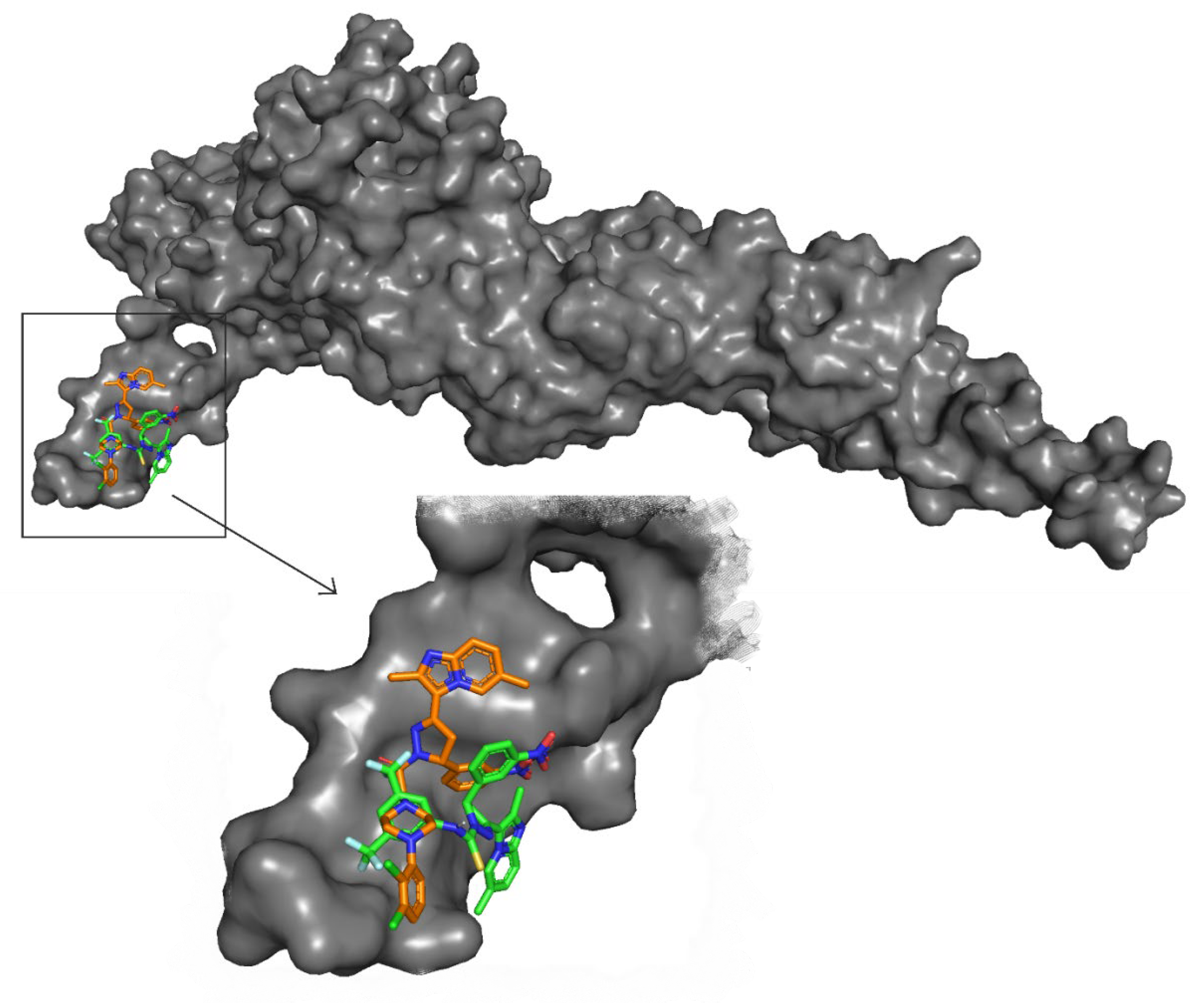
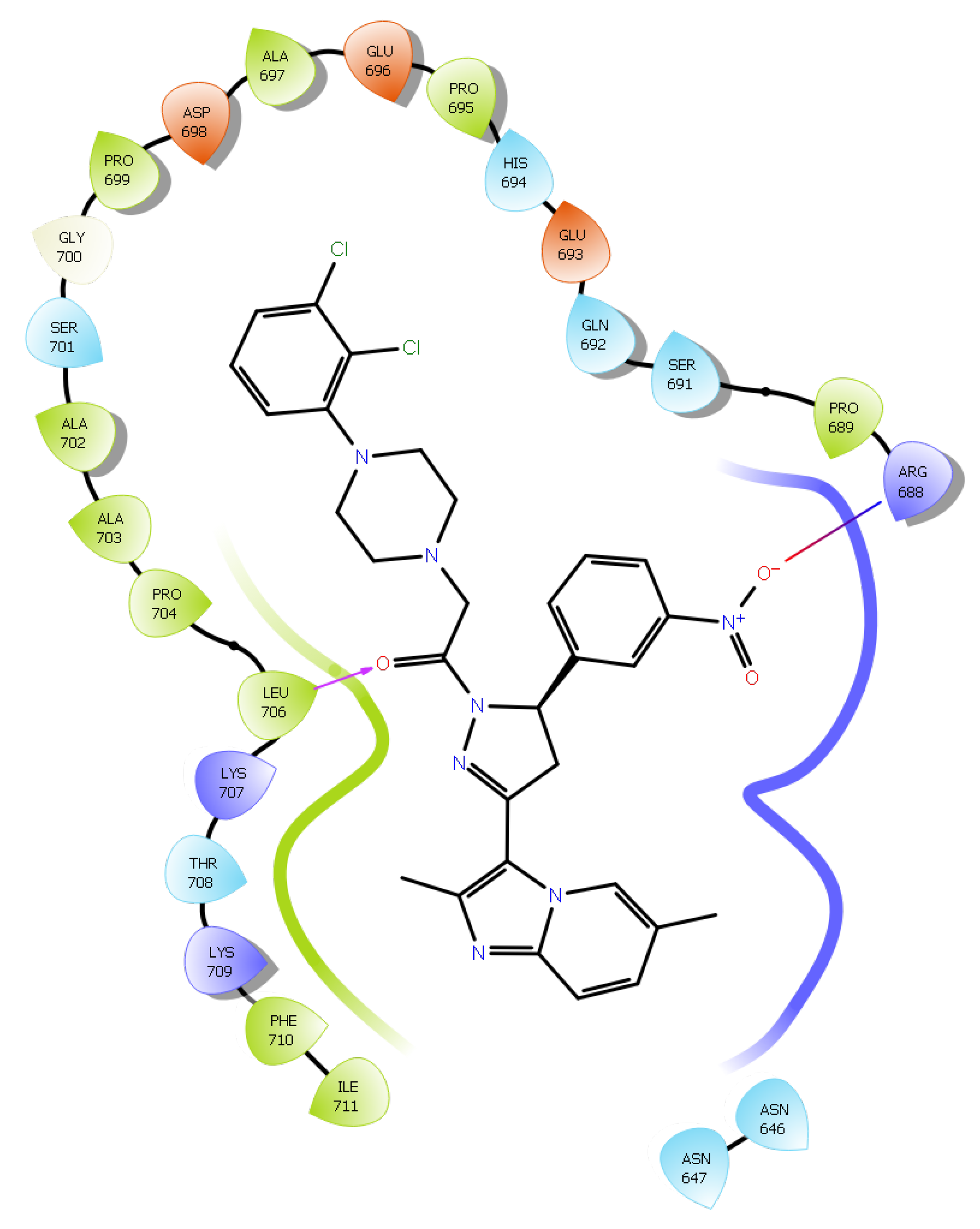
3.26. Docking Simulation
3.27. Immunocytochemistry Assay
4. Conclusions
Supplementary Materials
Author Contributions
Funding
Institutional Review Board Statement
Informed Consent Statement
Data Availability Statement
Conflicts of Interest
Sample Availability
References
- Ghoncheh, M.; Pournamdar, Z.; Salehiniya, H. Incidence and Mortality and Epidemiology of Breast Cancer in the World. Asian Pac. J. Cancer Prev. 2016, 17, 43–46. [Google Scholar] [CrossRef] [PubMed] [Green Version]
- Bray, F.; Ferlay, J.; Soerjomataram, I.; Siegel, R.L.; Torre, L.A.; Jemal, A. Global cancer statistics 2018: GLOBOCAN estimates of incidence and mortality worldwide for 36 cancers in 185 countries. CA Cancer J. Clin. 2018, 68, 394–424. [Google Scholar] [CrossRef] [PubMed] [Green Version]
- Wenger, C.R.; Clark, G.M. S-phase fraction and breast cancer—A decade of experience. Breast Cancer Res Treat. 1998, 51, 255–265. [Google Scholar] [CrossRef] [PubMed]
- Pandya, V.; Githaka, J.M.; Patel, N.; Veldhoen, R.; Hugh, J.; Damaraju, S.; McMullen, T.; Mackey, J.; Goping, I.S. BIK drives an aggressive breast cancer phenotype through sublethal apoptosis and predicts poor prognosis of ER-positive breast cancer. Cell Death Dis. 2020, 11, 448. [Google Scholar] [CrossRef] [PubMed]
- van Kuilenburg, A.B.; Maring, J.G. Evaluation of 5-fluorouracil pharmacokinetic models and therapeutic drug monitoring in cancer patients. Pharmacogenomics 2013, 14, 799–811. [Google Scholar] [CrossRef] [PubMed]
- Osborne, C.K. Tamoxifen in the treatment of breast cancer. N. Engl. J. Med. 1998, 339, 1609–1618. [Google Scholar] [CrossRef] [PubMed]
- Gourmelon, C.; Bourien, H.; Augereau, P.; Patsouris, A.; Frenel, J.S.; Campone, M. Vinflunine for the treatment of breast cancer. Expert Opin. Pharmacother. 2016, 17, 1817–1823. [Google Scholar] [CrossRef] [PubMed]
- Tutt, A.N.J.; Garber, J.E.; Kaufman, B.; Viale, G.; Fumagalli, D.; Rastogi, P.; Gelber, R.D.; de Azambuja, E.; Fielding, A.; Balmaña, J.; et al. Clinical Trial Steering Committee and Investigators. Adjuvant Olaparib for Patients with BRCA1- or BRCA2-Mutated Breast Cancer. N. Engl. J. Med. 2021, 384, 2394–2405. [Google Scholar] [CrossRef] [PubMed]
- Symmans, F.W. Breast cancer response to paclitaxel in vivo. Drug Resist. Updat. 2001, 4, 297–302. [Google Scholar] [CrossRef] [PubMed]
- Cuzick, J.; Sestak, I.; Forbes, J.F.; Dowsett, M.; Cawthorn, S.; Mansel, R.E.; Loibl, S.; Bonanni, B.; Evans, D.G.; Howell, A. IBIS-II investigators. Use of anastrozole for breast cancer prevention (IBIS-II): Long-term results of a randomised controlled trial. Lancet 2020, 395, 117–122. [Google Scholar] [CrossRef] [PubMed] [Green Version]
- Cuzick, J.; Sestak, I.; Baum, M.; Buzdar, A.; Howell, A.; Dowsett, M.; Forbes, J.F. ATAC/LATTE investigators. Effect of anastrozole and tamoxifen as adjuvant treatment for early-stage breast cancer: 10-year analysis of the ATAC trial. Lancet Oncol. 2010, 11, 1135–1141. [Google Scholar] [CrossRef] [PubMed]
- Hortobagyi, G.N.; Stemmer, S.M.; Burris, H.A.; Yap, Y.S.; Sonke, G.S.; Paluch-Shimon, S.; Campone, M.; Blackwell, K.L.; André, F.; Winer, E.P.; et al. Ribociclib as First-Line Therapy for HR-Positive, Advanced Breast Cancer. N. Engl. J. Med. 2016, 375, 1738–1748. [Google Scholar] [CrossRef] [PubMed]
- Jia, L.Y.; Shanmugam, M.K.; Sethi, G.; Bishayee, A. Potential role of targeted therapies in the treatment of triple-negative breast cancer. Anticancer. Drugs 2016, 27, 147–155. [Google Scholar] [CrossRef] [PubMed]
- Lee, J.H.; Kim, C.; Kim, S.H.; Sethi, G.; Ahn, K.S. Farnesol inhibits tumor growth and enhances the anticancer effects of bortezomib in multiple myeloma xenograft mouse model through the modulation of STAT3 signaling pathway. Cancer Lett. 2015, 360, 280–293. [Google Scholar] [CrossRef] [PubMed]
- Furtek, S.L.; Backos, D.S.; Matheson, C.J.; Reigan, P. Strategies and Approaches of Targeting STAT3 for Cancer Treatment. ACS Chem. Biol. 2016, 11, 308–318. [Google Scholar] [CrossRef] [PubMed]
- Garg, M.; Shanmugam, M.K.; Bhardwaj, V.; Goel, A.; Gupta, R.; Sharma, A.; Baligar, P.; Kumar, A.P.; Goh, B.C.; Wang, L.; et al. The pleiotropic role of transcription factor STAT3 in oncogenesis and its targeting through natural products for cancer prevention and therapy. Med. Res. Rev. 2021, 41, 1291–1336. [Google Scholar] [CrossRef] [PubMed]
- Lee, J.H.; Kim, C.; Baek, S.H.; Ko, J.H.; Lee, S.G.; Yang, W.M.; Um, J.Y.; Sethi, G.; Ahn, K.S. Capsazepine inhibits JAK/STAT3 signaling, tumor growth, and cell survival in prostate cancer. Oncotarget 2017, 8, 17700–17711. [Google Scholar] [CrossRef] [PubMed]
- Baek, S.H.; Ko, J.H.; Lee, H.; Jung, J.; Kong, M.; Lee, J.W.; Lee, J.; Chinnathambi, A.; Zayed, M.E.; Alharbi, S.A.; et al. Resveratrol inhibits STAT3 signaling pathway through the induction of SOCS-1: Role in apoptosis induction and radiosensitization in head and neck tumor cells. Phytomedicine 2016, 23, 566–577. [Google Scholar] [CrossRef] [PubMed]
- Ma, J.H.; Qin, L.; Li, X. Role of STAT3 signaling pathway in breast cancer. Cell Commun. Signal. 2020, 18, 33. [Google Scholar] [CrossRef] [PubMed] [Green Version]
- Hartenfeller, M.; Schneider, G. De novo drug design. Methods Mol. Biol. 2011, 672, 299–323. [Google Scholar] [CrossRef] [PubMed]
- Khatun, S.; Singh, A.; Bader, G.N.; Sofi, F.A. Imidazopyridine, a promising scaffold with potential medicinal applications and structural activity relationship (SAR): Recent advances. J. Biomol. Struct. Dyn. 2022, 40, 14279–14302. [Google Scholar] [CrossRef] [PubMed]
- Brown, D.G.; Wobst, H.J. A Decade of FDA-Approved Drugs (2010–2019): Trends and Future Directions. J. Med. Chem. 2021, 64, 2312–2338. [Google Scholar] [CrossRef] [PubMed]
- He, L.J.; Yang, D.L.; Chen, H.Y.; Huang, J.H.; Zhang, Y.J.; Qin, H.X.; Wang, J.L.; Tang, D.Y.; Chen, Z.Z. A Novel Imidazopyridine Derivative Exhibits Anticancer Activity in Breast Cancer by Inhibiting Wnt/β-catenin Signaling. Onco. Targets Ther. 2020, 13, 10111–10121. [Google Scholar] [CrossRef] [PubMed]
- Su, J.C.; Chang, C.H.; Wu, S.H.; Shiau, C.W. Novel imidazopyridine suppresses STAT3 activation by targeting SHP-1. J. Enzyme Inhib. Med. Chem. 2018, 33, 1248–1255. [Google Scholar] [CrossRef] [PubMed] [Green Version]
- Godse, P.; Kumar, P.; Yewalkar, N.; Deore, V.; Lohar, M.; Mundada, R.; Padgaonkar, A.; Manohar, S.; Joshi, A.; Bhatia, D.; et al. Discovery of P3971 an orally efficacious novel anticancer agent targeting HIF-1α and STAT3 pathways. Anticancer. Agents Med. Chem. 2013, 13, 1460–1466. [Google Scholar] [CrossRef] [PubMed]
- Nichols, W.C.; Kvols, L.K.; Ingle, J.N.; Edmonson, J.H.; Ahmann, D.L.; Rubin, J.; O’Connell, M.J. Phase II study of triazinate and pyrazofurin in patients with advanced breast cancer previously exposed to cytotoxic chemotherapy. Cancer Treat Rep. 1978, 62, 837–839. [Google Scholar] [PubMed]
- Tołoczko-Iwaniuk, N.; Dziemiańczyk-Pakieła, D.; Nowaszewska, B.K.; Celińska-Janowicz, K.; Miltyk, W. Celecoxib in Cancer Therapy and Prevention—Review. Curr. Drug Targets. 2019, 20, 302–315. [Google Scholar] [CrossRef] [PubMed]
- Nehra, B.; Rulhania, S.; Jaswal, S.; Kumar, B.; Singh, G.; Monga, V. Recent advancements in the development of bioactive pyrazoline derivatives. Eur. J. Med. Chem. 2020, 205, 112666. [Google Scholar] [CrossRef] [PubMed]
- Santoro, A.; Pisanti, S.; Grimaldi, C.; Izzo, A.A.; Borrelli, F.; Proto, M.C.; Malfitano, A.M.; Gazzerro, P.; Laezza, C.; Bifulco, M. Rimonabant inhibits human colon cancer cell growth and reduces the formation of precancerous lesions in the mouse colon. Int. J. Cancer 2009, 125, 996–1003. [Google Scholar] [CrossRef] [PubMed]
- Mamytbeková, A.; Hájícek, J.; Grimová, J.; Rezábek, K. Reductive effect of lonazolac on lung metastasis formation in mice. Neoplasma 1990, 37, 349–355. [Google Scholar] [PubMed]
- Srinivasa, V.; Li, F.; Siveen, K.S.; Dai, X.; Swamy, S.N.; Sethi, G.; Mantelingu, K.; Bender, A.; Rangappa, K.S. Synthesis and biological evaluation of tetrahydropyridinepyrazoles (‘PFPs’) as inhibitors of STAT3 phosphorylation. Med. Chem. Commun. 2014, 5, 32–40. [Google Scholar]
- Zhang, L.; Peterson, T.E.; Lu, V.M.; Parney, I.F.; Daniels, D.J. Antitumor activity of novel pyrazole-based small molecular inhibitors of the STAT3 pathway in patient derived high grade glioma cells. PLoS ONE 2019, 14, e0220569. [Google Scholar] [CrossRef] [PubMed] [Green Version]
- Wang, F.; Feng, K.R.; Zhao, J.Y.; Zhang, J.W.; Shi, X.W.; Zhou, J.; Gao, D.; Lin, G.Q.; Tian, P. Identification of novel STAT3 inhibitors bearing 2-acetyl-7-phenylamino benzofuran scaffold for antitumour study. Bioorg. Med. Chem. 2020, 28, 115822. [Google Scholar] [CrossRef] [PubMed]
- Anilkumar, N.C.; Sundaram, M.S.; Mohan, C.D.; Rangappa, S.; Bulusu, K.C.; Fuchs, J.E.; Girish, K.S.; Bender, A.; Basappa Rangappa, K.S. A One Pot Synthesis of Novel Bioactive Tri-Substitute-Condensed-Imidazopyridines that Targets Snake Venom Phospholipase A2. PLoS ONE 2015, 10, e0131896. [Google Scholar] [CrossRef] [PubMed] [Green Version]
- Kuthyala, S.; Hanumanthappa, M.; Kumar, S.M.; Sheik, S.; Karikannar, N.G.; Prabhu, A. Crystal, Hirshfeld, ADMET, drug-like and anticancer study of some newly synthesized imidazopyridine containing pyrazoline derivatives. J. Mol. Struct. 2019, 1197, 65–72. [Google Scholar] [CrossRef]
- Basappa, B.; Chumadathil Pookunoth, B.; Shinduvalli Kempasiddegowda, M.; Knchugarakoppal Subbegowda, R.; Lobie, P.E.; Pandey, V. Novel Biphenyl Amines Inhibit Oestrogen Receptor (ER)-α in ER-Positive Mammary Carcinoma Cells. Molecules 2021, 26, 783. [Google Scholar] [CrossRef] [PubMed]
- Bharathkumar, H.; Mohan, C.D.; Ananda, H.; Fuchs, J.E.; Li, F.; Rangappa, S.; Surender, M.; Bulusu, K.C.; Girish, K.S.; Sethi, G.; et al. Microwave-assisted synthesis, characterization and cytotoxic studies of novel estrogen receptor α ligands towards human breast cancer cells. Bioorg. Med. Chem. Lett. 2015, 25, 1804–1807. [Google Scholar] [CrossRef] [PubMed]
- Kalakoti, Y.; Yadav, S.; Sundar, D. Deep Neural Network-Assisted Drug Recommendation Systems for Identifying Potential Drug-Target Interactions. ACS Omega 2022, 7, 12138–12146. [Google Scholar] [CrossRef] [PubMed]
- Sebastian, A.; Pandey, V.; Mohan, C.D.; Chia, Y.T.; Rangappa, S.; Mathai, J.; Baburajeev, C.P.; Paricharak, S.; Mervin, L.H.; Bulusu, K.C.; et al. Novel Adamantanyl-Based Thiadiazolyl Pyrazoles Targeting EGFR in Triple-Negative Breast Cancer. ACS Omega 2016, 1, 1412–1424. [Google Scholar] [CrossRef] [PubMed] [Green Version]
- Lee, J.H.; Mohan, C.D.; Deivasigamani, A.; Jung, Y.Y.; Rangappa, S.; Basappa, S.; Chinnathambi, A.; Alahmadi, T.A.; Alharbi, S.A.; Garg, M.; et al. Brusatol suppresses STAT3-driven metastasis by downregulating epithelial-mesenchymal transition in hepatocellular carcinoma. J. Adv. Res. 2020, 26, 83–94. [Google Scholar] [CrossRef] [PubMed]
- Fleming, I. Molecular Orbitals and Organic Chemical Reactions; Wiley: Hoboken, NJ, USA, 2010; ISBN 9780470746585. [Google Scholar]
- Ansari, M.F.; Siddiqui, S.M.; Ahmad, K.; Avecilla, F.; Dharavath, S.; Gourinath, S.; Azam, A. Synthesis, antiamoebic and molecular docking studies of furan-thiazolidinone hybrids. Eur. J. Med. Chem. 2016, 124, 393–406. [Google Scholar] [CrossRef] [PubMed]
- Becker, S.; Groner, B.; Müller, C.W. Three-dimensional structure of the Stat3beta homodimer bound to DNA. Nature 1998, 394, 145–151. [Google Scholar] [CrossRef] [PubMed]
- Pandey, V.; Wang, B.; Mohan, C.D.; Raquib, A.R.; Rangappa, S.; Srinivasa, V.; Fuchs, J.E.; Girish, K.S.; Zhu, T.; Bender, A.; et al. Discovery of a small-molecule inhibitor of specific serine residue BAD phosphorylation. Proc. Natl. Acad. Sci. USA 2018, 115, E10505–E10514. [Google Scholar] [CrossRef] [PubMed] [Green Version]
- Barash, U.; Rangappa, S.; Mohan, C.D.; Vishwanath, D.; Boyango, I.; Basappa, B.; Vlodavsky, I.; Rangappa, K.S. New Heparanase-Inhibiting Triazolo-Thiadiazoles Attenuate Primary Tumor Growth and Metastasis. Cancers 2021, 13, 2959. [Google Scholar] [CrossRef] [PubMed]
- Zhang, J.; Sikka, S.; Siveen, K.S.; Lee, J.H.; Um, J.Y.; Kumar, A.P.; Chinnathambi, A.; Alharbi, S.A.; Basappa Rangappa, K.S.; Sethi, G.; et al. Cardamonin represses proliferation, invasion, and causes apoptosis through the modulation of signal transducer and activator of transcription 3 pathway in prostate cancer. Apoptosis 2017, 22, 158–168. [Google Scholar] [CrossRef] [PubMed]
- Mohan, C.D.; Bharathkumar, H.; Bulusu, K.C.; Pandey, V.; Rangappa, S.; Fuchs, J.E.; Shanmugam, M.K.; Dai, X.; Li, F.; Deivasigamani, A.; et al. Development of a novel azaspirane that targets the Janus kinase-signal transducer and activator of transcription (STAT) pathway in hepatocellular carcinoma in vitro and in vivo. J. Biol. Chem. 2014, 289, 34296–34307. [Google Scholar] [CrossRef] [PubMed] [Green Version]
- Frisch, M.J.; Trucks, G.W.; Schlegel, H.B.; Scuseria, G.E.; Robb, M.A.; Cheeseman, J.R. Gaussian 09; Gaussian Inc.: Wallingford, CT, USA, 2009. [Google Scholar]
- Lee, W.C.; Yang, R.G. Parr, Development of the Colle-Salvetti correlation-energy formula into a functional of the electron density. Phys. Rev. B 1988, 37, 785. [Google Scholar] [CrossRef] [Green Version]
- Ananda, S.; Khamees, H.A.; Mahendra, M.; Kumara, C.; Jagadeesh Prasad, D.; Hegde, T.A.; Vinitha, G. Structural, thermal, dielectric, nonlinear optical properties and DFT investigations of a novel material 2-(6-chloropyridin-3-yl)-N'-(2, 3-dihydro-1, 4-benzodioxin-6-ylmethylidene) acetohydrazide for optoelectronic applications. J. Mater. Sci. Mater. Electron. 2021, 32, 14677–14702. [Google Scholar] [CrossRef]
- Schrödinger, L.; DeLano, W. PyMOL. 2020. Available online: http://www.pymol.org/pymol (accessed on 1 October 2022).
- Sanner, M.F. Python: A programming language for software integration and development. J Mol Graph Model 1999, 17, 57–61. [Google Scholar]
- Abbasi-Radmoghaddam, Z.; Riahi, S.; Gharaghani, S.; Mohammadi-Khanaposhtanai, M. Design of potential anti-tumor PARP-1 inhibitors by QSAR and molecular modeling studies. Mol. Divers. 2021, 25, 263–277. [Google Scholar] [CrossRef]
- Schrödinger, LLC. Schrödinger Release 2020-1: Maestro; Schrödinger, LLC: New York, NY, USA, 2020. [Google Scholar]
- Kim, J.W.; Gautam, J.; Kim, J.E.; Kim, J.A.; Kang, K.W. Inhibition of tumor growth and angiogenesis of tamoxifen-resistant breast cancer cells by ruxolitinib, a selective JAK2 inhibitor. Oncol. Lett. 2019, 17, 3981–3989. [Google Scholar] [CrossRef] [Green Version]


| Compound Code | R1 or R2 | Melting Point in °C | Yield in % |
|---|---|---|---|
| 3a | Piperidone | 170–172 | 97 |
| 3b | 1-Boc piperazine | 166–168 | 96 |
| 3c | Piperazine | 176–178 | 35 |
| 3d | 1-acetyl piperazine | 208–210 | 98 |
| 3e | 1-(4-chlorophenyl)piperazine | 180–182 | 96 |
| 3f | 1-(2,3-dichlorophenyl)piperizine | 122–124 | 95 |
| 3g | 1-(4-chloro-2-fluorophenyl)piperazine | 196–198 | 95 |
| 3h | 1-(3,4-difluorophenyl)piperazine | 192–194 | 96 |
| 3i | 1-(2-fluorophenyl)piperazine | 160–162 | 98 |
| 4a | Aniline | 210–212 | 98 |
| 4b | 4-nitrophenylaniline | 230–232 | 95 |
| 4c | Cyclohexylamine | 204–206 | 98 |
| 4d | 4-chlorophenylaniline | 240–242 | 98 |
| 4e | 3-chlorophenylaniline | 242–244 | 98 |
| 4f | 4-methylphenylaniline | 256–258 | 97 |
| 4g | 3,5-bis(triflourophenyl)aniline | 230–232 | 98% |
| 4h | n-butylamine | 246–248 | 96% |
| Entry | IC50 (μM) | ||||
|---|---|---|---|---|---|
| MCF-7 | T47D | BT-474 | SK-BR-3 | MCF-10A | |
| 2 | >100 | >100 | 97.82±1.76 | >100 | >100 |
| 3a | 22.81 ± 1.36 | 23.31 ± 1.28 | 4.109 ± 0.41 | >100 | >100 |
| 3b | 22.29 ± 1.35 | 15.15 ± 1.13 | 21.29 ± 1.32 | 28.28 ± 1.34 | >100 |
| 3c | 29.27 ± 1.47 | 39.95 ± 1.49 | 26.75 ± 1.37 | 95.17 ± 1.79 | >100 |
| 3d | 21.03 ± 1.32 | >100 | 8.04 ± 0.47 | 32.14 ± 1.46 | >100 |
| 3e | 13.24 ± 1.12 | 29.9 ± 1.05 | 17.6 ± 0.87 | 39.13 ± 1.46 | 38.34 ± 1.44 |
| 3f | 9.27 ± 0.97 | 23.51 ± 1.25 | 13.91 ± 1.32 | >100 | >100 |
| 3g | 19.40 ± 1.29 | 86.55 ± 1.95 | 63.16 ± 1.75 | 99.96 ± 1.98 | 8.69 ± 0.69 |
| 3h | 19.56 ± 1.29 | 12.5 ± 1.01 | 3.061 ± 0.32 | 38.81 ± 1.42 | 6.27 ± 0.26 |
| 3i | 63.69 ± 1.80 | >100 | >100 | >100 | >100 |
| 4a | 81.37 ± 1.91 | >100 | >100 | >100 | >100 |
| 4b | 65.36 ± 1.82 | 45.61 ± 1.43 | 7.259 ± 0.54 | 69.02 ± 1.41 | >100 |
| 4c | 46.08 ± 1.66 | >100 | 52.83 ± 1.54 | >100 | >100 |
| 4d | >100 | >100 | >100 | >100 | >100 |
| 4e | >100 | >100 | >100 | >100 | >100 |
| 4f | 56.92 ± 1.39 | >100 | >100 | >100 | >100 |
| 4g | 10.90 ± 1.01 | >100 | >100 | >100 | >100 |
| 4h | >100 | 54.42 ± 1.68 | >100 | >100 | 90.59 ± 1.98 |
| Olaparib | 3.28 ± 0.68 | ||||
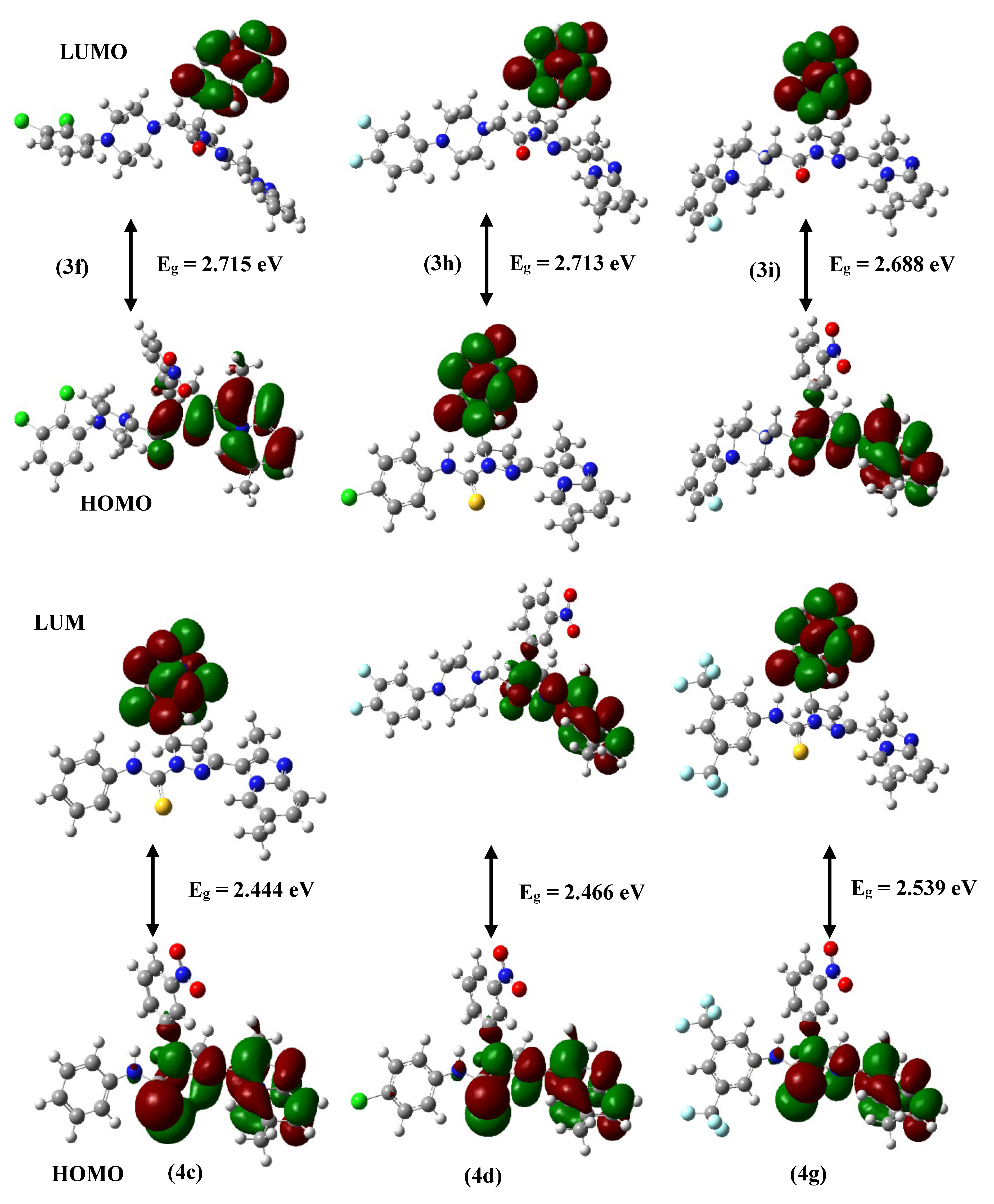
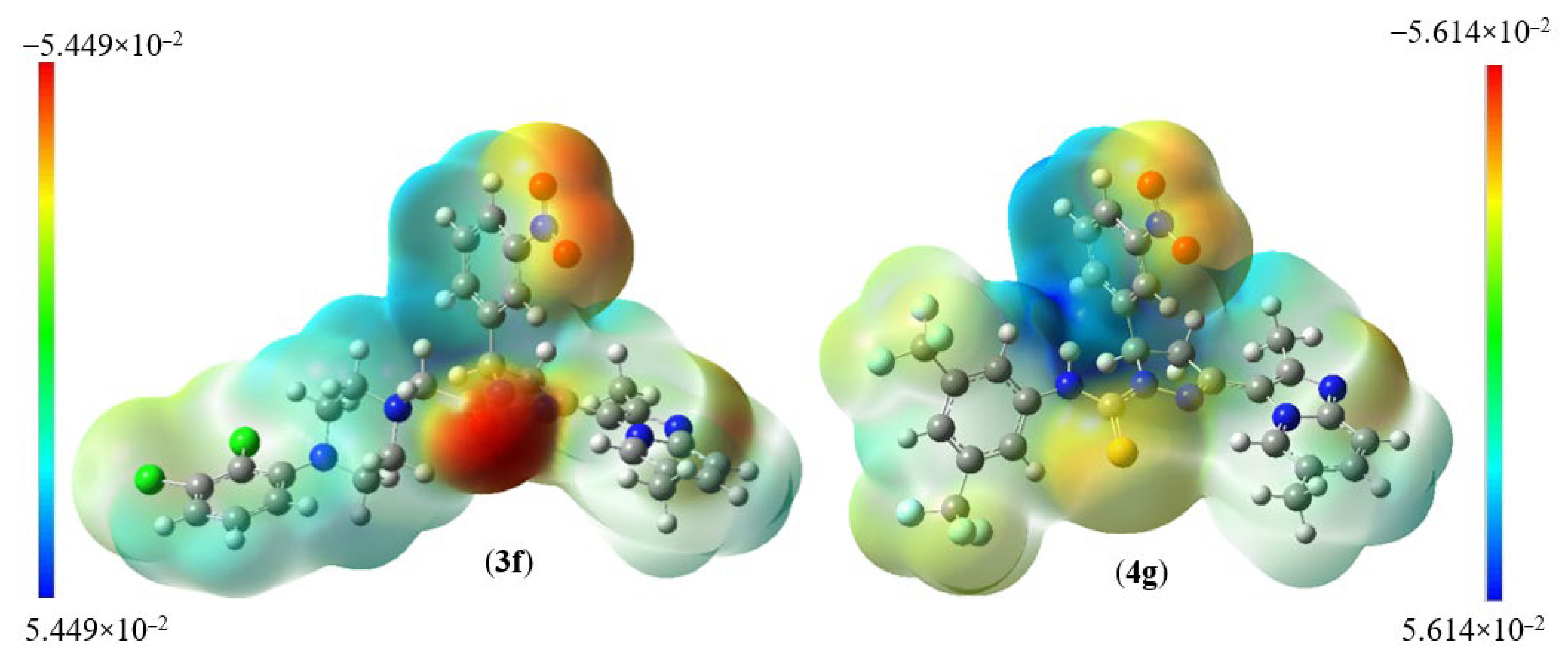
| Global Parameters | 3f | 3h | 3i | 4c | 4d | 4g |
|---|---|---|---|---|---|---|
| EHOMO (eV) | −5.764 | −5.799 | −5.704 | −5.645 | −5.733 | −5.905 |
| ELUMO (eV) | −3.049 | −3.086 | −3.016 | −3.201 | −3.267 | −3.366 |
| ΔELUMO–HOMO (eV) | 2.715 | 2.713 | 2.688 | 2.444 | 2.466 | 2.539 |
| Ionization potential (I) (eV) | 5.764 | 5.799 | 5.704 | 5.645 | 5.733 | 5.905 |
| Electron affinity (A) (eV) | 3.049 | 3.086 | 3.016 | 3.201 | 3.267 | 3.366 |
| Hardness (η) (eV) | 1.357 | 1.356 | 1.344 | 1.222 | 1.233 | 1.269 |
| Softness (S) (eV)−1 | 0.368 | 0.368 | 0.372 | 0.409 | 0.405 | 0.393 |
| Chemical potential (μ) (eV) | −4.406 | −4.442 | −4.360 | −4.423 | −4.500 | −4.635 |
| Electronegativity (χ) (eV) | 4.406 | 4.442 | 4.360 | 4.423 | 4.500 | 4.635 |
| Electrophilicity (ψ) (eV) | 7.151 | 7.274 | 7.072 | 8.004 | 8.211 | 8.463 |
Disclaimer/Publisher’s Note: The statements, opinions and data contained in all publications are solely those of the individual author(s) and contributor(s) and not of MDPI and/or the editor(s). MDPI and/or the editor(s) disclaim responsibility for any injury to people or property resulting from any ideas, methods, instructions or products referred to in the content. |
© 2023 by the authors. Licensee MDPI, Basel, Switzerland. This article is an open access article distributed under the terms and conditions of the Creative Commons Attribution (CC BY) license (https://creativecommons.org/licenses/by/4.0/).
Share and Cite
Ravish, A.; Shivakumar, R.; Xi, Z.; Yang, M.H.; Yang, J.-R.; Swamynayaka, A.; Nagaraja, O.; Madegowda, M.; Chinnathambi, A.; Alharbi, S.A.; et al. De Novo Design of Imidazopyridine-Tethered Pyrazolines That Target Phosphorylation of STAT3 in Human Breast Cancer Cells. Bioengineering 2023, 10, 159. https://doi.org/10.3390/bioengineering10020159
Ravish A, Shivakumar R, Xi Z, Yang MH, Yang J-R, Swamynayaka A, Nagaraja O, Madegowda M, Chinnathambi A, Alharbi SA, et al. De Novo Design of Imidazopyridine-Tethered Pyrazolines That Target Phosphorylation of STAT3 in Human Breast Cancer Cells. Bioengineering. 2023; 10(2):159. https://doi.org/10.3390/bioengineering10020159
Chicago/Turabian StyleRavish, Akshay, Rashmi Shivakumar, Zhang Xi, Min Hee Yang, Ji-Rui Yang, Ananda Swamynayaka, Omantheswara Nagaraja, Mahendra Madegowda, Arunachalam Chinnathambi, Sulaiman Ali Alharbi, and et al. 2023. "De Novo Design of Imidazopyridine-Tethered Pyrazolines That Target Phosphorylation of STAT3 in Human Breast Cancer Cells" Bioengineering 10, no. 2: 159. https://doi.org/10.3390/bioengineering10020159