Next Issue
Volume 10, March
Previous Issue
Volume 10, January


Bioengineering, Volume 10, Issue 2 (February 2023) – 155 articles

Cover Story (view full-size image): 
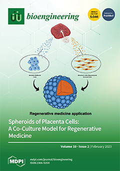
Given the significant limitations of exogenous insulin therapy in treating type 1 diabetes, there is a pressing need to explore alternative therapeutic options. Stem cell therapy and 3D cell cultures are emerging as potential regenerative medicine approaches. Perinatal cells, with their immunomodulatory activity and differentiative potential, are an ideal cell source for cell therapy. This study aimed to create 3D spheroids by co-culturing amniotic epithelial cells and Wharton’s jelly mesenchymal stromal cells. Our results suggest that co-culture spheroids are stable in long-term culture, still viable, and show consistent extracellular matrix production, indicating the potential for differentiation into endopancreatic cells for regenerative medicine applications in type 1 diabetes. View this paper
  • Issues are regarded as officially published after their release is announced to the table of contents alert mailing list.
  • You may sign up for e-mail alerts to receive table of contents of newly released issues.
  • PDF is the official format for papers published in both, html and pdf forms. To view the papers in pdf format, click on the "PDF Full-text" link, and use the free Adobe Reader to open them.
Order results
Result details
Section
Select all
Export citation of selected articles as:
20 pages, 3481 KiB  
Article
Liver Ultrasound Histotripsy: Novel Analysis of the Histotripsy Site Cell Constituents with Implications for Histotripsy Application in Cell Transplantation and Cancer Therapy
by Saied Froghi, Matheus Oliveira de Andrade, Layla Mohammad Hadi, Pierre Gelat, Hassan Rashidi, Alberto Quaglia, Barry Fuller, Nader Saffari and Brian Davidson
Bioengineering 2023, 10(2), 276; https://doi.org/10.3390/bioengineering10020276 - 20 Feb 2023
Cited by 2 | Viewed by 2218
Abstract
Introduction: Allogenic hepatocyte transplantation is an attractive alternative to whole-organ transplantation, particularly for the treatment of metabolic disorders and acute liver failure. However, the shortage of human donor organs for cell isolation, the low cell yield from decellularisation regimes, and low engraftment rates [...] Read more.
Introduction: Allogenic hepatocyte transplantation is an attractive alternative to whole-organ transplantation, particularly for the treatment of metabolic disorders and acute liver failure. However, the shortage of human donor organs for cell isolation, the low cell yield from decellularisation regimes, and low engraftment rates from portal administration of donor cells have restricted its clinical application. Using ultrasound histotripsy to provide a nidus in the liver for direct cell transplantation offers a new approach to overcoming key limitations in current cell therapy. We have analysed the liver cavity constituents to assess their potential as a site for cell delivery and implantation. Methods: Using human organ retrieval techniques, pig livers were collected from the abattoir and transported in ice-cold storage to the laboratory. Following 2 h of cold storage, the livers were flushed with organ preservation solution and placed on an organ perfusion circuit to maintain viability. Organs were perfused with Soltran™ organ preservation solution via the portal vein at a temperature of 24–30 °C. The perfusion circuit was oxygenated through equilibration with room air. Perfused livers (n=5) were subjected to ultrasound histotripsy, producing a total of 130 lesions. Lesions were generated by applying 50 pulses at 1 Hz pulse repetition frequency and 1% duty cycle using a single element 2 MHz bowl-shaped transducer (Sonic Concepts, H-148). Following histotripsy, a focal liver lesion was produced, which had a liquid centre. The fluid from each lesion was aspirated and cultured in medium (RPMI) at 37 °C in an incubator. Cell cultures were analysed at 1 and 7 days for cell viability and a live-dead assay was performed. The histotripsy sites were excised following aspiration and H&E staining was used to characterise the liver lesions. Cell morphology was determined by histology. Results: Histotripsy created a subcapsular lesion (~5 mm below the liver capsule; size ranging from 3 to 5 mm), which contained a suspension of cells. On average, 61×104 cells per mL were isolated. Hepatocytes were present in the aspirate, were viable at 24 h post isolation and remained viable in culture for up to 1 week, as determined by phalloidin/DAPI cell viability stains. Cultures up to 21 days revealed metabolically active live hepatocyte. Live-dead assays confirmed hepatocyte viability at 1 week (Day 1: 12% to Day 7: 45% live cells; p < 0.0001), which retained metabolic activity and morphology, confirmed on assay and microscopy. Cell Titre-GloTM showed a peak metabolic activity at 1 week (average luminescence 24.6 RLU; p < 0.0001) post-culture compared with the control (culture medium alone), reduced to 1/3 of peak level (7.85 RLU) by day 21. Conclusions: Histotripsy of the liver allows isolation and culture of hepatocytes with a high rate of viability after 1 week in culture. Reproducing these findings using human livers may lead to wide clinical applications in cell therapy. Full article
Show Figures

Figure 1

15 pages, 3197 KiB  
Article
Radiographic, Biomechanical and Histological Characterization of Femoral Fracture Healing in Aged CD-1 Mice
by Maximilian M. Menger, Ruben Manuschewski, Sabrina Ehnert, Mika F. Rollmann, Tanja C. Maisenbacher, Anne L. Tobias, Michael D. Menger, Matthias W. Laschke and Tina Histing
Bioengineering 2023, 10(2), 275; https://doi.org/10.3390/bioengineering10020275 - 20 Feb 2023
Cited by 4 | Viewed by 1359
Abstract
With a gradually increasing elderly population, the treatment of geriatric patients represents a major challenge for trauma and reconstructive surgery. Although, it is well established that aging affects bone metabolism, it is still controversial if aging impairs bone healing. Accordingly, we investigated fracture [...] Read more.
With a gradually increasing elderly population, the treatment of geriatric patients represents a major challenge for trauma and reconstructive surgery. Although, it is well established that aging affects bone metabolism, it is still controversial if aging impairs bone healing. Accordingly, we investigated fracture healing in young adult (3–4 months) and aged (16–18 months) CD-1 mice using a stable closed femoral fracture model. Bone healing was analyzed by radiographic, biomechanical and histological analysis at 1, 2, 3, 4 and 5 weeks after fracture. Our results demonstrated an increased callus diameter to femoral diameter ratio in aged animals at later time points of fracture healing when compared to young adult mice. Moreover, our biomechanical analysis revealed a significantly decreased bending stiffness at 3 and 4 weeks after fracture in aged animals. In contrast, at 5 weeks after fracture, the analysis showed no significant difference in bending stiffness between the two study groups. Additional histological analysis showed a delayed endochondral ossification in aged animals as well as a higher amounts of fibrous tissue at early healing time points. These findings indicate a delayed process of callus remodeling in aged CD-1 mice, resulting in a delayed fracture healing when compared to young adult animals. However, the overall healing capacity of the fractured femora was not affected by aging. Full article
(This article belongs to the Special Issue Advances in Fracture Healing Research)
Show Figures

Figure 1

16 pages, 4523 KiB  
Article
Influence of Inlet Boundary Conditions on the Prediction of Flow Field and Hemolysis in Blood Pumps Using Large-Eddy Simulation
by Wen-Jing Xiang, Jia-Dong Huo, Wei-Tao Wu and Peng Wu
Bioengineering 2023, 10(2), 274; https://doi.org/10.3390/bioengineering10020274 - 20 Feb 2023
Viewed by 1898
Abstract
Inlet boundary conditions (BC) are one of the uncertainties which may influence the prediction of flow field and hemolysis in blood pumps. This study investigated the influence of inlet BC, including the length of inlet pipe, type of inlet BC (mass flow rate [...] Read more.
Inlet boundary conditions (BC) are one of the uncertainties which may influence the prediction of flow field and hemolysis in blood pumps. This study investigated the influence of inlet BC, including the length of inlet pipe, type of inlet BC (mass flow rate or experimental velocity profile) and turbulent intensity (no perturbation, 5%, 10%, 20%) on the prediction of flow field and hemolysis of a benchmark centrifugal blood pump (the FDA blood pump) and a commercial axial blood pump (Heartmate II), using large-eddy simulation. The results show that the influence of boundary conditions on integral pump performance metrics, including pressure head and hemolysis, is negligible. The influence on local flow structures, such as velocity distributions, mainly existed in the inlet. For the centrifugal FDA blood pump, the influence of type of inlet BC and inlet position on velocity distributions can also be observed at the diffuser. Overall, the effects of position of inlet and type of inlet BC need to be considered if local flow structures are the focus, while the influence of turbulent intensity is negligible and need not be accounted for during numerical simulations of blood pumps. Full article
Show Figures

Figure 1

17 pages, 5716 KiB  
Article
An In Vivo Rat Study of Bioresorbable Mg-2Zn-2Ga Alloy Implants
by Alexey Drobyshev, Zaira Gurganchova, Nikolay Redko, Alexander Komissarov, Viacheslav Bazhenov, Eugene S. Statnik, Iuliia A. Sadykova, Eugeny Sviridov, Alexey I. Salimon, Alexander M. Korsunsky, Oleg Zayratyants, Denis Ushmarov and Oleg Yanushevich
Bioengineering 2023, 10(2), 273; https://doi.org/10.3390/bioengineering10020273 - 20 Feb 2023
Cited by 1 | Viewed by 1621
Abstract
In the present study, pins made from the novel Mg-2Zn-2Ga alloy were installed within the femoral bones of six Wistar rats. The level of bioresorption was assessed after 1, 3, and 6 months by radiography, histology, SEM, and EDX. Significant bioresorption was evident [...] Read more.
In the present study, pins made from the novel Mg-2Zn-2Ga alloy were installed within the femoral bones of six Wistar rats. The level of bioresorption was assessed after 1, 3, and 6 months by radiography, histology, SEM, and EDX. Significant bioresorption was evident after 3 months, and complete dissolution of the pins occurred at 6 months after the installation. No pronounced gas cavities could be found at the pin installation sites throughout the postoperative period. The animals’ blood parameters showed no signs of inflammation or toxication. These findings are sufficiently encouraging to motivate further research to broaden the experimental coverage to increase the number of observed animals and to conduct tests involving other, larger animals. Full article
(This article belongs to the Special Issue Advances in Tissue Engineering and Regenerative Medicine)
Show Figures

Figure 1

20 pages, 3951 KiB  
Article
The Effect of Blood Rheology and Inlet Boundary Conditions on Realistic Abdominal Aortic Aneurysms under Pulsatile Flow Conditions
by Konstantinos Tzirakis, Yiannis Kamarianakis, Nikolaos Kontopodis and Christos V. Ioannou
Bioengineering 2023, 10(2), 272; https://doi.org/10.3390/bioengineering10020272 - 20 Feb 2023
Cited by 4 | Viewed by 2011
Abstract
Background: The effects of non-Newtonian rheology and boundary conditions on various pathophysiologies have been studied quite extensively in the literature. The majority of results present qualitative and/or quantitative conclusions that are not thoroughly assessed from a statistical perspective. Methods: The finite volume method [...] Read more.
Background: The effects of non-Newtonian rheology and boundary conditions on various pathophysiologies have been studied quite extensively in the literature. The majority of results present qualitative and/or quantitative conclusions that are not thoroughly assessed from a statistical perspective. Methods: The finite volume method was employed for the numerical simulation of seven patient-specific abdominal aortic aneurysms. For each case, five rheological models and three inlet velocity boundary conditions were considered. Outlier- and heteroscedasticity-robust ANOVA tests assessed the simultaneous effect of rheological specifications and boundary conditions on fourteen variables that capture important characteristics of vascular flows. Results: The selection of inlet velocity profiles appears as a more critical factor relative to rheological specifications, especially regarding differences in the oscillatory characteristics of computed flows. Response variables that relate to the average tangential force on the wall over the entire cycle do not differ significantly across alternative factor levels, as long as one focuses on non-Newtonian specifications. Conclusions: The two factors, namely blood rheological models and inlet velocity boundary condition, exert additive effects on variables that characterize vascular flows, with negligible interaction effects. Regarding thrombus-prone conditions, the Plug inlet profile offers an advantageous hemodynamic configuration with respect to the other two profiles. Full article
(This article belongs to the Special Issue Feature Papers in Biomedical Engineering and Biomaterials)
Show Figures

Figure 1

14 pages, 2314 KiB  
Review
Investigation of Genome Biology by Synthetic Genome Engineering
by Hui Zhang, Yao Xiong, Wenhai Xiao and Yi Wu
Bioengineering 2023, 10(2), 271; https://doi.org/10.3390/bioengineering10020271 - 20 Feb 2023
Cited by 1 | Viewed by 2141
Abstract
Synthetic genomes were designed based on an understanding of natural genomic information, offering an opportunity to engineer and investigate biological systems on a genome-wide scale. Currently, the designer version of the M. mycoides genome and the E. coli genome, as well as most [...] Read more.
Synthetic genomes were designed based on an understanding of natural genomic information, offering an opportunity to engineer and investigate biological systems on a genome-wide scale. Currently, the designer version of the M. mycoides genome and the E. coli genome, as well as most of the S. cerevisiae genome, have been synthesized, and through the cycles of design–build–test and the following engineering of synthetic genomes, many fundamental questions of genome biology have been investigated. In this review, we summarize the use of synthetic genome engineering to explore the structure and function of genomes, and highlight the unique values of synthetic genomics. Full article
(This article belongs to the Section Biochemical Engineering)
Show Figures

Figure 1

17 pages, 3846 KiB  
Article
A Human Ovarian Tumor & Liver Organ-on-Chip for Simultaneous and More Predictive Toxo-Efficacy Assays
by Arianna Fedi, Chiara Vitale, Marco Fato and Silvia Scaglione
Bioengineering 2023, 10(2), 270; https://doi.org/10.3390/bioengineering10020270 - 18 Feb 2023
Cited by 4 | Viewed by 2702
Abstract
In oncology, the poor success rate of clinical trials is becoming increasingly evident due to the weak predictability of preclinical assays, which either do not recapitulate the complexity of human tissues (i.e., in vitro tests) or reveal species-specific outcomes (i.e., animal testing). Therefore, [...] Read more.
In oncology, the poor success rate of clinical trials is becoming increasingly evident due to the weak predictability of preclinical assays, which either do not recapitulate the complexity of human tissues (i.e., in vitro tests) or reveal species-specific outcomes (i.e., animal testing). Therefore, the development of novel approaches is fundamental for better evaluating novel anti-cancer treatments. Here, a multicompartmental organ-on-chip (OOC) platform was adopted to fluidically connect 3D ovarian cancer tissues to hepatic cellular models and resemble the systemic cisplatin administration for contemporarily investigating drug efficacy and hepatotoxic effects in a physiological context. Computational fluid dynamics was performed to impose capillary-like blood flows and predict cisplatin diffusion. After a cisplatin concentration screening using 2D/3D tissue models, cytotoxicity assays were conducted in the multicompartmental OOC and compared with static co-cultures and dynamic single-organ models. A linear decay of SKOV-3 ovarian cancer and HepG2 liver cell viability was observed with increasing cisplatin concentration. Furthermore, 3D ovarian cancer models showed higher drug resistance than the 2D model in static conditions. Most importantly, when compared to clinical therapy, the experimental approach combining 3D culture, fluid-dynamic conditions, and multi-organ connection displayed the most predictive toxicity and efficacy results, demonstrating that OOC-based approaches are reliable 3Rs alternatives in preclinic. Full article
(This article belongs to the Special Issue Microfluidics and Miniaturized Systems in Bioengineering)
Show Figures

Figure 1

22 pages, 6436 KiB  
Article
Oscillatory ERK Signaling and Morphology Determine Heterogeneity of Breast Cancer Cell Chemotaxis via MEK-ERK and p38-MAPK Signaling Pathways
by Kenneth K. Y. Ho, Siddhartha Srivastava, Patrick C. Kinnunen, Krishna Garikipati, Gary D. Luker and Kathryn E. Luker
Bioengineering 2023, 10(2), 269; https://doi.org/10.3390/bioengineering10020269 - 18 Feb 2023
Cited by 2 | Viewed by 1951
Abstract
Chemotaxis, regulated by oscillatory signals, drives critical processes in cancer metastasis. Crucial chemoattractant molecules in breast cancer, CXCL12 and EGF, drive the activation of ERK and Akt. Regulated by feedback and crosstalk mechanisms, oscillatory signals in ERK and Akt control resultant changes in [...] Read more.
Chemotaxis, regulated by oscillatory signals, drives critical processes in cancer metastasis. Crucial chemoattractant molecules in breast cancer, CXCL12 and EGF, drive the activation of ERK and Akt. Regulated by feedback and crosstalk mechanisms, oscillatory signals in ERK and Akt control resultant changes in cell morphology and chemotaxis. While commonly studied at the population scale, metastasis arises from small numbers of cells that successfully disseminate, underscoring the need to analyze processes that cancer cells use to connect oscillatory signaling to chemotaxis at single-cell resolution. Furthermore, little is known about how to successfully target fast-migrating cells to block metastasis. We investigated to what extent oscillatory networks in single cells associate with heterogeneous chemotactic responses and how targeted inhibitors block signaling processes in chemotaxis. We integrated live, single-cell imaging with time-dependent data processing to discover oscillatory signal processes defining heterogeneous chemotactic responses. We identified that short ERK and Akt waves, regulated by MEK-ERK and p38-MAPK signaling pathways, determine the heterogeneous random migration of cancer cells. By comparison, long ERK waves and the morphological changes regulated by MEK-ERK signaling, determine heterogeneous directed motion. This study indicates that treatments against chemotaxis in consider must interrupt oscillatory signaling. Full article
(This article belongs to the Special Issue Engineering-Inspired Cancer Research)
Show Figures

Figure 1

20 pages, 17249 KiB  
Article
Development of a Hip Joint Socket by Finite-Element-Based Analysis for Mechanical Assessment
by Ana Karen González, Juvenal Rodríguez-Reséndiz, José Eli Eduardo Gonzalez-Durán, Juan Manuel Olivares Ramírez and Adyr A. Estévez-Bén
Bioengineering 2023, 10(2), 268; https://doi.org/10.3390/bioengineering10020268 - 18 Feb 2023
Cited by 1 | Viewed by 1417
Abstract
This article evaluates a hip joint socket design by finite element method (FEM). The study was based on the needs and characteristics of a patient with an oncological amputation; however, the solution and the presented method may be generalized for patients with similar [...] Read more.
This article evaluates a hip joint socket design by finite element method (FEM). The study was based on the needs and characteristics of a patient with an oncological amputation; however, the solution and the presented method may be generalized for patients with similar conditions. The research aimed to solve a generalized problem, taking a typical case from the study area as a reference. Data were collected on the use of the current improving prosthesis—specifically in interaction with its socket—to obtain information on the new approach design: this step constituted the work’s starting point, where the problems to be solved in conventional designs were revealed. Currently, the development of this type of support does not consider the functionality and comfort of the patient. Research has reported that 58% of patients with sockets have rejected their use, because they do not fit comfortably and functionally; therefore, patients’ low acceptance or rejection of the use of the prosthesis socket has been documented. In this study, different designs were evaluated, based on the FEM as scientific support for the results obtained, for the development of a new ergonomic fit with a 60% increase in patient compliance, that had correct gait performance when correcting postures, improved fit–user interaction, and that presented an esthetic fit that met the usability factor. The validation of the results was carried out through the physical construction of the prototype. The research showed how the finite element method improved the design, analyzing the structural behavioral, and that it could reduce cost and time instead of generating several prototypes. Full article
(This article belongs to the Special Issue Computational Biomechanics)
Show Figures

Figure 1

24 pages, 7768 KiB  
Article
K2S Challenge: From Undersampled K-Space to Automatic Segmentation
by Aniket A. Tolpadi, Upasana Bharadwaj, Kenneth T. Gao, Rupsa Bhattacharjee, Felix G. Gassert, Johanna Luitjens, Paula Giesler, Jan Nikolas Morshuis, Paul Fischer, Matthias Hein, Christian F. Baumgartner, Artem Razumov, Dmitry Dylov, Quintin van Lohuizen, Stefan J. Fransen, Xiaoxia Zhang, Radhika Tibrewala, Hector Lise de Moura, Kangning Liu, Marcelo V. W. Zibetti, Ravinder Regatte, Sharmila Majumdar and Valentina Pedoiaadd Show full author list remove Hide full author list
Bioengineering 2023, 10(2), 267; https://doi.org/10.3390/bioengineering10020267 - 18 Feb 2023
Cited by 5 | Viewed by 3553
Abstract
Magnetic Resonance Imaging (MRI) offers strong soft tissue contrast but suffers from long acquisition times and requires tedious annotation from radiologists. Traditionally, these challenges have been addressed separately with reconstruction and image analysis algorithms. To see if performance could be improved by treating [...] Read more.
Magnetic Resonance Imaging (MRI) offers strong soft tissue contrast but suffers from long acquisition times and requires tedious annotation from radiologists. Traditionally, these challenges have been addressed separately with reconstruction and image analysis algorithms. To see if performance could be improved by treating both as end-to-end, we hosted the K2S challenge, in which challenge participants segmented knee bones and cartilage from 8× undersampled k-space. We curated the 300-patient K2S dataset of multicoil raw k-space and radiologist quality-checked segmentations. 87 teams registered for the challenge and there were 12 submissions, varying in methodologies from serial reconstruction and segmentation to end-to-end networks to another that eschewed a reconstruction algorithm altogether. Four teams produced strong submissions, with the winner having a weighted Dice Similarity Coefficient of 0.910 ± 0.021 across knee bones and cartilage. Interestingly, there was no correlation between reconstruction and segmentation metrics. Further analysis showed the top four submissions were suitable for downstream biomarker analysis, largely preserving cartilage thicknesses and key bone shape features with respect to ground truth. K2S thus showed the value in considering reconstruction and image analysis as end-to-end tasks, as this leaves room for optimization while more realistically reflecting the long-term use case of tools being developed by the MR community. Full article
(This article belongs to the Special Issue AI in MRI: Frontiers and Applications)
Show Figures

Graphical abstract

19 pages, 21111 KiB  
Article
Toward a Physiologically Relevant 3D Helicoidal-Oriented Cardiac Model: Simultaneous Application of Mechanical Stimulation and Surface Topography
by Fatemeh Navaee, Philippe Renaud, Niccolò Piacentini, Mathilde Durand, Dara Zaman Bayat, Diane Ledroit, Sarah Heub, Stephanie Boder-Pasche, Alexander Kleger, Thomas Braschler and Gilles Weder
Bioengineering 2023, 10(2), 266; https://doi.org/10.3390/bioengineering10020266 - 17 Feb 2023
Cited by 1 | Viewed by 2140
Abstract
Myocardium consists of cardiac cells that interact with their environment through physical, biochemical, and electrical stimulations. The physiology, function, and metabolism of cardiac tissue are affected by this dynamic structure. Within the myocardium, cardiomyocytes’ orientations are parallel, creating a dominant orientation. Additionally, local [...] Read more.
Myocardium consists of cardiac cells that interact with their environment through physical, biochemical, and electrical stimulations. The physiology, function, and metabolism of cardiac tissue are affected by this dynamic structure. Within the myocardium, cardiomyocytes’ orientations are parallel, creating a dominant orientation. Additionally, local alignments of fibers, along with a helical organization, become evident at the macroscopic level. For the successful development of a reliable in vitro cardiac model, evaluation of cardiac cells’ behavior in a dynamic microenvironment, as well as their spatial architecture, is mandatory. In this study, we hypothesize that complex interactions between long-term contraction boundary conditions and cyclic mechanical stimulation may provide a physiological mechanism to generate off-axis alignments in the preferred mechanical stretch direction. This off-axis alignment can be engineered in vitro and, most importantly, mirrors the helical arrangements observed in vivo. For this purpose, uniaxial mechanical stretching of dECM-fibrin hydrogels was performed on pre-aligned 3D cultures of cardiac cells. In view of the potential development of helical structures similar to those in native hearts, the possibility of generating oblique alignments ranging between 0° and 90° was explored. Indeed, our investigations of cell alignment in 3D, employing both mechanical stimulation and groove constraint, provide a reliable mechanism for the generation of helicoidal structures in the myocardium. By combining cyclic stretch and geometric alignment in grooves, an intermediate angle toward favored direction can be achieved experimentally: while cyclic stretch produces a perpendicular orientation, geometric alignment is associated with a parallel one. In our 2D and 3D culture conditions, nonlinear cellular addition of the strains and strain avoidance concept reliably predicted the preferred cellular alignment. The 3D dECM-fibrin model system in this study shows that cyclical stretching supports cell survival and development. Using mechanical stimulation of pre-aligned heart cells, maturation markers are augmented in neonatal cardiomyocytes, while the beating culture period is prolonged, indicating an improved model function. We propose a simplified theoretical model based on numerical simulation and nonlinear strain avoidance by cells to explain oblique alignment angles. Thus, this work lays a possible rational basis for understanding and engineering oblique cellular alignments, such as the helicoidal layout of the heart, using approaches that simultaneously enhance maturation and function. Full article
(This article belongs to the Special Issue Advances in Cardiovascular Tissue-Engineering)
Show Figures

Figure 1

21 pages, 6713 KiB  
Article
Multilayer In Vitro Human Skin Tissue Platforms for Quantitative Burn Injury Investigation
by Sean Brocklehurst, Neda Ghousifam, Kameel Zuniga, Danielle Stolley and Marissa Nichole Rylander
Bioengineering 2023, 10(2), 265; https://doi.org/10.3390/bioengineering10020265 - 17 Feb 2023
Cited by 1 | Viewed by 1896
Abstract
This study presents a multilayer in vitro human skin platform to quantitatively relate predicted spatial time–temperature history with measured tissue injury response. This information is needed to elucidate high-temperature, short-duration burn injury kinetics and enables determination of relevant input parameters for computational models [...] Read more.
This study presents a multilayer in vitro human skin platform to quantitatively relate predicted spatial time–temperature history with measured tissue injury response. This information is needed to elucidate high-temperature, short-duration burn injury kinetics and enables determination of relevant input parameters for computational models to facilitate treatment planning. Multilayer in vitro skin platforms were constructed using human dermal keratinocytes and fibroblasts embedded in collagen I hydrogels. After three seconds of contact with a 50–100 °C burn tip, ablation, cell death, apoptosis, and HSP70 expression were spatially measured using immunofluorescence confocal microscopy. Finite element modeling was performed using the measured thermal characteristics of skin platforms to determine the temperature distribution within platforms over time. The process coefficients for the Arrhenius thermal injury model describing tissue ablation and cell death were determined such that the predictions calculated from the time–temperature histories fit the experimental burn results. The activation energy for thermal collagen ablation and cell death was found to be significantly lower for short-duration, high-temperature burns than those found for long-duration, low-temperature burns. Analysis of results suggests that different injury mechanisms dominate at higher temperatures, necessitating burn research in the temperature ranges of interest and demonstrating the practicality of the proposed skin platform for this purpose. Full article
(This article belongs to the Special Issue Advances in Organs-on-a-Chip Engineering)
Show Figures

Figure 1

10 pages, 2337 KiB  
Article
The Effects of Habitual Foot Strike Patterns on the Morphology and Mechanical Function of the Medial Gastrocnemius–Achilles Tendon Unit
by Lu Li, Kaicheng Wu, Liqin Deng, Cuixian Liu and Weijie Fu
Bioengineering 2023, 10(2), 264; https://doi.org/10.3390/bioengineering10020264 - 17 Feb 2023
Cited by 1 | Viewed by 1579
Abstract
As a crucial and vulnerable component of the lower extremities, the medial gastrocnemius–Achilles tendon unit (gMTU) plays a significant role in sport performance and injury prevention during long-distance running. However, how habitual foot strike patterns influence the morphology of the gMTU remains unclear. [...] Read more.
As a crucial and vulnerable component of the lower extremities, the medial gastrocnemius–Achilles tendon unit (gMTU) plays a significant role in sport performance and injury prevention during long-distance running. However, how habitual foot strike patterns influence the morphology of the gMTU remains unclear. Therefore, this study aimed to explore the effects of two main foot strike patterns on the morphological and mechanical characteristics of the gMTU. Long-distance male runners with habitual forefoot (FFS group, n = 10) and rearfoot strike patterns (RFS group, n = 10) and male non-runners (NR group, n = 10) were recruited. A Terason uSmart 3300 ultrasonography system was used to image the medial gastrocnemius (MG) and Achilles tendon, Image J software to analyze the morphology, and a dynamometer to determine plantar flexion torque during maximal voluntary isometric contractions. The participants first performed a 5-minute warm up; then, the morphological measurements of MG and AT were recorded in a static condition; finally, the MVICs test was conducted to investigate the mechanical function of the gMTU. One-way ANOVA and nonparametric tests were used for data analysis. The significance level was set at a p value of <0.05. The muscle fascicle length (FL) (FFS: 67.3 ± 12.7, RFS: 62.5 ± 7.6, NRs: 55.9 ± 2.0, η2 = 0.187), normalized FL (FFS: 0.36 ± 0.48, RFS: 0.18 ± 0.03, NRs: 0.16 ± 0.01, η2 = 0.237), and pennation angle (PA) (FFS: 16.2 ± 1.9, RFS: 18.9 ± 2.8, NRs: 19.3 ± 2.4, η2 = 0.280) significantly differed between the groups. Specifically, the FL and normalized FL were longer in the FFS group than in the NR group (p < 0.05), while the PA was smaller in the FFS group than in the NR group (p < 0.05). Conclusion: Long-term running with a forefoot strike pattern could significantly affect the FL and PA of the MG. A forefoot strike pattern could lead to a longer FL and a smaller PA, indicating an FFS pattern could protect the MG from strain under repetitive high loads. Full article
Show Figures

Figure 1

18 pages, 3288 KiB  
Article
An In Vivo Platform for Rebuilding Functional Neocortical Tissue
by Alexandra Quezada, Claire Ward, Edward R. Bader, Pavlo Zolotavin, Esra Altun, Sarah Hong, Nathaniel J. Killian, Chong Xie, Renata Batista-Brito and Jean M. Hébert
Bioengineering 2023, 10(2), 263; https://doi.org/10.3390/bioengineering10020263 - 16 Feb 2023
Viewed by 3321
Abstract
Recent progress in cortical stem cell transplantation has demonstrated its potential to repair the brain. However, current transplant models have yet to demonstrate that the circuitry of transplant-derived neurons can encode useful function to the host. This is likely due to missing cell [...] Read more.
Recent progress in cortical stem cell transplantation has demonstrated its potential to repair the brain. However, current transplant models have yet to demonstrate that the circuitry of transplant-derived neurons can encode useful function to the host. This is likely due to missing cell types within the grafts, abnormal proportions of cell types, abnormal cytoarchitecture, and inefficient vascularization. Here, we devised a transplant platform for testing neocortical tissue prototypes. Dissociated mouse embryonic telencephalic cells in a liquid scaffold were transplanted into aspiration-lesioned adult mouse cortices. The donor neuronal precursors differentiated into upper and deep layer neurons that exhibited synaptic puncta, projected outside of the graft to appropriate brain areas, became electrophysiologically active within one month post-transplant, and responded to visual stimuli. Interneurons and oligodendrocytes were present at normal densities in grafts. Grafts became fully vascularized by one week post-transplant and vessels in grafts were perfused with blood. With this paradigm, we could also organize cells into layers. Overall, we have provided proof of a concept for an in vivo platform that can be used for developing and testing neocortical-like tissue prototypes. Full article
(This article belongs to the Special Issue Novel Advances in Stem Cell Therapy for Neurological Diseases)
Show Figures

Figure 1

28 pages, 1162 KiB  
Review
Curcumin Release from Biomaterials for Enhanced Tissue Regeneration Following Injury or Disease
by Adelle E. Hamilton and Ryan J. Gilbert
Bioengineering 2023, 10(2), 262; https://doi.org/10.3390/bioengineering10020262 - 16 Feb 2023
Cited by 6 | Viewed by 3288
Abstract
Curcumin, a bioactive phenol derived from turmeric, is an antioxidant, anti-inflammatory, and antibacterial molecule. Although curcumin exhibits beneficial effects in its innate form, it is highly hydrophobic, which leads to poor water solubility and, consequently, low bioavailability. The lack of bioavailability limits curcumin’s [...] Read more.
Curcumin, a bioactive phenol derived from turmeric, is an antioxidant, anti-inflammatory, and antibacterial molecule. Although curcumin exhibits beneficial effects in its innate form, it is highly hydrophobic, which leads to poor water solubility and, consequently, low bioavailability. The lack of bioavailability limits curcumin’s effectiveness as a treatment and restricts its use in clinical applications. Furthermore, to achieve beneficial, clinically relevant results, high doses of curcumin are required for systemic administration. Many researchers have utilized biomaterial carriers, including electrospun fibers, nanoparticles, hydrogels, and composite scaffolds, to overcome curcumin’s principle therapeutic limitation of low bioavailability. By using biomaterials to deliver curcumin directly to injury sites, researchers have harnessed the beneficial natural properties of curcumin while providing scaffolding to support tissue regeneration. This review will provide an in-depth overview of the literature that utilizes biomaterial delivery of curcumin for tissue regeneration in injury and disease models. Full article
(This article belongs to the Special Issue Advances in Biomimetic Materials and Biomedical Devices)
Show Figures

Graphical abstract

14 pages, 5618 KiB  
Article
Immunosensing for Early Detection of Rheumatoid Arthritis Biomarkers: Anti-Cyclic Citrullinated Peptide Antibodies Based on Tilted-Fiber Bragg Grating Biosensor
by Hsin-Yi Wen, Chia-Chin Chiang, Rou-Yu Chen, Wei-Zhi Ni, Yu-Qiao Weng, Yao-Tsung Yeh and Hsiang-Cheng Hsu
Bioengineering 2023, 10(2), 261; https://doi.org/10.3390/bioengineering10020261 - 16 Feb 2023
Cited by 4 | Viewed by 2171
Abstract
Rheumatoid arthritis (RA) is regarded as a chronic, immune-mediated disease that leads to the damage of various types of immune cells and signal networks, followed by inappropriate tissue repair and organ damage. RA is primarily manifested in the joints, but also manifests in [...] Read more.
Rheumatoid arthritis (RA) is regarded as a chronic, immune-mediated disease that leads to the damage of various types of immune cells and signal networks, followed by inappropriate tissue repair and organ damage. RA is primarily manifested in the joints, but also manifests in the lungs and the vascular system. This study developed a method for the in vitro detection of RA through cyclic citrullinated peptide (CCP) antibodies and antigens. The diameter of a tilted-fiber Bragg grating (TFBG) biosensor was etched to 50 μm and then bonded with CCP antigens and antibodies. The small variations in the external refractive index and the optical fiber cladding were measured. The results indicated that the self-assembled layer of the TFBG biosensor was capable of detecting pre- and post-immune CCP antigen and CCP peptide concentrations within four minutes. A minimum CCP concentration of 1 ng/mL was detected with this method. This method is characterized by the sensor’s specificity, ability to detect CCP reactions, user-friendliness, and lack of requirement for professional analytical skills, as the detections are carried out by simply loading and releasing the test samples onto the platform. This study provides a novel approach to medical immunosensing analysis and detection. Although the results for the detection of different concentrations of CCP antigen are not yet clear, it was possible to prove the concept that the biosensor is feasible even if the measurement is not easy and accurate at this stage. Further study and improvement are required. Full article
Show Figures

Figure 1

14 pages, 2172 KiB  
Article
Whole-Body Vibration Training on Oxidative Stress Markers, Irisin Levels, and Body Composition in Women with Fibromyalgia: A Randomized Controlled Trial
by Jousielle Márcia dos Santos, Redha Taiar, Vanessa Gonçalves César Ribeiro, Vanessa Kelly da Silva Lage, Pedro Henrique Scheidt Figueiredo, Henrique Silveira Costa, Vanessa Pereira Lima, Borja Sañudo, Mário Bernardo-Filho, Danúbia da Cunha de Sá-Caputo, Marco Fabrício Dias Peixoto, Vanessa Amaral Mendonça, Amandine Rapin and Ana Cristina Rodrigues Lacerda
Bioengineering 2023, 10(2), 260; https://doi.org/10.3390/bioengineering10020260 - 16 Feb 2023
Cited by 3 | Viewed by 1875
Abstract
(1) Background: Mitochondrial dysfunction and redox imbalance seem to be involved in fibromyalgia (FM) pathogenesis. The results of our previous studies suggest that whole-body vibration training (WBVT) would improve redox status markers, increase blood irisin levels, and ameliorate the body composition of women [...] Read more.
(1) Background: Mitochondrial dysfunction and redox imbalance seem to be involved in fibromyalgia (FM) pathogenesis. The results of our previous studies suggest that whole-body vibration training (WBVT) would improve redox status markers, increase blood irisin levels, and ameliorate the body composition of women with FM. (2) Objective: The current study aimed to investigate WBVT on oxidative stress markers, plasma irisin levels, and body composition in women with FM. (3) Methods: Forty women with FM were randomized into WBVT or untrained (UN) groups. Before and after 6 weeks of WBVT, body composition was assessed by dual-energy radiological absorptiometry (DXA), and inflammatory marker activities were measured by enzymatic assay. (4) Results: Body composition, blood irisin levels, and oxidative stress markers were similar between UN and WBVT groups before the intervention. After 6 weeks of intervention, the WBVT group presented higher irisin levels (WBVT: 316.98 ± 109.24 mg·dL³, WBVT: 477.61 ± 267.92 mg·dL³, p = 0.01) and lower TBARS levels (UN: 0.39 ± 0.02 nmol MDA/mg protein, WBVT: 0.24 ± 0.06 nmol MDA/mg protein, p = 0.001) and visceral adipose tissue mass (UN: 1.37 ± 0.49 kg, WBVT: 0.69 ± 0.54 kg, p = 0.001) compared to the UN group. (5) Conclusions: Six weeks of WBVT improves blood redox status markers, increases irisin levels, and reduces visceral adipose tissue mass, favoring less cell damage and more outstanding oxidative balance in women with FM. Full article
(This article belongs to the Special Issue Biomechanics, Health, Disease and Rehabilitation)
Show Figures

Figure 1

13 pages, 4284 KiB  
Article
Spatial Scattering Radiation to the Radiological Technologist during Medical Mobile Radiography
by Kazuki Otomo, Yohei Inaba, Keisuke Abe, Mana Onodera, Tomohiro Suzuki, Masahiro Sota, Yoshihiro Haga, Masatoshi Suzuki, Masayuki Zuguchi and Koichi Chida
Bioengineering 2023, 10(2), 259; https://doi.org/10.3390/bioengineering10020259 - 16 Feb 2023
Cited by 3 | Viewed by 3111
Abstract
Mobile radiography allows for the diagnostic imaging of patients who cannot move to the X-ray examination room. Therefore, mobile X-ray equipment is useful for patients who have difficulty with movement. However, staff are exposed to scattered radiation from the patient, and they can [...] Read more.
Mobile radiography allows for the diagnostic imaging of patients who cannot move to the X-ray examination room. Therefore, mobile X-ray equipment is useful for patients who have difficulty with movement. However, staff are exposed to scattered radiation from the patient, and they can receive potentially harmful radiation doses during radiography. We estimated occupational exposure during mobile radiography using phantom measurements. Scattered radiation distribution during mobile radiography was investigated using a radiation survey meter. The efficacy of radiation-reducing methods for mobile radiography was also evaluated. The dose decreased as the distance from the X-ray center increased. When the distance was more than 150 cm, the dose decreased to less than 1 μSv. It is extremely important for radiological technologists (RTs) to maintain a sufficient distance from the patient to reduce radiation exposure. The spatial dose at eye-lens height increases when the bed height is high, and when the RT is short in stature and abdominal imaging is performed. Maintaining sufficient distance from the patient is also particularly effective in limiting radiation exposure of the eye lens. Our results suggest that the doses of radiation received by staff during mobile radiography are not significant when appropriate radiation protection is used. To reduce exposure, it is important to maintain a sufficient distance from the patient. Therefore, RTs should bear this is mind during mobile radiography. Full article
(This article belongs to the Section Biomedical Engineering and Biomaterials)
Show Figures

Figure 1

26 pages, 848 KiB  
Review
Protective Role and Functional Engineering of Neuropeptides in Depression and Anxiety: An Overview
by Nathalie Okdeh, Georges Mahfouz, Julien Harb, Jean-Marc Sabatier, Rabih Roufayel, Eddie Gazo Hanna, Hervé Kovacic and Ziad Fajloun
Bioengineering 2023, 10(2), 258; https://doi.org/10.3390/bioengineering10020258 - 16 Feb 2023
Cited by 4 | Viewed by 2168
Abstract
Behavioral disorders, such as anxiety and depression, are prevalent globally and touch children and adults on a regular basis. Therefore, it is critical to comprehend how these disorders are affected. It has been demonstrated that neuropeptides can influence behavior, emotional reactions, and behavioral [...] Read more.
Behavioral disorders, such as anxiety and depression, are prevalent globally and touch children and adults on a regular basis. Therefore, it is critical to comprehend how these disorders are affected. It has been demonstrated that neuropeptides can influence behavior, emotional reactions, and behavioral disorders. This review highlights the majority of the findings demonstrating neuropeptides’ behavioral role and functional engineering in depression and anxiety. Gut–brain peptides, hypothalamic releasing hormone peptides, opioid peptides, and pituitary hormone peptides are the four major groups of neuropeptides discussed. Some neuropeptides appear to promote depression and anxiety-like symptoms, whereas others seem to reduce it, all depending on the receptors they are acting on and on the brain region they are localized in. The data supplied here are an excellent starting point for future therapy interventions aimed at treating anxiety and depression. Full article
Show Figures

Figure 1

14 pages, 5289 KiB  
Article
A Novel and Noninvasive Risk Assessment Score and Its Child-to-Adult Trajectories to Screen Subclinical Renal Damage in Middle Age
by Chen Chen, Guanzhi Liu, Chao Chu, Wenling Zheng, Qiong Ma, Yueyuan Liao, Yu Yan, Yue Sun, Dan Wang and Jianjun Mu
Bioengineering 2023, 10(2), 257; https://doi.org/10.3390/bioengineering10020257 - 15 Feb 2023
Cited by 1 | Viewed by 1193
Abstract
This study aimed to develop a noninvasive, economical and effective subclinical renal damage (SRD) risk assessment tool to identify high-risk asymptomatic people from a large-scale population and improve current clinical SRD screening strategies. Based on the Hanzhong Adolescent Hypertension Cohort, SRD-associated variables were [...] Read more.
This study aimed to develop a noninvasive, economical and effective subclinical renal damage (SRD) risk assessment tool to identify high-risk asymptomatic people from a large-scale population and improve current clinical SRD screening strategies. Based on the Hanzhong Adolescent Hypertension Cohort, SRD-associated variables were identified and the SRD risk assessment score model was established and further validated with machine learning algorithms. Longitudinal follow-up data were used to identify child-to-adult SRD risk score trajectories and to investigate the relationship between different trajectory groups and the incidence of SRD in middle age. Systolic blood pressure, diastolic blood pressure and body mass index were identified as SRD-associated variables. Based on these three variables, an SRD risk assessment score was developed, with excellent classification ability (AUC value of ROC curve: 0.778 for SRD estimation, 0.729 for 4-year SRD risk prediction), calibration (Hosmer—Lemeshow goodness-of-fit test p = 0.62 for SRD estimation, p = 0.34 for 4-year SRD risk prediction) and more potential clinical benefits. In addition, three child-to-adult SRD risk assessment score trajectories were identified: increasing, increasing-stable and stable. Further difference analysis and logistic regression analysis showed that these SRD risk assessment score trajectories were highly associated with the incidence of SRD in middle age. In brief, we constructed a novel and noninvasive SRD risk assessment tool with excellent performance to help identify high-risk asymptomatic people from a large-scale population and assist in SRD screening. Full article
Show Figures

Figure 1

19 pages, 9621 KiB  
Article
Adjustment of Micro- and Macroporosity of ß-TCP Scaffolds Using Solid-Stabilized Foams as Bone Replacement
by Lukas Dufner, Bettina Oßwald, Jan Eberspaecher, Bianca Riedel, Chiara Kling, Frank Kern and Michael Seidenstuecker
Bioengineering 2023, 10(2), 256; https://doi.org/10.3390/bioengineering10020256 - 15 Feb 2023
Cited by 2 | Viewed by 1540
Abstract
To enable rapid osteointegration in bioceramic implants and to give them osteoinductive properties, scaffolds with defined micro- and macroporosity are required. Pores or pore networks promote the integration of cells into the implant, facilitating the supply of nutrients and the removal of metabolic [...] Read more.
To enable rapid osteointegration in bioceramic implants and to give them osteoinductive properties, scaffolds with defined micro- and macroporosity are required. Pores or pore networks promote the integration of cells into the implant, facilitating the supply of nutrients and the removal of metabolic products. In this paper, scaffolds are created from ß-tricalciumphosphate (ß-TCP) and in a novel way, where both the micro- and macroporosity are adjusted simultaneously by the addition of pore-forming polymer particles. The particles used are 10–40 wt%, spherical polymer particles of polymethylmethacrylate (PMMA) (Ø = 5 µm) and alternatively polymethylsilsesquioxane (PMSQ) (Ø = 2 µm), added in the course of ß-TCP slurry preparation. The arrangement of hydrophobic polymer particles at the interface of air bubbles was incorporated during slurry preparation and foaming of the slurry. The foam structures remain after sintering and lead to the formation of macro-porosity in the scaffolds. Furthermore, decomposition of the polymer particles during thermal debindering results in the formation of an additional network of interconnecting micropores in the stabilizing structures. It is possible to adjust the porosity easily and quickly in a range of 1.2–140 μm with a relatively low organic fraction. The structures thus prepared showed no cytotoxicity nor negative effects on the biocompatibility. Full article
(This article belongs to the Special Issue Recent Advances of Bone Bioengineering)
Show Figures

Graphical abstract

19 pages, 2343 KiB  
Review
Gait Analysis to Monitor Fracture Healing of the Lower Leg
by Elke Warmerdam, Marcel Orth, Tim Pohlemann and Bergita Ganse
Bioengineering 2023, 10(2), 255; https://doi.org/10.3390/bioengineering10020255 - 15 Feb 2023
Cited by 4 | Viewed by 2175
Abstract
Fracture healing is typically monitored by infrequent radiographs. Radiographs come at the cost of radiation exposure and reflect fracture healing with a time lag due to delayed fracture mineralization following increases in stiffness. Since union problems frequently occur after fractures, better and timelier [...] Read more.
Fracture healing is typically monitored by infrequent radiographs. Radiographs come at the cost of radiation exposure and reflect fracture healing with a time lag due to delayed fracture mineralization following increases in stiffness. Since union problems frequently occur after fractures, better and timelier methods to monitor the healing process are required. In this review, we provide an overview of the changes in gait parameters following lower leg fractures to investigate whether gait analysis can be used to monitor fracture healing. Studies assessing gait after lower leg fractures that were treated either surgically or conservatively were included. Spatiotemporal gait parameters, kinematics, kinetics, and pedography showed improvements in the gait pattern throughout the healing process of lower leg fractures. Especially gait speed and asymmetry measures have a high potential to monitor fracture healing. Pedographic measurements showed differences in gait between patients with and without union. No literature was available for other gait measures, but it is expected that further parameters reflect progress in bone healing. In conclusion, gait analysis seems to be a valuable tool for monitoring the healing process and predicting the occurrence of non-union of lower leg fractures. Full article
(This article belongs to the Special Issue Advanced Engineering Technology in Orthopaedic Research)
Show Figures

Graphical abstract

18 pages, 4478 KiB  
Article
Are Activity Wrist-Worn Devices Accurate for Determining Heart Rate during Intense Exercise?
by Pilar Martín-Escudero, Ana María Cabanas, María Luisa Dotor-Castilla, Mercedes Galindo-Canales, Francisco Miguel-Tobal, Cristina Fernández-Pérez, Manuel Fuentes-Ferrer and Romano Giannetti
Bioengineering 2023, 10(2), 254; https://doi.org/10.3390/bioengineering10020254 - 15 Feb 2023
Cited by 3 | Viewed by 3342
Abstract
The market for wrist-worn devices is growing at previously unheard-of speeds. A consequence of their fast commercialization is a lack of adequate studies testing their accuracy on varied populations and pursuits. To provide an understanding of wearable sensors for sports medicine, the present [...] Read more.
The market for wrist-worn devices is growing at previously unheard-of speeds. A consequence of their fast commercialization is a lack of adequate studies testing their accuracy on varied populations and pursuits. To provide an understanding of wearable sensors for sports medicine, the present study examined heart rate (HR) measurements of four popular wrist-worn devices, the (Fitbit Charge (FB), Apple Watch (AW), Tomtom runner Cardio (TT), and Samsung G2 (G2)), and compared them with gold standard measurements derived by continuous electrocardiogram examination (ECG). Eight athletes participated in a comparative study undergoing maximal stress testing on a cycle ergometer or a treadmill. We analyzed 1,286 simultaneous HR data pairs between the tested devices and the ECG. The four devices were reasonably accurate at the lowest activity level. However, at higher levels of exercise intensity the FB and G2 tended to underestimate HR values during intense physical effort, while the TT and AW devices were fairly reliable. Our results suggest that HR estimations should be considered cautiously at specific intensities. Indeed, an effective intervention is required to register accurate HR readings at high-intensity levels (above 150 bpm). It is important to consider that even though none of these devices are certified or sold as medical or safety devices, researchers must nonetheless evaluate wrist-worn wearable technology in order to fully understand how HR affects psychological and physical health, especially under conditions of more intense exercise. Full article
(This article belongs to the Special Issue Sports Biomechanics and Wearable Technology)
Show Figures

Graphical abstract

14 pages, 670 KiB  
Review
Mesenchymal and Neural Stem Cell-Derived Exosomes in Treating Alzheimer’s Disease
by Hongmin Wang, Christa C. Huber and Xiao-Ping Li
Bioengineering 2023, 10(2), 253; https://doi.org/10.3390/bioengineering10020253 - 15 Feb 2023
Cited by 5 | Viewed by 4065
Abstract
As the most common form of dementia and a progressive neurodegenerative disorder, Alzheimer’s disease (AD) affects over 10% world population with age 65 and older. The disease is neuropathologically associated with progressive loss of neurons and synapses in specific brain regions, deposition of [...] Read more.
As the most common form of dementia and a progressive neurodegenerative disorder, Alzheimer’s disease (AD) affects over 10% world population with age 65 and older. The disease is neuropathologically associated with progressive loss of neurons and synapses in specific brain regions, deposition of amyloid plaques and neurofibrillary tangles, neuroinflammation, blood–brain barrier (BBB) breakdown, mitochondrial dysfunction, and oxidative stress. Despite the intensive effort, there is still no cure for the disorder. Stem cell-derived exosomes hold great promise in treating various diseases, including AD, as they contain a variety of anti-apoptotic, anti-inflammatory, and antioxidant components. Moreover, stem cell-derived exosomes also promote neurogenesis and angiogenesis and can repair damaged BBB. In this review, we will first outline the major neuropathological features associated with AD; subsequently, a discussion of stem cells, stem cell-secreted exosomes, and the major exosome isolation methods will follow. We will then summarize the recent data involving the use of mesenchymal stem cell- or neural stem cell-derived exosomes in treating AD. Finally, we will briefly discuss the challenges, perspectives, and clinical trials using stem cell-derived exosomes for AD therapy. Full article
(This article belongs to the Special Issue Stem Cell Therapy for Cerebrovascular Disease)
Show Figures

Figure 1

17 pages, 4287 KiB  
Article
Detection of Suspicious Cardiotocographic Recordings by Means of a Machine Learning Classifier
by Carlo Ricciardi, Francesco Amato, Annarita Tedesco, Donatella Dragone, Carlo Cosentino, Alfonso Maria Ponsiglione and Maria Romano
Bioengineering 2023, 10(2), 252; https://doi.org/10.3390/bioengineering10020252 - 15 Feb 2023
Cited by 4 | Viewed by 2045
Abstract
Cardiotocography (CTG) is one of the fundamental prenatal diagnostic methods for both antepartum and intrapartum fetal surveillance. Although it has allowed a significant reduction in intrapartum and neonatal mortality and morbidity, its diagnostic accuracy is, however, still far from being fully satisfactory. In [...] Read more.
Cardiotocography (CTG) is one of the fundamental prenatal diagnostic methods for both antepartum and intrapartum fetal surveillance. Although it has allowed a significant reduction in intrapartum and neonatal mortality and morbidity, its diagnostic accuracy is, however, still far from being fully satisfactory. In particular, the identification of uncertain and suspicious CTG traces remains a challenging task for gynecologists. The introduction of computerized analysis systems has enabled more objective evaluations, possibly leading to more accurate diagnoses. In this work, the problem of classifying suspicious CTG recordings was addressed through a machine learning approach. A machine-based labeling was proposed, and a binary classification was carried out using a support vector machine (SVM) classifier to distinguish between suspicious and normal CTG traces. The best classification metrics showed accuracy, sensitivity, and specificity values of 92%, 92%, and 90%, respectively. The main results were compared both with results obtained by considering a more unbalanced dataset and with relevant literature studies in the field. The use of the SVM proved to be promising in the field of CTG classification. However, appropriate feature selection and dataset balancing are crucial to achieve satisfactory performance of the classifier. Full article
Show Figures

Figure 1

9 pages, 244 KiB  
Article
Machine Learning and BMI Improve the Prognostic Value of GAP Index in Treated IPF Patients
by Donato Lacedonia, Cosimo Carlo De Pace, Gaetano Rea, Ludovica Capitelli, Crescenzio Gallo, Giulia Scioscia, Pasquale Tondo and Marialuisa Bocchino
Bioengineering 2023, 10(2), 251; https://doi.org/10.3390/bioengineering10020251 - 14 Feb 2023
Cited by 3 | Viewed by 1445
Abstract
Patients affected by idiopathic pulmonary fibrosis (IPF) have a high mortality rate in the first 2–5 years from diagnosis. It is therefore necessary to identify a prognostic indicator that can guide the care process. The Gender-Age-Physiology (GAP) index and staging system is an [...] Read more.
Patients affected by idiopathic pulmonary fibrosis (IPF) have a high mortality rate in the first 2–5 years from diagnosis. It is therefore necessary to identify a prognostic indicator that can guide the care process. The Gender-Age-Physiology (GAP) index and staging system is an easy-to-calculate prediction tool, widely validated, and largely used in clinical practice to estimate the risk of mortality of IPF patients at 1–3 years. In our study, we analyzed the GAP index through machine learning to assess any improvement in its predictive power in a large cohort of IPF patients treated either with pirfenidone or nintedanib. In addition, we evaluated this event through the integration of additional parameters. As previously reported by Y. Suzuki et al., our data show that inclusion of body mass index (BMI) is the best strategy to reinforce the GAP performance in IPF patients under treatment with currently available anti-fibrotic drugs. Full article
15 pages, 2214 KiB  
Article
Synthetic CT in Carbon Ion Radiotherapy of the Abdominal Site
by Giovanni Parrella, Alessandro Vai, Anestis Nakas, Noemi Garau, Giorgia Meschini, Francesca Camagni, Silvia Molinelli, Amelia Barcellini, Andrea Pella, Mario Ciocca, Viviana Vitolo, Ester Orlandi, Chiara Paganelli and Guido Baroni
Bioengineering 2023, 10(2), 250; https://doi.org/10.3390/bioengineering10020250 - 14 Feb 2023
Cited by 5 | Viewed by 1482
Abstract
The generation of synthetic CT for carbon ion radiotherapy (CIRT) applications is challenging, since high accuracy is required in treatment planning and delivery, especially in an anatomical site as complex as the abdomen. Thirty-nine abdominal MRI-CT volume pairs were collected and a three-channel [...] Read more.
The generation of synthetic CT for carbon ion radiotherapy (CIRT) applications is challenging, since high accuracy is required in treatment planning and delivery, especially in an anatomical site as complex as the abdomen. Thirty-nine abdominal MRI-CT volume pairs were collected and a three-channel cGAN (accounting for air, bones, soft tissues) was used to generate sCTs. The network was tested on five held-out MRI volumes for two scenarios: (i) a CT-based segmentation of the MRI channels, to assess the quality of sCTs and (ii) an MRI manual segmentation, to simulate an MRI-only treatment scenario. The sCTs were evaluated by means of similarity metrics (e.g., mean absolute error, MAE) and geometrical criteria (e.g., dice coefficient). Recalculated CIRT plans were evaluated through dose volume histogram, gamma analysis and range shift analysis. The CT-based test set presented optimal MAE on bones (86.03 ± 10.76 HU), soft tissues (55.39 ± 3.41 HU) and air (54.42 ± 11.48 HU). Higher values were obtained from the MRI-only test set (MAEBONE = 154.87 ± 22.90 HU). The global gamma pass rate reached 94.88 ± 4.9% with 3%/3 mm, while the range shift reached a median (IQR) of 0.98 (3.64) mm. The three-channel cGAN can generate acceptable abdominal sCTs and allow for CIRT dose recalculations comparable to the clinical plans. Full article
(This article belongs to the Special Issue Artificial Intelligence in Medical Image Processing and Segmentation)
Show Figures

Figure 1

13 pages, 1740 KiB  
Article
Identification of Coronary Artery Diseases Using Photoplethysmography Signals and Practical Feature Selection Process
by Amjed S. Al Fahoum, Ansam Omar Abu Al-Haija and Hussam A. Alshraideh
Bioengineering 2023, 10(2), 249; https://doi.org/10.3390/bioengineering10020249 - 13 Feb 2023
Cited by 23 | Viewed by 2435
Abstract
A low-cost, fast, dependable, repeatable, non-invasive, portable, and simple-to-use vascular screening tool for coronary artery diseases (CADs) is preferred. Photoplethysmography (PPG), a low-cost optical pulse wave technology, is one method with this potential. PPG signals come from changes in the amount of blood [...] Read more.
A low-cost, fast, dependable, repeatable, non-invasive, portable, and simple-to-use vascular screening tool for coronary artery diseases (CADs) is preferred. Photoplethysmography (PPG), a low-cost optical pulse wave technology, is one method with this potential. PPG signals come from changes in the amount of blood in the microvascular bed of tissue. Therefore, these signals can be used to figure out anomalies within the cardiovascular system. This work shows how to use PPG signals and feature selection-based classifiers to identify cardiorespiratory disorders based on the extraction of time-domain features. Data were collected from 360 healthy and cardiovascular disease patients. For analysis and identification, five types of cardiovascular disorders were considered. The categories of cardiovascular diseases were identified using a two-stage classification process. The first stage was utilized to differentiate between healthy and unhealthy subjects. Subjects who were found to be abnormal were then entered into the second stage classifier, which was used to determine the type of the disease. Seven different classifiers were employed to classify the dataset. Based on the subset of features found by the classifier, the Naïve Bayes classifier obtained the best test accuracy, with 94.44% for the first stage and 89.37% for the second stage. The results of this study show how vital the PPG signal is. Many time-domain parts of the PPG signal can be easily extracted and analyzed to find out if there are problems with the heart. The results were accurate and precise enough that they did not need to be looked at or analyzed further. The PPG classifier built on a simple microcontroller will work better than more expensive ones and will not make the patient nervous. Full article
Show Figures

Figure 1

24 pages, 6293 KiB  
Article
Biogenic Synthesis of Silver Nanoparticles Using Pantoea stewartii and Priestia aryabhattai and Their Antimicrobial, Larvicidal, Histopathological, and Biotoxicity Potential
by Jeyaraj John Wilson, Thangamariyappan Harimuralikrishnaa, Thangavel Sivakumar, Shunmugiah Mahendran, Ponnirul Ponmanickam, Ramasamy Thangaraj, Subramanian Sevarkodiyone, Naiyf S. Alharbi, Shine Kadaikunnan, Baskar Venkidasamy, Muthu Thiruvengadam and Rajakumar Govindasamy
Bioengineering 2023, 10(2), 248; https://doi.org/10.3390/bioengineering10020248 - 13 Feb 2023
Cited by 4 | Viewed by 1931
Abstract
In recent years, green nanotechnology has gained considerable importance for the synthesis of nanoparticles due to its economic viability and biosafety. In the current study, silver nanoparticles were synthesized using two bacterial isolates, H2 and H3, which were isolated from soil samples collected [...] Read more.
In recent years, green nanotechnology has gained considerable importance for the synthesis of nanoparticles due to its economic viability and biosafety. In the current study, silver nanoparticles were synthesized using two bacterial isolates, H2 and H3, which were isolated from soil samples collected from the Western Ghats, Tamil Nadu, and identified at the species level as Pantoeastewartii (H2) and Priestiaaryabhattai (H3) by sequencing their 16s rRNA genes. Intracellularly synthesized silver nanoparticles were characterized by UV–visible spectroscopy, Fourier transform infrared spectroscopy, atomic force microscopy, and particle size analysis. AFM studies show that both of the bacterial synthesized Ag NPs were circular-shaped and disaggregated, with an average size distribution of 4 nm for Pantoeastewartii and 3.6 nm for Priestiaaryabhattai. Furthermore, their larvicidal activity, antimicrobial, histopathological, and biotoxicity effects were determined. The synthesized Ag NPs exhibited potent larvicidal activity against fourth instars of Ae.aegypti, An.stephensi, and Cx.quinquefasciatus exposed to a 50 µg/mL concentration for 24 h based on their LC50 and LC90 values. Histopathological studies of the affected mosquito larvae clearly show damage to the epithelial cells, food bolus, basement membrane, muscles, and midgut parts. The maximum antimicrobial activity of Priestiaaryabhattai-synthesized Ag NPs was observed for Streptomyces varsoviensis MTCC-1537, and that of Pantoea stewartii-synthesized Ag NPs was against Escherichia coli MTCC-43. The toxicity test on non-target organisms such as Artemia nauplii and zebrafish embryos indicates no visible abnormalities or mortality after their exposure for 48h. It is concluded that silver nanoparticles can easily be synthesized using Pantoea stewartii (H2) and Priestia aryabhattai (H3) as capping and reducing agents. Silver nanoparticles showed potent larvicidal activities and could potentially be used in integrated vector control programs because they are safe for other inhabitants of the same aquatic environment as mosquito larvae. Full article
Show Figures

Figure 1

15 pages, 1151 KiB  
Article
Adverse Pathology after Radical Prostatectomy of Patients Eligible for Active Surveillance—A Summary 7 Years after Introducing mpMRI-Guided Biopsy in a Real-World Setting
by Benedikt Ebner, Maria Apfelbeck, Nikolaos Pyrgidis, Tobias Nellessen, Stephan Ledderose, Paulo Leonardo Pfitzinger, Yannic Volz, Elena Berg, Benazir Enzinger, Severin Rodler, Michael Atzler, Troya Ivanova, Dirk-André Clevert, Christian Georg Stief and Michael Chaloupka
Bioengineering 2023, 10(2), 247; https://doi.org/10.3390/bioengineering10020247 - 13 Feb 2023
Viewed by 1246
Abstract
Objective: Over the last decade, active surveillance (AS) of low-risk prostate cancer has been increasing. The mpMRI fusion-guided biopsy of the prostate (FBx) is considered to be the gold standard in preoperative risk stratification. However, the role of FBx remains unclear in terms [...] Read more.
Objective: Over the last decade, active surveillance (AS) of low-risk prostate cancer has been increasing. The mpMRI fusion-guided biopsy of the prostate (FBx) is considered to be the gold standard in preoperative risk stratification. However, the role of FBx remains unclear in terms of risk stratification of low-risk prostate cancer outside high-volume centers. The aim of this study was to evaluate adverse pathology after radical prostatectomy (RP) in a real-world setting, focusing on patients diagnosed with Gleason score (GS) 6 prostate cancer (PCa) and eligible for AS by FBx. Subjects and Methods: Between March 2015 and March 2022, 1297 patients underwent FBx at the Department of Urology, Ludwig-Maximilians-University of Munich, Germany. MpMRI for FBx was performed by 111 different radiology centers. FBx was performed by 14 urologists from our department with different levels of experience. In total, 997/1297 (77%) patients were diagnosed with prostate cancer; 492/997 (49%) of these patients decided to undergo RP in our clinic and were retrospectively included. Univariate and multivariable logistic regression analyses were performed to evaluate clinical and histopathological parameters associated with adverse pathology comparing FBx and RP specimens. To compare FBx and systematic randomized biopsies performed in our clinic before introducing FBx (SBx, n = 2309), we performed a propensity score matching on a 1:1 ratio, adjusting for age, number of positive biopsy cores, and initial PSA (iPSA). Results: A total of 492 patients undergoing FBx or SBx was matched. In total, 55% of patients diagnosed with GS 6 by FBx were upgraded to clinically significant PCa (defined as GS ≥ 7a) after RP, compared to 52% of patients diagnosed by SBx (p = 0.76). A time delay between FBx and RP was identified as the only correlate associated with upgrading. A total of 5.9% of all FBx patients and 6.1% of all SBx patients would have been eligible for AS (p > 0.99) but decided to undergo RP. The positive predictive value of AS eligibility (diagnosis of low-risk PCa after biopsy and after RP) was 17% for FBx and 6.7% for SBx (p = 0.39). Conclusions: In this study, we show, in a real-world setting, that introducing FBx did not lead to significant change in ratio of adverse pathology for low-risk PCa patients after RP compared to SBx. Full article
(This article belongs to the Special Issue Metabolic Imaging and Cancers)
Show Figures

Figure 1

Previous Issue
Next Issue
Back to TopTop