Article

Cefotaxime-, Ciprofloxacin-, and Extensively Drug-Resistant Escherichia coli O157:H7 and O55:H7 in Camel Meat

1
Department of Food Hygiene and Control, Faculty of Veterinary Medicine, Mansoura University, Mansoura 35516, Egypt
2
Food Hygiene Department, Animal Health Research Institute (AHRI), Dokki, Giza, Cairo 12618, Egypt
3
Department of Animal Production and Veterinary Public Health, Faculty of Veterinary Medicine, University of Life Sciences “King Mihai I” from Timișoara, 300645 Timișoara, Romania
4
Department of Infectious Diseases and Preventive Medicine, Faculty of Veterinary Medicine, University of Life Sciences “King Mihai I” from Timişoara, 300645 Timișoara, Romania
*
Authors to whom correspondence should be addressed.
Foods 2023, 12(7), 1443; https://doi.org/10.3390/foods12071443
Submission received: 14 March 2023 / Revised: 25 March 2023 / Accepted: 27 March 2023 / Published: 29 March 2023
(This article belongs to the Special Issue Antibiotic Resistance from Farm-to-Fork: Prevention and Containment)

Abstract

:
The present study aimed to explore for the first time the occurrence and the antimicrobial resistance profiles of E. coli O157:H7 and O55:H7 isolates in camel meat in Egypt. Among the 110 camel meat samples examined using standardized microbiological techniques, 10 (9.1%) and 32 (29.1%) were positive for E. coli O157:H7 and E. coli O55:H7, respectively. In total, 24 isolates were verified as E. coli O157:H7, while 102 isolates were confirmed serologically as E. coli O55:H7. Multiplex PCR revealed the existence of eaeA, stx1, stx2, and EHEC-hlyA among E. coli O157:H7 and O55:H7 isolates (n = 126) at various percentages. According to their resistance against 14 antibiotics, 16.7% and 83.3% of O157:H7 isolates and 8.6% and 76.5% of O55:H7 isolates were classified into extensively drug-resistant and multi-drug-resistant, respectively, whereas 29.4% and 22.2% of E. coli isolates were resistant to cefotaxime and ciprofloxacin, respectively. The study results emphasize that camel meat may be a vehicle for multi- and extensively drug-resistant E. coli O157:H7 and O55:H7 strains, indicating a potential threat to public health. Further studies based on the molecular evidence of the antimicrobial resistance genes and enrolling a larger number of samples are recommended for a better understanding of the antimicrobial resistance phenomenon of camel-meat-originating pathogenic E. coli strains.

1. Introduction

Enterohemorrhagic Escherichia coli (EHEC) O157 serotype is a well-documented human foodborne pathogen able to produce diarrhea, hemorrhagic colitis (HC), and hemolytic uremic syndrome (HUS) in consumers in both developing and industrialized countries [1]. The main elements associated with the EHEC O157 pathogenicity include Shiga toxins 1 and 2 (encoded by stx1 and stx2 genes), intimin (encoded by eae gene), and EHEC hemolysin (encoded by Ehly gene). Most of the Shiga-toxin-producing Escherichia coli (STEC) O157 isolates have eae and stx2 genes [2].
Although the EHEC O157:H7 have been incriminated in most outbreaks of HC and HUS, several outbreaks caused by STEC serotype O55:H7 have been recorded in many countries. Outbreaks of HUS caused by STEC serotype O55:H7 occurred in England between 2014 and 2018 [3]. Sporadic outbreaks with E. coli O55:H7 have been reported in the Czech Republic, Italy, and Germany [4]. In July 2014, in Dorset County, England, 31 cases were linked to the O55:H7 outbreak: 13 (42%) of those patients suffered from HUS, with 8 (66%) of them having neurologic complications and 11 (90%) necessitating long-term treatment for kidney transplantation [5]. Again, in England, in 2017, in the county of Surrey, O55:H7 was found as the implicated organism in human infection in two people and suspected in a further five [6]; additionally, in 2018, in Leicestershire, O55:H7 was reported in two children who developed HUS, terminating in death [7].
E. coli O55:H7 strains have a chromosome backbone very similar to that of EHEC O157:H7 [8,9]. It has been presumed that the usual STEC O157:H7 clone developed from the enteropathogenic E. coli serotype O55:H7. Various studies have suggested the evolutionary models predicting the gradual acquirement of a Shiga toxin (Stx)-encoding bacteriophage of the enteropathogenic E. coli O55:H7 ancestor strain with subsequent replacement of the rfb locus encoding the antigen of the somatic O55, with that encoding the O157 antigen [8,9,10].
Food of animal origin is considered one of the most important sources of STEC infection in humans. Among the domesticated ruminants, cattle are recognized as major natural reservoirs for the STEC and play important roles in the dissemination of such pathogen to humans through contaminated animal-origin foodstuffs [11,12].
Egypt’s camel meat production in 2019 was 8664 metric tons, which puts the country at number 12 in the rankings among camel-meat-producing countries in the world. Egypt is overtaken by Niger, which was ranked number 11 with 10,725 metric tons. Saudi Arabia ranked the highest with 108,679 metric tons, while Kenya, Somalia, and the United Arab Emirates ranked at numbers 2, 3, and 4, respectively.
Camel meat has an exceptional feature, as it contains low fat, good-quality protein, and cholesterol contents with moderate amounts of polyunsaturated fatty acids. Camel meat can transmit diseases and infections through consumption even though the foodborne illness-outbreak rate linked with the ingestion of camel meat is somewhat low. The consumption of unsafe raw or undercooked meat products such as ground beef and beef-burger hamburger has been frequently associated with several human infections [11]. Contamination of ruminant carcasses, including camels, with STEC occurs throughout slaughtering procedures during the hide removal or the evisceration process as well as during butchering, which enables the organisms to be entirely mixed into the meat, especially when minced into beef burgers. Skinning, evisceration, and carcass splitting, if occurring on the abattoir floor, lead to high opportunities for meat contamination from the skin, intestines, and polluted ground. Several studies published from many countries including Egypt have demonstrated the contamination of ruminant meat and derived products with E. coli O157:H7 and O55:H7 [13,14,15,16].
Antimicrobial resistance has become a significant threat to human and animal health worldwide and hinders the usage of conventional antibiotics for the treatment of specific infections. Food-producing animals generally require antimicrobials as a treatment or prophylactic course against various bacterial infections. The over- and misuse of antimicrobials at a worldwide level induced the emergence of multidrug-resistant bacterial strains, which disseminate in human food of animal origin and constitute a great threat to human health. Nowadays, the increased speed of the emergence of multidrug-resistant bacterial pathogens constitute one of the greatest challenges in the management of human as well as animal infections. Moreover, the frequently occurring treatment failures reflect the necessity of new, potent, and safe antimicrobial agents.
Until recently, almost no studies have been available about the prevalence of E. coli O157:H7 and O55:H7 in camel meat in Egypt. Therefore, the present study aimed to investigate the prevalence of E. coli O157:H7 and E. coli O55:H7 strains in the marketed camel meat distributed in camel-butcher shops in Beheira Province, Egypt, and to explore their antimicrobial resistance profile against 14 antimicrobials from different categories. Additionally, the presence of certain virulence markers in the isolated strains including eae, stx2, stx1, and hlyA genes were evaluated.

2. Materials and Methods

2.1. Collection of Samples

A total of 110 camel meat samples taken from 110 different camel carcasses were collected throughout November 2021 to March 2022 from different camel-butcher shops distributed in Beheira Governorate, Egypt. The samples (500 g each) were taken from the thigh region and were individually and aseptically packaged into a plastic bag, labeled with a number, and transferred in an icebox to the Laboratory of Food Hygiene and Control Department, Faculty of Veterinary Medicine, Mansoura University, where the microbiological examinations were performed.

2.2. Isolation and Identification of E. coli Strains

A total of 25 g from each harvested sample was aseptically homogenized with 225 mL of modified tryptone soya broth (CM0989; Oxoid-Thermo Fisher Scientific Inc., St. Leon Roth, Germany) containing vancomycin at a final concentration of 40 mg/mL. The resultant homogenate was subsequently incubated at 37 °C for 18 h. Next, the enriched culture was streaked onto sorbitol Mac Conkey agar (CM0813; Oxoid-Thermo Fisher Scientific Inc.) previously supplemented with cefixime and potassium tellurite (SR0172; Oxoid-Thermo Fisher Scientific Inc.). The plates were then incubated for 24 h at 37 °C and the grown presumptive E. coli O157:H7 and E. coli O55:H7 colorless colonies (sorbitol nonfermenting) were selected. Five to ten colonies were taken and sub-cultured onto nutrient agar (CM0003; Oxoid-Thermo Fisher Scientific Inc.) slopes for further characterization. A total of 280 colonies (colorless) were exposed to biochemical tests designed for E. coli identification, including indole, methyl red, Voges–Proskauer, and citrate tests. Typical E. coli O157:H7 and E. coli O55:H7 isolates exhibited positive reactions for indole production and methyl red tests as well as negative reactions for each of the Voges–Proskauer and citrate utilization tests. The enterohemolytic phenotype of the strains isolated was determined through the streaking of E. coli colonies on blood agar plates that contained 5% washed sheep erythrocytes, followed by incubation at 37 °C. The occurrence of a narrow hemolysis zone within 18–24 h clarifies a positive result [17].

2.3. Serological Identification of E. coli Isolates

E. coli O157:H7 and E. coli O55:H7 isolates were serologically characterized by using a rapid diagnostic E. coli antisera kit (Denka Seiken Co., Ltd., Tokyo, Japan) designed for diagnosis of the enteropathogenic serotypes, according to the manufacturer instructions. The serological characterization was performed at the Food Analysis Center, Faculty of Veterinary Medicine, Benha University, Egypt.

2.4. Molecular Characterization of E. coli Isolates

2.4.1. Isolation of Genomic DNA

The genomic DNA of E. coli O157:H7 and E. coli O55:H7 isolates as well as from the positive (E. coli O157:H7 reference strains) and negative (E. coli K12 DH5α) controls was isolated using the commercially available DNA extraction kits (Roche Applied Science, Basel, Switzerland), according to the manufacturer’s recommendations.

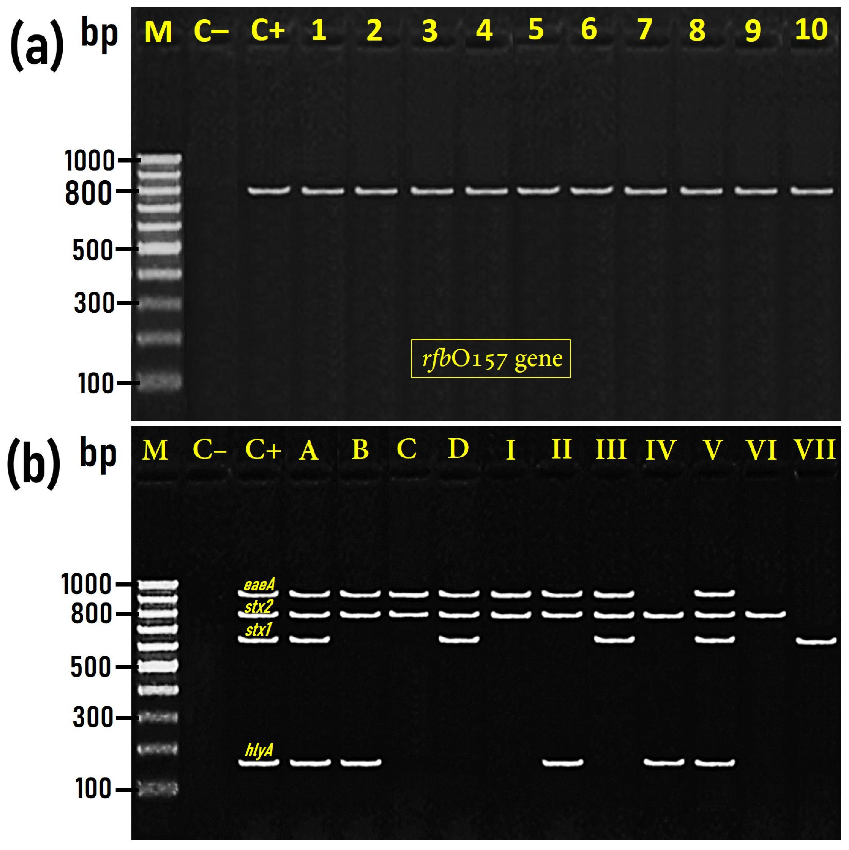
2.4.2. PCR for Detection of rfbO157 Gene Specific for E. coli O157

The biochemically confirmed E. coli strains were further tested by PCR for identification of the rfbO157 marker gene of E. coli O157 using the specific primer set: F: 5’-GCGGAACAAAACCATGTGCA-3’ and R: 5’-ACTGGCCTTGTTTCGATGAG-3’, which was previously constructed by Sallam et al. [13] to yield an 800 bp DNA size specific for the rfbO157 gene. A twenty-five-microliter reaction mixture comprising 12.5 μL of DreamTaq Green Master Mix (Thermo Scientific, St. Leon Roth, Germany), 1 μL from each of the forward and reverse primer (10 pmol each), 1 μL of the extracted genomic DNA as a template, and 9.5 μL RNase free water added to the 25 μL final volume. The PCR amplification condition included an initial denaturation step at 96 °C for 4 min, followed by 30 cycles of 96 °C for 30 s, 60 °C for 45 s, and 72 °C for 90 s. The final cycle was followed by an extension step of 72 °C for 5 min. The SimpliAmp Thermal Cycler (Thermo Fisher Scientific Inc.) was used for PCR reactions.

2.4.3. Detection of stx1, stx2, eaeA, and hylA Virulence Genes with PCR

E. coli O157:H7 and E. coli O55:H7 strains (n = 126) isolated from camel meat were examined for the evidence of some virulence genes specific for pathotyping of the diarrheagenic E. coli as eaeA, stx1, stx2, and EHEC-hlyA. The primer sequences constructed for PCR reactions of the stx1 (614 bp) and stx2 (779 bp) genes, according to Dhanashree and Mallya [18], were 5’-acactggatgatctcagtgg-3’ for stx1 forward, 5’-ctgaatccccctccattatg-3’ for stx1 reverse, 5’-ccatgacaacggacagcagtt-3’ for stx2 forward, and 5’-cctgtcaactgagcagcactttg-’3 for stx2 reverse. Moreover, the constructed sequences of the primer set used for PCR amplification of each gene were as follows: 5’-gtggcgaatactggcgagact-3’ for sense and 5’-ccccattctttttcaccgtcg-3’for antisense to yield an 890-bp DNA fragments for eaeA [19], whereas the primer set sequences for PCR amplification of hylA gene were 5’-acgatgtggtttattctgga-’3 for sense and 5’-cttcacgtgaccatacatat-’3 for antisense to yield a DNA size of 165 bp [20].
Multiplex PCR of 50 μL volume containing 25 μL of DreamTaq Green Master Mix (Thermo Scientific, St. Leon Roth, Germany); 1 μL from each the sense and antisense primer (10 pmol each) specific for each of the stx1, stx2, eaeA, and hylA virulent genes (8 μL in total); 2.5 μL of E. coli genomic DNA as a template; and 14.5 μL RNase-free water added to a final volume of 50 μL. Multiplex PCR for stx1, stx2, eaeA, and hylA genes consisted of an initial denaturation step for 3 min at 95 °C, followed by 35 cycles of denaturation at 95 °C for 20 s, annealing at 58 °C for 30 s, and polymerization at 72 °C for 90 s. The final cycle was followed by 72 °C for 5 min as an extension step.
An E. coli O157:H7 Sakai reference strain was employed as a positive control (positive for stx1, stx2, eaeA, and hylA), while E. coli K12 DH5α was used as a negative control non-pathogenic strain not harboring any virulence gene. The resulted amplicons (6 μL aliquots from each reaction mixture) were separated within the agarose gel (1.5%) electrophoresis at 100 V for 50 min and stained with ethidium bromide solution for 25 min. The agarose gel was then subjected to an ultra-violet (UV) transilluminator for visualization and photographing the separated PCR products.

2.5. Antimicrobial Resistance Profile and Multiple Antibiotic Resistance (MAR) index of E. coli O157:H7 and E. coli O55:H7 Isolated Strains

The antimicrobial susceptibility testing for the isolated E. coli O157:H7 and E. coli O55:H7 strains was performed with the disk diffusion susceptibility technique applied according to the guidelines specified by the Clinical and Laboratory Standards Institute [21]. Fourteen antibiotics susceptibility discs (Oxoid Limited, Basingstoke, Hampshire, UK) from nine antimicrobial classes were used with the following variable concentrations: aminoglycosides: amikacin (30 μg), gentamicin (10 μg); carbapenemes: imipenem (10 μg); cephalosporins: cefotaxime (30 μg), ceftriaxone (30 μg), cephalothin (30 μg); fluoroquinolones: levofloxacin (5 μg), ciprofloxacin (5 μg); Macrolides: erythromycin (15 μg); lincomycins: clindamycin (10 μg); penicillins: ampicillin (10 μg), penicillin (10 IU); sulfonamides: sulfamethoxazole (25 μg); and tetracyclines: tetracycline (30 μg). The strains tested were categorized into susceptible, intermediate, and resistant. The MAR index for each isolated strain was calculated according to the formula set by Singh et al. [22] as follows:
MAR   index = The   number   of   resistant   isolates Total   number   of   tested   antibiotics .

3. Results and Discussion

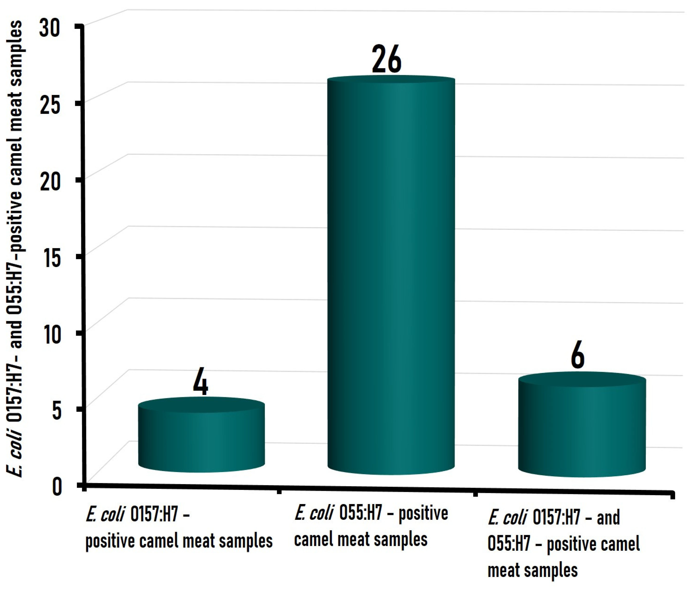
3.1. Prevalence of the Isolated E. coli Strains in Camel Meat Samples

Among the 110 camel meat samples tested, 10 (9.1%) samples were positive for E. coli O157:H7, which was present either alone (n = 4) or coexisting with E. coli O55:H7 (n = 6), while 32 (29.1%) camel meat samples were found positive for E. coli O55:H7, which existed either alone (n = 26) or as a mixed contamination (n = 6) with E. coli O157:H7 strains (Figure 1). Data about the prevalence of E. coli O157:H7 and/or E. coli O55:H7 in camel meat are very limited. A study conducted on 50 camel meat samples in Iran [23] indicated that only one (2.0%) sample was contaminated with E. coli O157. Likewise, Hassan et al. [24] could not detect the enteropathogenic E. coli in any of the 15 camel meat samples examined in Qaliubiya Governorate, Egypt, although they could detect it in 13.3% (2/15), 13.3% (2/15), and 33.3% (5/15) of camel spleen, liver, and kidney samples, respectively. In the fecal sample taken from healthy camels, however, E. coli O157:H7 was isolated from 19 (19%) of 100 camels’ fecal specimens collected from 100 apparently healthy camels in the middle of Iraq [25], while it was present in 3.3% from the fecal samples taken from 140 healthy camels at a slaughterhouse in the United Arab Emirates [26].
The relatively higher incidence of E. coli O157:H7 (9.1%) and E. coli O55:H7 (28.6%) among the tested camel meat samples in this study may be attributed to the cross-contamination of such organisms that normally inhabitant the gut of food animals including camels and are shed out with the animal feces, and therefore, it is very likely that E. coli can pollute the meat during the slaughtering process, especially if a faulty evisceration takes place, and also from the contaminated hide. In this context, Elder et al. [27] revealed a significant association between fecal and hide prevalence of EHEC O157 and cattle carcass contamination by such organisms.

3.2. Molecular Characterization and Virulence Genes Distribution among E. coli O157:H7 and E. coli O55:H7 Isolated from Camel Meat

Serological identification of E. coli isolates indicated that 102 isolates were serotyped as O55:H7, while only 24 isolates were serotyped as O157:H7. The twenty-four E. coli O157:H7 isolates were further confirmed by PCR as positive for the existence of the rfbO157-specific gene, which was detected at 800 bp size (Figure 2a). Both E. coli O157:H7 and E. coli O55:H7 isolates (n = 126) were examined by multiplex PCR for the existence of eaeA, stx1, stx2, and EHEC-hlyA, which were identified at the expected molecular sizes of 890 bp, 614 bp, 779 bp, and 165 bp, respectively (Figure 2b).
The association of the four virulent genes determined in the 126 E. coli isolates obtained from camel meat in the present investigation varied among the isolates.
Based on the presence of these four representative virulence genes, E. coli O157:H7 (n = 24) were categorized into four groups (A–D), while E. coli O55:H7 (n = 102) were categorized into seven groups (I–VII) (Table 1). Interestingly, groups A (n = 13), B (n = 6), C (n = 3), and D (n = 2) of O157:H7 isolates exhibited the same existence pattern of the virulence gene in comparison to groups V (n = 5), II (n = 21), I (n = 46), and III (n = 12) of O55:H7 isolates, respectively. Precisely, groups A and V harbored stx1, stx2, eaeA, and hlyA; groups B and II harbored stx2, eaeA, and hlyA; group C and I harbored stx2 and eaeA, while group D and III harbored stx1, stx2, and eaeA genes (Table 1). The comparable pattern of the virulence genes distribution in both E. coli O157:H7 and E. coli O55:H7 isolates is not surprising since E. coli O55:H7 and E. coli O157:H7 strains have a very similar chromosome backbone [7,8], and many studies have indicated that the STEC O157:H7 clone developed from E. coli O55:H7.

3.2.1. Shiga Toxin Genes (stx1 and stx2)

Shiga toxin 2 (stx2) was the most prevalent virulence gene detected in STEC O157:H7 and O55:H7 isolated from camel meat in the current investigation, where it was identified in 97.6% (123/126) of the isolates. On the other hand, only 27.8% (35/126) of the isolates harbored stx1 (Table 1). Among the 123 stx2-positive E. coli O157:H7 and O55:H7 strains tested in the present study, 32 (25.4%) strains harbored both stx1 and stx2 genes, while 91 (72.2%) strains harbored stx2 while lacking stx1 (Table 1). It was previously pointed out that the strains harboring the stx2 gene are possibly more severe than those harboring the stx1 gene or even the strains containing both stx1 and stx2 [28,29]. Studies have revealed that stx2 is the most significant virulence element linked with severe illnesses and that stx2-carrying strains are more commonly correlated with HUS than the strains carrying stx1 [30,31]. Actually, stx2 is described to be 1000 times more cytotoxic compared to stx1 towards the micro-vascular endothelial cells of human kidneys [32].
The existence rate along with the association of stx1 and stx2 genes in STEC strains from food of animal origin varied from one study to another. Lee et al. [33], in Korea, found that 64% of the STEC strains obtained from fresh beef carried stx2, whereas 14% carried both stx1 and stx2. Beutin et al. [34], in Germany, identified stx2 in 81% and stx1 in 40% of the STEC strains recovered from fresh meat samples. Mora et al. [35], in Spain, showed that 28 (29%) of the 96 STEC isolates recovered from minced beef possessed stx1 genes, 49 (51%) carried stx2 genes, and 19 (20%) carried both stx1 and stx2. Slanec et al. [36], in another study in Germany, reported that 70.7%, 9.3%, and 20.0% of the 75 STEC strains isolated from animal origin foodstuffs carried stx2, stx1, and both stx1 and stx2, respectively. Additionally, Eid et al. [37] found that all of the three (100%) instances of E. coli O55:H7 obtained from broiler chicken in Egypt were positive for stx1, while two (66.7%) of them were positive for the stx2 gene. Contrary to our results, Rahimi et al. [23] revealed that the sole E. coli O157 isolate recovered from camel meat in Iran was lacking stx1 and stx2 virulent genes. Even if the striking findings of the virulence pattern of the isolated E. coli strains in the present study can be considered suggestive, strengthening the occurrence of positive results recorded in other surveys with different detection rates, further investigations that focus on the evidence of other virulence genes/factors (e.g., elt, est, daaD, invE, Eagg, or astA genes) and enrolling a larger number of isolates are still necessary for a comprehensive understanding of the pathogenicity of the camel meat origin E. coli strains.

3.2.2. Intimin (eaeA) and Enterohemolysin (hlyA) Genes

The eaeA is the gene that encodes the intimin adherence factor, which is considered an outer membrane protein necessary for the intimate attachment of E. coli to the intestinal mucosa of the host. The human pathogenic E. coli isolates are mostly positive for the eae gene. In the present study, the intimin (eaeA) gene was the second most prevalent virulence gene after stx2, where it was detected in 85.7% (108/126) of the isolates. On the other hand, 43.7% (55/126) of the isolates harbored hlyA genes (Table 1). The eaeA gene existed in all of the 24 (100%) E. coli O157:H7 and in 84 (82.4%) of the 102 E. coli O55:H7 isolates isolated from the camel meat investigated in this study. Likewise, Sallam et al. [13], in Egypt, indicated that 93.3% (14/15) of the E. coli O157 strains recovered from beef products were positive for the eaeA gene. Similarly, Cagney et al. [38] identified the eae gene in 95.3% (41/43) of E. coli O157:H7 isolates obtained from meat products in Ireland. Moreover, Chapman et al. [39] detected the eae gene in all (100%) of the 72 E. coli O157:H7 strains recovered from raw beef and lamb products in the UK. Conversely, a low existence rate of 26% (25/96) was determined for the eae gene in STEC strains from minced beef in Spain [35], while a much lower prevalence rate of 5% was found among the isolated STEC strains recovered from samples of fresh meat harboring eaeA genes [34]. Nonetheless, Eid et al. [37] found that one (33.3%) of the three E. coli O55:H7 strains recovered from broiler chicken in Egypt was positive for the eaeA gene, while Barlow et al. [40] failed to detect the eaeA gene in the any of the meat-products-originating STEC strains (n = 184) obtained in Australia. A strong association has been clarified by several publications between the presence of the eae gene and the competency of STEC isolates to induce severe infections in humans, especially HUS and bloody diarrhea [28,41,42]. In the present study, the association of eaeA and stx2 genes was determined in all (100%) of the 24 E. coli O157:H7 and in 84 (82.4%) of the 102 E. coli O55:H7, while the association of the eaeA gene and stx1 was determined in 15 (62.5%) of the 24 E. coli O157:H7 and in 17 (16.7%) of the 102 E. coli O55:H7. Lee et al. [33] found that 7% and 14% of the STEC strains recovered from beef harbored both stx1 and eae and both stx2 and eae, respectively.
The EHEC-hlyA is another principal agent implicated in the virulence of E. coli. In the present survey, 19 (79.2%) of the 24 E. coli O157:H7 and 36 (35.3%) of the 102 E. coli O55:H7 isolates were positive for EHEC-hlyA gene, with a general prevalence of 43.7% (55/126) among the 126 STEC isolates tested (Table 1). EHEC-hlyA gene was detected at a higher prevalence rate of 93.3% (14/15) among the isolated E. coli O157 strains from beef products in Egypt [13]. A study conducted on beef and mutton meat in Hamedan, Iran, indicated that the single E. coli O157:H7 isolate as well as the nine (9/57; 15.8%) non-O157:H7 isolated strains were positive for the hlyA gene [43]. A lower detection rate of 25% (2/8) was also detected for the hlyA gene among the STEC strains obtained from cattle meat in Iran [16]. Similarly, a lower positive rate of 18.2% (2/11) for hlyA was determined in E. coli O55:H7 isolated from different meat products in Saudi Arabia [14]. Contrary to our findings, Rahimi et al. [23] revealed that the sole E. coli O157 isolate from camel meat in Iran was deficient in eaeA and ehlyA virulent genes. In the same country, the markedly genetic resistance of the human-originating E. coli strains was recently demonstrated in a study conducted by Jomehzadeh et al. [44].
The most frequent profile of the virulence gene detected in E. coli isolates from camel meat was stx2 and eaeA. Therefore, our isolated strains should be considered pathogenic for humans since the capability of STEC to induce severe illness and their potential to cause outbreaks of infection are associated with stx2-positive strains.

3.3. Antimicrobial Resistance Profile and MAR Index of E. coli O157:H7 and E. coli O55:H7 Strains

Antimicrobial-resistant bacteria are considered a serious problem that has received significant attention worldwide due to their ability to obstruct the treatment of severe infections in human patients [45]. In the present study, all (100%) of the obtained E. coli O157:H7 and E. coli O55:H7 isolates displayed absolute resistance to at least three antibiotics from different classes, namely clindamycin (lincomycins class), penicillin (penicillins class), and erythromycin (macrolydes class) (Table 2), which are commonly used in the veterinary medicine.
Furthermore, very high resistance rates of 92.9%, 71.4%, and 48.4% was observed regarding tetracycline, ampicillin, and sulfamethoxazole-trimethoprim, respectively (Table 2). Tetracyclines and beta-lactam antibiotics such as ampicillin are still frequently used, especially in livestock treatment in Egypt [46]. The recorded high resistance towards tetracycline, ampicillin, and sulfamethoxazole-trimethoprim in this survey was consistent with previous studies on E. coli strains isolated from beef in Egypt [47,48]. Furthermore, the aminoglycosides, including amikacin and gentamicin, are extensively used in animal husbandry as broad-spectrum antibiotics and as growth promoters. The recovered E. coli O157:H7 and E. coli O55:H7 isolates displayed a resistance rate of 39.7% and 16.7% towards amikacin and gentamicin, respectively (Table 2). In a previous study in Ethiopia, 100% of E. coli O157:H7 isolated from raw cattle meat were susceptible to amikacin and gentamicin [49].
Third-generation cephalosporin antibiotics such as cefotaxime and ceftriaxone have been widely used in the management of infections caused by Gram-negative bacteria [50]. In the present study, 84.9%, 29.4%, and 3.97% of the identified E. coli O157:H7 and E. coli O55:H7 isolates were resistant to cephalothin (first-generation cephalosporin), cefotaxime, and ceftriaxone (Table 2), respectively. On the contrary, all E. coli O157:H7 isolates from cattle in Tunisia were sensitive to cefotaxime [51]. Our results are in line with those reported in a previous study conducted in Italy by Grispoldi et al. [52], who found that 89.7% of E. coli from bovine lymph nodes were resistant to cephalothin, which is recognized as an effective antimicrobial against Gram-negative as well as Gram-positive bacteria. Furthermore, a relatively lower resistance rate of 57% was observed in E. coli serovars isolated from bovine origin meat samples (four cows and three buffaloes) in Egypt against cephalothin [53]. Interestingly, despite the two third-generation cephalosporins, namely ceftriaxone and cefotaxime, being similar in their antibacterial spectrum, indications, and route of administration, the isolates examined showed a resistance rate of 29.4% against cefotaxime and a resistance rate of only 3.97% against ceftriaxone (Table 2), which may be attributed to the difference in the administration dose and elimination half-life between the two antibiotics (ceftriaxone: 2 g once a day with half-life elimination of 8.8 h versus cefotaxime: 2 g every 4 h, 1.2 h) [54,55]. Likewise, Gums et al. [56] revealed that the nonmeningeal Streptococcus pneumoniae isolates were more susceptible to ceftriaxone than to cefotaxime. The higher resistance rate of the identified E. coli O157:H7 and E. coli O55:H7 isolates in the present study against cefotaxime constitutes a potential threat for public health and indicates the crucial requirement for the cautious administration of antibiotics in the veterinary medicine.
Fluoroquinolones are extensively used against a wide variety of Gram-positive and Gram-negative bacteria. In this study, the identified E. coli O157:H7 and E. coli O55:H7 isolates (n = 126) revealed high resistance rates of 22.2% and 8.7% toward ciprofloxacin (second-generation quinolone) and levofloxacin (third-generation quinolone), respectively (Table 2), which is in agreement with the resistance rates found in a previous study conducted in Saudi Arabia by El-Ghareeb et al. [57], who found 17.6% of E. coli isolates obtained from minced camel meat were resistant to ciprofloxacin. In contrast, Sabala et al. [46], in Egypt, found that all of the E. coli isolates (n = 150) from raw beef samples were susceptible to ciprofloxacin. The resistance rates of the identified E. coli O157:H7 and E. coli O55:H7 isolates (n = 126) against imipenem, which is recognized as a first-line drug in the treatment of human infections, were 5.56%. Conversely, none of the examined E. coli isolates (n = 150) from raw beef samples were resistant to carbapenem antimicrobials (imipenem and meropenem) [46].
Interestingly, 16.7% (4/24) and 83.3% (20/24) of the identified E. coli O157 isolates in the present study were categorized, based on their resistance profiles against the 14 different antimicrobials tested, into extensively drug-resistant (XDR) and multi-drug resistant (MDR), respectively, with a mean MAR index of 0.497 (Table 3), while 4.9%, 18.6%, and 76.5% of the identified E. coli O55:H7 isolates (n = 102) were categorized, according to their resistance profiles toward the 14 different antibiotics examined, into pan-drug resistant (PDR), extensively drug-resistant (XDR), and multi-drug resistant (MDR), respectively, with a mean MAR index of 0.527 (Table 3). Surprisingly, 3.97% (5/126) of E. coli isolates (all belong to E. coli O55:H7 serovars) exhibited resistance towards all of the 14 tested antibiotics, with a MAR index equal to 1.0 (Table 3). This finding could constitute a potential threat to public health. The average MAR index for E. coli O157:H7 and O55:H7 isolates in the present study was 0.514 (Table 3). E. coli isolates with a MAR index of >0.2 indicate a misuse and overuse of antibiotics, while E. coli isolates with a MAR index of 0.4 and above indicate human fecal contamination [58]. Therefore, it is crucial to establish monitoring systems that impose a rational usage of antibiotics in veterinary medicine for protecting public health from the spreading of multidrug-resistant bacteria to humans via animal-origin foodstuffs.
Altogether, the recorded overall antimicrobial resistance pattern of the isolated camel-meat-originating pathogenic E. coli strains can be considered worrisome in the present study, which largely agrees with results of other studies carried out in other regions of the world. These results strengthen the fact that the antimicrobial resistance phenomenon has become a significant threat to human and veterinary medicine. However, the recorded differences in terms of susceptibility towards the tested antimicrobials can be markedly influenced by the number of enrolled samples, study design, the used testing methodologies, and guidelines in interpreting the results, etc. Furthermore, supplementary studies focusing on continuous monitoring for the presence of pathogenic E. coli strains in camel meat combined with the testing of their resistance towards a wider variety of antimicrobials are still recommended.

4. Conclusions

The results of the present survey highlighted that the camel meat marketed in Beheira Governorate, Egypt, is contaminated with multi- and extensively drug-resistant Shiga toxigenic Escherichia coli (STEC) O157:H7 and O55:H7 strains, with an average MAR index for the isolates of 0.497 and 0.527, respectively, which emphasizes the necessity to optimize the use of the antibiotics in human and veterinary medicine. However, for a better understanding of the antimicrobial resistance phenomenon, in the case of the camel-meat-originating E. coli pathogenic strains, further investigations based on the screening of the antimicrobial resistance genes in a larger number of samples are still required. The study also elucidated that the most frequent profile of the virulence gene detected in E. coli O157:H7 and O55:H7 isolates from camel meat included stx2 and eaeA, indicating that the isolated strains have the potential to cause severe infection in humans.

Author Contributions

Conceptualization, resources, project administration, investigation, and writing—review and editing, K.I.S.; data curation, investigation, methodology, resources, and writing—original draft, Y.A.-E.; data curation, investigation, and formal analysis, M.T.R.; supervision, resources, and writing—review and editing, K.I.; formal analysis, validation, visualization, and data curation, A.M.; funding acquisition and software, V.H.; writing–original draft, editing, and formal analysis, H.A.Z. All authors have read and agreed to the published version of the manuscript.

Funding

This research received no external funding.

Institutional Review Board Statement

Not applicable.

Informed Consent Statement

Not applicable.

Data Availability Statement

The data presented in this study are available on request from the corresponding author.

Acknowledgments

This research paper is supported by the project “Increasing the impact of excellence research on the capacity for innovation and technology transfer within USAMVB Timișoara” code 6PFE, submitted in the competition Program 1—Development of the national system of research—development, Subprogram 1.2—Institutional performance, Institutional development projects—Development projects of excellence in RDI.

Conflicts of Interest

The authors declare no conflict of interest.

References

  1. Ameer, M.A.; Wasey, A.; Salen, P. Escherichia coli (E. coli O157:H7). In Stat Pearls; Stat-Pearls Publishing: Treasure Island, FL, USA, 2022. Available online: https://www.ncbi.nlm.nih.gov/books/NBK507845/ (accessed on 18 November 2022).
  2. Karch, H.; Bielaszewska, M. Sorbitol-fermenting Shiga toxin producing Escherichia coli O157:H-strains: Epidemiology, phenotypic and molecular characteristics, and microbiological diagnosis. J. Clin. Microbiol. 2001, 39, 2043–2049. [Google Scholar] [CrossRef] [PubMed] [Green Version]
  3. Sawyer, C.; Vishram, B.; Jenkins, C.; Jorgensen, F.; Byrne, L.; Mikhail, A.F.W.; Dallman, T.J.; Carroll, K.; Ahyow, L.; Vahora, Q.; et al. Epidemiological investigation of recurrent outbreaks of haemolytic uraemic syndrome caused by Shiga toxin-producing Escherichia coli serotype O55:H7 in England, 2014–2018. Epidemiol. Infect. 2021, 149, E108. [Google Scholar] [CrossRef] [PubMed]
  4. Hughes, J.M.; Wilson, M.E.; Johnson, K.E.; Thorpe, C.M.; Sears, C.L. The emerging clinical importance of non-O157 Shiga toxin-producing Escherichia coli. Clin. Infect. Dis. 2006, 43, 1587–1595. [Google Scholar] [CrossRef] [PubMed]
  5. McFarland, N.; Bundle, N.; Jenkins, C.; Godbole, G.; Mikhail, A.; Dallman, T.; O’Connor, C.; McCarthy, N.; O’Connell, E.; Treacy, J.; et al. Recurrent seasonal outbreak of an emerging serotype of Shiga toxin-producing Escherichia coli (STEC O55:H7 Stx2a) in the south west of England, July 2014 to September 2015. Euro Surveill. 2017, 22, 30610. [Google Scholar] [CrossRef] [PubMed] [Green Version]
  6. Brock, A. Guildford woman critically ill in hospital after suffering organ failure from ‘confirmed case of E. coli’ infection. Get. Surrey 2017. Available online: https://www.getsurrey.co.uk (accessed on 29 November 2022).
  7. Available online: https://www.bbc.com/news/uk-england-leicestershire-45838401 (accessed on 11 January 2023).
  8. Iguchi, A.; Ooka, T.; Ogura, Y.; Nakayama, K.; Frankel, G.; Hayashi, T. Genomic comparison of the O-antigen biosynthesis gene clusters of Escherichia coli O55 strains belonging to three distinct lineages. Microbiology 2008, 154, 559–570. [Google Scholar] [CrossRef] [Green Version]
  9. Zhou, Z.; Li, X.; Liu, B.; Beutin, L.; Xu, J.; Ren, Y.; Feng, L.; Lan, R.; Reeves, P.R.; Wang, L. Derivation of Escherichia coli O157:H7 from its O55:H7 precursor. PLoS ONE 2010, 5, e8700. [Google Scholar] [CrossRef] [Green Version]
  10. Wick, L.M.; Qi, W.; Lacher, D.W.; Whittam, T.S. Evolution of genomic content in the stepwise emergence of Escherichia coli O157:H7. J. Bacteriol. 2005, 187, 1783–1791. [Google Scholar] [CrossRef] [Green Version]
  11. Persad, A.K.; LeJeune, J.T. Animal Reservoirs of Shiga toxin-producing Escherichia coli. Microbiol. Spectr. 2014, 2, HEC-0027. [Google Scholar] [CrossRef]
  12. Gautam, R.; Kulow, M.; Park, D.; Gonzales, T.K.; Dahm, J.; Shiroda, M.; Stasic, A.J.; Döpfer, D.; Kaspar, C.W.; Ivanek, R. Transmission of Escherichia coli O157:H7 in cattle is influenced by the level of environmental contamination. Epidemiol. Infect. 2015, 143, 274–287. [Google Scholar] [CrossRef] [Green Version]
  13. Sallam, K.I.; Mohammed, M.; Ahdy, A.M.; Tamura, T. Prevalence, genetic characterization and virulence genes of sorbitol-fermenting Escherichia coli O157:H- and E. coli O157:H7 isolated from retail beef. Int. J. Food Microbiol. 2013, 165, 295–301. [Google Scholar] [CrossRef]
  14. Mohammed, M.A.; Sallam, K.I.; Eldaly, E.A.Z.; Ahdy, A.M.; Tamura, T. Occurrence, serotypes and virulence genes of non-O157 Shiga toxin-producing Escherichia coli in fresh beef, ground beef, and beef burger. Food Control 2014, 37, 182–187. [Google Scholar] [CrossRef]
  15. Hessain, A.M.; Al-Arfaj, A.A.; Zakri, A.M.; El-Jakee, J.K.; Al-Zogibi, O.G.; Hemeg, H.A.; Ibrahim, I.M. Molecular characterization of Escherichia coli O157:H7 recovered from meat and meat products relevant to human health in Riyadh, Saudi Arabia. Saudi J. Biol. Sci. 2015, 22, 725–729. [Google Scholar] [CrossRef] [Green Version]
  16. Babolhavaeji, K.; Shokoohizadeh, L.; Yavari, M.; Moradi, A.; Alikhani, M.Y. Prevalence of Shiga toxin-producing Escherichia coli o157 and non-o157 serogroups isolated from fresh raw beef meat samples in an industrial slaughterhouse. Int. J. Microbiol. 2021, 2021, 1978952. [Google Scholar] [CrossRef]
  17. ISO 16649-2:2001; Microbiology of Food and Animal Feeding Stuffs—Horizontal Method for the Enumeration of Beta-Glucuronidase-Positive Escherichia coli—Part 2: Colony Count Technique at 44 Degrees C Using 5-Bromo-4-Chloro-3-Indolyl Beta-D-Glucuronide. International Organization for Standardization: Geneva, Switzerland, 2001.
  18. Dhanashree, B.; Mallya, P.S. Detection of Shiga-toxigenic Escherichia coli (STEC) in diarrhoeagenic stool & meat samples in Mangalore, India. Indian J. Med. Res. 2008, 128, 271–277. [Google Scholar]
  19. Mazaheri, S.; Ahrabi, S.; Aslani, M. Shiga toxin-producing Escherichia coli isolated from lettuce samples in Tehran, Iran. Jundishapur J. Microbiol. 2014, 7, e12346. [Google Scholar] [CrossRef] [Green Version]
  20. Fratamico, P.M.; Sackitey, S.K.; Wiedmann, M.; Deng, M.Y. Detection of Escherichia coli O157: H7 by multiplex PCR. J. Clin. Microbiol. 1995, 33, 2188–2191. [Google Scholar] [CrossRef] [Green Version]
  21. CLSI Standard M02; Performance Standards for Antimicrobial Disk Susceptibility Tests. 13th ed. Clinical and Laboratory Standards Institute: Wayne, PA, USA, 2018.
  22. Singh, S.; Yadav, A.S.; Singh, S.M.; Bharti, P. Prevalence of Salmonella in chicken eggs collected from poultry farms and marketing channels and their antimicrobial resistance. Food Res. Int. 2010, 43, 2027–2030. [Google Scholar] [CrossRef]
  23. Rahimi, E.; Kazemeini, H.R.; Salajegheh, M. Escherichia coli O157: H7/NM prevalence in raw beef, camel, sheep, goat, and water buffalo meat in Fars and Khuzestan provinces, Iran. Vet. Res. Forum 2012, 3, 15–17. [Google Scholar]
  24. Hassan, M.A.; Heikal, G.I.; Barhoma, R.M. Traceability of enteropathogenic E. coli in cattle and camel carcasses. Benha Vet. Med. J. 2016, 31, 50–55. [Google Scholar] [CrossRef]
  25. Al-Gburi, N.M. Prevalence of Escherichia coli O157:H7 in camels’ fecal samples. J. Genet. Environ. Res. Conserv. 2016, 4, 46–50. [Google Scholar]
  26. Al Ajmi, D.S.; Banu, S.; Rahman, S. A Study on prevalence of Escherichia coli O157 in healthy camels, cattle, sheep and goat from slaughterhouse in Al Ain, the United Arab Emirates. J. Trauma Crit. Care 2019, 3, 41. [Google Scholar] [CrossRef]
  27. Elder, R.O.; Keen, J.E.; Siragusa, G.R.; Barkocy-Gallagher, G.A.; Koohmaraie, M.; Laegreid, W.W. Correlation of enterohemorrhagic Escherichia coli O157 prevalence in feces, hides, and carcasses of beef cattle during processing. Proc. Natl. Acad. Sci. USA 2000, 97, 2999–3003. [Google Scholar] [CrossRef] [PubMed]
  28. Ludwig, K.; Sarkim, V.; Bitzan, M.; Karmali, M.A.; Bobrowski, C.; Ruder, H.; Laufs, R.; Sobottka, I.; Petric, M.; Karch, H.; et al. Shiga toxin-producing Escherichia coli infection and antibodies against Stx2 and Stx1 in household contacts of children with enteropathic hemolytic-uremic syndrome. J. Clin. Microbiol. 2002, 40, 1773–1782. [Google Scholar] [CrossRef] [PubMed] [Green Version]
  29. Thorpe, C.M.; Ritchie, J.M.; Acheson, D.W.K. Enterohemorrhagic and other Shiga toxin-producing Escherichia coli. In Escherichia coli: Virulence Mechanisms of a Versatile Pathogen; Donnenberg, M.S., Ed.; Academic Press: Boston, MA, USA, 2002; pp. 119–154. [Google Scholar]
  30. Brooks, J.T.; Sowers, E.G.; Wells, J.G.; Greene, K.D.; Griffin, P.M.; Hoekstra, R.M.; Strockbine, N.A. Non-O157 Shiga toxin–producing Escherichia coli infections in the United States, 1983–2002. J. Infect. Dis. 2005, 192, 1422–1429. [Google Scholar] [CrossRef] [Green Version]
  31. Pradel, N.; Bertin, Y.; Martin, C.; Livrelli, V. Molecular analysis of Shiga toxin-producing Escherichia coli strains isolated from hemolytic-uremic syndrome patients and dairy samples in France. Appl. Environ. Microbiol. 2008, 74, 2118–2128. [Google Scholar] [CrossRef] [Green Version]
  32. Louise, C.B.; Obrig, T.G. Specific interaction of Escherichia coli O157:H7-derived Shiga-like toxin II with human renal endothelial cells. J. Infect. Dis. 1995, 172, 1397–1401. [Google Scholar] [CrossRef]
  33. Lee, G.Y.; Jang, H.I.; Hwang, I.G.; Rhee, M.S. Prevalence and classification of pathogenic Escherichia coli isolated from fresh beef, poultry, and pork in Korea. Int. J. Food Microbiol. 2009, 134, 196–200. [Google Scholar] [CrossRef]
  34. Beutin, L.; Miko, A.; Krause, G.; Pries, K.; Haby, S.; Steege, K.; Albrecht, N. Identification of human-pathogenic strains of Shiga toxin-producing Escherichia coli from food by a combination of serotyping and molecular typing of Shiga toxin genes. Appl. Environ. Microbiol. 2007, 73, 4769–4775. [Google Scholar] [CrossRef] [Green Version]
  35. Mora, A.; Blanco, M.; Blanco, J.E.; Dahbi, G.; López, C.; Justel, P.; Alonso, M.P.; Echeita, A.; Bernárdez, M.I.; González, E.A.; et al. Serotypes, virulence genes and intimin types of Shiga toxin (verocytotoxin)-producing Escherichia coli isolates from minced beef in Lugo (Spain) from 1995 through 2003. BMC Microbiol. 2007, 7, 13. [Google Scholar] [CrossRef] [Green Version]
  36. Slanec, T.; Fruth, A.; Creuzburg, K.; Schmidt, H. Molecular analysis of virulence profiles and Shiga toxin genes in food-borne Shiga toxin-producing Escherichia coli. Appl. Environ. Microbiol. 2009, 75, 6187–6197. [Google Scholar] [CrossRef] [Green Version]
  37. Eid, H.I.; Algammal, A.M.; Nasef, S.A.; Elfeil, W.K.; Mansour, G.H. Genetic variation among avian pathogenic E. coli strains isolated from broiler chickens. Asian J. Anim. Vet. Adv. 2016, 11, 350–356. [Google Scholar] [CrossRef] [Green Version]
  38. Cagney, C.; Crowley, H.; Duffy, G.; Sheridan, J.J.; O’Brien, S.; Carney, E.; Anderson, W.; McDowell, D.A.; Blair, I.S.; Bishop, R.H. Prevalence and numbers of Escherichia coli O157:H7 in minced beef and beef burgers from butcher shops and supermarkets in the Republic of Ireland. Food Microbiol. 2004, 21, 203–212. [Google Scholar] [CrossRef]
  39. Chapman, P.A.; Siddons, C.A.; Cerdan Malo, A.T.; Harkin, M.A. A one year study of Escherichia coli O157 in raw beef and lamb products. Epidemiol. Infect. 2000, 124, 207–213. [Google Scholar] [CrossRef]
  40. Barlow, R.S.; Hirst, R.G.; Norton, R.E.; Ashhurst-Smith, C.; Bettelheim, K.A. A novel serotype of enteropathogenic Escherichia coli (EPEC) as a major pathogen in an outbreak of infantile diarrhoea. J. Med. Microbiol. 1999, 48, 1123–1125. [Google Scholar] [CrossRef] [Green Version]
  41. Beutin, L.; Krause, G.; Zimmermann, S.; Kaulfuss, S.; Gleier, K. Characterization of Shiga toxin-producing Escherichia coli strains isolated from human patients in Germany over a 3-year period. J. Clin. Microbiol. 2004, 42, 1099–1108. [Google Scholar] [CrossRef] [Green Version]
  42. Blanco, M.; Blanco, J.E.; Mora, A.; Dahbi, G.; Alonso, M.P.; González, E.A.; Bernárdez, M.I.; Blanco, J. Serotypes, virulence genes, and intimin types of Shiga toxin (verotoxin)-producing Escherichia coli isolates from cattle in Spain and identification of a new intimin variant gene (eae-ξ). J. Clin. Microbiol. 2004, 42, 645–651. [Google Scholar] [CrossRef] [Green Version]
  43. Binandeh, F.; Pajohi-Alamoti, M.; Mahmoodi, P.; Ahangari, A. Evaluation of stx1, stx2, hlyA, and eaeA virulence genes in Escherichia coli O157:H7 isolated from meat (beef and mutton) in Hamedan, Iran, during 2015–2016. Int. J. Enteric Pathog. 2020, 8, 55–59. [Google Scholar] [CrossRef]
  44. Jomehzadeh, N.; Saki, M.; Ahmadi, K.; Zandi, G. The prevalence of plasmid-mediated quinolone resistance genes among Escherichia coli strains isolated from urinary tract infections in southwest Iran. Mol. Biol. Rep. 2022, 49, 3757–3763. [Google Scholar] [CrossRef]
  45. Da Costa, P.M.; Loureiro, L.; Matos, A.J. Transfer of multidrug-resistant bacteria between intermingled ecological niches: The interface between humans, animals and the environment. Int. J. Environ. Res. Public Health 2013, 10, 278–294. [Google Scholar] [CrossRef] [Green Version]
  46. Sabala, R.F.; Usui, M.; Tamura, Y.; Abd-Elghany, S.M.; Sallam, K.I.; Elgazzar, M.M. Prevalence of colistin-resistant Escherichia coli harbouring mcr-1 in raw beef and ready-to-eat beef products in Egypt. Food Control 2021, 119, 107436. [Google Scholar] [CrossRef]
  47. Ahmed, A.M.; Shimamoto, T. Molecular analysis of multidrug resistance in Shiga toxin-producing Escherichia coli O157:H7 isolated from meat and dairy products. Int. J. Food Microbiol. 2015, 193, 6873. [Google Scholar] [CrossRef] [PubMed]
  48. Moawad, A.A.; Hotzel, H.; Awad, O.; Tomaso, H.; Neubauer, H.; Hafez, H.M.; El-Adawy, H. Occurrence of Salmonella enterica and Escherichia coli in raw chicken and beef meat in northern Egypt and dissemination of their antibiotic resistance markers. Gut Pathog. 2017, 9, 57. [Google Scholar] [CrossRef] [PubMed] [Green Version]
  49. Hiko, A.; Asrat, D.; Zewde, G. Occurrence of Escherichia coli O157:H7 in retail raw meat products in Ethiopia. J. Infect. Dev. Ctries. 2008, 2, 389–393. [Google Scholar] [CrossRef] [PubMed] [Green Version]
  50. Angulo, F.J.; Collinon, P.; Powers, J.H.; Chiller, T.M.; Aidara-Kane, A.; Aarestrup, F.M. World Health Organization ranking of antimicrobials according to their importance in human medicine: A critical step for developing risk management strategies for the use of antimicrobials in food production animals. Clin. Inf. Dis. 2009, 49, 132–141. [Google Scholar] [CrossRef] [Green Version]
  51. Tayh, G.; Boubaker, S.M.; Khedher, R.B.; Jbeli, M.; Chehida, F.B.; Mamlouk, A.; Dâaloul-Jedidi, M.; Messadi, L. Prevalence, virulence genes, and antimicrobial profiles of Escherichia coli O157: H7 isolated from healthy cattle in Tunisia. J. Infect. Dev. Ctries. 2022, 16, 1308–1316. [Google Scholar] [CrossRef]
  52. Grispoldi, L.; Karama, M.; Hadjicharalambous, C.; De Stefani, F.; Ventura, G.; Ceccarelli, M.; Revoltella, M.; Sechi, P.; Crotti, C.; D’Innocenzo, A.; et al. Bovine lymph nodes as a source of Escherichia coli contamination of the meat. Int. J. Food Microbiol. 2020, 331, 108715. [Google Scholar] [CrossRef]
  53. El-Ashmony, A.L.; Mostafa, A.E.; Tarabees, R. The antimicrobial pattern of E. coli isolated from various animal sources at El Beheira Governorate. Alex. J. Vet. Sci. 2022, 73, 7–19. [Google Scholar] [CrossRef]
  54. Nath, S.K.; Foster, G.A.; Mandell, L.A.; Rotstein, C. Antimicrobial activity of ceftriaxone compared with cefotaxime in the presence of serum albumin. Can. J. Infect. Dis. 1995, 6, 21–27. [Google Scholar] [CrossRef] [Green Version]
  55. Smith, C.R.; Petty, B.G.; Hendrix, C.W.; Kernan, W.N.; Garver, P.L.; Fox, K.; Beamer, A.; Carbone, K.; Threlkeld, M.; Lietman, P.S. Ceftriaxone compared with cefotaxime for serious bacterial infections. J. Infect. Dis. 1989, 160, 442–447. [Google Scholar] [CrossRef]
  56. Gums, J.G.; Boatwright, D.W.; Camblin, M.; Halstead, D.C.; Jones, M.E.; Sanderson, R. Differences between ceftriaxone and cefotaxime: Microbiological inconsistencies. Ann. Pharmacother. 2008, 42, 71–79. [Google Scholar] [CrossRef]
  57. El-Ghareeb, W.R.; Abdel-Raheem, S.M.; Al-Marri, T.M.; Alaql, F.A.; Fayez, M. Isolation and identification of extended spectrum β-lactamases (ESBLs) Escherichia coli from minced camel meat in Eastern Province, Saudi Arabia. Thai J. Vet. Med. 2020, 50, 155–161. [Google Scholar]
  58. Gessew, G.T.; Desta, A.F.; Adamu, E. High burden of multidrug resistant bacteria detected in Little Akaki River. Comp. Immunol. Microbiol. Infect. Dis. 2022, 80, 101723. [Google Scholar] [CrossRef]
Figure 1. Number of camel meat samples contaminated with Escherichia coli O157:H7 and Escherichia coli O55:H7, from the total of 110 investigated.
Foods 12 01443 g001
Figure 2. Representative agarose gel electrophoresis image for PCR amplicon of the rfbO157 gene (800 bp) characteristic for Escherichia coli O157:H7 (Panel (a)) and for the multiplex PCR products of stx1 (614 bp), stx2 (779 bp), eaeA (890 bp), and hlyA (165 bp) virulence genetic markers in both E. coli O157:H7 and E. coli O55:H7 (Panel (b)). M, 100 bp ladder as a molecular DNA marker; Lane C–, PCR runs resulted from using a template of E. coli K12 DH5α genome as a negative control strain (does not harbor any virulence gene); Lane C+, control positive E. coli O157:H7 Sakai as positive reference strain for rfbO157 gene (a) and stx1, stx2, eaeA, and hlyA genes (b). Lanes 1–10 representative isolates for the 10 O157:H7-positive camel meat samples. Lanes A (O157:H7, group A; 13 isolates positive for stx1, stx2, eaeA, and hlyA genes); Lane B (O157:H7, group B; 6 isolates positive for stx2, eaeA, and hlyA genes); Lane C (O157:H7, group C; 3 isolates positive for stx2 and eaeA genes); Lanes D (O157:H7, group D; 2 isolates positive for stx1, stx2, and eaeA genes). Lane I (O55:H7, group I; 46 isolates positive for stx2 and eaeA genes); Lane II (O55:H7, group II; 21 isolates positive for stx2, eaeA, and hlyA genes); Lane III (O55:H7, group III; 12 isolates positive for stx1, stx2, and eaeA genes); Lane IV (O55:H7, group IV; 10 isolates positive for stx2 and hlyA genes); Lane V (O55:H7, group V; 5 isolates positive for stx1, stx2, eaeA, and hlyA genes); Lane VI (O55:H7, group VI; 3 isolates positive only for stx2 gene); Lane VII (O55:H7, group VII; 3 isolates positive only for stx1 gene).
Foods 12 01443 g002
Table 1. Distribution of virulence genes among the obtained E. coli O157:H7 and O55:H7 isolates (n = 126).
SerotypeGroupNumber of E. coli IsolatesVirulence Genes
stx1stx2eaeAhlyA
O157:H7A13++++
B6+++
C3++
D2+++
O55:H7I46++
II21+++
III12+++
IV10++
V5++++
VI5+
VII3+
Total1263512310855
Note: stx1, Shiga-toxin 1 gene; stx2, Shiga-toxin 2 gene; eaeA, intimin gene; hylA, hemolysin gene.
Table 2. Antimicrobial susceptibility of E. coli O157:H7 and O55:H7 strains (n = 126).
NoAntimicrobial Agent (Acronym)SensitiveIntermediateResistant
NO%NO%NO%
1Clindamycin (CL)126100
2Penicillin (P)126100
3Erythromycin (E)126100
4Tetracycline (T)75.5621.5911792.9
5Cephalothin (CN)1511.943.1710784.9
6Ampicillin (AM)2923.075.569071.4
7Sulfamethoxazole (SXT)6047.653.976148.4
8Amikacin (AK)6853.9786.355039.7
9Cefotaxime (CF)7962.7107.943729.4
10Ciprofloxacin (CP)9877.82822.2
11Gentamicin (G)10079.453.972116.7
12Levofloxacin (L)10986.564.76118.7
13Imipenem (IPM)11591.343.1775.56
14Ceftriaxone (C)12196.0353.97
Note: CL, clindamycin; P, penicillin; E, erythromycin; T, tetracycline; CN, cephalothin; AM, ampicillin; SXT, sulfamethoxazole; AK, amikacin; CF, cefotaxime; CP, ciprofloxacin; G, gentamicin, L, levofloxacin; IPM, imipenem; C, ceftriaxone.
Table 3. Classification of E. coli O157:H7 and O55:H7 (n = 126) based on their antimicrobial resistance degree against 14 antimicrobials tested.
E. coli
Strains
Number of IsolatesAntimicrobial Resistance ProfileClasses with ResistanceMar IndexClassification of Strains
No. and (%)
O157:H7
(n = 24)
2CL, P, E, T, CN, AM, SXT, AK, CF, CP, G, L, IPMMacrolides, lincomycins, penicillins, tetracyclines, cephalosporins, aminoglycosides, sulfonamides, fluoroquinolones, carbapenemes0.929Extensively drug-resistant4 (16.7%)
2CL, P, E, T, CN, AM, SXT, AK, CF, CP, GMacrolides, lincomycins, penicillins, tetracyclines, cephalosporins, aminoglycosides, sulfonamides, fluoroquinolones0.786
2CL, P, E, T, CN, AM, SXT, AK, CFMacrolides, lincomycins, penicillins, tetracyclines, cephalosporins, aminoglycosides, sulfonamides0.643Multidrug-resistant20 (83.3.0%)
4CL, P, E, T, CN, AM, SXT, AKMacrolides, lincomycins, penicillins, tetracyclines, cephalosporins, aminoglycosides, sulfonamides0.571
5CL, P, E, T, CN, AMMacrolides, lincomycins, penicillins, tetracyclines, cephalosporins, aminoglycosides0.429
6CL, P, E, T Macrolides, lincomycins, penicillins, tetracyclines0.357
3CL, P, EMacrolides, lincomycins, penicillins0.214
Sum 24Average MAR Index for O157:H7 0.497
O55:H7
(n = 102)
5CL, P, E, T, CN, AM, SXT, AK, CF, CP, G, L, IPM, CAminoglycosides, carbapenemes, cephalosporins, fluoroquinolones, macrolides, lincomycins, penicillins, sulfonamides, tetracyclines1.000Pan-drug resistant5 (4.9%)
4CL, P, E, T, CN, AM, SXT, AK, CF, CP, G, LMacrolides, lincomycins, penicillins, tetracyclines, cephalosporins, aminoglycosides, sulfonamides, fluoroquinolones0.857Extensively drug-resistant19 (18.6%)
8CL, P, E, T, CN, AM, SXT, AK, CF, CP, GMacrolides, lincomycins, penicillins, tetracyclines, cephalosporins, aminoglycosides, sulfonamides, fluoroquinolones0.786
7CL, P, E, T, CN, AM, SXT, AK, CF, CPMacrolides, lincomycins, penicillins, tetracyclines, cephalosporins, aminoglycosides, sulfonamides, fluoroquinolones0.714
7CL, P, E, T, CN, AM, SXT, AK, CFMacrolides, lincomycins, penicillins, tetracyclines, cephalosporins, aminoglycosides, sulfonamides, fluoroquinolones0.643Multidrug-resistant78 (76.5%)
9CL, P, E, T, CN, AM, SXT, AKMacrolides, lincomycins, penicillins, tetracyclines, cephalosporins, aminoglycosides, sulfonamides0.571
11CL, P, E, T, CN, AM, SXTMacrolides, lincomycins, penicillins, tetracyclines, cephalosporins, aminoglycosides, sulfonamides0.500
24CL, P, E, T, CN, AMMacrolides, lincomycins, penicillins, tetracyclines, cephalosporins, aminoglycosides0.429
17CL, P, E, T, CNMacrolides, lincomycins, penicillins, tetracyclines, cephalosporins0.357
4CL, P, E, TMacrolides, lincomycins, penicillins, tetracyclines0.286
6CL, P, EMacrolides, lincomycins, penicillins,0.214
Sum 102Average MAR Index for O55:H7 isolates 0.527
Total126Average MAR index for O157:H7 and O55:H7 isolates Type of resistance0.514
Note: CL, clindamycin; P, penicillin; E, erythromycin; T, tetracycline; CN, cephalothin; AM, ampicillin; SXT, sulfamethoxazole; AK, amikacin; CF, cefotaxime; CP, ciprofloxacin; G, gentamicin, L, levofloxacin; IPM, imipenem; C, ceftriaxone.
Disclaimer/Publisher’s Note: The statements, opinions and data contained in all publications are solely those of the individual author(s) and contributor(s) and not of MDPI and/or the editor(s). MDPI and/or the editor(s) disclaim responsibility for any injury to people or property resulting from any ideas, methods, instructions or products referred to in the content.

