Article

Genomic and Transcriptomic Analysis Reveal Multiple Strategies for the Cadmium Tolerance in Vibrio parahaemolyticus N10-18 Isolated from Aquatic Animal Ostrea gigas Thunberg

1
Key Laboratory of Quality and Safety Risk Assessment for Aquatic Products on Storage and Preservation (Shanghai), Ministry of Agriculture and Rural Affairs of the People’s Republic of China, College of Food Science and Technology, Shanghai Ocean University, Shanghai 201306, China
2
Lab of Food Function and Nutrigenomics, College of Food Science and Technology, Hunan Agricultural University, Changsha 410128, China
*
Authors to whom correspondence should be addressed.
These authors contributed equally to this work.
Foods 2022, 11(23), 3777; https://doi.org/10.3390/foods11233777
Submission received: 17 September 2022 / Revised: 5 November 2022 / Accepted: 16 November 2022 / Published: 23 November 2022
(This article belongs to the Special Issue Contaminants in Seafood: Prevention, Control, and Detection)

Abstract

:
The waterborne Vibrio parahaemolyticus can cause acute gastroenteritis, wound infection, and septicemia in humans. Pollution of heavy metals in aquatic environments is proposed to link high incidence of the multidrug-resistant (MDR) pathogen. Nevertheless, the genome evolution and heavy metal tolerance mechanism of V. parahaemolyticus in aquatic animals remain to be largely unveiled. Here, we overcome the limitation by characterizing an MDR V. parahaemolyticus N10-18 isolate with high cadmium (Cd) tolerance using genomic and transcriptomic techniques. The draft genome sequence (4,910,080 bp) of V. parahaemolyticus N10-18 recovered from Ostrea gigas Thunberg was determined, and 722 of 4653 predicted genes had unknown function. Comparative genomic analysis revealed mobile genetic elements (n = 11) and heavy metal and antibiotic-resistance genes (n = 38 and 7). The bacterium significantly changed cell membrane structure to resist the Cd2+ (50 μg/mL) stress (p < 0.05). Comparative transcriptomic analysis revealed seven significantly altered metabolic pathways elicited by the stress. The zinc/Cd/mercury/lead transportation and efflux and the zinc ATP-binding cassette (ABC) transportation were greatly enhanced; metal and iron ABC transportation and thiamine metabolism were also up-regulated; conversely, propanoate metabolism and ribose and maltose ABC transportation were inhibited (p < 0.05). The results of this study demonstrate multiple strategies for the Cd tolerance in V. parahaemolyticus.

1. Introduction

Vibrio parahaemolyticus is a Gram-negative bacterium that thrives in marine, riverine, and aquaculture environments worldwide [1,2]. The bacterium can cause acute gastroenteritis, wound infection, and septicemia in humans [2]. V. parahaemolyticus was first identified as a foodborne pathogen in Japan in the 1950s [3]. Since then, pathogenic V. parahaemolyticus has been reported in Asian countries and subsequently in Africa, America, and Europe, arguing a pandemic of V. parahaemolyticus worldwide [4]. It was estimated that V. parahaemolyticus is responsible for roughly 35,000 human infection cases each year in the United States [5]. The bacterium has been identified as the leading cause of the foodborne diarrhea disease in China since the 1990s [6]. The crucial virulence determinants in pathogenic V. parahaemolyticus are thermostable-direct hemolysin (TDH) and TDH-related hemolysin (TRH) [7].
V. parahaemolyticus is frequently isolated from aquatic products worldwide, including crustaceans, fish, and shellfish [1,8,9,10]. For example, recently, Li et al. analyzed food samples (n = 905) collected from 15 provinces in China and found that 14.17% of fish (n = 204), 15.34% of shrimp (n = 365), and 3.67% of ready-to-eat food (n = 300) samples were detected positive for potential V. parahaemolyticus contamination [8]. Antibiotics effectively control infectious diseases caused by pathogenic microorganisms in aquaculture [11]. Nevertheless, during the past few decades, the inappropriate application of antibiotics in medical treatment and aquaculture has resulted in the emergence and spread of multidrug resistant (MDR) pathogenic bacteria, which pose serious threats to therapeutic options for human diseases [9]. On the other hand, rapid industrialization and modernization may lead to the increased heavy metal pollution in the aquatic environment, such as cadmium (Cd), chromium (Cr), copper (Cu), lead (Pb), mercury (Hg), nickel (Ni), and zinc (Zn). Heavy metal residues are detected in various aquatic environments and aquatic products [12,13]. The high bioaccumulation of heavy metals through the food chain poses grave risks to human health. For example, Cd is classified into Group 1 as carcinogenic to humans by the International Agency for Research on Cancer (IARC) [14]. Moreover, heavy metals have been proposed to enhance selection for antibiotic resistance in the environment and vice versa [15]. Recently, Fang et al. isolated V. parahaemolyticus strains (n = 112) from Pacific mackerel (Pneumatophorus japonicus) samples (n = 360) collected from different markets in Zhejiang Province, China. They found that most strains showed resistance to the antibiotics ampicillin (AMP) (n = 108) and streptomycin (STR) (n = 101), as well as the heavy metals Cd2+ (n = 88) and Pb2+ (n = 58) [16].
The co-selection is highly favored when diverse resistance genes are located on the same mobile genetic elements (MGEs) [17,18]. Genetic diversity of MGEs and genome plasticity in V. parahaemolyticus may affect the survival of the pathogen in the environment [19]. With the increased breakthrough of sequencing technology [20], approximately 1740 V. parahaemolyticus isolates have been sequenced, among which complete genomes of 64 V. parahaemolyticus isolates are available in the GenBank database (https://www.ncbi.nlm.nih.gov/, accession date: 29 January 2022). Recently, draft genome sequences of six V. parahaemolyticus isolates recovered from six species of aquatic animals, Paphia undulate, Perna viridis, Mactra veneriformis, Aristichthys nobilis, Carassius auratu, and Litopenaeus vannamei, were determined by our research group; a complete genome sequence of V. parahaemolyticus CHN25 was also obtained [21,22].
In our previous studies, a number of V. parahaemolyticus strains were isolated and characterized from various species of aquatic animals [23]. Of these, V. parahaemolyticus N10-18 isolate showed MDR and high tolerance to Cd2+ and Zn2+. Therefore, we asked what the genome features of this bacterium could be and what the molecular mechanism underlying the resistance phenotype could be. Thus, the major objectives of this study were (1) to determine the draft genome sequence of V. parahaemolyticus N10-18 isolate recovered from the aquatic animal Ostrea gigas Thunberg using Illumina Hiseq × 10 sequencing technique; (2) to identify MGEs and virulence- and resistance-related genes in the V. parahaemolyticus N10-18 genome; (3) to examine the survival of V. parahaemolyticus N10-18 at different concentrations of Cd2+ stress; (4) to decipher the possible molecular mechanism of the Cd2+ tolerance in V. parahaemolyticus N10-18 by comparative transcriptomics analysis. The results of this study will enrich genome data of V. parahaemolyticus and facilitate the risk control of the leading seafood-borne pathogen in edible aquatic animals.

2. Materials and Methods

2.1. V. parahaemolyticus Strain and Cultural Conditions

V. parahaemolyticus N10-18 strain was isolated from O. gigas Thunberg and identified in our previous research [23] (Table S1). The bacterium was routinely incubated in Tryptic Soy Broth (TSB) medium (3% NaCl, pH 8.5, Beijing Land Bridge Technology Co., Ltd., Beijing, China) at 37 °C with shaking at 180 rpm. The bacterial growth was examined as described in our previous study [24].

2.2. Genomic DNA Preparation, Sequencing, Assembly, and Annotation

V. parahaemolyticus N10-18 incubated in the TSB medium to the middle-logarithmic growth phase (mid-LGP) was harvested by centrifugation at 8000× g for 1 min. Genomic DNA of the bacterial cell pellet was extracted using the MiniBEST DNA extraction kit (Japan TaKaRa BIO, Dalian Company, Dalian, China) according to the manufacture’s instructions. DNA concentrations and purity (A260/A280) of the extracted samples were examined as described previously [25]. High-quality DNA samples were subjected to genome DNA sequencing, which was conducted by Shanghai Majorbio Bio-pharm Technology Co., Ltd., Shanghai, China, using the Illumina HiSeq × 10 sequencing platform (Illumina, Santiago, CA, USA). The average length of sequencing reads was 150 bp. Raw sequencing reads were analyzed using the FastQC software (https://www.bioinformatics.babraham.ac.uk/projects//fastqc/, accessed on 30 January 2022) [26] with the parameters described in our previous research [21]. High-quality reads were assembled using the SOAPdenovo (version 2.04) software [21]. V. parahaemolyticus RIMD2210633 (GenBank accession numbers for chromosomes 1 and 2: NC_004603.1 and NC_004605.1) was used for the reference genome. Coding sequences (CDSs), rRNA, and tRNA genes were predicted using the software Glimmer (version 3.02) [27], Barrnap tool (https://github.com/tseemann/barrnap, accessed on 30 January 2022), and tRNAscan-SE (version 2.0) [28] with 80% identity and 90% coverage at E ≤ 1 × 10⁻⁵, respectively.
Functional assignments of the predicted CDSs were inferred [29]. If the CDS did not match the clusters of orthologous groups (COG) function, it was assigned as unknown protein. The programs were run with default parameters.

2.3. Comparative Genome Analysis

The average nucleotide identity (ANI) value was calculated using the JspeciesWS software (http://jspecies.ribohost.com/jspeciesws/, accessed on 28 August 2022). MGEs were described in our previous studies [21,24,30], and genome islands (GIs), prophages, integrons (INs), and insertion sequences (ISs) were predicted using the software IslandViewer (version 1.2) [31], Phage_Finder [32], Integron_Finder (version 2.0) [33], and ISEScan (version 1.7.2.1) [34], respectively. The virulence factor database (https://www.mgc.ac.cn/VFs, accessed on 28 August 2022), heavy metal resistance gene database BacMet (http://bacmet.biomedicine.gu.se/, accessed on 28 August 2022), and antibiotic resistance gene database (http://ardb.cbcb.umd.edu/, accessed on 28 August 2022) were employed to analyze virulence-, heavy metal-, and antibiotic-resistance-related genes, respectively.
Gene loci of somatic (O) antigens and capsular polysaccharide (K) antigens are located between dgkA and gmhD genes and between gmhD and rjg genes in V. parahaemolyticus genomes, respectively [35,36]. Of these, the wvaG, wvaR, VP0208, orf16, wvcA, wvcJ, wvcN, wvdG, wvaH, wvcP/orf9, wvdB, and wvcP genes are responsible for the O1 to O12 serotype antigens, respectively [35], while the VP0214 to VP0238 genes are for the K serotype antigens, respectively [36]. Serotype sequences of 12 O antigens and 43 K antigens were collected from V. parahaemolyticus ATCC17802, RIMD221063, and 20-082a3 strains that are available in current literature [35,36,37,38,39,40]. The strains with untypeable (UT) antigenic markers by the Basic Local Alignment Search Tool (BLAST) (https://www.ncbi.nlm.nih.gov/BLAST, accessed on 30 August 2022) analysis were designated as OUT (O untypeable) or KUT (K untypeable).
Based on seven conserved core genes dnaE, gyrB, recA, dtdS, pntA, pyrC, and tnaA in V. parahaemolyticus [41], the multi-locus sequence typing (MLST) analysis was performed using the MLST software (version 2.19.0, http://github.com/tseemann/mlst, accessed on 15 February 2022).

2.4. Phylogenetic Tree Analysis

Complete gene sequences of 64 V. parahaemolyticus strains were downloaded from the GenBank database (Table S2). Amino acid data sets of single-copy orthologs in V. parahaemolyticus genomes were analyzed using the software OrthoFinder (version 2.2.6) [42]. The FastTree (version 2.1.11) software was used to build a phylogenetic tree using the method and parameters described in our recent research [21].

2.5. Determination of Minimum Inhibitory Concentrations (MICs) of Antibiotics and Heavy Metals

The MICs of antibiotics and heavy metals against V. parahaemolyticus N10-18 were measured using the broth dilution testing (microdilution) according to the guidelines of the Clinical and Laboratory Standards Institute (CLSI, M2-A9, 2006), including the CdCl2, ZnCl2, AMP, kanamycin (KAN), and STR (Sinopharm Chemical Reagent Co., Ltd., Shanghai, China). Escherichia coli K-12 was used as a quality control strain in the tests [23].

2.6. Stress Conditions

The fresh cell culture of V. parahaemolyticus N10-18 was individually inoculated into the TSB medium supplemented with different concentrations (0, 50, 100, 200, and 400 μg/mL) of CdCl2 and then incubated at 37 °C for 48 h. Bacterial growth curves were measured [24]. Bacterial survival rates were calculated using the standard colony counting method [43].

2.7. Cell Membrane Permeability, Fluidity, and Surface Hydrophobicity Assays

V. parahaemolyticus N10-18 was incubated in the TSB medium (3% NaCl, pH 8.5) to the mid-LGP at 37 °C. A final concentration of CdCl2 (50 μg/mL) was added and then incubated at 37 °C for 2 h. The outer cell membrane permeability was examined using the method described by Harman et al. [44]. The N-phenyl-1-naphthylamine (NPN) was purchased from the Shanghai Labtop Bio-Technology Co., Ltd., Shanghai, China. The inner membrane permeability was examined using the method described by Ibrahim et al. [45]. The O-nitrophenyl-β-D galactopyranoside (ONPG) was purchased from Beijing Solarbio Science & Technology Co., Ltd., Beijing, China.
The membrane fluidity assay was performed using the method described by Voss and Montville [46]. The cell surface hydrophobicity assay was performed using the method described by Yan et al. [47]. The n-hexadecane was purchased from China National Pharmaceutical Group Corporation Co., Ltd., Shanghai, China.

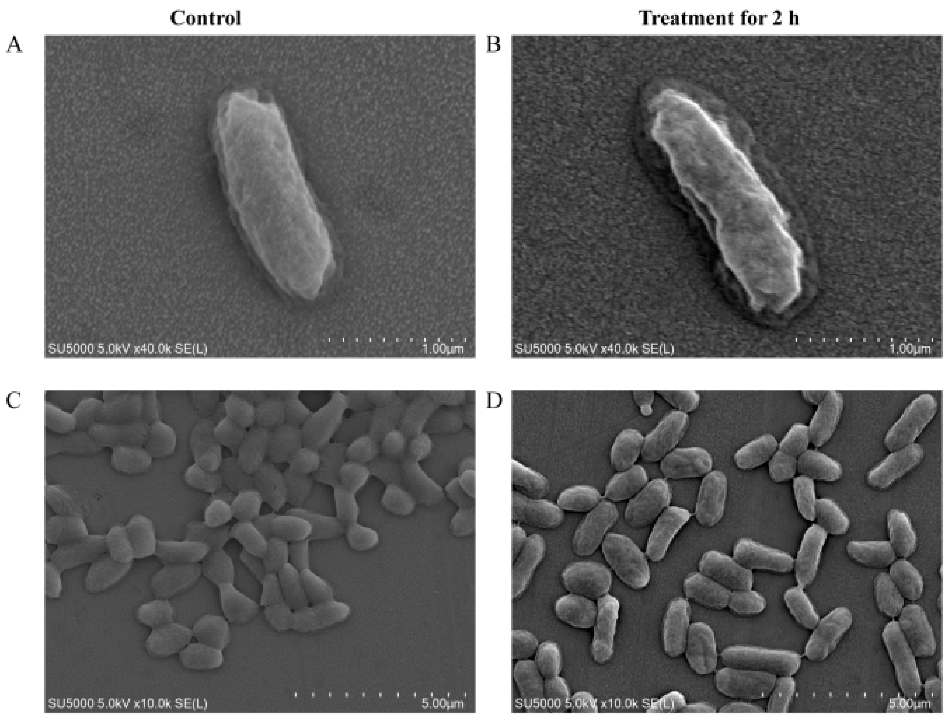
2.8. Scanning Electron Microscope (SEM) Assay

The SEM assay was performed according to the method described previously [48]. Briefly, a final concentration (50 μg/mL) of CdCl2 was added into V. parahaemolyticus N10-18 culture grown in the TSB medium (pH 8.5, 3% NaCl) at mid-LGP and then continuously incubated at 37 °C for 2 h. An amount of 1.5 mL of the cell suspension was collected, washed, dehydrated, dried, and gold-covered by cathodic spraying and observed using the thermal field emission SEM (Hitachi, SU5000, Tokyo, Japan) with accelerating voltages of 5–10 kV [48].

2.9. Illumina RNA Sequencing and Analysis

V. parahaemolyticus N10-18 was incubated in the TSB medium (pH 8.5, 3% NaCl) to the mid-LGP at 37 °C. A final concentration of CdCl2 (50 μg/mL) was added and then incubated at 37 °C for 2 h. Controls were cultures also exposed to no cadmium for the same time period and collected as treatments. The bacterial cells were collected by centrifugation and subjected for the Illumina RNA sequencing. The RNA extraction and quality control, sequencing library construction, and Illumina sequencing were conducted by Shanghai Majorbio Bio-pharm Technology Co., Ltd., Shanghai, China, using Illumina HiSeq 2500 platform (Illumina, Santiago, CA, USA). Three replicates were conducted for each sample.
Expression of each gene was calculated, and differentially expressed genes (DEGs) were defined and used for gene set enrichment analysis (GSEA) as described previously [25]. Representative DEGs were examined using real-time reverse-transcription PCR (RT-qPCR) assay [25,48].

2.10. Statistical Analysis

The data were analyzed using the SPSS software (version 22, IBM, Armonk, NY, USA). All tests in this study were conducted in triplicate.

3. Results

3.1. Genotype and Phenotype of V. parahaemolyticus N10-18

V. parahaemolyticus N10-18 isolate was recovered from O. gigas Thunberg [23]. The bacterium tested negative for the toxic tdh and trh genes but positive for the species-specific gene tlh [49]. The results also showed that V. parahaemolyticus N10-18 was tolerant to the heavy metals Cd2+ and Zn2+, as well as the antimicrobial agents AMP, KAN, and STR (Table S1).

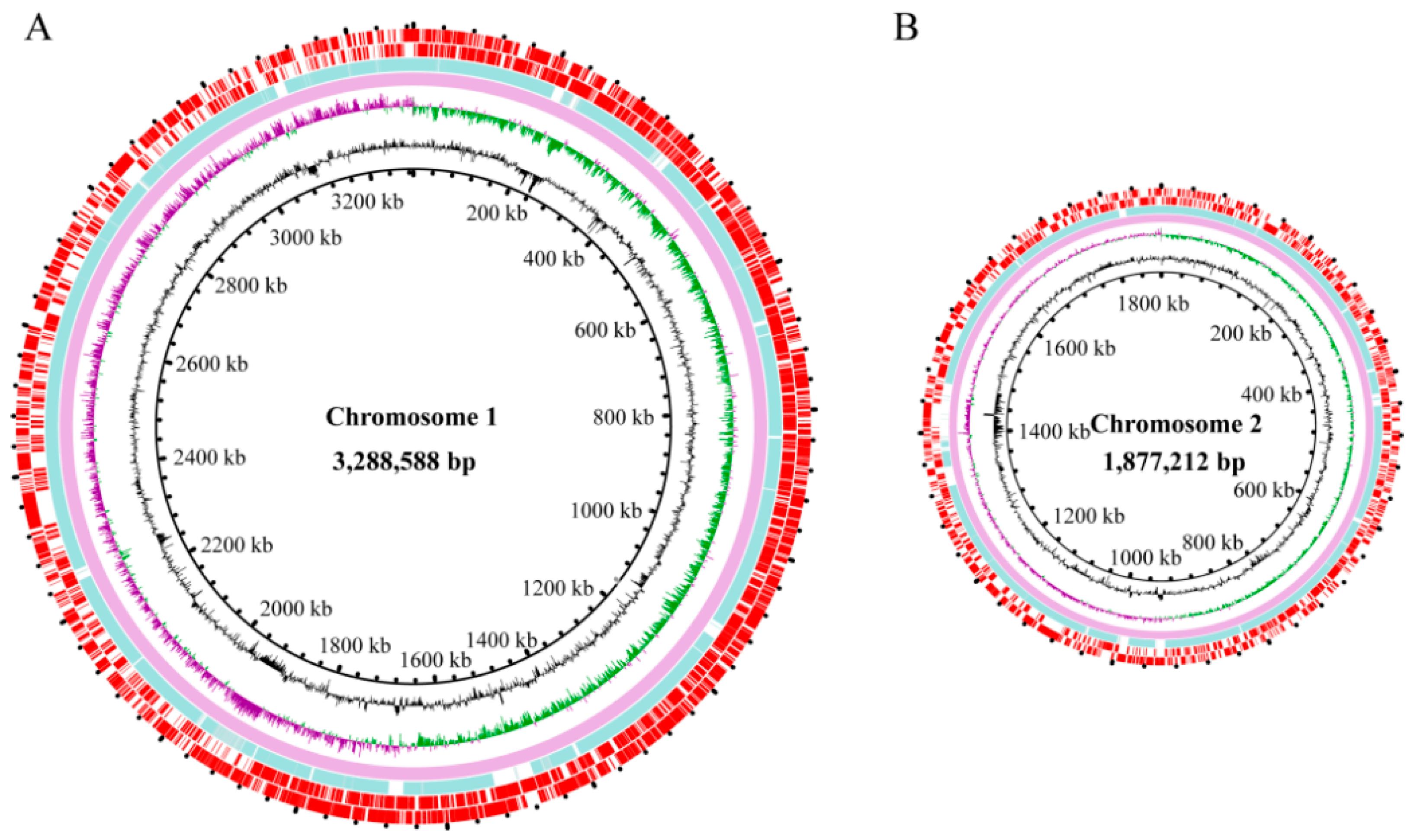
3.2. Genome Features of V. parahaemolyticus N10-18

The ANI value of the V. parahaemolyticus N10-18 genome was determined, which was higher (98.22%) than the threshold (94–96%) for species determination [50]. The draft genome sequence of V. parahaemolyticus N10-18 was determined using the Illumina Hiseq × 10 sequencing technique (Figure 1), and approximately 438,181 clean single reads were obtained. The assembly generated 70 scaffolds with a sequencing depth (on average) of 319.27-fold. V. parahaemolyticus N10-18 showed a clean single peak in the frequency of observed unique 17-mers within the sequencing data and varied as a typical Poisson distribution, suggesting less repetitive DNA in the V. parahaemolyticus N10-18 genome (Figure S1). The obtained V. parahaemolyticus N10-18 genome sequence has been deposited in the GenBank database under the assigned accession number JALGSE000000000.
The obtained genome size of V. parahaemolyticus N10-18 was 4,910,080 bp with 45.46% of the GC content. A total of 4653 genes were predicted, among which approximately 4565 coded for proteins. Remarkably, approximately 722 proteins-coding genes had unknown function, while 3843 were classified into 21 functional catalogues against the COG database (Table 1).
The V. parahaemolyticus N10-18 genome contained transposase genes (n = 10) and MGEs, including GIs (n = 2), INs (n = 8), and ISs (n = 1), suggesting possible horizontal gene transfer (HGT) mediated by the MGEs during the V. parahaemolyticus N10-18 genome evolution. The identified MGEs were absent from the ends of the scaffolds (Table S3), which indicated that the draft genome contained all such elements.

3.3. Serotype and ST of V. parahaemolyticus N10-18

The BLAST analysis of the antigen gene loci revealed that the V. parahaemolyticus N10-18 genome contained the O antigen loci orf16/wvdB and specific loci wzc for K4 polymorphic sites [51], indicating that the serotype of V. parahaemolyticus N10-18 was O4/O11:K4. Additionally, the ST by the MLST analysis showed that the bacterium belonged to the ST-499.

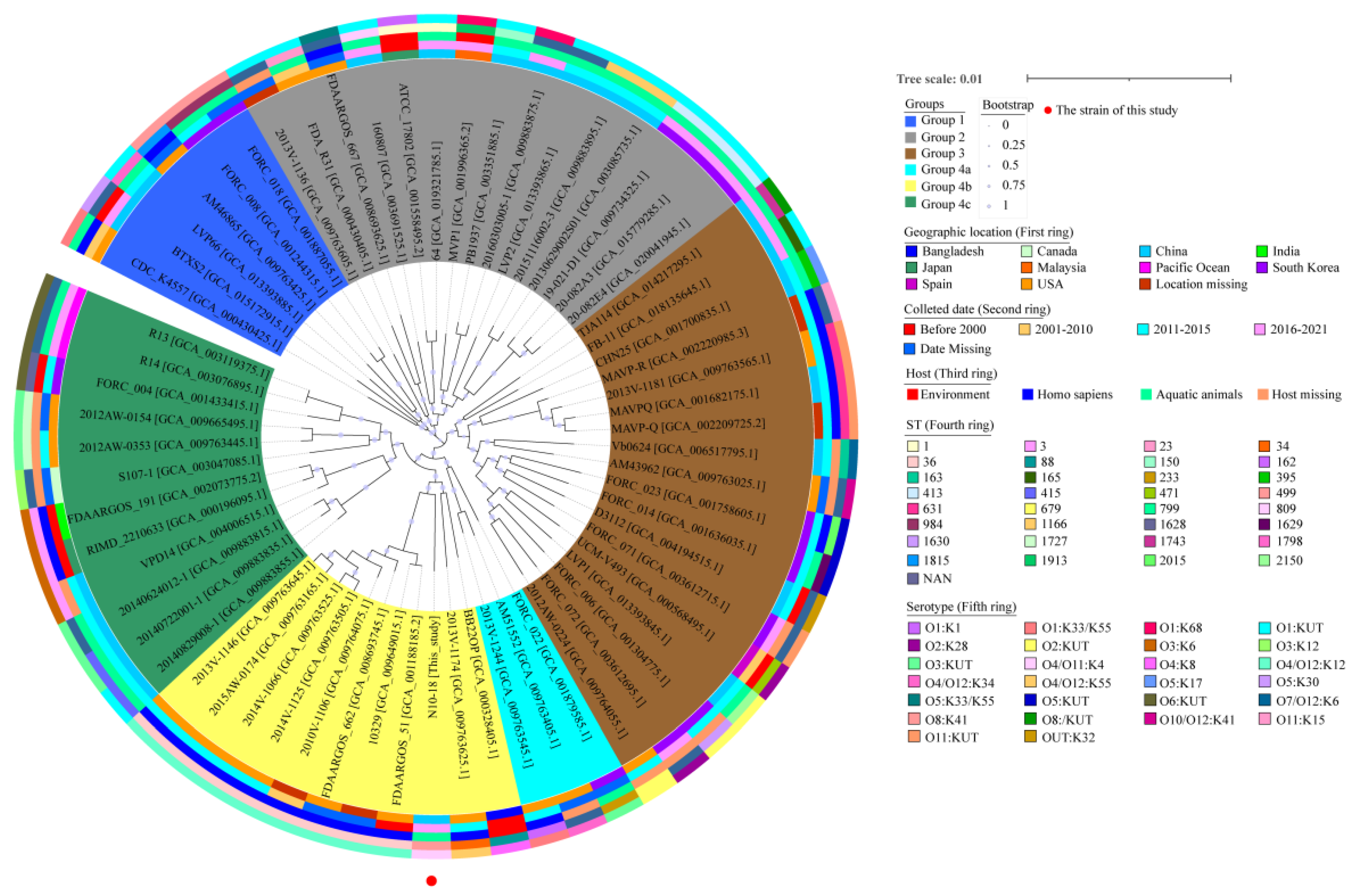
3.4. Phylogenetic Relatedness of V. parahaemolyticus N10-18

Approximately 1485 homologous single-copy amino acid sequences were identified from 64 V. parahaemolyticus genomes available in the GenBank database and the V. parahaemolyticus N10-18 genome determined in this study. A phylogenetic tree was construed (Figure 2), in which 20 V. parahaemolyticus strains were recovered from homo sapiens, 7 from the environment, 26 from aquatic animals (Penaeus, crayfish, fine spotted flounder, marinated crab, oyster, seabass, shrimps, and toothfish), and 12 from an unknown source (Table S2). This analysis revealed four distinct groups, designated as Groups 1 to 4. Group 4 was further classified into three subgroups (Groups 4a, 4b, and 4c) (Figure 2).
Although V. parahaemolyticus N10-18 (O4/O11:K4; ST-499; GenBank accession no. JALGSE000000000) was classified into Group 4b, the bacterium fell into a single sub-branch and was found to be phylogenetically distant from the other V. parahaemolyticus strains originating in aquatic animals. Moreover, V. parahaemolyticus N10-18 showed the closest evolutionary distance with the V. parahaemolyticus strains FDAARGOS_51, 10,329, FDAARGOS_662, 2010V-1106, 2014V-1125, 2014V-1066, 2015AW-0174, and 2013V-1146 with the GenBank assembly accession nos. GCA_001188185.2, GCA_009649015.1, GCA_008693745.1, GCA_009764075.1, GCA_009763505.1, GCA_009763525.1, GCA_009763165.1, and GCA_009763645.1, respectively. These strains were isolated from Homo sapiens between 1998 and 2015 in the USA (except V. parahaemolyticus 2010V-1106 and 10,329 strains with unknown isolation location) and belonged to O4/O12:K12, and ST-36 (Figure 2). These results demonstrated the unique genome trait of V. parahaemolyticus N10-18 with the resistance phenotype and provided additional evidence for the genome variation of V. parahaemolyticus in aquatic animals.

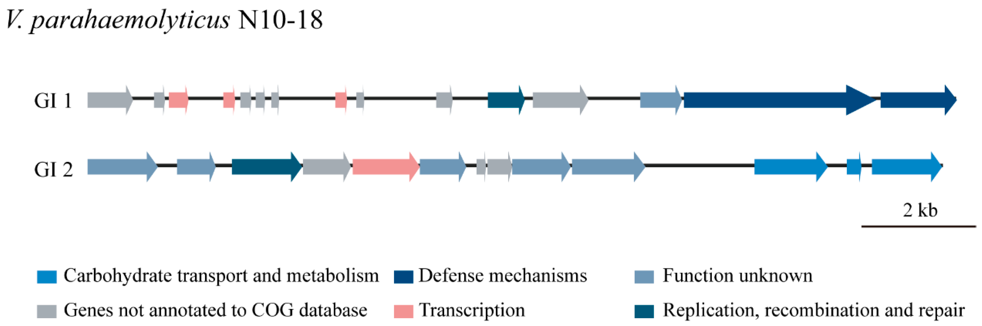
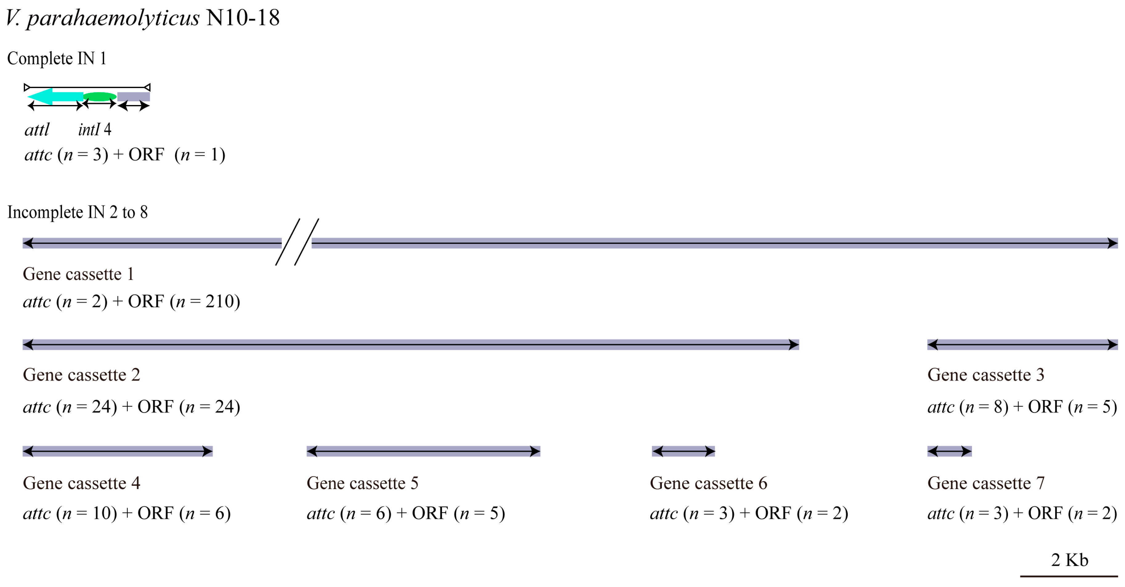
3.5. MGEs in the V. parahaemolyticus N10-18 Genome

3.5.1. GIs

GIs play a critical role in V. parahaemolyticus genome evolution by the acquisition of novel biological traits through HGT [52]. In this study, two GIs (GI 1 to GI 2) were identified in the V. parahaemolyticus N10-18 genome (Figure 3). GI 1 (15,200 bp) contained 15 genes, wherein six had known functions, encoding a serine/threonine protein phosphatase (Vp_N10_18_3246), a transcriptional regulator (Vp_N10_18_3248), cold-shock proteins (Vp_N10_18_3249, Vp_N10_18_3253), a resolvase (Vp_N10_18_3256), and a deoxyribonuclease HsdR (Vp_N10_18_3259). The other nine genes coded for unknown proteins. GI 2 (14,954 bp) had 13 genes, wherein seven had known functions, encoding a short-chain dehydrogenase (Vp_N10_18_3808), an integrase (Vp_N10_18_3810), a transcriptional regulator (Vp_N10_18_3812), a P-loop ATPase (Vp_N10_18_3817), a dehydrogenase (Vp_N10_18_3818), a phosphotransferase system (PTS) system cellobiose-specific IIB component (Vp_N10_18_3819), and a PTS sugar transporter (Vp_N10_18_3820). The other six genes coded for unknown proteins.

3.5.2. INs

Mobile INs are prevalent in human-dominated ecosystems with prolonged exposure to selective agents such as detergents, antibiotics, and heavy metals [53]. INs are generally classified according to integrase genes intI 1, intI 2, intI 3, and intI 4 into type I, type II, type III, and super integron, respectively [54]. In this study, eight INs (IN 1 to IN 8) were identified in the V. parahaemolyticus N10-18 genome, which ranged from 910 bp to 227,599 bp and carried 2 to 210 genes. Of these, there was one complete IN (IN 1) and seven gene cassettes (IN 2 to IN 8) (Figure 4). Typically, gene cassettes consist of a promoterless open reading frame (orf) and a recombination site (attC) necessary for integration. They can exist free as circular molecules or mobilized in INs [55].
In this study, the complete IN 1 (2566 bp) contained a hypothetical protein-encoding gene (Vp_N10_18_2516) and an integrase gene IntI 4 (Vp_N10_18_2515). The latter showed sequence identity (99.38%) with the super IN IntI 4 (NR reference sequence: AHI99301.1) [56], which indicated that IN 1 was a super IN in V. parahaemolyticus N10-18.
Among the seven incomplete INs, the largest, IN 2 (227,599 bp), contained 210 genes, of which 54 genes coded for hypothetical proteins. IN 3 (15,877 bp) carried 24 genes encoding 15 hypothetical proteins and 9 proteins with known functions including an adenylate kinase and related kinase (Vp_N10_18_4543), a GCN5-related N-acetyltransferase (GNAT) (Vp_N10_18_4551), a histone acetyltransferase (Vp_N10_18_4552), plasmid stabilization proteins (Vp_N10_18_4545 and Vp_N10_18_4553), prevent-host-death family proteins (Vp_N10_18_4544 and Vp_N10_18_4554), a putative membrane protein (Vp_N10_18_4549), and a site-specific DNA-methyltransferase (Vp_N10_18_4538). Additionally, IN 4 (3898 bp) carried five genes encoding four hypothetical proteins and a vco30 (Vp_N10_18_4598); IN 5 (3866 bp) encoded five hypothetical proteins and an acetyltransferase (Vp_N10_18_4601); IN 6 (4779 bp) encoded four hypothetical proteins and a methyltransferase (Vp_N10_18_4620); IN 7 (1300) encoded two hypothetical proteins; and IN 8 (910 bp) encoded a hypothetical protein and a plasmid stabilization protein ParE (Vp_N10_18_4638) (Figure 4).
Virulence-related genes were also detected in the INs in V. parahaemolyticus N10-18, such as the GNAT (Vp_N10_18_4551, IN 3), prevent-host-death family proteins (Vp_N10-18_4544 and Vp_N10-18_4554, IN 3), and plasmid stabilization proteins (Vp_N10_18_4545 and Vp_N10_18_4553, IN 3; Vp_N10-18_4638, IN 8).

3.5.3. ISs

A single short IS can transfer one or more resistance-related genes in Gram-negative bacteria and affect bacterial resistance phenotype [21,22]. In this study, only one IS110 (1327 bp) was identified in the V. parahaemolyticus N10-18 genome, encoding a IS110 family transposase (Table S3).

3.6. Putative Virulence-Associated Genes

We identified approximately 45 virulence-associated genes in the V. parahaemolyticus N10-18 genome by the BLAST analysis (Table S4). Of these, 36 genes encoded type III secretion system 1 (T3SS1)-related proteins, including VecA, YscO, VcrDGHRV, VopBDNQRS, and VscCDFGHIJKLNPQRSTUVWXY. T3SS1 is an essential virulence determinant for V. parahaemolyticus survival in the environment [57,58]. Interestingly, the exsACD gene cascade was also present in the V. parahaemolyticus N10-18 genome. T3SS1 expression is regulated by this cascade, in which the master transcription factor ExsA positively regulates T3SS1 expression, whereas ExsD negatively regulates its expression [59]. The other virulence-associated genes functioning in bacterial adhesion or epithelial cell invasion also existed in the V. parahaemolyticus N10-18 genome, e.g., ilpA [60], MAM7 (multivalent adhesion molecule 7) [61], gmhA [62], gmd [63], and kdsA [64] (Table S4).

3.7. Heavy Metal and Antibiotic Resistance-Associated Genes

Approximately 38 heavy metal tolerance-associated genes were identified in the V. parahaemolyticus N10-18 genome by the BLAST analysis (Table 2). For example, the cadC gene and dsbABC gene cluster, which are responsible for the bacterial tolerance to Cd, Zn, and Pb, as well as Cd, Zn, Hg, and Cu, respectively [65], were present in the V. parahaemolyticus N10-18 genome. Moreover, the zntAR, znuABC, zur, and smtA genes for the Zn and Hg tolerance [65,66,67] were also identified (Table 2). These results were consistent with the heavy metal tolerance phenotype of V. parahaemolyticus N10-18.
Antimicrobial resistance-associated genes (n = 7) also existed in the V. parahaemolyticus N10-18 genome (Table 2), such as an elongation factor Tu (tuf) [68], a cAMP-activated global transcriptional regulator CRP (crp) [69], a DNA-directed RNA polymerase subunit beta (rpoB) [70], and a hexose-6-phosphate (uhpT) [71], tet34 and tet35 [72], and β-lactamase (blaCARB-21), consistent with the MDR phenotype of V. parahaemolyticus N10-18.

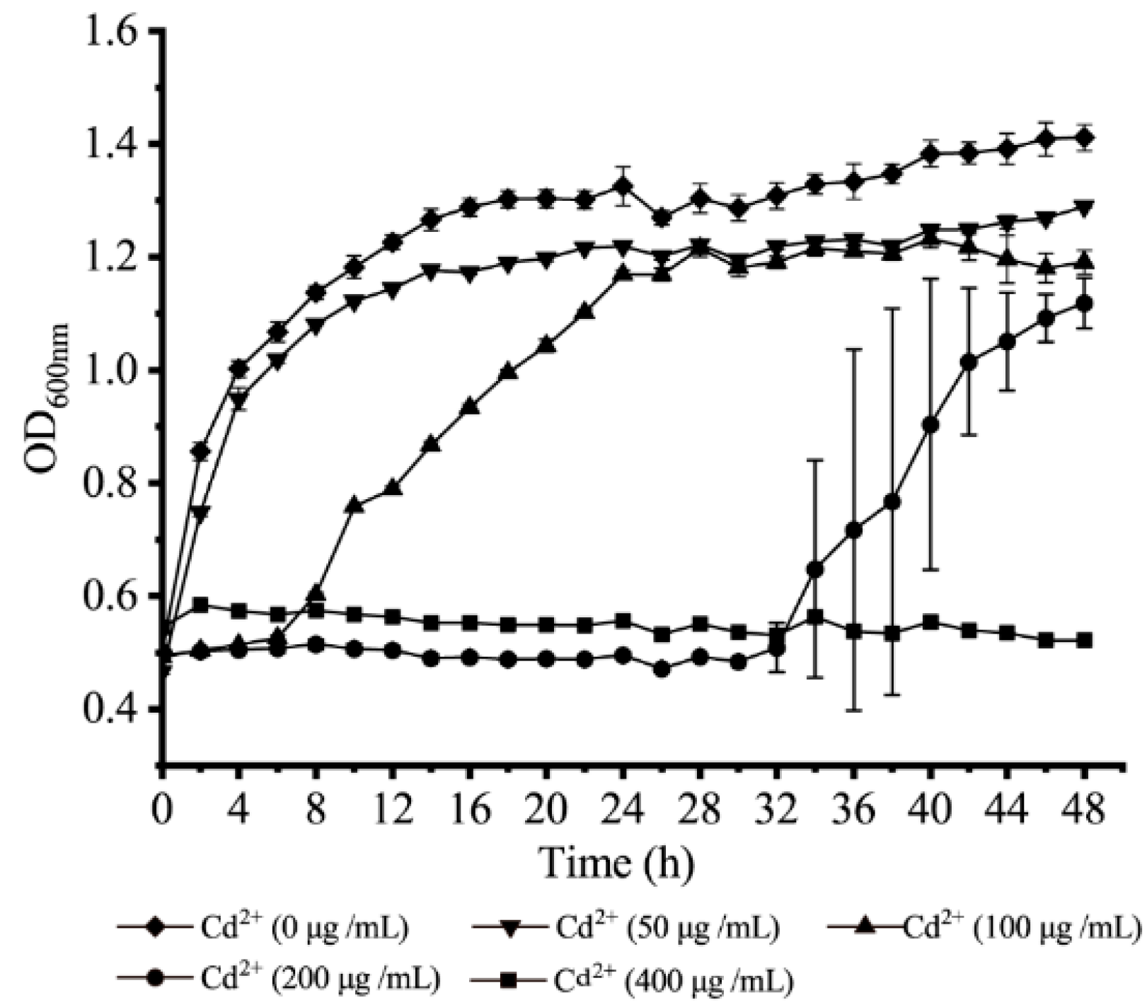
3.8. Survival of V. parahaemolyticus N10-18 under the Cd2+ (50 μg/mL) Stress

Based on the above results, MIC values of the heavy metals and antibiotics against V. parahaemolyticus N10-18 were determined (Table S1). Remarkably, the observed MICs of Cd2+ and Zn2+ were 400 μg/mL and 1600 μg/mL, respectively. Given that Zn2+ is essential for the growth and development of aquatic animals and often used as feed additives, the survival of V. parahaemolyticus N10-18 to resist the high level of Cd2+ was further investigated in this study.
Growth curves of V. parahaemolyticus N10-18 at different concentrations of CdCl2 were determined in the TSB medium at 37 °C. As shown in Figure 5, at the concentration of 400 μg/mL of Cd2+, the growth of V. parahaemolyticus N10-18 was fully inhibited. At 200 μg/mL and 100 μg/mL of Cd2+, the bacterial growth was retarded, showing a longer lag phase of 32 h and 6 h, respectively. Moreover, the bacterial biomass reached the maximum with OD600 nm values of 1.178 and 1.216 at 48 h and 28 h, respectively. At 50 μg/mL of Cd2+, only a slight decrease in growth was observed, when compared with the control (0 μg/mL of Cd2+). Under this treatment, the observed fatality rate of V. parahaemolyticus N10-18 was 10.73%.

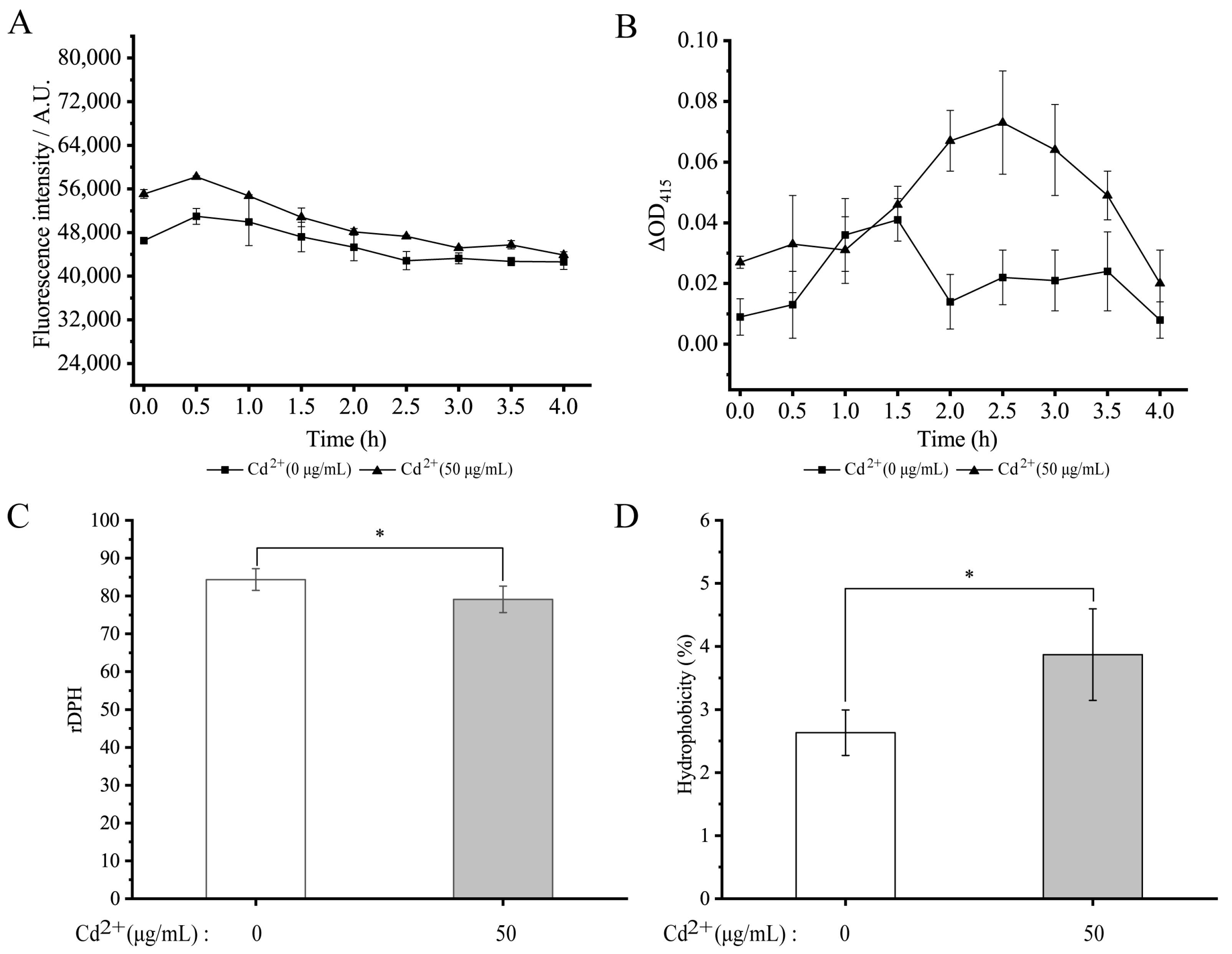
3.9. Changes in Cell Membrane Permeability and Fluidity and Cell Surface Hydrophobicity of V. parahaemolyticus N10-18 under the Cd2+ (50 μg/mL) Stress

Bacterial cell membrane permeability and fluidity and cell surface hydrophobicity are key parameters of the cell membrane responding to environmental challenges [80,81]. Cd is a heavy metal whose cations often cause toxicity to both eukaryotic and prokaryotic cells even at low concentrations [82]. In this study, the outer cell membrane permeability was examined using the NPN probe. As shown in Figure 6A, the probe fluorescence intensity showed an overall downward trend after treatment with 50 μg/mL of Cd2+ for 4 h. Additionally, we used the ONPG as a probe to examine the inner cell membrane permeability, and no significant difference in the inner membrane permeability was also observed after the Cd2+ stress for 1.5 h compared with the control group (p > 0.05). However, the extended treatment time (2.0 to 4.0 h) increased the bacterial inner membrane permeability by 2.04- to 4.96-fold (p < 0.05) (Figure 6B).
Cytoplasmic membrane fluidity also influences the ability of most compounds (nutrients and antibiotics) and ions to cross the bacterial cytoplasmic membrane by diffusion and active transport [80]. As shown in Figure 6C, cell membrane fluidity of V. parahaemolyticus N10-18 was significantly decreased by 1.07-fold after being treated with 50 μg/mL of Cd2+ for 2 h, compared with the control group. Cell surface hydrophobicity is crucial in bacterial adhesion to abiotic and biological surfaces [83]. As shown in Figure 6D, cell surface hydrophobicity of V. parahaemolyticus N10-18 was significantly increased by 1.47-fold after being treated with 50 μg/mL of Cd2+ for 2 h (p < 0.05).
Cell structure changes of V. parahaemolyticus N10-18 under the Cd2+ (50 μg/mL) stress were also observed by the SEM analysis. As shown in Figure 7, the treatment with Cd2+ (50 μg/mL) for 2 h resulted in the cell surface shrinking of certain V. parahaemolyticus N10-18 cells compared to the control group.

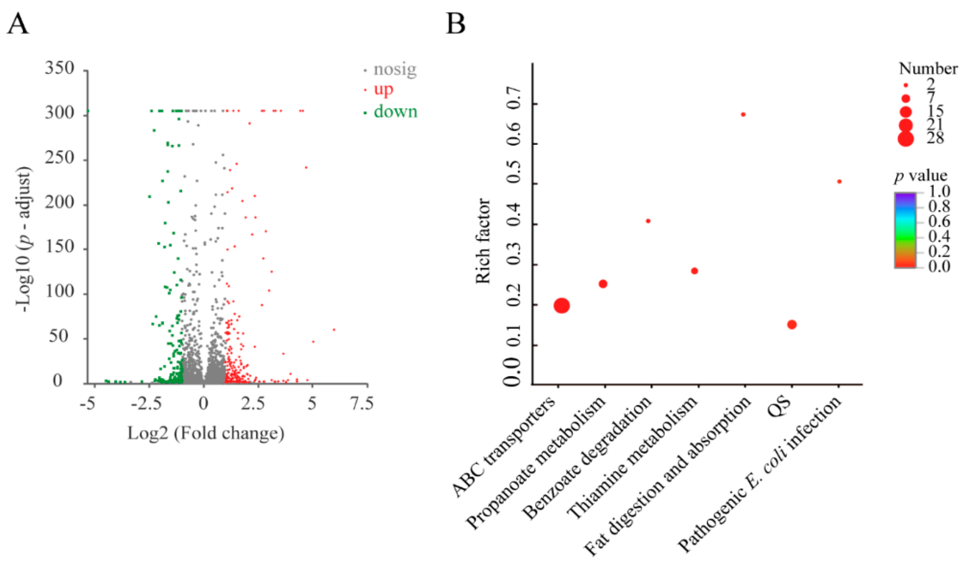
3.10. The Major Changed Metabolic Pathways Medicated by the Cd2+ (50 μg/mL) Stress in V. parahaemolyticus N10-18

Based on the obtained results, V. parahaemolyticus N10-18 grown at the mid-LGP in the TSB medium at 37 °C was treated with the Cd2+ (50 μg/mL) for 2 h, and gene expression changes at the global genome level of V. parahaemolyticus N10-18 induced by the Cd2+ stress were determined using the Illumina HiSeq 2500 sequencing technology.
Approximately 8.3% (377 of 4565 genes) of the bacterial genes were differentially expressed under the treatment, when compared to the control group. Of these, 217 DEGs showed higher transcriptional levels (fold change ≥ 2.0), whereas 160 were significantly down-regulated (fold change ≤ 0.5) (Figure 8A). Approximately seven significantly altered metabolic pathways were identified in V. parahaemolyticus N10-18, including the ATP-binding cassette (ABC) transporters, propanoate metabolism, benzoate degradation, thiamine metabolism, fat digestion and absorption, quorum sensing (QS), and pathogenic E. coli infection (Figure 8B, Table 3).
Remarkably, the expression of approximately 28 DEGs of the ABC transporters was significantly changed at the transcription level (0.061- to 11.609-fold) (p < 0.05). Of these, the DEGs encoding the maltose and ribose transporters and some amino acid transporters were significantly inhibited (0.061- to 0.500-fold). For example, the malEK gene cluster (Vp_N10_18_1557, Vp_N10_18_1556), which encoded a maltose ABC transporter substrate-binding protein MalE and a maltose/maltodextrin import ATP-binding protein MalK, respectively, was significantly down-regulated. The rbsBCD gene cluster (Vp_N10_18_3026, Vp_N10_18_3025, and Vp_N10_18_3023), which encoded a ribose ABC transporter substrate-binding protein RbsB, a ribose ABC transporter permease, and a D-ribose pyranase, respectively, was significantly down-regulated as well (p < 0.05). Additionally, the expression of the livH gene (Vp_N10_18_2959), which encoded a branched-chain amino acid ABC transporter permease, was remarkably down-regulated (0.061-fold) at the transcriptional level.
In the propanoate metabolism, expression of approximately seven DEGs was also significantly inhibited (0.069- to 0.438-fold) (p < 0.05), e.g., the prpCEF, and acnD genes (Vp_N10_18_0015, Vp_N10_18_0011, Vp_N10_18_0012, and Vp_N10_18_0013) involved in the 2-methylcitric acid cycle (2-MCC). The 2-MCC in the propionate catabolic pathway is used to oxidize the Cα methylene of propionate to a keto group yielding pyruvate [84]. These results indicated that V. parahaemolyticus N10-18 greatly reduced the branched-chain amino acid transportation, inhibited the maltose and ribose transportation, and inactivated to utilize the propionic acid as a carbon source under the Cd2+ (50 μg/mL) stress.
Conversely, the DEGs encoding the Zn/Cd/Hg/Pb-transporting ATPase (zntA, Vp_N10_18_0526) and heavy metal efflux resistance-nodulation-cell division (RND) transporter of the CusA/CzcA family (cusA, Vp_N10_18_0582) were greatly up-regulated by 23.639- and 8.649-fold, respectively (p < 0.05). Moreover, the znuABC gene cluster (Vp_N10_18_4099, Vp_N10_18_1679, Vp_N10_18_1681, Vp_N10_18_4101, and Vp_N10_18_1680) involved in Zn uptake was highly expressed at the transcriptional level (2.594- to 11.609-fold) (p < 0.05). Of these, the znuA gene (Vp_N10_18_1679) was remarkably up-regulated by 11.609-fold. Cd is chemically similar to Zn, both of which belong to the IIB transition elements. It is probably common in microbial species that Cd2+ is imported via the Zn channels [67]. Likewise, with expression of three DEGs for the iron (III) transportation, which encoded a Fe3+-hydroxamate ABC transporter permease FhuB (fhuB, Vp_N10_18_1520), an iron (III) ABC transporter ATP-binding protein (Vp_N10_18_1522), and an iron ABC transporter substrate-binding protein (afuA, Vp_N10_18_1887), all were significantly increased (2.243- to 3.891-fold) (p < 0.05). Additionally, the artIMP gene cluster (Vp_N10_18_0733, Vp_N10_18_0735, and Vp_N10_18_0732) for the arginine ABC transportation was also significantly up-regulated (3.101- to 4.015-fold) (p < 0.05), which encoded an arginine ABC transporter substrate-binding protein, an arginine transporter permease subunit ArtM, and an arginine ABC transporter ATP-binding protein ArtP in the arginine transportation. Arginine is a structurally stabilizing factor that contains side chains to form salt bridges and hydrogen bonds [85].
In the QS, the DEGs encoding a sugar ABC transporter ATP-binding protein (Vp_N10_18_1219), a polyamine ABC transporter substrate-binding protein (Vp_N10_18_0181), and ABC transporter permeases (Vp_N10_18_1876 and Vp_N10_18_2783) were also significantly up-regulated by 2.140- to 9.727-fold (p < 0.05).
In the thiamine metabolism, approximately five DEGs were also up-regulated at the transcriptional level (2.116- to 2.740-fold) (p < 0.05), which encoded a phosphomethylpyrimidine synthase (thic, Vp_N10_18_4412), a hydroxymethylpyrimidine kinase/phosphomethylpyrimidine kinase (thiD, Vp_N10_18_1089), thiamine phosphate synthases (thiE, Vp_N10_18_1096 and Vp_N10_18_4413), and a thiaminase II (tenA, Vp_N10_18_1094). Thiamine is a precursor of thiamine pyrophosphate (TPP), an essential coenzyme in the central metabolism. Bacterial thiamine biosynthesis and salvage genes are controlled at the RNA level by TPP-responsive riboswitches that include the ABC family transporter ThiXYZ [86]. In this study, the thiXYZ genes (Vp_N10_18_1091, Vp_N10_18_1092, and Vp_N10_18_1090) were also up-regulated by 2.400- to 3.567-fold. These results suggested that the Cd2+ (50 μg/mL) stress enhanced the thiamine metabolism in V. parahaemolyticus N10-18 to increase the biosynthesis of TPP.
Interestingly, the DEGs encoding the T3SS needle filament protein VscF (Vp_N10_18_0060) and glyceraldehyde-3-phosphate dehydrogenase (gapA, Vp_N10_18_3876) in pathogenic E. coli infection were significantly up-regulated by 5.836- and 2.086-fold, respectively (p < 0.05). The VscF in T3SS1 helps to translocate VPA0226 that can be secreted into the host cell cytoplasm via T3SS1 in V. parahaemolyticus [58]. The gapA gene was only expressed under certain stress conditions, and overproduction of GapA led to increased resistance to H2O2 in Lactococcus lactis MG1363 [87]. Additionally, expression of representative DEGs was examined by the RT-PCR assay, and the resulting data were generally consistent with the transcriptomic analysis (Tables S6 and S7).
Taken together, under the Cd2+ (50 μg/mL) stress, V. parahaemolyticus N10-18 employed multiple strategies for efficient transportation and exocytosis of Cd2+ to alleviate its cytotoxicity: (1) greatly enhanced the Zn/Cd/Hg/Pb-transportation and efflux; (2) up-regulated metal and iron ABC transportation; (3) enhanced the biosynthesis of TPP in the thiamine metabolism; (4) up-regulated the expression of stress-related proteins, such as GapA, and structurally stabilizing factor arginine; (5) conversely, greatly reduced the branched-chain amino acid transportation; (6) inhibited the maltose and ribose ABC transportation; and (7) down-regulated the propanoate metabolism, in order to survive in the adverse niche.

4. Discussion

The pollution of heavy metals in aquatic environments has led to heavy metal residues in aquatic products, which poses a huge hidden danger in human health [88,89]. Cd is classified into Group 1 as carcinogenic to humans by the IARC [14]. This toxic element possibly results in short-term or long-term disorders in the body, such as degenerative bone disease, kidney dysfunction, lung injuries, disorders in Zn and Cu metabolism, and cancer [14]. The heavy metal pollution has also been supposed to link high incidence of the MDR V. parahaemolyticus, which is a challenging issue in the clinical treatment. This study was the first to characterize the MDR V. parahaemolyticus N10-18 with high tolerance to Cd2+ and Zn2+. The observed MIC values of Cd2+, Zn2+, AMP, KAN, and STR against V. parahaemolyticus N10-18 were 400 μg/mL, 1600 μg/mL, 50,000 μg/mL, 128 μg/mL, and 128 μg/mL, respectively, which suggested possible antibiotic and heavy metal pollution in the aquaculture environment of O. gigas Thunberg, consistent with previous reports [8,15,21,23].
In this study, the draft genome sequence (4,910,080 bp) of V. parahaemolyticus N10-18 was determined using the Illumina Hiseq × 10 sequencing technique. Some repeats were located at the end of scaffolds (n = 23, <1.1 Kb) (Table S5), indicating that the genome assembly was incomplete and contained some gaps, due to limitations of the second-generation Illumina short-read sequencing technique. Approximately 4653 genes were predicted, of which 722 encoded unknown proteins. In our previous study, we found a number of unknown-function genes in V. parahaemolyticus isolates from the six species of aquatic animals (P. undulate, P.a viridis, M.veneriformis, A. nobilis, C. auratu, and L. vannamei) [21]. These results demonstrated genome variation and plasticity of V. parahaemolyticus in aquatic animals.
Intercellular transmissibility of MGEs may have constituted important driving forces in V. parahaemolyticus genome evolution and formation of ecotypes and speciation [90]. In this study, we identified 11 MGEs in the V. parahaemolyticus N10-18 genome. It cannot exclude that additional MGEs may be located in the gaps between scaffolds [21]. In this study, we found two GIs that contained 28 genes in the V. parahaemolyticus N10-18 genome, which facilitated the bacterium to better fit into the niche. For example, GI 1 contained the genes encoding cold-shock proteins (CSPs) (Vp_N10_18_3249, Vp_N10_18_3253). The CSPs served as global gene expression regulators to respond to different stress conditions [22].
INs allow bacteria to capture, stockpile, express, and exchange genes embedded within gene cassettes [91]. In this study, one super IN and seven incomplete INs were identified in the V. parahaemolyticus N10-18 genome. Although approximately 33.20% of the INs-carrying genes (n = 85) encoded unknown proteins, the identified INs endowed the bacterium with diverse environmental adaptability. For instance, several gene cassettes were found to carry virulence-related genes, such as the GNAT super family proteins, prevent-host-death family proteins, and plasmid stabilization proteins. Of these, the GNAT (Vp_N10_18_4551) belongs to type II toxin of toxin–antitoxin systems. The GNAT toxin blocks protein translation by acetylating the amino group of charged tRNAs, thus preventing tRNA from participating in peptidyl ribosomal transferase [92].
Some V. parahaemolyticus isolates lacking the tdh and/or trh genes are also highly cytotoxic to human gastrointestinal cells, which indicates that other virulence factors exist. In this study, we found potential virulence-related genes (n = 45) in the V. parahaemolyticus N10-18 genome, e.g., ilpA, MAM7, exsACD, gmhA, gmd, kdsA, and T3SS1-related genes, which are involved in bacterial adhesion or epithelial cell invasion. For example, the ilpA gene encodes an immunogenic lipoprotein A in Vibrio vulnificus, an adhesion molecule that can induce cytokine production in human immune cells [60]. MAM7 enables Gram-negative pathogens to establish high-affinity binding to host cells during the early stages of infection and is crucial for the delivery of virulence factors into hosts [61]. In this study, we identified 36 genes in T3SS1 in the V. parahaemolyticus N10-18 genome, which are important determinants of the pathogenicity of V. parahaemolyticus. Of these genes, VscCD genes not only activated bacterial resistance to acid stress, H2O2, and antibiotics but also enhanced the colonization ability and pathogenicity of Vibrio harveyi [93]. These results suggested a health risk in consuming O. gigas Thunberg contaminated by V. parahaemolyticus N10-18.
Bacterial MDR is regarded as an emerging pollutant in different food production avenues including aquaculture [94]. Resistance factors have been reported in pathogenic bacteria [68,69,70,71,72,95,96,97,98]. In this study, we identified seven antibiotic-resistance-related genes in the V. parahaemolyticus N10-18 genome, consistent with the observed MDR phenotype of the bacterium. Remarkably, 38 heavy metal tolerance-associated genes existed in V. parahaemolyticus N10-18. For instance, the gene (Vp_N10_18_3808, GI 2) encoding a short-chain dehydrogenase/reductase SDR family member was identified, which functions in the Cd2+ stress in Pleurotus eryngii [99]. Moreover, the cadC, dsbABC, zntAR, znuABC, zur, and smtA genes related to the Cd and Zn resistance were also present in the V. parahaemolyticus N10-18 genome, consistent with the high Cd and Zn tolerance phenotype of the bacterium. For instance, the dsbABC gene cluster involved in the degradation of pyrimidine ribonucleosides was found to be related to the resistance and absorbing of Cd in Enterococcus faecalis LZ-11, which was isolated from Lanzhou reach of the Yellow River in China [100]. The diversity of resistance genes, gene variance, and selective pressure from the environment may result in the difference between resistance phenotype and resistance genotype.
In this study, the constructed phylogenetic tree showed that the 65 V. parahaemolyticus genomes were clustered into four large clusters, among which V. parahaemolyticus N10-18 fell into a single sub-branch in Group 4b. The bacterium is located phylogenetically distant from the other V. parahaemolyticus strains originating in aquatic animals but showed the closest evolutionary distance with 8 V. parahaemolyticus strains isolated from Homo sapiens between 1998 and 2015 in the USA. Until 1996, V. parahaemolyticus infection cases were sporadic, occurred in certain countries, and could be related to diverse serovars [101]. Location and isolation time of V. parahaemolyticus strains were not associated with evolutionary taxa, suggesting that the widespread global trade in aquatic products over the past 30 years may have contributed to the cross-regional spread of the pathogen, leading to an increased risk of edible aquatic animals.
Based on the findings in this study, the molecular mechanism underlying the heavy metal Cd2+ tolerance of V. parahaemolyticus N10-18 was further explored. Under the Cd2+ (50 μg/mL) stress, the bacterium significantly changed cell membrane permeability and fluidity and cell surface hydrophobicity (p < 0.05). Cell osmotic changes have been disclosed as stressors that can affect biophysical properties and the composition of the membrane and consequently transport mechanisms (permeability) and cell shape and integrity [80]. In this study, after the Cd2+ (50 μg/mL) treatment, the cell surface of V. parahaemolyticus N10-18 was observed shrinking to a certain extent.
Comparative transcriptomic analysis revealed seven significantly altered metabolic pathways in V. parahaemolyticus N10-18 under the Cd2+ stress. Remarkably, the DEGs encoding the Zn/Cd/Hg/Pb-transporting ATPase (zntA), and heavy metal efflux RND transporter of the CusA/CzcA family (cusA) were greatly up-regulated by 23.639- and 8.649-fold, respectively (p < 0.05). The zntA gene was originally described as a Zn-transporting ATPase in E. coli, but it also confers resistance to Cd [66]. RND efflux pumps are essential for the expulsion of a plethora of potentially small lethal agents or compounds such as detergents, solvents, heavy metals, antibiotics, and toxic secondary metabolites [102]. The CusC(F)BA complex exports copper (I) and silver (I) and mediates resistance to these two metal ions in E. coli [102]. Interestingly, in this study, the ABC transporter encoded by the znuABC genes for high-affinity Zn2+ uptake [66,67] was also highly increased at the transcriptional level (2.594- to 11.609-fold) (p < 0.05). This was consistent with the high tolerance of V. parahaemolyticus N10-18 to Zn2+. Given that Cd is chemically similar to Zn, both of which belong to the IIB transition elements [67], our result provided evidence for the expulsion of Cd2+ via the Zn2+ channels in V. parahaemolyticus N10-18. On the other hand, the DEGs (afuA and fhuB) encoding the ion and metal transporters were also up-regulated (p < 0.05). For instance, the fhuB gene encoded a Fe3+-hydroxamate ABC transporter permease FhuB. Iron (III) hydroxamate transport across the cytoplasmic membrane is catalyzed by the very hydrophobic FhuB protein and the membrane-associated FhuC protein [103]. These results indicated highly enhanced expulsion of Cd2+ by V. parahaemolyticus N10-18 to alleviate its cytotoxicity.
In the QS, expression of five DEGs was also significantly up-regulated by 2.14- to 9.727-fold (p < 0.05). Additionally, the pcaC gene (Vp_N10_18_2971) encoding the carboxymuconolactone decarboxylase family protein in benzoate degradation and the atoB gene (Vp_N10_18_2988) encoding the thiolase family protein in the fat digestion and absorption were also significantly enhanced by 2.003- and 4.215-fold, respectively (p < 0.05). These results suggested possibly increased substance absorption for energy conservation and stringent response regulation in V. parahaemolyticus N10-18 under the Cd2+ (50 μg/mL) stress.
In contrast, the DEGs involved in the branched-chain amino acid transportation and maltose and ribose transportation were significantly repressed (0.061- to 0.500-fold) (p < 0.05). Meanwhile, the propanoate metabolism was also significantly inhibited (0.069- to 0.438-fold) (p < 0.05). These results suggested possible repressed energy consumption and nucleotide and ribosome biosynthesis under the Cd2+ adverse condition. It will be interesting to further investigate the DEGs using proteomic, cell, and animal mode techniques and methods in the future research.

5. Conclusions

This study was the first to characterize the MDR V. parahaemolyticus N10-18 with high tolerance to Cd2+ and Zn2+ (MICS: 400 μg/mL and 1600 μg/mL) using genomic and transcriptomic techniques. The draft genome sequence (4,910,080 bp) of V. parahaemolyticus N10-18 was determined, and 722 of 4653 predicted genes had unknown function. Comparative genomic analyses revealed MGEs, including GIs (n = 2), INs (n = 8), and ISs (n = 1). Heavy metal and antibiotic-resistance genes (n = 38 and 7) and virulence-associated genes (n = 45) were also found in the V. parahaemolyticus N10-18 genome. The bacterial growth was slightly decreased under the 50 μg/mL of Cd2+. V. parahaemolyticus N10-18 significantly changed cell membrane permeability and fluidity and surface hydrophobicity under the Cd2+ (50 μg/mL) stress (p < 0.05). Meanwhile, comparative transcriptomic analysis revealed seven significantly altered metabolic pathways. Under the Cd2+ stress, V. parahaemolyticus N10-18 employed multiple strategies for efficient transportation and exocytosis of Cd2+ to alleviate its cytotoxicity, including greatly enhanced Zn/Cd/Hg/Pb transportation and efflux and significantly up-regulated metal and iron ABC transportation, thiamine metabolism, and stress-related protein expression (e.g., GapA and arginine); in contrast, it greatly reduced the branched-chain amino acid transportation and significantly inhibited the maltose and ribose ABC transportation and propanoate metabolism, in order to resist and survive in the adverse Cd2+ environment. The results also provided evidence for the expulsion of Cd2+ via the Zn2+ channels in V. parahaemolyticus N10-18. Overall, the results of this study enriched genome data of V. parahaemolyticus from aquatic animals and revealed multiple strategies for the cadmium tolerance in the leading seafood-borne pathogen worldwide.

Supplementary Materials

The following supporting information can be downloaded at: https://www.mdpi.com/article/10.3390/foods11233777/s1, Figure S1: The k-mer analysis for V. parahaemolyticus N10-18 subread data based on the number of unique 17-mers; Table S1: The genotype and phenotype of V. parahaemolyticus N10-18 isolate used in this study; Table S2: The 65 V. parahaemolyticus strains analyzed in the phylogenetic tree; Table S3: The identified GIs, Ins, and ISs in the V. parahaemolyticus N10-18 genome; Table S4: The identified repeats in the V. parahaemolyticus N10-18 genome [104,105,106,107]; Table S5: The potential virulence-associated genes identified in the V. parahaemolyticus N10-18 genome; Table S6: Oligonucleotide primers used in the RT-PCR assay; Table S7: Expression of representative DEGs by the RT-PCR assay.

Author Contributions

Conceptualization, L.C.; Data curation, P.Y., L.Y. and J.W.; Funding acquisition, L.C.; Investigation, P.Y., L.Y. and C.S.; Supervision, S.Q. and C.Z.; Writing—original draft, P.Y. and L.Y.; Writing—review & editing, L.C. All authors have read and agreed to the published version of the manuscript.

Funding

This work was supported by the Science and Technology Commission of Shanghai Municipality, grant number 17050502200, and the National Natural Science Foundation of China, grant number 31671946.

Data Availability Statement

The draft genome of V. parahaemolyticus N10-18 is available in the GenBank database under the accession number JALGSE000000000. A complete list of DEGs in the strain under the Cd2+ stress is available in the National Center for Biotechnology Information (NCBI, https://www.ncbi.nlm.nih.gov, accessed on 21 April 2022) SRA database under the accession number PRJNA825334. Other data is contained within the article or Supplementary Materials.

Acknowledgments

We would like to express gratitude to Dingxiang Xu from Shanghai Ocean University for her help in comparative genomic analysis.

Conflicts of Interest

The authors declare no conflict of interest.

References

  1. Liu, J.; Bai, L.; Li, W.; Han, H.; Fu, P.; Ma, X.; Bi, Z.; Yang, X.; Zhang, X.; Zhen, S.; et al. Trends of foodborne diseases in China: Lessons from laboratory-based surveillance since 2011. Front. Med. 2017, 12, 48–57. [Google Scholar] [CrossRef] [PubMed]
  2. Broberg, C.A.; Calder, T.J.; Orth, K. Vibrio parahaemolyticus cell biology and pathogenicity determinants. Microbes Infect. 2011, 13, 992–1001. [Google Scholar] [CrossRef] [PubMed] [Green Version]
  3. Fujino, T.; Okuno, Y.; Nakada, D.; Aoyama, A.; Fukai, K.; Mukai, T.; Ueho, T. On the bacteriological examination of shirasu-food poisoning. Med. J. Osaka Univ. 1953, 4, 299–304. [Google Scholar]
  4. Ghenem, L.; Elhadi, N.; Alzahrani, F.; Nishibuchi, M. Vibrio parahaemolyticus: A Review on Distribution, Pathogenesis, Virulence Determinants and Epidemiology. Saudi J. Med. Med. Sci. 2017, 5, 93–103. [Google Scholar] [PubMed]
  5. Scallan, E.; Hoekstra, R.M.; Angulo, F.J.; Tauxe, R.V.; Widdowson, M.-A.; Roy, S.L.; Jones, J.L.; Griffin, P.M. Foodborne illness acquired in the United States—Major pathogens. Emerg. Infect. Dis. 2011, 17, 7–15. [Google Scholar] [CrossRef] [PubMed]
  6. Song, X.; Zang, J.; Yu, W.; Shi, X.; Wu, Y. Occurrence and Identification of Pathogenic Vibrio Contaminants in Common Seafood Available in a Chinese Traditional Market in Qingdao, Shandong Province. Front. Microbiol. 2020, 11, 1488. [Google Scholar] [CrossRef] [PubMed]
  7. Eraghunath, P. Roles of thermostable direct hemolysin (TDH) and TDH-related hemolysin (TRH) in Vibrio parahaemolyticus. Front. Microbiol. 2015, 5, 805. [Google Scholar] [CrossRef]
  8. Li, Y.; Xie, T.; Pang, R.; Wu, Q.; Zhang, J.; Lei, T.; Xue, L.; Wu, H.; Wang, J.; Ding, Y.; et al. Food-Borne Vibrio parahaemolyticus in China: Prevalence, Antibiotic Susceptibility, and Genetic Characterization. Front. Microbiol. 2020, 11, 1670. [Google Scholar] [CrossRef]
  9. Dewi, R.R.; Hassan, L.; Daud, H.M.; Matori, M.F.; Nordin, F.; Ahmad, N.I.; Zakaria, Z. Prevalence and Antimicrobial Resistance of Escherichia coli, Salmonella and Vibrio Derived from Farm-Raised Red Hybrid Tilapia (Oreochromis spp.) and Asian Sea Bass (Lates calcarifer, Bloch 1970) on the West Coast of Peninsular Malaysia. Antibiotics 2022, 11, 136. [Google Scholar] [CrossRef]
  10. Prescott, J.; Barkovskii, A.L. In situ dynamics of Vibrio parahaemolyticus and Vibrio vulnificus in water, sediment and triploid Crassostrea virginica oysters cultivated in floating gear. J. Appl. Microbiol. 2021, 132, 3343–3354. [Google Scholar] [CrossRef]
  11. Pepi, M.; Focardi, S. Antibiotic-Resistant Bacteria in Aquaculture and Climate Change: A Challenge for Health in the Mediterranean Area. Int. J. Environ. Res. Public Health 2021, 18, 5723. [Google Scholar] [CrossRef]
  12. Ma, T.; Zhang, Y.; Hu, Q.; Han, M.; Li, X.; Zhang, Y.; Li, Z.; Shi, R. Accumulation Characteristics and Pollution Evaluation of Soil Heavy Metals in Different Land Use Types: Study on the Whole Region of Tianjin. Int. J. Environ. Res. Public Health 2022, 19, 10013. [Google Scholar] [CrossRef] [PubMed]
  13. Xu, X.; Pan, B.; Shu, F.; Chen, X.; Xu, N.; Ni, J. Bioaccumulation of 35 metal(loid)s in organs of a freshwater mussel (Hyriopsis cumingii) and environmental implications in Poyang Lake, China. Chemosphere 2022, 307, 136150. [Google Scholar] [CrossRef] [PubMed]
  14. Balali-Mood, M.; Naseri, K.; Tahergorabi, Z.; Khazdair, M.R.; Sadeghi, M. Toxic Mechanisms of Five Heavy Metals: Mercury, Lead, Chromium, Cadmium, and Arsenic. Front. Pharmacol. 2021, 12, 643972. [Google Scholar] [CrossRef] [PubMed]
  15. Jiang, H.; Yu, T.; Yang, Y.; Yu, S.; Wu, J.; Lin, R.; Li, Y.; Fang, J.; Zhu, C. Co-occurrence of Antibiotic and Heavy Metal Resistance and Sequence Type Diversity of Vibrio parahaemolyticus Isolated From Penaeus vannamei at Freshwater Farms, Seawater Farms, and Markets in Zhejiang Province, China. Front. Microbiol. 2020, 11, 1294. [Google Scholar] [CrossRef]
  16. Fang, J.; Cheng, H.; Yu, T.; Jiang, H. Occurrence of Virulence Factors and Antibiotic and Heavy Metal Resistance in Vibrio parahaemolyticus Isolated from Pacific Mackerel at Markets in Zhejiang, China. J. Food Prot. 2020, 83, 1411–1419. [Google Scholar] [CrossRef] [PubMed]
  17. Kang, C.-H.; Shin, Y.; Yu, H.; Kim, S.; So, J.-S. Antibiotic and heavy-metal resistance of Vibrio parahaemolyticus isolated from oysters in Korea. Mar. Pollut. Bull. 2018, 135, 69–74. [Google Scholar] [CrossRef] [PubMed]
  18. Lu, J.; Zhang, X.; Wang, C.; Li, M.; Chen, J.; Xiong, J. Responses of sediment resistome, virulence factors and potential pathogens to decades of antibiotics pollution in a shrimp aquafarm. Sci. Total Environ. 2021, 794, 148760. [Google Scholar] [CrossRef] [PubMed]
  19. Prithvisagar, K.S.; Kumar, B.K.; Kodama, T.; Rai, P.; Iida, T.; Karunasagar, I.; Karunasagar, I. Whole genome analysis unveils genetic diversity and potential virulence determinants in Vibrio parahaemolyticus associated with disease outbreak among cultured Litopenaeus vannamei (Pacific white shrimp) in India. Virulence 2021, 12, 1936–1949. [Google Scholar] [CrossRef]
  20. Levy, S.E.; Boone, B.E. Next-Generation Sequencing Strategies. Cold Spring Harb. Perspect. Med. 2018, 9, a025791. [Google Scholar] [CrossRef] [PubMed]
  21. Xu, D.; Peng, X.; Xie, L.; Chen, L. Survival and Genome Diversity of Vibrio parahaemolyticus Isolated from Edible Aquatic Animals. Diversity 2022, 14, 350. [Google Scholar] [CrossRef]
  22. Zhu, C.; Sun, B.; Liu, T.; Zheng, H.; Gu, W.; He, W.; Sun, F.; Wang, Y.; Yang, M.; Bei, W.; et al. Genomic and transcriptomic analyses reveal distinct biological functions for cold shock proteins (VpaCspA and VpaCspD) in Vibrio parahaemolyticus CHN25 during low-temperature survival. BMC Genom. 2017, 18, 436. [Google Scholar] [CrossRef] [Green Version]
  23. Su, C.; Chen, L. Virulence, resistance, and genetic diversity of Vibrio parahaemolyticus recovered from commonly consumed aquatic products in Shanghai, China. Mar. Pollut. Bull. 2020, 160, 111554. [Google Scholar] [CrossRef]
  24. Yao, W.; Yang, L.; Shao, Z.; Xie, L.; Chen, L. Identification of salt tolerance-related genes of Lactobacillus plantarum D31 and T9 strains by genomic analysis. Ann. Microbiol. 2020, 70, 10. [Google Scholar] [CrossRef] [Green Version]
  25. Yang, L.; Wang, Y.; Yu, P.; Ren, S.; Zhu, Z.; Jin, Y.; Yan, J.; Peng, X.; Chen, L. Prophage-Related Gene VpaChn25_0724 Contributes to Cell Membrane Integrity and Growth of Vibrio parahaemolyticus CHN25. Front. Cell. Infect. Microbiol. 2020, 10, 595709. [Google Scholar] [CrossRef]
  26. Davis, E.M.; Sun, Y.; Liu, Y.; Kolekar, P.; Shao, Y.; Szlachta, K.; Mulder, H.L.; Ren, D.; Rice, S.V.; Wang, Z.; et al. SequencErr: Measuring and suppressing sequencer errors in next-generation sequencing data. Genome Biol. 2021, 22, 37. [Google Scholar] [CrossRef]
  27. Delcher, A.L.; Bratke, K.A.; Powers, E.C.; Salzberg, S.L. Identifying bacterial genes and endosymbiont DNA with Glimmer. Bioinformatics 2007, 23, 673–679. [Google Scholar] [CrossRef] [PubMed] [Green Version]
  28. Chan, P.P.; Lowe, T.M. tRNAscan-SE: Searching for tRNA Genes in Genomic Sequences. Methods Mol. Biol. 2019, 1962, 1–14. [Google Scholar] [CrossRef]
  29. Jensen, L.J.; Julien, P.; Kuhn, M.; von Mering, C.; Muller, J.; Doerks, T.; Bork, P. eggNOG: Automated construction and annotation of orthologous groups of genes. Nucleic Acids Res. 2007, 36, D250–D254. [Google Scholar] [CrossRef] [PubMed]
  30. Gong, L.; Yu, P.; Zheng, H.; Gu, W.; He, W.; Tang, Y.; Wang, Y.; Dong, Y.; Peng, X.; She, Q.; et al. Comparative genomics for non-O1/O139 Vibrio cholerae isolates recovered from the Yangtze River Estuary versus V. cholerae representative isolates from serogroup O1. Mol. Genet. Genom. 2018, 294, 417–430. [Google Scholar] [CrossRef] [PubMed] [Green Version]
  31. Bertelli, C.; Laird, M.R.; Williams, K.P.; Simon Fraser University Research Computing Group; Lau, B.Y.; Hoad, G.; Winsor, G.L.; Brinkman, F.S.L. IslandViewer 4: Expanded prediction of genomic islands for larger-scale datasets. Nucleic Acids Res. 2017, 45, W30–W35. [Google Scholar] [CrossRef] [PubMed]
  32. Fouts, D.E. Phage_Finder: Automated identification and classification of prophage regions in complete bacterial genome sequences. Nucleic Acids Res. 2006, 34, 5839–5851. [Google Scholar] [CrossRef]
  33. Cury, J.; Jové, T.; Touchon, M.; Néron, B.; Rocha, E.P. Identification and analysis of integrons and cassette arrays in bacterial genomes. Nucleic Acids Res. 2016, 44, 4539–4550. [Google Scholar] [CrossRef] [PubMed] [Green Version]
  34. Siguier, P.; Gourbeyre, E.; Chandler, M. Bacterial insertion sequences: Their genomic impact and diversity. FEMS Microbiol. Rev. 2014, 38, 865–891. [Google Scholar] [CrossRef] [Green Version]
  35. Li, M.; Jiang, Y.; Shi, X.; Li, Y.; Jiang, M.; Lin, Y.; Qiu, Y.; Zuo, L.; Deng, Y.; Lin, Z.; et al. Simultaneous Identification of Clinically Common Vibrio parahaemolyticus Serotypes Using Probe Melting Curve Analysis. Front. Cell. Infect. Microbiol. 2019, 9, 385. [Google Scholar] [CrossRef]
  36. Bian, S.; Zeng, W.; Li, Q.; Li, Y.; Wong, N.-K.; Jiang, M.; Zuo, L.; Hu, Q.; Li, L. Genetic Structure, Function, and Evolution of Capsule Biosynthesis Loci in Vibrio parahaemolyticus. Front. Microbiol. 2021, 11, 546150. [Google Scholar] [CrossRef]
  37. Chen, Y.; Dai, J.; Jr, J.G.M.; Johnson, J.A. Genetic analysis of the capsule polysaccharide (K antigen) and exopolysaccharide genes in pandemic Vibrio parahaemolyticus O3:K6. BMC Microbiol. 2010, 10, 374. [Google Scholar] [CrossRef] [Green Version]
  38. Lun, J.; Zhang, S.; Dong, Y.; Xie, S.; Mai, H.; Mei, Y.; Hu, Z. Immunological cross-reactivity and cross-protection of outer membrane protein OmpU among Vibrio species. Acta Microbiol. Sin. 2016, 56, 867–879. [Google Scholar]
  39. Guo, X.; Liu, B.; Chen, M.; Wang, Y.; Wang, L.; Chen, H.; Wang, Y.; Tu, L.; Zhang, X.; Feng, L. Genetic and serological identification of three Vibrio parahaemolyticus strains as candidates for novel provisional O serotypes. Int. J. Food Microbiol. 2017, 245, 53–58. [Google Scholar] [CrossRef] [PubMed]
  40. Chen, X.; Li, Y.; Yao, W.; Wu, T.; Zhu, Q.; Zhang, Y.; Ye, H.; Wang, R.; Zheng, S.; Yu, F.; et al. A new emerging serotype of Vibrio parahaemolyticus in China is rapidly becoming the main epidemic strain. Clin. Microbiol. Infect. 2019, 26, 644.e1–644.e7. [Google Scholar] [CrossRef]
  41. González-Escalona, N.; Martinez-Urtaza, J.; Romero, J.; Espejo, R.T.; Jaykus, L.-A.; DePaola, A. Determination of Molecular Phylogenetics of Vibrio parahaemolyticus Strains by Multilocus Sequence Typing. J. Bacteriol. 2008, 190, 2831–2840. [Google Scholar] [CrossRef] [PubMed] [Green Version]
  42. Emms, D.M.; Kelly, S. OrthoFinder: Phylogenetic orthology inference for comparative genomics. Genome Biol. 2019, 20, 238. [Google Scholar] [CrossRef] [PubMed]
  43. Buck, J. The Plate Count in Aquatic Microbiology. Nativ. Aquat. Bact. Enumer. Act. Ecol. 1979, 1, 19–28. [Google Scholar] [CrossRef]
  44. Harman, R.M.; Yang, S.; He, M.K.; Van de Walle, G.R. Antimicrobial peptides secreted by equine mesenchymal stromal cells inhibit the growth of bacteria commonly found in skin wounds. Stem Cell Res. Ther. 2017, 8, 157. [Google Scholar] [CrossRef] [PubMed] [Green Version]
  45. Ibrahim, H.R.; Sugimoto, Y.; Aoki, T. Ovotransferrin antimicrobial peptide (OTAP-92) kills bacteria through a membrane damage mechanism. Biochim. Biophys. Acta (BBA) Gen. Subj. 2000, 1523, 196–205. [Google Scholar] [CrossRef]
  46. Voss, D.; Montville, T.J. 1,6-Diphenyl-1,3,5-hexatrine as a reporter of inner spore membrane fluidity in Bacillus subtilis and Alicyclobacillus acidoterrestris. J. Microbiol. Methods 2014, 96, 101–103. [Google Scholar] [CrossRef]
  47. Yan, F.; Dang, Q.; Liu, C.; Yan, J.; Wang, T.; Fan, B.; Cha, D.; Li, X.; Liang, S.; Zhang, Z. 3,6-O-[N-(2-Aminoethyl)-acetamide-yl]-chitosan exerts antibacterial activity by a membrane damage mechanism. Carbohydr. Polym. 2016, 149, 102–111. [Google Scholar] [CrossRef] [PubMed]
  48. Fu, J.; Wang, Y.; Sun, M.; Xu, Y.; Chen, L. Antibacterial Activity and Components of the Methanol-Phase Extract from Rhizomes of Pharmacophagous Plant Alpinia officinarum Hance. Molecules 2022, 27, 4308. [Google Scholar] [CrossRef] [PubMed]
  49. Almuhaideb, E.; Chintapenta, L.K.; Abbott, A.; Parveen, S.; Ozbay, G. Assessment of Vibrio parahaemolyticus levels in oysters (Crassostrea virginica) and seawater in Delaware Bay in relation to environmental conditions and the prevalence of molecular markers to identify pathogenic Vibrio parahaemolyticus strains. PLoS ONE 2020, 15, e0242229. [Google Scholar] [CrossRef] [PubMed]
  50. Ciufo, S.; Kannan, S.; Sharma, S.; Badretdin, A.; Clark, K.; Turner, S.; Brover, S.; Schoch, C.L.; Kimchi, A.; DiCuccio, M. Using average nucleotide identity to improve taxonomic assignments in prokaryotic genomes at the NCBI. Int. J. Syst. Evol. Microbiol. 2018, 68, 2386–2392. [Google Scholar] [CrossRef] [PubMed]
  51. Pang, Y.; Guo, X.; Tian, X.; Liu, F.; Wang, L.; Wu, J.; Zhang, S.; Li, S.; Liu, B. Developing a novel molecular serotyping system based on capsular polysaccharide synthesis gene clusters of Vibrio parahaemolyticus. Int. J. Food Microbiol. 2019, 309, 108332. [Google Scholar] [CrossRef]
  52. Chi, P.-S.; Wong, H.-C. Variation of genomic islands and flanking fragments in Vibrio parahaemolyticus isolates from environmental and clinical sources in Taiwan. Int. J. Food Microbiol. 2017, 259, 68–76. [Google Scholar] [CrossRef] [PubMed]
  53. An, X.-L.; Chen, Q.; Zhu, D.; Zhu, Y.-G.; Gillings, M.R.; Su, J.-Q. Impact of Wastewater Treatment on the Prevalence of Integrons and the Genetic Diversity of Integron Gene Cassettes. Appl. Environ. Microbiol. 2018, 84, e02766-17. [Google Scholar] [CrossRef] [PubMed] [Green Version]
  54. Sabbagh, P.; Rajabnia, M.; Maali, A.; Ferdosi-Shahandashti, E. Integron and its role in antimicrobial resistance: A literature review on some bacterial pathogens. Iran. J. Basic Med. Sci. 2021, 24, 136–142. [Google Scholar] [CrossRef] [PubMed]
  55. Hall, R.M.; Collis, C.M. Mobile gene cassettes and integrons: Capture and spread of genes by site-specific recombination. Mol. Microbiol. 1995, 15, 593–600. [Google Scholar] [CrossRef] [PubMed]
  56. Mazel, D.; Dychinco, B.; Webb, V.A.; Davies, J. A Distinctive Class of Integron in the Vibrio cholerae Genome. Science 1998, 280, 605–608. [Google Scholar] [CrossRef]
  57. De Nisco, N.J.; Casey, A.K.; Kanchwala, M.; Lafrance, A.E.; Coskun, F.S.; Kinch, L.N.; Grishin, N.V.; Xing, C.; Orth, K. Manipulation of IRE1-Dependent MAPK Signaling by a Vibrio Agonist-Antagonist Effector Pair. mSystems 2021, 6, e00872-20. [Google Scholar] [CrossRef] [PubMed]
  58. Lian, L.; Xue, J.; Li, W.; Ren, J.; Tang, F.; Liu, Y.; Xue, F.; Dai, J. VscF in T3SS1 Helps to Translocate VPA0226 in Vibrio parahaemolyticus. Front. Cell. Infect. Microbiol. 2021, 11, 652432. [Google Scholar] [CrossRef] [PubMed]
  59. Gu, D.; Zhang, Y.; Wang, Q.; Zhou, X. S-nitrosylation-mediated activation of a histidine kinase represses the type 3 secretion system and promotes virulence of an enteric pathogen. Nat. Commun. 2020, 11, 5777. [Google Scholar] [CrossRef]
  60. Rahman, M.M.; Machuca, M.A.; Roujeinikova, A. Bioinformatics analysis and biochemical characterisation of ABC transporter-associated periplasmic substrate-binding proteins ModA and MetQ from Helicobacter pylori strain SS1. Biophys. Chem. 2021, 272, 106577. [Google Scholar] [CrossRef] [PubMed]
  61. Krachler, A.M.; Ham, H.; Orth, K. Outer membrane adhesion factor multivalent adhesion molecule 7 initiates host cell binding during infection by Gram-negative pathogens. Proc. Natl. Acad. Sci. USA 2011, 108, 11614–11619. [Google Scholar] [CrossRef] [Green Version]
  62. Yu, C.-K.; Wang, C.-J.; Chew, Y.; Wang, P.-C.; Yin, H.-S.; Kao, M.-C. Functional characterization of Helicobacter pylori 26695 sedoheptulose 7-phosphate isomerase encoded by hp0857 and its association with lipopolysaccharide biosynthesis and adhesion. Biochem. Biophys. Res. Commun. 2016, 477, 794–800. [Google Scholar] [CrossRef] [PubMed]
  63. Eisemann, T.; Langelier, M.-F.; Pascal, J.M. Structural and functional analysis of parameters governing tankyrase-1 interaction with telomeric repeat-binding factor 1 and GDP-mannose 4,6-dehydratase. J. Biol. Chem. 2019, 294, 14574–14590. [Google Scholar] [CrossRef]
  64. Cesur, M.F.; Siraj, B.; Uddin, R.; Durmuş, S.; Çakır, T. Network-Based Metabolism-Centered Screening of Potential Drug Targets in Klebsiella pneumoniae at Genome Scale. Front. Cell. Infect. Microbiol. 2020, 9, 447. [Google Scholar] [CrossRef] [Green Version]
  65. Pal, C.; Bengtsson-Palme, J.; Rensing, C.; Kristiansson, E.; Larsson, D.G.J. BacMet: Antibacterial biocide and metal resistance genes database. Nucleic Acids Res. 2013, 42, D737–D743. [Google Scholar] [CrossRef] [PubMed] [Green Version]
  66. Lee, S.W.; Glickmann, E.; Cooksey, D.A. Chromosomal locus for cadmium resistance in Pseudomonas putida consisting of a cadmium-transporting ATPase and a MerR family response regulator. Appl. Environ. Microbiol. 2001, 67, 1437–1444. [Google Scholar] [CrossRef] [PubMed] [Green Version]
  67. Chen, M.; Wang, L.; Zheng, X.; Cohen, M.; Li, X. Cross-Kingdom Comparative Transcriptomics Reveals Conserved Genetic Modules in Response to Cadmium Stress. mSystems 2021, 6, e0118921. [Google Scholar] [CrossRef]
  68. Che, R.-X.; Xing, X.-X.; Liu, X.; Qu, Q.-W.; Chen, M.; Yu, F.; Ma, J.-X.; Chen, X.-R.; Zhou, Y.-H.; God’Spower, B.-O.; et al. Analysis of multidrug resistance in Streptococcus suis ATCC 700794 under tylosin stress. Virulence 2019, 10, 58–67. [Google Scholar] [CrossRef] [Green Version]
  69. Kary, S.C.; Yoneda, J.R.K.; Olshefsky, S.C.; Stewart, L.A.; West, S.B.; Cameron, A.D.S. The Global Regulatory Cyclic AMP Receptor Protein (CRP) Controls Multifactorial Fluoroquinolone Susceptibility in Salmonella enterica Serovar Typhimurium. Antimicrob. Agents Chemother. 2017, 61, e01666-17. [Google Scholar] [CrossRef] [Green Version]
  70. Wang, B.-W.; Zhu, J.-H.; Javid, B. Clinically relevant mutations in mycobacterial LepA cause rifampicin-specific phenotypic resistance. Sci. Rep. 2020, 10, 8402. [Google Scholar] [CrossRef]
  71. Ortiz-Padilla, M.; Portillo-Calderón, I.; Maldonado, N.; Rodríguez-Martínez, J.; de Gregorio-Iaria, B.; Merino-Bohórquez, V.; Rodríguez-Baño, J.; Pascual, Á.; Docobo-Pérez, F. Role of inorganic phosphate concentrations in in vitro activity of fosfomycin. Clin. Microbiol. Infect. 2021, 28, 302.e1–302.e4. [Google Scholar] [CrossRef]
  72. El-Razik, K.A.; Arafa, A.; Hedia, R.H.; Ibrahim, E. Tetracycline resistance phenotypes and genotypes of coagulase-negative staphylococcal isolates from bubaline mastitis in Egypt. Veter World 2017, 10, 702–710. [Google Scholar] [CrossRef] [PubMed]
  73. Moseng, M.A.; Lyu, M.; Pipatpolkai, T.; Glaza, P.; Emerson, C.C.; Stewart, P.L.; Stansfeld, P.J.; Yu, E.W. Cryo-EM Structures of CusA Reveal a Mechanism of Metal-Ion Export. mBio 2021, 12, e00452-21. [Google Scholar] [CrossRef] [PubMed]
  74. Ackerley, D.F.; Gonzalez, C.F.; Keyhan, M.; Blake, R., 2nd; Matin, A. Mechanism of chromate reduction by the Escherichia coli protein, NfsA, and the role of different chromate reductases in minimizing oxidative stress during chromate reduction. Environ. Microbiol. 2004, 6, 851–860. [Google Scholar] [CrossRef]
  75. Alboghobeish, H.; Tahmourespour, A.; Doudi, M. The study of Nickel Resistant Bacteria (NiRB) isolated from wastewaters polluted with different industrial sources. J. Environ. Health Sci. Eng. 2014, 12, 44. [Google Scholar] [CrossRef] [Green Version]
  76. Gu, R.; Gao, J.; Dong, L.; Liu, Y.; Li, X.; Bai, Q.; Jia, Y.; Xiao, H. Chromium metabolism characteristics of coexpression of ChrA and ChrT gene. Ecotoxicol. Environ. Saf. 2020, 204, 111060. [Google Scholar] [CrossRef] [PubMed]
  77. Pombinho, R.; Camejo, A.; Vieira, A.; Reis, O.; Carvalho, F.; Almeida, M.T.; Pinheiro, J.C.; Sousa, S.; Cabanes, D. Listeria monocytogenes CadC Regulates Cadmium Efflux and Fine-tunes Lipoprotein Localization to Escape the Host Immune Response and Promote Infection. J. Infect. Dis. 2017, 215, 1468–1479. [Google Scholar] [CrossRef] [Green Version]
  78. Decré, D.; Arlet, G.; Bergogne-Bérézin, E.; Philippon, A. Identification of a carbenicillin-hydrolyzing beta-lactamase in Alcaligenes denitrificans subsp. xylosoxydans. Antimicrob. Agents Chemother. 1995, 39, 771–774. [Google Scholar] [CrossRef] [Green Version]
  79. Håkonsholm, F.; Lunestad, B.T.; Aguirre Sánchez, J.R.; Martinez-Urtaza, J.; Marathe, N.P.; Svanevik, C.S. Vibrios from the Norwegian marine environment: Characterization of associated antibiotic resistance and virulence genes. Microbiologyopen 2020, 9, e1093. [Google Scholar] [CrossRef] [PubMed]
  80. Bessa, L.J.; Ferreira, M.; Gameiro, P. Evaluation of membrane fluidity of multidrug-resistant isolates of Escherichia coli and Staphylococcus aureus in presence and absence of antibiotics. J. Photochem. Photobiol. B Biol. 2018, 181, 150–156. [Google Scholar] [CrossRef] [PubMed]
  81. Rogers, J.R.; Garcia, G.E.; Geissler, P.L. Membrane hydrophobicity determines the activation free energy of passive lipid transport. Biophys. J. 2021, 120, 3718–3731. [Google Scholar] [CrossRef]
  82. Zhengwei, Z.; Fang, W.; Lee, H.Y.; Yang, Z. Responses of Azorhizobium caulinodans to cadmium stress. FEMS Microbiol. Ecol. 2005, 54, 455–461. [Google Scholar] [CrossRef]
  83. Liu, Y.; Yang, L.; Liu, P.; Jin, Y.; Qin, S.; Chen, L. Identification of Antibacterial Components in the Methanol-Phase Extract from Edible Herbaceous Plant Rumex madaio Makino and Their Antibacterial Action Modes. Molecules 2022, 27, 660. [Google Scholar] [CrossRef]
  84. Garvey, G.S.; Rocco, C.J.; Escalante-Semerena, J.C.; Rayment, I. The three-dimensional crystal structure of the PrpF protein of Shewanella oneidensis complexed withtrans-aconitate: Insights into its biological function. Protein Sci. 2007, 16, 1274–1284. [Google Scholar] [CrossRef] [Green Version]
  85. Adekoya, O.A.; Helland, R.; Willassen, N.-P.; Sylte, I. Comparative sequence and structure analysis reveal features of cold adaptation of an enzyme in the thermolysin family. Proteins Struct. Funct. Bioinform. 2005, 62, 435–449. [Google Scholar] [CrossRef] [PubMed]
  86. Rodionov, D.A.; Leyn, S.A.; Li, X.; Rodionova, I.A. A novel transcriptional regulator related to thiamine phosphate synthase controls thiamine metabolism genes in Archaea. J. Bacteriol. 2017, 199, e00743. [Google Scholar] [CrossRef] [Green Version]
  87. Liu, Y.; de Groot, A.; Boeren, S.; Abee, T.; Smid, E.J. Lactococcus lactis Mutants Obtained from Laboratory Evolution Showed Elevated Vitamin K2 Content and Enhanced Resistance to Oxidative Stress. Front. Microbiol. 2021, 12, 746770. [Google Scholar] [CrossRef]
  88. Tchounwou, P.B.; Yedjou, C.G.; Patlolla, A.K.; Sutton, D.J. Heavy metal toxicity and the environment. Mol. Clin. Environ. Toxicol. 2012, 101, 133–164. [Google Scholar] [CrossRef] [Green Version]
  89. Boyd, R.S. Heavy Metal Pollutants and Chemical Ecology: Exploring New Frontiers. J. Chem. Ecol. 2010, 36, 46–58. [Google Scholar] [CrossRef]
  90. Chibani, C.M.; Roth, O.; Liesegang, H.; Wendling, C.C. Genomic variation among closely related Vibrio alginolyticus strains is located on mobile genetic elements. BMC Genom. 2020, 21, 354. [Google Scholar] [CrossRef]
  91. Baltazar, M.; Bourgeois-Nicolaos, N.; Larroudé, M.; Couet, W.; Uwajeneza, S.; Doucet-Populaire, F.; Ploy, M.-C.; Da Re, S. Activation of class 1 integron integrase is promoted in the intestinal environment. PLoS Genet. 2022, 18, e1010177. [Google Scholar] [CrossRef]
  92. He, Z.; Li, T.; Wang, J.; Luo, D.; Ning, N.; Li, Z.; Chen, F.; Wang, H. AtaT Improves the Stability of Pore-Forming Protein EspB by Acetylating Lysine 206 to Enhance Strain Virulence. Front. Microbiol. 2021, 12, 627141. [Google Scholar] [CrossRef]
  93. Zhang, Y.; Deng, Y.; Feng, J.; Guo, Z.; Chen, H.; Wang, B.; Hu, J.; Lin, Z.; Su, Y. Functional characterization of VscCD, an important component of the type III secretion system of Vibrio harveyi. Microb. Pathog. 2021, 157, 104965. [Google Scholar] [CrossRef]
  94. Nadella, R.K.; Panda, S.K.; Badireddy, M.R.; Kurcheti, P.P.; Raman, R.P.; Mothadaka, M.P. Multi-drug resistance, integron and transposon-mediated gene transfer in heterotrophic bacteria from Penaeus vannamei and its culture environment. Environ. Sci. Pollut. Res. 2022, 29, 37527–37542. [Google Scholar] [CrossRef]
  95. Liu, B.-T.; Liao, X.-P.; Yang, S.-S.; Wang, X.-M.; Li, L.-L.; Sun, J.; Yang, Y.-R.; Fang, L.-X.; Li, L.; Zhao, D.-H.; et al. Detection of mutations in the gyrA and parC genes in Escherichia coli isolates carrying plasmid-mediated quinolone resistance genes from diseased food-producing animals. J. Med. Microbiol. 2012, 61, 1591–1599. [Google Scholar] [CrossRef]
  96. Liu, B.-T.; Liao, X.-P.; Liao, S.-D.; Sun, N.; Zhang, M.-J.; Yang, S.-S.; Li, L.-L.; Sun, J.; Yang, Y.-R.; Liu, Y.-H. Plasmid-mediated quinolone resistance determinant qepA1 and extended-spectrum β-lactamase gene bla CTX-M-14 co-located on the same plasmid in two Escherichia coli strains from China. J. Med. Microbiol. 2012, 61, 603–605. [Google Scholar] [CrossRef]
  97. Liu, Q.; Han, L.; Li, B.; Sun, J.; Ni, Y. Virulence Characteristic and MLST-agr Genetic Background of High-Level Mupirocin-Resistant, MRSA Isolates from Shanghai and Wenzhou, China. PLoS ONE 2012, 7, e37005. [Google Scholar] [CrossRef] [Green Version]
  98. Liu, S.-W.; Chang, H.-J.; Chia, J.-H.; Kuo, A.-J.; Wu, T.-L.; Lee, M.-H. Outcomes and characteristics of ertapenem-nonsusceptible Klebsiella pneumoniae bacteremia at a university hospital in Northern Taiwan: A matched case-control study. J. Microbiol. Immunol. Infect. 2012, 45, 113–119. [Google Scholar] [CrossRef] [Green Version]
  99. Zhao, C.; Bao, Z.; Feng, H.; Chen, L.; Li, Q. Nitric oxide enhances resistance of Pleurotus eryngii to cadmium stress by alleviating oxidative damage and regulating of short-chain dehydrogenase/reductase family. Environ. Sci. Pollut. Res. 2022, 29, 53036–53049. [Google Scholar] [CrossRef]
  100. Khan, Z.; Rehman, A.; Nisar, M.A.; Zafar, S.; Zerr, I. Biosorption behavior and proteomic analysis of Escherichia coli P4 under cadmium stress. Chemosphere 2017, 174, 136–147. [Google Scholar] [CrossRef]
  101. Bhuiyan, N.A.; Ansaruzzaman, M.; Kamruzzaman, M.; Alam, K.; Chowdhury, N.R.; Nishibuchi, M.; Faruque, S.M.; Sack, D.A.; Takeda, Y.; Nair, G.B. Prevalence of the Pandemic Genotype of Vibrio parahaemolyticus in Dhaka, Bangladesh, and Significance of Its Distribution across Different Serotypes. J. Clin. Microbiol. 2002, 40, 284–286. [Google Scholar] [CrossRef] [PubMed] [Green Version]
  102. Delmar, J.A.; Su, C.-C.; Yu, E.W. Structural mechanisms of heavy-metal extrusion by the Cus efflux system. BioMetals 2013, 26, 593–607. [Google Scholar] [CrossRef] [PubMed]
  103. Cabrera, G.; Xiong, A.; Uebel, M.; Singh, V.K.; Jayaswal, R.K. Molecular Characterization of the Iron-Hydroxamate Uptake System in Staphylococcus aureus. Appl. Environ. Microbiol. 2001, 67, 1001–1003. [Google Scholar] [CrossRef] [PubMed] [Green Version]
  104. Abushattal, S.; Vences, A.; Osorio, C.R. A virulence gene typing scheme for Photobacterium damselae subsp. piscicida, the causative agent of fish photobacteriosis, reveals a high prevalence of plasmid-encoded virulence factors and of type III secretion system genes. Aquaculture 2020, 521, 735057. [Google Scholar] [CrossRef]
  105. Akeda, Y.; Okayama, K.; Kimura, T.; Dryselius, R.; Kodama, T.; Oishi, K.; Iida, T.; Honda, T. Identification and characterization of a type III secretion-associated chaperone in the type III secretion system 1 of Vibrio parahaemolyticus. FEMS Microbiol. Lett. 2009, 296, 18–25. [Google Scholar] [CrossRef] [Green Version]
  106. Tam, V.; Suzuki, M.; Coughlin, M.; Saslowsky, D.; Biswas, K.; Lencer, W.; Faruque, S.M.; Mekalanos, J.J. Functional Analysis of VopF Activity Required for Colonization in Vibrio cholerae. mBio 2010, 1, e00289-10. [Google Scholar] [CrossRef]
  107. Chen, J.; Zhang, R.; Qi, X.; Zhou, B.; Wang, J.; Chen, Y.; Zhang, H. Epidemiology of foodborne disease outbreaks caused by Vibrio parahaemolyticus during 2010–2014 in Zhejiang Province, China. Food Control. 2017, 77, 110–115. [Google Scholar] [CrossRef]
Figure 1. Genome circle maps of V. parahaemolyticus N10-18. (A,B) represent the larger and smaller chromosomes of V. parahaemolyticus N10-18, respectively. Circles from the inside to outside: GC contents (outward part means higher than average, inward part means lower than average); GC skew (purple value is greater than zero, green value is less than zero); the reference genome of V. parahaemolyticus RIMD2210633 (GenBank accession numbers: NC_004603.1 and NC_004605.1) and V. parahaemolyticus N10-18 genome (GenBank accession no. JALGSE000000000), respectively; and CDSs on the positive and negative chains, respectively.
Foods 11 03777 g001
Figure 2. A phylogenetic tree construed on the basis of genome-wide homologous single-copy genes in 65 V. parahaemolyticus strains. The isolation time and location, serotypes, STs, and host information of these strains were integrated into the tree.
Foods 11 03777 g002
Figure 3. The gene organization of the GIs identified in the V. parahaemolyticus N10-18 genome. Different colors refer to COG classification to mark gene function, and genes with unknown function are displayed in grey color.
Foods 11 03777 g003
Figure 4. The structure diagram of the INs identified in the V. parahaemolyticus N10-18 genome. The complete IN and incomplete gene cassettes are shown with the predicted attc/attl sites and ORFs.
Foods 11 03777 g004
Figure 5. The survival of V. parahaemolyticus N10-18 under different concentrations of heavy metal Cd2+. Three replicates were assessed at each concentration.
Foods 11 03777 g005
Figure 6. The changes in cell membrane permeability and fluidity and cell surface hydrophobicity of V. parahaemolyticus N10-18 under the Cd2+ (50 μg/mL) stress. (AD) The outer and inner membrane permeability, membrane fluidity, and cell surface hydrophobicity, respectively. * p < 0.05.
Foods 11 03777 g006
Figure 7. The SEM observation of cell surface structure of V. parahaemolyticus N10-18 under Cd2+ (50 μg/mL) stress. (A,C) The control groups with 0 μg/mL of Cd2+ (observed by ×40.0 k, and ×10.0 k). (B,D) The treatment groups with 50 μg/mL of Cd2+ (observed by ×40.0 k, and ×10.0 k).
Foods 11 03777 g007
Figure 8. The major changed metabolic pathways in V. parahaemolyticus N10-18 under the Cd2+ (50 μg/mL) stress. (A) The volcano plot of differential gene expression. (B) The significantly altered metabolic pathways in the bacterium. The pathway of ‘pathogenic E. coli infection’ was labeled according to the catalog in the Kyoto Encyclopedia of Genes and Genomes (KEGG) database.
Foods 11 03777 g008
Table 1. General features of the V. parahaemolyticus N10-18 genome.
Genome FeatureV. parahaemolyticus N10-18
Genome size (bp)4,910,080
G + C (%)45.46
DNA Scaffold70
Total predicted gene4653
Protein-coding gene4565
RNA gene143
Genes assigned to COG3843
Genes with unknown function722
Transposase gene10
GI2
Prophage0
IN8
IS1
Table 2. The heavy metal and antibiotic resistance-related genes identified in the V. parahaemolyticus N10-18 genome.
Heavy Metal and Antibiotic AgentResistance GeneReference
Heavy metal
AsarsCR, pstABCS[65]
CuactP, copAB, corC, cueR, cusABRS, cutAC[65,73]
CrnfsA[65,74]
NinirBD[65,75]
ZnzntAR, znuABC, zur[66,67]
Zn, HgsmtA[65]
Cr, FechrAR[65,76]
W, MomodABC[65]
Cr, Te, SerecG, ruvB[65]
Cd, Zn, PbcadC[65,77]
Cd, Zn, Hg, CudsbABC[65]
Antimicrobial agent
Beta-lactamasesblaCARB-21[78]
Elfamycintuf[68]
Fluoroquinolonecrp[69]
FosfomycinUhpT[71]
Peptide, rifamycinrpoB[70]
TetracyclineTet (34), Tet (35)[72,79]
Table 3. The major altered metabolic pathways in V. parahaemolyticus N10-18 under the Cd2+ (50 μg/mL) stress.
Metabolic PathwayGeneGene IDFold ChangeDescription
ABC transporterslivHVp_N10_18_29590.061Branched-chain amino acid ABC transporter permease
znuBVp_N10_18_41010.081Metal ABC transporter permease
malEVp_N10_18_15570.252Maltose ABC transporter substrate-binding protein MalE
malKVp_N10_18_15560.263Maltose/maltodextrin import ATP-binding protein MalK
rbsBVp_N10_18_30260.325Ribose ABC transporter substrate-binding protein RbsB
aapPVp_N10_18_25270.355Arginine ABC transporter ATP-binding protein
rbsDVp_N10_18_30230.378D-ribose pyranase
rbsCVp_N10_18_30250.428Ribose ABC transporter permease
aapJVp_N10_18_25300.441Amino acid ABC transporter substrate-binding protein
oppFVp_N10_18_21540.478Hypothetical protein VIBHAR_00643
yejAVp_N10_18_21560.494Extracellular solute-binding protein
aapQVp_N10_18_25290.496Amino acid ABC transporter permease
mlaFVp_N10_18_27200.500ATP-binding cassette domain-containing protein
proVVp_N10_18_00942.147Glycine betaine/L-proline transport ATP binding subunit
afuAVp_N10_18_18872.243Iron ABC transporter substrate-binding protein
fhuBVp_N10_18_15202.270Fe3+-hydroxamate ABC transporter permease FhuB
thiYVp_N10_18_10922.400Hypothetical protein
oppBVp_N10_18_34302.402Oligopeptide ABC transporter permease OppB
znuAVp_N10_18_40992.594Metal ABC transporter substrate-binding protein
artIVp_N10_18_07333.101Arginine ABC transporter substrate-binding protein
artMVp_N10_18_07353.204Arginine transporter permease subunit ArtM
thiZVp_N10_18_10903.271Hydrogenase expression protein
thiXVp_N10_18_10913.567ABC transporter permease
-Vp_N10_18_15223.891Iron (III) ABC transporter ATP-binding protein
artPVp_N10_18_07324.015Arginine ABC transporter ATP-binding protein ArtP
znuBVp_N10_18_16816.403Zinc ABC transporter permease subunit ZnuB
znuCVp_N10_18_16809.190Zinc ABC transporter ATP-binding protein ZnuC
znuAVp_N10_18_167911.609Zinc ABC transporter substrate-binding protein ZnuA
Propanoate metabolismpuuEVp_N10_18_29020.069Aspartate aminotransferase family protein
prpEVp_N10_18_00110.330AMP-binding protein
acnDVp_N10_18_00130.370Fe/S-dependent 2-methylisocitrate dehydratase AcnD
pdhBVp_N10_18_07420.382Alpha-ketoacid dehydrogenase subunit beta
prpFVp_N10_18_00120.4312-Methylaconitate cis-trans isomerase PrpF
gabTVp_N10_18_01390.4324-Aminobutyrate--2-oxoglutarate transaminase
prpCVp_N10_18_00150.4382-Methylcitrate synthase
Benzoate degradationpcaHVp_N10_18_07370.49Dioxygenase family protein
pcaCVp_N10_18_29712.003Carboxymuconolactone decarboxylase family protein
Thiamine metabolismthiCVp_N10_18_44122.116Phosphomethylpyrimidine synthase ThiC
thiEVp_N10_18_44132.247Thiamine phosphate synthase
thiDVp_N10_18_10892.555Bifunctional hydroxymethylpyrimidine Kinase/phosphomethylpyrimidine kinase
tenAVp_N10_18_10942.615Thiaminase II
thiEVp_N10_18_10962.740Thiamine phosphate synthase
Fat digestion and absorptionatoBVp_N10_18_38490.3713-Ketoacyl-CoA thiolase @ Acetyl-CoA Acetyltransferase
atoBVp_N10_18_29884.215Thiolase family protein
Quorum sensing-Vp_N10_18_21550.424ABC transporter ATP-binding protein
ribAVp_N10_18_12170.453GTP cyclohydrolase II
-Vp_N10_18_18790.472ABC transporter ATP-binding protein
-Vp_N10_18_26320.486Sigma 54-interacting transcriptional regulator
-Vp_N10_18_12192.140Sugar ABC transporter ATP-binding protein
-Vp_N10_18_01812.378Polyamine ABC transporter substrate-binding protein
ribAVp_N10_18_24682.675GTP cyclohydrolase II
-Vp_N10_18_18762.918ABC transporter permease
-Vp_N10_18_27839.727ABC transporter permease
Pathogenic Escherichia coli infectiongapAVp_N10_18_38762.086Glyceraldehyde-3-phosphate dehydrogenase
yscFVp_N10_18_00605.836Type III secretion system needle filament protein VscF
Publisher’s Note: MDPI stays neutral with regard to jurisdictional claims in published maps and institutional affiliations.

