
Recent Trends of Microfluidics in Food Science and Technology: Fabrications and Applications
1
College of Food Science, Fujian Agriculture and Forestry University, Fuzhou 350002, China
2
Key Laboratory of Subtropical Characteristic Fruits, Vegetables and Edible Fungi Processing (Co-Construction by Ministry and Province), Ministry of Agriculture and Rural Affairs, Shanghai 201106, China
3
Department of Engineering Mechanics, Tsinghua University, Beijing 100084, China
4
School of Food Science and Biotechnology, Zhejiang Gongshang University, Hangzhou 310018, China
*
Authors to whom correspondence should be addressed.
Foods 2022, 11(22), 3727; https://doi.org/10.3390/foods11223727
Submission received: 27 September 2022
/
Revised: 10 November 2022
/
Accepted: 17 November 2022
/
Published: 20 November 2022
(This article belongs to the Section Food Engineering and Technology)
Abstract
:The development of novel materials with microstructures is now a trend in food science and technology. These microscale materials may be applied across all steps in food manufacturing, from raw materials to the final food products, as well as in the packaging, transport, and storage processes. Microfluidics is an advanced technology for controlling fluids in a microscale channel (1~100 μm), which integrates engineering, physics, chemistry, nanotechnology, etc. This technology allows unit operations to occur in devices that are closer in size to the expected structural elements. Therefore, microfluidics is considered a promising technology to develop micro/nanostructures for delivery purposes to improve the quality and safety of foods. This review concentrates on the recent developments of microfluidic systems and their novel applications in food science and technology, including microfibers/films via microfluidic spinning technology for food packaging, droplet microfluidics for food micro-/nanoemulsifications and encapsulations, etc.
1. Introduction
Food quality and safety are growing concerns in the food industry [1]. All steps during food processing are critical for achieving high-quality and safe food products [2,3]. Firstly, most food products are dispersed systems that contain various components. The development of food emulsions is one of the most efficient methods to enhance the quality of food systems [4,5]. In addition to traditional emulsions formed through homogenization, novel technologies have long been applied to produce emulsions in food systems, such as nanoemulsions produced through high-pressure microfluidization [6]. In food systems, there are also various minor components, such as polyphenols [7], probiotics [8], anthocyanins [9], etc., that are sensitive to stimuli during food processing. Microencapsulation is a common solution to increase the quality of food products [10,11]. Furthermore, food packaging is indispensable for processed whole food products [12,13]. It is a protection outside food systems and can effectively promote the product quality and safety of the food [14]. In addition, food analysis is the final step in processed food chains to ensure product quality and safety [15]. Various advanced methods have been applied in food analysis such as spectroscopic [8], electrochemical [16], and chromatographic methods [17]. However, all these techniques are relatively slow and require long times for sample preparation. The demand for cheap, high-throughput, and portable analytical systems has encouraged the development of new technologies and more suitable analytical methods.
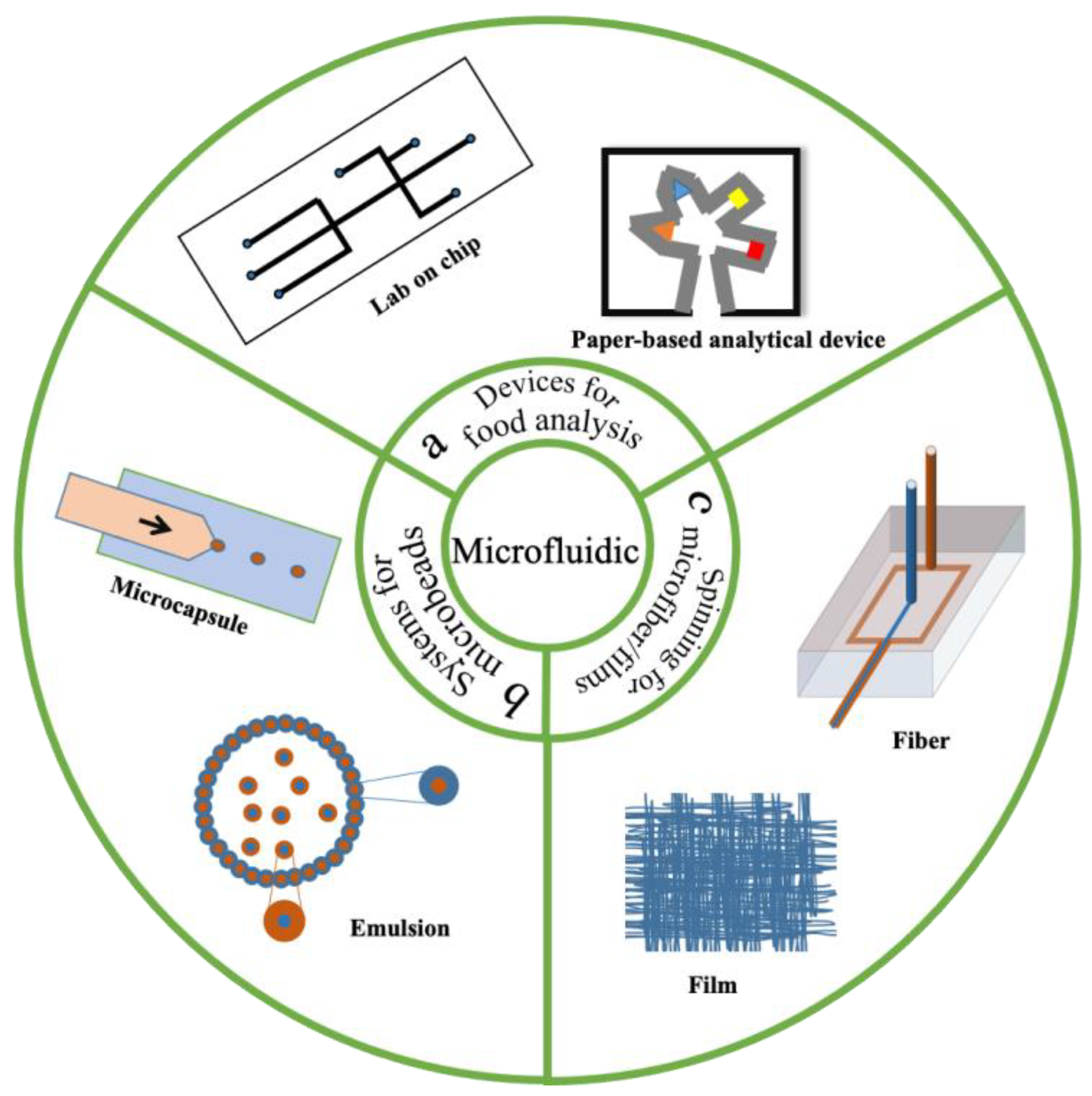
Microfluidics is an advanced technology for manipulating fluid flows in microscale channels (1~100 μm) [18]. A microfluidic system can be defined as a fluid element or chip in which there are many channels on the nanometer to micron scale [19]. These tiny channels give the fluid an interesting and unique character and a wide range of applications in different fields [20]. Microfluidic technology has shown great potential as a new tool in food science (Figure 1) [21,22]. Microfluidic technology can produce various shapes of micro–nano structures, including particles and fibers. Microfibers fabricated via microfluidic spinning have been used as microreactors for analyzing the chemical components [23], and microfilms have potential applications as functional packaging for food products [24]. Droplet-based microfluidics is an advanced technology to produce particles [25]. By controlling the wetting of the channel walls, this technology can generate stable emulsions that are widely applied in food systems [26]. In addition, droplet microfluidic strategy is used to manufacture advanced particles, which are useful structures for the encapsulation and release of active substances [27].
One previous review summarized the applications of microfluidic systems in food analysis [28]. Microfluidic technologies provide high-throughput and large-scale analysis via the integration of multiple steps, multiplexing, and the parallelization of analyses on a single device. Most analytical methods of components in food systems have been reviewed, such as major/minor nutrients, pathogens, toxins, and allergens, and thus are not included in this review. This review investigates the materials, design, and microchannel arrangement of microfluidic chips and their applications. This review focuses on the construction of microfluidic systems and their applications on micro-/nanofabrications in food quality assurance. The system is divided into two parts: The first one is microfluidic spinning technology, which can produce micro-/nanofibers and films. The second is droplet microfluidics, which can produce micro-/nanoemulsions and capsules.
2. Systems for Microfluidics
2.1. Microchannel Arrangements in Microfluidic Platforms
Microfluidics integrates basic operation units of sample fabrication, reaction, separation, and detection in the process of biological, chemical, and medical analysis into a microchip to automatically complete the whole analysis or preparation process [29]. The whole system for microfluidics is a combination of various unit devices that may include injectors (with a micropump), tubes and chips for the fluid flows, external stimuli systems for solidifications, and detection systems to collect signals from analysis [30]. The design of the microchannels of microfluidic chips is the main in many applications [31,32,33]. It is the key for achieving rapid analysis and multiple chemical reactions to produce various fibers and particles. The devices used for microfluidics have been developed using various materials. These devices exist in many forms, such as pulled-glass micropipettes, PDMS microchannels, metal needles, and tubes. Table 1 summarizes the platforms and microchannels applied (or potentially applied) in the fields of food processing and analysis.
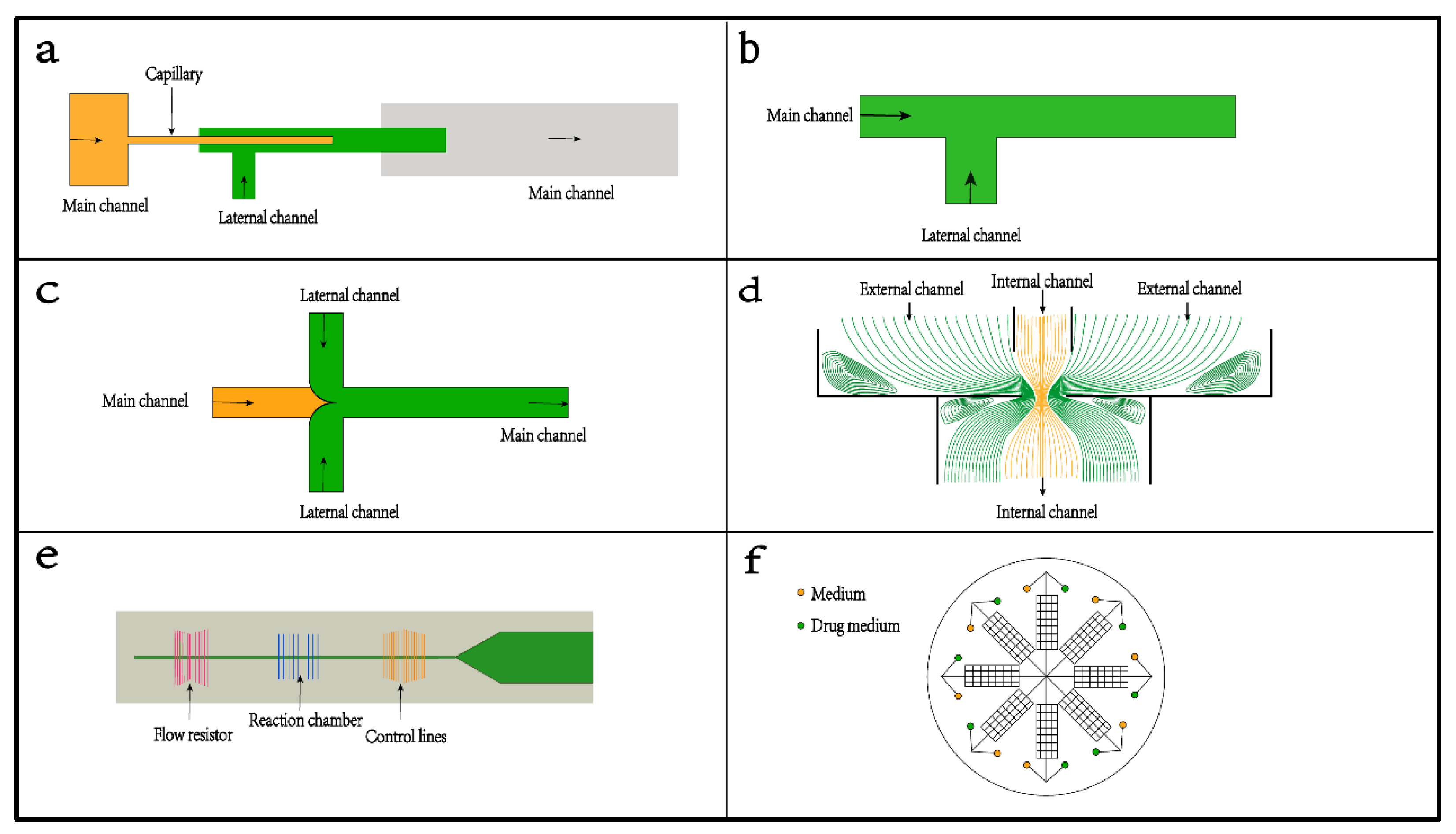
Microchannels are helpful to control the flow parameters in the process of coaxial flow [65]. Figure 2 illustrates various designs of microfluidic platforms. Capillary-based platforms (Figure 2a) are favored for creating coaxial flows for the production of microparticles and reactions in channels [66,67,68]. It is very simple; only two kinds of glass capillary tubes with different pipe diameters, glass sheets, syringes, and glue are needed to construct a capillary device. It should be noted that the axes of the two glass capillaries should be coaxial, as far as possible, to ensure a more stable and uniform liquid drop. In general, dispersed-phase fluid is introduced into the small-diameter capillary tube, and continuous-phase fluid is introduced outside of the small-diameter capillary tube. However, the fabrication of drawn glass micropipettes is labor-intensive and requires a high level of skill. In addition, those superthin tubes are easily cracked during the experiments [69]. The most developed microfluidic devices are PDMS channels or PDMS-based multifunctional platforms (Figure 2b–d). PDMS has high flexibility and chemical and thermal stability and is biocompatible with biological and medical applications [70,71]. The frequently used geometries are the T-connection (Figure 2b) and the cross-connection of four cross channels (Figure 2c). The T-connection uses the geometric structure at the intersection of the microchannel to make the front edge of the dispersed-phase fluid vertically enter the continuous-phase fluid. At the corner of the intersection, the momentum of the dispersed-phase fluid changes under the action of the continuous-phase fluid and finally becomes unstable. It is continuously sheared into many droplets [72]. Compared with the T-connection, the dispersed-phase in a cross-connection is subjected to continuous and symmetrical shear forces on both sides to produce a focusing effect and is extruded into droplets [73]. Compared with the T-connection, the cross-connections are more stable, convenient to operate, produce a wide range of droplet sizes, and are more likely to produce uniform small droplets much smaller than the channel’s characteristic size. The cross-connections are always filled with solutions and can solidify the polymers in the main channels. Both arrangements can produce microfibers [74,75,76] and microcapsules [77,78,79].
Two (or more) channels can be arranged concentrically to produce a strong extensional flow (Figure 2d). This arrangement is commonly applied for the formation of multiple emulsions and foams, which can be realized by one-step emulsification. Moreover, the stability of droplets can be improved. It has been proven that the coaxial channels have multifunctional advances: (1) Coaxial channels allow multiphase flows by confinement in microcapillaries and emulsification to multiple emulsion drops [80]. (2) A microfluidic platform with a coaxial annular chip interface is favored for the high-throughput production of emulsion droplets with controllable sizes and internal compositions [81]. (3) Coaxial channels can exert longer forces, allow larger deflections, and improve reliability [82]. In addition, a capillary-driven microfluidic chip with various and sophisticated arrangements is a common system for analysis or bioassay (Figure 2e,f). For example, Ezgi Ahi et al. fabricated a capillary-driven microfluidic chip with four continuous chambers for SERS-based hCG detection [83]. Li et al. prepared a stretch-driven microfluidic channel with multiple wing structures for nucleic acid detection [84].
2.2. Materials for Microfluidic Chips
The application of new materials and the combination and configuration of new technologies promote the development of microfluidic systems, which makes them more functional [85,86]. The materials for synthesizing microfluidic chips are mainly divided into the following categories: inorganic materials, organic materials, and composite materials (Table 2).
Inorganic materials were first used as substrates in the early stage of microfluidics and usually had high surface stability, adjustable thermal conductivity, and solvent compatibility. Silicon and glass are the two most commonly used raw materials for chips. Silicon was the most ideal material due to its high possibility and advancement in micromachining technology [87,88,89]. However, silicon is an opaque material and has great limitations in optical detection [90,91]. In contrast, glass has strong light permeability with good surface chemical properties and high pressure resistance [92,93]. Various configurations of glass capillaries are often used in the microfluidic processing of functional microparticles [94]. Although glass materials have many advantages, the real challenge is how to use amorphous glass to prepare high specific surface area and anisotropic structural materials. Low-temperature cofired ceramic (LTCC) with laminar characteristic is another common inorganic material that can be utilized to prepare complex devices [95]. Currently, an LTCC device is usually applied in pharmaceutical analysis and sensors [96].
Organic materials are flexible with low costs. Compared to inorganic materials, they can make the process of microfluidic spin faster and simpler. The commonly used organic materials for microfluidic devices are polystyrene (PS), polyvinyl chloride (PVC), polymethyl methacrylate (PMMA), cycloolefin copolymers, polycarbonate (PC), and polydimethylsiloxane (PDMS) These materials have good surface modification, low thermal conductivity, and compatibility in biomedical applications [97,98]. PDMS microfluidic equipment has permeability to gas, and can therefore be used for long-term cell culture. According to their physicochemical properties, the organic materials used in microfluidic systems can be divided into the following categories: elastic [99], thermoplastic [100], plastic [101], hydrogel [102], and paper-based platforms [103]. While organic materials have many advantages, there are still some challenges in their applications, such as aging, chemical resistance, and their mechanical, optical, and thermal properties [101]. PDMS has high resistance to short-wavelength fluorescence detection, so the sensitivity of detection is much lower than that of glass materials [104].
The application of single materials such as silicon, glass, elastomers, and hydrogels can be combined into a hybrid chip to give full access to their advantages. The well-designed multifunctional complex system is suitable for different environments. Organic modified ceramics is a typical example that is beneficial to many applications of biological microfluidic technology [105]. Currently, the most widely used hybrid materials are based on the combination of PDMS with other materials, such as glass [106,107], SU-8 [108,109], polycarbonate [110], PMMA [111], hydrogel [112] and biodegradable materials [113,114,115]. Those hybrids can be applied in different states. A PDMS/polycarbonate microfluidic system uses a polycarbonate nanoporous membrane to control the fluid flow, which is also suitable for cell culture as a gas diffusion barrier [116]. Recently, scientists have developed an ethylene propylene polyimide film for the synthesis of organic materials. This film has high hardness under high pressures and good operability under low temperatures and is also chemically inert and will not react with most solvents [117].

Table 2.
Summary of the common materials for microfluidic platforms.
| Materials | Optical Clarity | Mechanics | Biocompatibility | Thermostability | Bonding Performance | Formability | Refs | |
|---|---|---|---|---|---|---|---|---|
| Inorganic | Silicon | Good | Medium | Bad | Good | Difficult | Difficult | [118,119] |
| Glass | Bad | Medium | Bad | Good | Difficult | Difficult | [120,121] | |
| LTCC | Bad | Medium | Good | Good | Difficult | Difficult | [95,96] | |
| Organic | PDMS | Good | - | Good | Good | Easy | Easy | [122] |
| PMMA | Good | Good | Good | Medium | Easy | Easy | [123,124] | |
| PC | Good | Good | Medium | Medium | Easy | Medium | [119,125] | |
| PS | Good | Medium | Bad | Medium | Easy | Easy | [118,119] | |
| PVC | Good | Good | Good | Medium | Easy | Medium | [118,119] | |
| SU-8 | Medium | Good | Good | Good | Easy | Easy | [126,127] | |
| Paper | - | Bad | Bad | Medium | Medium | - | - | [128,129] |
3. Microfluidic Spinning Technology for Micro-/Nanofibers and Films
Microfluidic spinning technology (MST) refers to the preparation of microfibers with different sizes and morphologies from materials with a certain viscosity under the action of gravity by changing the fluid driving force and the drawing force of the receiver. MST has become a powerful and widely used platform due to its high specific surface area, effective heat transfer, and high reaction rate [130]. Microstructure fiber is very important because of its wide application, such as in microreactors, optical sensors, and biomaterials [23]. The multifunctional properties of microfluidic spun fiber show many potential applications in food science and technology. [131]. The advantages of microfluid devices for the fabrication of microfibers are as follows: (1) They are good systems to produce core–shell fibers or emulsion fibers to protect many sensitive components. (2) It is possible for the composites of different materials to enhance the mechanics of fibers and therefore form films for food packaging. (3) To make the best use of microfilms, MST can introduce bioactive molecules into the fibers to improve their functionality (such as antioxidation and antimicrobial properties). (4) Functional microfibers/films produced from MST are also good systems for food analysis and solvent purification. Figure 3 and Table 3 briefly illustrate the fabrication of microfibers via microfluidic spinning technology and its potential applications in food processing and analysis.
3.1. Solidification Methods for Microfiber Generation
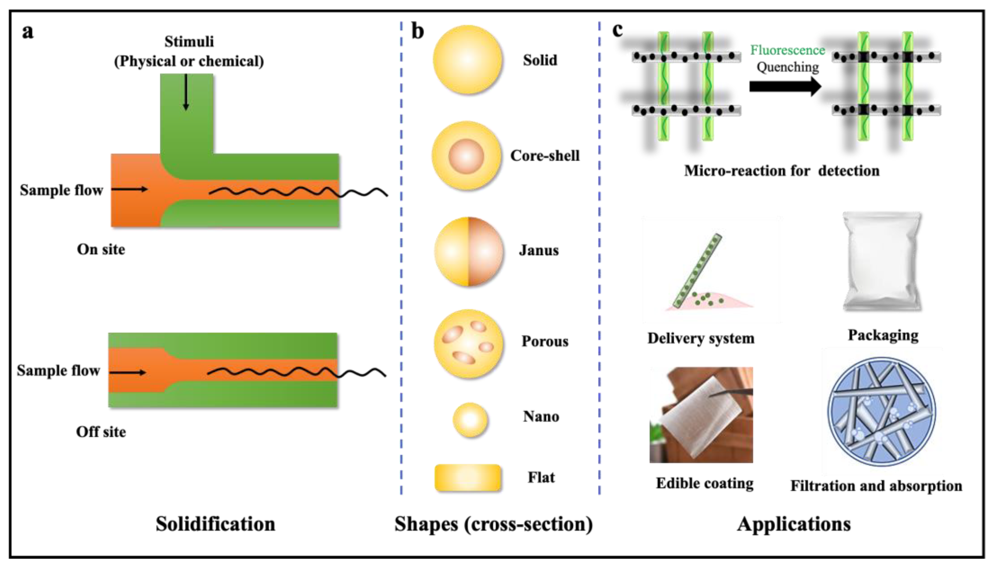
Solidification is critical for the formation of microfibers. The diversity of approaches to solidify polymer solutions to generate functional microfibers/films has been investigated for a long time. Two reviews simply divided the methods of solidification into photopolymerization and chemical reactions [132]. Photopolymerization mainly uses UV light to crosslink prepolymers such as poly (ethylene glycol), diacrylate (PEG-DA), and 4-hydroxybutyl acrylate (4-HBA) then generate the solidified microfibers. Chemical reactions always introduce a crosslinking agent to react with prepolymers and form solid fibers. For example, sodium alginate is crosslinked due to the diffusion of calcium ions into a sodium alginate solution. In their review, the solidification approaches were divided into chemical reactions and physical processes. In this classification, photopolymerization was collected into the chemical reactions, as chemical reactions happen during the polymerizations. The physical processes include ionic crosslinking, solvent exchange, non-solvent-induced phase separation, and solvent evaporation. Based on the solidification time during the fabrication of the microfibers, we divided them into two groups (Figure 3a). The on-site solidification shows that the microfibers are solidified inside the devices. A physical or chemical stimulus (such as UV light, ions, reactive agents, etc.) is applied during the fabrication of the microfibers. The off-site solidification does not apply any physical or chemical approach inside the devices. This fabrication approach always needs a solid receiver for the microfibers because they can only be solidified after solvent evaporation. Many food materials have been used for the solidification of microfibers, such as alginate, chitosan, glucomannan, and silk proteins. Those fibers with various shapes and functions were applied in different areas of science and engineering.
3.2. Design Principle of Fibers with Different Shapes
MST is distinguished by constructing micro-/nanofibers with complex shapes (solid, core shell, Janus, hollow, nano, flat, etc.) by simply regulating the channel shape, solvent type, solidification methods, flow rates, types, concentration of spinning solution, etc. [133].
It is convenient to prepare solid-shaped fibers through a single channel and a coaxial multichannel. The size of fibers is commonly influenced by the dimensions of channels, the flow rates of spinning solutions, and the solidification methods. In general, the size of a fiber mostly depends on the size of the spinning fluid in the channel in the range of micrometers to one hundred micrometers [130]. When using different solidification methods, such as on-site photopolymerization, it can also effectively adjust the size of fibers by altering the intensity of irradiation and irradiation time [134]. The on-site ionic crosslinking-assisted fabrication of biological fibers presents potential advantages in the fast gelation process. The Ca2+ cross-linker is one of the most commonly applied agents for this gelation process. It has been used for the solidification of mixed carbon nanotubes and sodium alginate to fabricate nanofiber-based macroscopic cables [135]. In addition, the size of fibers was also influenced by the types of solvents and the concentrations of spinning solutions [136]. One recent study prepared silk fibers with sizes ranging from 54.1 to 102.2 nm by non-solvent-induced phase separation [137].
It is necessary to add other capillary tubes for the injection of different fluids to generate core–shell and hollow fibers. In a previous report, researchers constructed hollow fibers utilizing three microchannels, where the innermost tube was filled with a CaCl2 solution. The second inner tube contained a sodium alginate solution, and the outermost flowed with a CaCl2 solution. The hollow fiber was formed by two stages of gelation at the intersection of the tubes, where the core sodium alginate solution gelled with the CaCl2 solution in the innermost tube, followed by gelation with the CaCl2 solution in the outermost tube [138]. Based on hollow fibers, core–shell fibers can also be prepared by MST by combining a curable core fluid and a polymerizable shell fluid. For instance, Daniele et al. produced UV-assisted core–shell fibers composed of polyethylene glycol dimethacrylate (shell) and gelatin (core) [139]. To further precisely manipulate the thickness of each layer of the core–shell fibers, the multiphase flow rates should be strictly controlled. Researchers found that decreasing the concentration of gelatin could lead to a shape change for the fibers. Janus fibers can be generated by multiphase microfluidic platforms composed of outer sheath flow inlets and sample flow inlets. One common approach for the preparation of Janus fibers is to inject a photocurable PU solution into the central microchannel and simultaneously inject a sodium dodecyl sulfate solution into the outer sheath microchannels. Hence, Janus fibers were formed under UV irradiation [140]. Flat-shaped fibers can also easily be produced by MST. Lee and coworkers investigated whether MST can fabricate microbelt-shaped fibers consisting of poly(ethylene glycol) diacrylate. During the processing, several poly(ethylene glycol) diacrylate streams were simultaneously introduced into inlets and generated multiphase jetting without the engulfment phenomenon due to their similar compositions [141].
3.3. Applications of Microfibers in Analysis and Encapsulations
Microfibers with diverse shapes (such as cylindrical, flat, grooved, porous, core–shell, etc.) (Figure 3b) fabricated via MST perform many functions (Table 3). The most easily prepared cylindrical microfibers allow chemical reactions to be performed inside to form microreactors or sensors for analysis. Yu et al. described a high-throughput photofluidic platform of mosaic patterned microfibers by generating a layered laminar flow and prepared mosaic microfibers with the desired configuration for multiple biomolecular analyses [142]. Mu et al. proposed an MST for the fabrication of microfibers with KGM and sodium polyacrylate (PAAS) [23].
The prepared microfibers can be easily arranged into microarrays and microgrids, which provides a useful platform for amine molecular recognition. The function of microfluidic fibers also depends on the inclusion of functional components. The encapsulation of functional components is one of the commonly used methods for the functionalization of microfibers, which can further expand the application prospect of microfibers. In general, functional components are added to a curable precursor solution to obtain a uniform suspension or a uniform solution as a sample stream. Then, the fibers with expected properties are obtained by MST [130]. By encapsulating various functional components, those microfibers obtain unique properties that are applied in tissue engineering and the biomedical area. It has been well-investigated that the microfibers produced via MST are good delivery systems for cells. One study successfully prepared chitosan–alginate fibers carrying human liver cancer cells (HepG2) using a coaxial flow microfluidic chip [143]. Another recent work described a new alginate microfiber production method using microfluidic technology to precisely regulate microfibers, thereby improving the vitality and function of embedded cells [144]. In order to protect the transplanted islets immunologically, an immunoprotected ultrafine fiber has been newly designed for successful islet transplantation [145]. The most commonly used alginate-Ca2+ solidification produced many functional microfibers that can potentially be applied in food science. For example, Chaurasia and Sajjadi made alginate fibers loaded with oily objects through the arranged internal capillaries, showing the potential for better triggering reactions [146]. Meng et al. reported an effective microfluidic method for the successive production of hollow calcium alginate microfibers with controlled structure and function, and the addition of active components to the core stream for encapsulation [147]. Pham et al. proposed a simple method for the self-assembly of hollow alginate microfibers based on a PDMS microfluidic device [148]. The inner diameter and wall thickness of the microfibers were controlled by changing the flow velocity of the core and sheath in the microfluidic channel.
Table 3.
Summary of the applications of micro-/nanofibers and films in food processing and analysis.
Table 3.
Summary of the applications of micro-/nanofibers and films in food processing and analysis.
| Materials | Solidification | Shape | Functional Component | Applications | Refs |
|---|---|---|---|---|---|
| Konjac glucomannan/polyvinylidene fluoride | Off-site | Solid | Epigallocatechin-3-gallate | Drug release | [63] |
| Konjac glucomannan/sodium polyacrylate | Off-site | Solid | Ofloxacin | Microreactors | [23] |
| Konjac glucomannan/polylactic acid | Off-site | Solid | Trans-cinnamic | Food packaging | [24] |
| Konjac glucomannan/poly(ε-caprolactone) | Off-site | Solid | Silver nanoparticles | Food packaging | [149] |
| Konjac glucomannan/poly (methyl methacrylate) | Off-site | Solid | Chlorogenic acid | Food packaging | [150] |
| Ethyl cellulose/polyvinylpyrrolidone | Off-site | Solid | - | Food packaging | [64] |
| Polyurethane/sodium dodecyl sulfate | On-site | Janus | - | 3D scaffold | [83] |
| Graphene oxide/ bacterial cellulose | On-site | Core–shell | Silver nanoparticles | Antibacterial | [151] |
3.4. Applications of Microfilms as Food Packaging and Purification Systems
Microfibers can be further integrated to form functional films and exhibit various properties such as high mechanical, antioxidation, and antimicrobial properties. These microfilms have potential applications in food packaging [152]. In our group, we prepared microfilms based on a new kind of biopolymer, KGM, which also promotes wound healing through new blood vessel formation and the advanced development of hair follicles [153]. We further adopted simple and green strategies and built a series of triple-layer microfilms, such as a konjac glucomannan/polylactic acid/anti-cinnamic acid micromembrane (KPTMF) [24] and a konjac glucomannan/poly (methyl methacrylate)/chlorogenic acid food packing film inspired by the amphiphilic theory [150]. In addition to the packaging, microfilms produced via MST are also applied in purifications. Inspired by the conformation of spider silk, He et al. manufactured ultrafine fibers with adjustable magnetic spindle-knotted microfibers [154]. Those fibers contained magnetic Fe3O4 nanoparticles for controlled 3D assembly and water collection. Wu et al. utilized microfluidic emulsification and spinning synergy technology to prepare spindle-knotted graphene microfibers that can absorb oil in the environment of a water–oil mixture [155]. Graphene oxide (GO) fibers with a spindle junction structure and photothermal response phase change behavior are manufactured in microfluidics through spinning and emulsification programs, which can realize fog capture and near-infrared-triggered water collection applications [156].
4. Droplet Microfluidics for Micro-/Nanoemulsions and Capsules
The continuous production and processing of monodisperse microparticles have always been a scientific and technical issue. The first advantage of microfluidic equipment in the fabrication of food products is that it can provide a suitable processing environment for fluids. In microchannels, food scientists can obtain stable microparticles in a homogeneous emulsion by controlling the fluid to give them corresponding physical and chemical properties [157,158]. Therefore, microfluidics has become a widely used advanced technology for the preparation of microparticles. Another advantage is that this technology can not only prepare single-component gel particles but can also prepare mixed-component particles with specific structures and characteristics. In materials science, droplet microfluidics has been applied to fabricate “Janus” microbeads [159], core–shell microcapsules [42], porous microparticles [160], and photosensitive [161] and thermosensitive [162] capsule particles. The third advantage is that the stop and flow technology of a microfluidic system in a flat plate is creating a revolutionary change in the field of material processing [163]. Traditional material processing technology can only synthesize spherical materials because of the effect of surface tension in the emulsion system. In microfluidics, fluid can be projected into different patterns in the process of microchannel chip flow [164]. This technology has been applied to the preparation of many microparticles with various shapes, including cell-loaded microparticles, some column-shaped evolution particles, high-throughput particle processing, and combination with fluid aggregation or a large-scale array [165]. Additionally, the use of a microfluidic nebulizer can achieve the application of ultrasonic spray-drying technology, synthesized RNA-loaded lipid nanoparticles, and microreactors for controlling the physical and chemical changes in the material synthesis process [166]. Figure 4 and Table 4 illustrate droplet microfluidics for emulsions and microcapsules.
4.1. Principle of Droplet Microfluidics
The fluid behavior in droplet microfluidics is mainly divided into laminar flow and droplets [167]. Hence, it is necessary to introduce a driving force to push the fluid into a microchannel. Pressure is the one of the common driving forces urging the flow at the import and export of the channel. We can simply adjust the parameters of the syringe pump to control the driving forces applied to the fluid. Apart from external driving forces, the flow of fluid can be promoted by its gravity [168]. When introducing multiphase fluids into a microchannel, a droplet is generated by the energy from the breakup of fluids, which provides interfacial energy for the production of emulsions [169]. In this process, there are two types of methods: passive methods (e.g., coflow, cross-flow, and flow-focusing) and active methods (e.g., electrical, magnetic, thermal, and mechanical methods), which are mainly distinguished by the existence of external energy. The principle of droplet microfluidics was reviewed in detail in a previous report [26].
Microcapsules and emulsions generated by droplet microfluidics have been widely applied in the field of encapsulation and release. In order to construct microcapsules and emulsions, a T-junction or cross-junction is usually needed, where the core materials flowing into the main tube are wrapped by the core materials flowing into lateral tubes. It is feasible to regulate the size and number of inner droplets by controlling the flow rates. Apart from the assembly of droplets, designing the multistructure of droplet microfluidic devices is another effective strategy. A capillary device combined with continuous narrowing expansion junctions is an applicable example. In brief, oil droplets are first produced by flow-focusing. Then, oil droplets flow across continuous narrowing expansion junctions, where the spherical oil droplets are first squeezed at the narrow export, allowing the inclusion of water, then relaxed into a spherical shape at the expansive import, and eventually formed into double emulsions [170]. Another strategy is to combine “co-flow” and “flow-focusing into” in one capillary device, where two immiscible fluids flow coaxially and another outer immiscible fluid flows in the opposite direction [171]. When three fluids flow simultaneously through a tapered tube, double emulsions are formed.
4.2. Droplet Microfluidics for Microcapsules
Microparticles are becoming increasingly important tools for a wide range of applications in food science, such as probiotic and nutrient delivery [78,172]. Microencapsulation is an important technology for sustaining cell viability and nutrient activity during food processing [10,11]. Microencapsulation fixes the surface of the emulsion to enhance the stability of the emulsion and cover the solid, liquid, or gas in a shell to protect or isolate the material in the shell, prevent the external environment from producing adverse or toxic effects on the material in the shell, and control the release of the material in the shell [173]. The core of a microcapsule is generally the cavity or the active substance dispersed in the cavity, which can be one or multiple substances. The shells may be single, double, or multiple layers and are usually made of organic polymers [174]. The capsule shell can not only protect the active ingredient from the damage of pH change, oxidation, water, and other environmental pressures but also regulates the release of the active ingredient. The microcapsule shape is relatively rich, including spherical, a grain shape, and amorphous, but a spherical structure is the most common structure. Microcapsules can be divided into mononuclear microcapsules, multicompartment microcapsules, irregular microcapsules, etc.
The development of a new type of biomaterial is an urgent need for the application of food science. Especially in the field of food science, using low-cost and biocompatible raw materials is a challenging task [175]. Polysaccharides are a kind of robust biopolymer extracted from natural sources. Several of them are ideal materials for application in microfluidic technology, such as sodium alginate [78], chitosan [176], and konjac glucomannan [24]. Among various possibilities, alginate gel particles are widely used. They are nontoxic, biocompatible, biodegradable, inexpensive, and relatively simple to produce, especially as carriers of microencapsulated compounds [177,178,179].
4.3. Droplet Microfluidics for Emulsions
Food emulsions exist in most food systems [180,181,182]. The easiest double emulsion system from microfluidics has long been applied to stabilize the sensitive compounds in food. For example, Xu and Nisisako fabricated double emulsions using a microcapillary device containing a single internal droplet in a core–shell geometry [183]. Nam et al. utilized poly(ethylene glycol) diacrylate (PEGDA) and droplet microfluidics to obtain the direct encapsulation of water and oil droplets in PEG microcapsules [184]. Recently, one study compared the emulsion systems between microfluidics and the conventional homogenization method. The microcapsules obtained via microfluidics exhibited many advances, such as monodispersity, the stability of physical and chemical properties, and the tracking ability of shell crosslinking [44]. To obtain more stability and various functions of the emulsions, multiple layers of emulsions were developed in food systems. For example, Kantak et al. described a new microfluidic technology that utilizes a microcolumn in the channel to continuously generate, encapsulate, and guide layer-by-layer (LBL) polyelectrolyte microcapsules [185]. In less than 3 min, six hydrogen-bonded polyelectrolytes (three double layers) were adsorbed on each droplet. A new technology recently introduced an ultrathin water layer between two phases in a triple emulsion and achieved high packaging efficiency of hydrophobic goods in a hydrophobic polymer shell directly dispersed in water [186].
Table 4.
Summary of the droplet microfluidics for emulsions and microcapsules in food processing and analysis.
Table 4.
Summary of the droplet microfluidics for emulsions and microcapsules in food processing and analysis.
| Shell Materials | Embedding Materials | Type | Applications | Refs |
|---|---|---|---|---|
| Polycaprolactone | Chlorophyll | Microparticle | Drug encapsulation | [47] |
| Starch | Nisin | Nanoparticle | Drug encapsulation | [48] |
| Silk fibroin/chondroitin sulfate/alginate | Bovine serum albumin/polystyrene latex | Microgel | Drug delivery | [51] |
| 4,4-methylenediphenyl diisocyanate/ethylenediamine | Pendimethalin | Microcapsule | Drug delivery | [187] |
| Chitosan | Curcumin/catechin | Microcapsule | Drug delivery | [188] |
| Sodium alginate | Sucralfate | Microcapsule | Intestinal barriers | [189] |
| Zein | Nisin | Microcapsule | Drug encapsulation | [190] |
| Zein | Lecithin | Microcapsule | Drug delivery | [191] |
| N-isopropylacrylamide/methacrylic acid | Lumogen Red | Microcapsule | Drug delivery | [192] |
| Liposomes | Plasminogen activator | Liposome | Drug encapsulation | [193] |
| Sodium alginate/gelatin | Vitamin A | O/W emulsion | Drug encapsulation | [194] |
| Sodium alginate/cellulose nanocrystals | Oil | O/W emulsion | Lipophilic compound delivery | [195] |
| Gelatin | β-carotene | O/W emulsion | Drug encapsulation | [196] |
| Polyvinyl alcohol | Rifampicin | W/O/W emulsion | Drug delivery | [27] |
| PDMS-b-PDMAEMA | Sucrose/catechin | W/O/W emulsion | Drug encapsulation | [34] |
| Sodium alginate | Phycocyanin | W/O/W emulsion | Drug delivery | [42] |
| Sodium alginate/calcium–ethylenediaminetetraacetic acid | Oil | O/W/O emulsion | Lipophilic compound delivery | [197] |
| Silica nanoparticles/poly(diallyldimethylammoniumchloride)/polystyrene sodium sulfate | Trypsin | W/W emulsion | Enzyme delivery | [198] |
5. Conclusions and Perspectives
Microfluidics has been defined for more than 60 years. Due to the unique fluid properties in microscale environments, this technology is widely applied in food processing. Microfluidics can produce fine droplets to stabilize the multicomponents of dispersed systems, which is very beneficial for the design of novel emulsion-based foods. Compared to the traditional phase-dispersion processing, microfluidics is more effective in the use of energy as well as the control of the shape, size, and size distribution of components. It has the potential to significantly change the processing of dispersed food systems. In addition to emulsions, microfluidics is also an efficient approach to prepare solidified microcapsules for protecting sensitive objects during food processing and storage. Microfibers/films fabricated by microfluidic spinning technology with high mechanical properties can potentially be applied as functional packaging for various food products. The bioactive components dispersed or embedded in the fibers/films are critical factors for food preservation and analysis. On-chip microfluidic systems also provide high-throughput and large-scale analysis by integrating multiple steps, multiplexing, and parallel analysis in a single device.
It is obvious that the microfluidics can be applied in many fields of food science. However, most current applications of this new technology are in the development of functional emulsions. Fibers and films are still in a lab-scale stage, and there are still many attempts expected from food scientists to scale-up and expand its influences in food science: (1) Microcapsules produced via microfluidics have been applied for embedding many sensitive components in food systems. However, those applications are used far less than traditional methods. More components can be stabilized by introducing this easy-to-use, stable, and cost-effective technology to produce functional foods. (2) Microfluidic spinning technology is an efficient way to fabricate microfibers/films. Their potential applications as functional packaging and microanalysis systems have already been demonstrated by recent studies from our groups and other peers. However, the solidification of biopolymers (such as proteins and polysaccharides) and their stabilities during the processing and application of micro-/nanofibers is still a challenge. More investigations need to be focused on the design of biopolymers to produce stable fibers and films. (3) There is no doubt that both MST and droplet microfluidics have aroused wide attention in the field of food science and technology. However, most applications are currently in the laboratory stage of development. One challenge for large-scale production in industry is the design of the microfluidic equipment. In a lab, a chip can be easily produced for the fabrication of microfibers and droplets. One cannot guarantee that the same design of microchannels will be applicable in industry. Another challenge is productivity of the microfluidic equipment. Demonstration is always needed in fundamental research, but mass production is needed in industry. Although some strategies can fulfill the requirement of ultrahigh throughput, much more effort should be devoted to exploring the manipulation of the fiber and droplet generation under complex flows in an extended scale of microchannels in industry.
Author Contributions
Conceptualization, R.M.; investigation, N.B.; Writing—original draft preparation, R.M. and N.B.; Writing—review and editing, L.W. and Y.Z.; Visualization, J.P. and Y.Z.; funding acquisition, R.M., L.W. and J.P. All authors have read and agreed to the published version of the manuscript.
Funding
This work was supported by the Key Project of the Natural Science Foundation of Fujian Province (2022J02021), the Natural Science Foundation from the Fujian Provincial Department of Science and Technology (2020J01556), the Scientific Research Foundation for Distinguished Young Scientists from Fujian Agriculture and Forestry University (xjq202006), and the China Postdoctoral Science Foundation (2022M712180).
Data Availability Statement
Not applicable.
Conflicts of Interest
The authors declare no conflict of interest.
References
- Onyeaka, H.; Passaretti, P.; Miri, T.; Al-Sharify, Z.T. The safety of nanomaterials in food production and packaging. Curr. Res. Food Sci. 2022, 5, 763–774. [Google Scholar] [CrossRef] [PubMed]
- Knorr, D.; Augustin, M.A. Food processing needs, advantages and misconceptions. Trends Food Sci. Technol. 2021, 108, 103–110. [Google Scholar] [CrossRef]
- Scholten, E. Composite foods: From structure to sensory perception. Food Funct. 2017, 8, 481–497. [Google Scholar] [CrossRef] [PubMed]
- Zhang, R.; Belwal, T.; Li, L.; Lin, X.; Xu, Y.; Luo, Z. Recent advances in polysaccharides stabilized emulsions for encapsulation and delivery of bioactive food ingredients: A review. Carbohydr. Polym. 2020, 242, 116388. [Google Scholar] [CrossRef] [PubMed]
- Cui, F.; Zhao, S.; Guan, X.; McClements, D.J.; Liu, X.; Liu, F.; Ngai, T. Polysaccharide-based Pickering emulsions: Formation, stabilization and applications. Food Hydrocoll. 2021, 119, 106812. [Google Scholar] [CrossRef]
- Hu, C.; Zhang, W. Micro/nano emulsion delivery systems: Effects of potato protein/chitosan complex on the stability, oxidizability, digestibility and β-carotene release characteristics of the emulsion. Innov. Food Sci. Emerg. Technol. 2022, 77, 102980. [Google Scholar] [CrossRef]
- Dai, J.; Sameen, D.E.; Zeng, Y.; Li, S.; Qin, W.; Liu, Y. An overview of tea polyphenols as bioactive agents for food packaging applications. LWT 2022, 167, 113845. [Google Scholar] [CrossRef]
- Wu, M.; Dong, Q.; Ma, Y.; Yang, S.; Zohaib Aslam, M.; Liu, Y.; Li, Z. Potential antimicrobial activities of probiotics and their derivatives against Listeria monocytogenes in food field: A review. Food Res. Int. 2022, 160, 111733. [Google Scholar] [CrossRef]
- Cao, G.; Bu, N.; Zeng, T.; Sun, R.; Mu, R.; Pang, J.; Wang, L. Development of pH-responsive konjac glucomannan/pullulan films incorporated with acai berry extract to monitor fish freshness. Int. J. Biol. Macromol. 2022, 219, 897–906. [Google Scholar] [CrossRef]
- Pan, L.-H.; Chen, L.-P.; Wu, C.-L.; Wang, J.-F.; Luo, S.-Z.; Luo, J.-P.; Zheng, Z. Microencapsulation of blueberry anthocyanins by spray drying with soy protein isolates/high methyl pectin combination: Physicochemical properties, release behavior in vitro and storage stability. Food Chem. 2022, 395, 133626. [Google Scholar] [CrossRef]
- Sridhar, K.; Bouhallab, S.; Croguennec, T.; Renard, D.; Lechevalier, V. Application of high-pressure and ultrasound technologies for legume proteins as wall material in microencapsulation: New insights and advances. Trends Food Sci. Technol. 2022, 127, 49–62. [Google Scholar] [CrossRef]
- Shlush, E.; Davidovich-Pinhas, M. Bioplastics for food packaging. Trends Food Sci. Technol. 2022, 125, 66–80. [Google Scholar] [CrossRef]
- Panda, P.K.; Sadeghi, K.; Seo, J. Recent advances in poly (vinyl alcohol)/natural polymer based films for food packaging applications: A review. Food Packag. Shelf Life 2022, 33, 100904. [Google Scholar] [CrossRef]
- Fu, Y.; Dudley, E.G. Antimicrobial-coated films as food packaging: A review. Compr. Rev. Food Sci. Food Saf. 2021, 20, 3404–3437. [Google Scholar] [CrossRef] [PubMed]
- He, Y.; Bai, X.; Xiao, Q.; Liu, F.; Zhou, L.; Zhang, C. Detection of adulteration in food based on nondestructive analysis techniques: A review. Crit. Rev. Food Sci. Nutr. 2021, 61, 2351–2371. [Google Scholar] [CrossRef]
- Torre, R.; Freitas, M.; Costa-Rama, E.; Nouws, H.P.A.; Delerue-Matos, C. Food allergen control: Tropomyosin analysis through electrochemical immunosensing. Food Chem. 2022, 396, 133659. [Google Scholar] [CrossRef]
- Tarhan, Ö.; Jafari, S.M. Chapter Seventeen—Spectroscopic and Chromatographic Analyses of Nanoencapsulated Food Ingredients. In Characterization of Nanoencapsulated Food Ingredients; Jafari, S.M., Ed.; Academic Press: Cambridge, MA, USA, 2020; pp. 585–615. [Google Scholar]
- Li, X.; You, B.; Shum, H.C.; Chen, C.-H. Future foods: Design, fabrication and production through microfluidics. Biomaterials 2022, 287, 121631. [Google Scholar] [CrossRef]
- Chen, K.-H.; Liu, C.-C.; Lu, S.-Y.; Chen, S.-J.; Sheu, F.; Fu, L.-M. Rapid microfluidic analysis detection system for sodium dehydroacetate in foods. Chem. Eng. J. 2022, 427, 131530. [Google Scholar] [CrossRef]
- Hong Tham Phan, T.; Kim, S.-J. Super-hydrophobic microfluidic channels fabricated via xurography-based polydimethylsiloxane (PDMS) micromolding. Chem. Eng. Sci. 2022, 258, 117768. [Google Scholar] [CrossRef]
- Jolvis Pou, K.R.; Raghavan, V.; Packirisamy, M. Microfluidics in smart packaging of foods. Food Res. Int. 2022, 161, 111873. [Google Scholar] [CrossRef]
- Schroën, K.; Wu, L.; Corstens, M. Food-grade microgel capsules tailored for anti-obesity strategies through microfluidic preparation. Curr. Opin. Food Sci. 2022, 45, 100816. [Google Scholar] [CrossRef]
- Mu, R.-J.; Ni, Y.; Wang, L.; Yuan, Y.; Yan, Z.; Pang, J.; Chen, S. Fabrication of ordered konjac glucomannan microfiber arrays via facile microfluidic spinning method. Mater. Lett. 2017, 196, 410–413. [Google Scholar] [CrossRef]
- Lin, W.; Ni, Y.; Liu, D.; Yao, Y.; Pang, J. Robust microfluidic construction of konjac glucomannan-based micro-films for active food packaging. Int. J. Biol. Macromol. 2019, 137, 982–991. [Google Scholar] [CrossRef] [PubMed]
- Shao, C.; Chi, J.; Shang, L.; Fan, Q.; Ye, F. Droplet microfluidics-based biomedical microcarriers. Acta Biomater. 2022, 138, 21–33. [Google Scholar] [CrossRef] [PubMed]
- Shang, L.; Cheng, Y.; Zhao, Y. Emerging Droplet Microfluidics. Chem. Rev. 2017, 117, 7964–8040. [Google Scholar] [CrossRef]
- Luo, Z.; Zhao, G.; Panhwar, F.; Akbar, M.F.; Shu, Z. Well-designed microcapsules fabricated using droplet-based microfluidic technique for controlled drug release. J. Drug Deliv. Sci. Technol. 2017, 39, 379–384. [Google Scholar] [CrossRef] [Green Version]
- Pu, H.; Xiao, W.; Sun, D.-W. SERS-microfluidic systems: A potential platform for rapid analysis of food contaminants. Trends Food Sci. Technol. 2017, 70, 114–126. [Google Scholar] [CrossRef]
- Escarpa, A. Lights and shadows on food microfluidics. Lab Chip 2014, 14, 3213–3224. [Google Scholar] [CrossRef]
- Lin, L.; Lin, J.-M. Design and Preparation of Microfluidics Device. In Cell Analysis on Microfluidics; Lin, J.-M., Ed.; Springer: Singapore, 2018; pp. 1–42. [Google Scholar]
- Purwanti, N.; Ichikawa, S.; Neves, M.A.; Uemura, K.; Nakajima, M.; Kobayashi, I. β-lactoglobulin as food grade surfactant for clove oil-in-water and limonene-in-water emulsion droplets produced by microchannel emulsification. Food Hydrocoll. 2016, 60, 98–108. [Google Scholar] [CrossRef]
- Nasseri, B.; Soleimani, N.; Rabiee, N.; Kalbasi, A.; Karimi, M.; Hamblin, M.R. Point-of-care microfluidic devices for pathogen detection. Biosens. Bioelectron. 2018, 117, 112–128. [Google Scholar] [CrossRef]
- Stoukatch, S.; Francis, L.A.; Dupont, F.; Kraft, M. Low-cost microfluidic device micromachining and sequential integration with SAW sensor intended for biomedical applications. Sens. Actuators A Phys. 2021, 319, 112526. [Google Scholar] [CrossRef]
- Bodin-Thomazo, N.; Malloggi, F.; Pantoustier, N.; Perrin, P.; Guenoun, P.; Rosilio, V. Formation and stabilization of multiple w/o/w emulsions encapsulating catechin, by mechanical and microfluidic methods using a single pH-sensitive copolymer: Effect of copolymer/drug interaction. Int. J. Pharm. 2022, 622, 121871. [Google Scholar] [CrossRef] [PubMed]
- Escareño, N.; Hassan, N.; Kogan, M.J.; Juárez, J.; Topete, A.; Daneri-Navarro, A. Microfluidics-assisted conjugation of chitosan-coated polymeric nanoparticles with antibodies: Significance in drug release, uptake, and cytotoxicity in breast cancer cells. J. Colloid Interface Sci. 2021, 591, 440–450. [Google Scholar] [CrossRef] [PubMed]
- Vit, F.F.; Nunes, R.; Wu, Y.T.; Prado Soares, M.C.; Godoi, N.; Fujiwara, E.; Carvalho, H.F.; Gaziola de la Torre, L. A modular, reversible sealing, and reusable microfluidic device for drug screening. Anal. Chim. Acta 2021, 1185, 339068. [Google Scholar] [CrossRef] [PubMed]
- Pang, B.; Fu, K.; Liu, Y.; Ding, X.; Hu, J.; Wu, W.; Xu, K.; Song, X.; Wang, J.; Mu, Y.; et al. Development of a self-priming PDMS/paper hybrid microfluidic chip using mixed-dye-loaded loop-mediated isothermal amplification assay for multiplex foodborne pathogens detection. Anal. Chim. Acta 2018, 1040, 81–89. [Google Scholar] [CrossRef] [PubMed]
- Zhao, M.; Li, X.; Zhang, Y.; Wang, Y.; Wang, B.; Zheng, L.; Zhang, D.; Zhuang, S. Rapid quantitative detection of chloramphenicol in milk by microfluidic immunoassay. Food Chem. 2021, 339, 127857. [Google Scholar] [CrossRef]
- Lin, J.J.; Yang, D.; Ou, S.J.L.; Mak, Y.Y.; Lee, D.P.S.; Lim, K.L.; Tai, E.S.; Liu, M.H.; Khan, S.A. Creating texturally tuneable, low calorie and palatable noodle-like food assemblies via microfluidics. Food Hydrocoll. 2022, 127, 107544. [Google Scholar] [CrossRef]
- Mesquita, C.R.S.; Charelli, L.E.; Baptista, L.S.; Naveira-Cotta, C.P.; Balbino, T.A. Continuous-mode encapsulation of human stem cell spheroids using droplet-based glass-capillary microfluidic device for 3D bioprinting technology. Biochem. Eng. J. 2021, 174, 108122. [Google Scholar] [CrossRef]
- Comunian, T.A.; Ravanfar, R.; Alcaine, S.D.; Abbaspourrad, A. Water-in-oil-in-water emulsion obtained by glass microfluidic device for protection and heat-triggered release of natural pigments. Food Res. Int. 2018, 106, 945–951. [Google Scholar] [CrossRef]
- Wang, X.; Zhu, M.; Wang, K.; He, S.; Shi, X.; Yuan, B.; Dong, B.; Wang, Z. Preparation of core-shell microcapsules based on microfluidic technology for the encapsulation, protection and controlled delivery of phycocyanin. J. Drug Deliv. Sci. Technol. 2022, 72, 103361. [Google Scholar] [CrossRef]
- Othman, R.; Vladisavljević, G.T.; Nagy, Z.K. Preparation of biodegradable polymeric nanoparticles for pharmaceutical applications using glass capillary microfluidics. Chem. Eng. Sci. 2015, 137, 119–130. [Google Scholar] [CrossRef] [Green Version]
- Ravanfar, R.; Comunian, T.A.; Dando, R.; Abbaspourrad, A. Optimization of microcapsules shell structure to preserve labile compounds: A comparison between microfluidics and conventional homogenization method. Food Chem. 2018, 241, 460–467. [Google Scholar] [CrossRef] [PubMed]
- Comunian, T.A.; Abbaspourrad, A.; Favaro-Trindade, C.S.; Weitz, D.A. Fabrication of solid lipid microcapsules containing ascorbic acid using a microfluidic technique. Food Chem. 2014, 152, 271–275. [Google Scholar] [CrossRef] [PubMed]
- Bandara, G.C.; Heist, C.A.; Remcho, V.T. Patterned polycaprolactone-filled glass microfiber microfluidic devices for total protein content analysis. Talanta 2018, 176, 589–594. [Google Scholar] [CrossRef]
- Hsiao, C.-J.; Lin, J.-F.; Wen, H.-Y.; Lin, Y.-M.; Yang, C.-H.; Huang, K.-S.; Shaw, J.-F. Enhancement of the stability of chlorophyll using chlorophyll-encapsulated polycaprolactone microparticles based on droplet microfluidics. Food Chem. 2020, 306, 125300. [Google Scholar] [CrossRef]
- Liu, X.; Ibarra-Sánchez, L.A.; Miller, M.J.; Lee, Y. Fabrication of zein-modified starch nanoparticle complexes via microfluidic chip and encapsulation of nisin. Curr. Res. Food Sci. 2022, 5, 1110–1117. [Google Scholar] [CrossRef]
- Xie, M.; Chen, T.; Xin, X.; Cai, Z.; Dong, C.; Lei, B. Multiplex detection of foodborne pathogens by real-time loop-mediated isothermal amplification on a digital microfluidic chip. Food Control 2022, 136, 108824. [Google Scholar] [CrossRef]
- Zhou, Z.; Kong, T.; Mkaouar, H.; Salama, K.N.; Zhang, J.M. A hybrid modular microfluidic device for emulsion generation. Sens. Actuators A Phys. 2018, 280, 422–428. [Google Scholar] [CrossRef]
- de Carvalho, B.G.; Taketa, T.B.; Garcia, B.B.M.; Han, S.W.; de la Torre, L.G. Hybrid microgels produced via droplet microfluidics for sustainable delivery of hydrophobic and hydrophilic model nanocarrier. Mater. Sci. Eng. C 2021, 118, 111467. [Google Scholar] [CrossRef]
- Sitkov, N.; Zimina, T.; Kolobov, A.; Sevostyanov, E.; Trushlyakova, V.; Luchinin, V.; Krasichkov, A.; Markelov, O.; Galagudza, M.; Kaplun, D. Study of the Fabrication Technology of Hybrid Microfluidic Biochips for Label-Free Detection of Proteins. Micromachines 2022, 13, 20. [Google Scholar] [CrossRef]
- Xie, L.; Zi, X.; Zeng, H.; Sun, J.; Xu, L.; Chen, S. Low-cost fabrication of a paper-based microfluidic using a folded pattern paper. Anal. Chim. Acta 2019, 1053, 131–138. [Google Scholar] [CrossRef] [PubMed]
- Dias, B.C.; Batista, A.D.; da Silveira Petruci, J.F. μOPTO: A microfluidic paper-based optoelectronic tongue as presumptive tests for the discrimination of alkaloid drugs for forensic purposes. Anal. Chim. Acta 2021, 1187, 339141. [Google Scholar] [CrossRef] [PubMed]
- Jin, S.-Q.; Guo, S.-M.; Zuo, P.; Ye, B.-C. A cost-effective Z-folding controlled liquid handling microfluidic paper analysis device for pathogen detection via ATP quantification. Biosens. Bioelectron. 2015, 63, 379–383. [Google Scholar] [CrossRef] [PubMed]
- Trouillon, R.; Gijs, M.A.M. Dynamic electrochemical quantitation of dopamine release from a cells-on-paper system. RSC Adv. 2016, 6, 31069–31073. [Google Scholar] [CrossRef]
- Li, C.; Boban, M.; Tuteja, A. Open-channel, water-in-oil emulsification in paper-based microfluidic devices. Lab Chip 2017, 17, 1436–1441. [Google Scholar] [CrossRef]
- Thompson, B.L.; Birch, C.; Li, J.; DuVall, J.A.; Le Roux, D.; Nelson, D.A.; Tsuei, A.-C.; Mills, D.L.; Krauss, S.T.; Root, B.E.; et al. Microfluidic enzymatic DNA extraction on a hybrid polyester-toner-PMMA device. Analyst 2016, 141, 4667–4675. [Google Scholar] [CrossRef]
- Kaba, A.M.; Jeon, H.; Park, A.; Yi, K.; Baek, S.; Park, A.; Kim, D. Cavitation-microstreaming-based lysis and DNA extraction using a laser-machined polycarbonate microfluidic chip. Sens. Actuators B Chem. 2021, 346, 130511. [Google Scholar] [CrossRef]
- Powell, L.; Wiederkehr, R.S.; Damascus, P.; Fauvart, M.; Buja, F.; Stakenborg, T.; Ray, S.C.; Fiorini, P.; Osburn, W.O. Rapid and sensitive detection of viral nucleic acids using silicon microchips. Analyst 2018, 143, 2596–2603. [Google Scholar] [CrossRef]
- Mishra, R.; Maiti, T.K.; Bhattacharyya, T.K. Development of SU-8 hollow microneedles on a silicon substrate with microfluidic interconnects for transdermal drug delivery. J. Micromech. Microeng. 2018, 28, 105017. [Google Scholar] [CrossRef]
- Ni, Y.; Lin, W.; Mu, R.-J.; Wang, L.; Zhang, X.; Wu, C.; Pang, J. Microfluidic fabrication of robust konjac glucomannan-based microfiber scaffolds with high antioxidant performance. J. Sol-Gel Sci. Technol. 2019, 90, 214–220. [Google Scholar] [CrossRef]
- Ni, Y.; Lin, W.; Mu, R.-J.; Wu, C.; Wang, L.; Wu, D.; Chen, S.; Pang, J. Robust microfluidic construction of hybrid microfibers based on konjac glucomannan and their drug release performance. RSC Adv. 2018, 8, 26432–26439. [Google Scholar] [CrossRef] [PubMed] [Green Version]
- Rao, J.; Shen, C.; Yang, Z.; Fawole, O.A.; Li, J.; Wu, D.; Chen, K. Facile microfluidic fabrication and characterization of ethyl cellulose/PVP films with neatly arranged fibers. Carbohydr. Polym. 2022, 292, 119702. [Google Scholar] [CrossRef] [PubMed]
- Nooranidoost, M.; Haghshenas, M.; Muradoglu, M.; Kumar, R. Cell encapsulation modes in a flow-focusing microchannel: Effects of shell fluid viscosity. Microfluid. Nanofluid. 2019, 23, 31. [Google Scholar] [CrossRef]
- Nikoo, A.H.; Malayeri, M.R. Incorporation of geochemical, geometrical and intermolecular interactions into modelling of scale removal in a capillary microchannel. J. Pet. Sci. Eng. 2022, 208, 109311. [Google Scholar] [CrossRef]
- Wang, L.; Zhong, Y.; Qian, C.; Yang, D.; Nie, J.; Ma, G. A natural polymer-based porous sponge with capillary-mimicking microchannels for rapid hemostasis. Acta Biomater. 2020, 114, 193–205. [Google Scholar] [CrossRef]
- Liu, Z.; Fontana, F.; Python, A.; Hirvonen, J.T.; Santos, H.A. Microfluidics for Production of Particles: Mechanism. Methodol. Appl. Small 2020, 16, 1904673. [Google Scholar] [CrossRef]
- Deshpande, S.; Dekker, C. On-chip microfluidic production of cell-sized liposomes. Nat. Protoc. 2018, 13, 856–874. [Google Scholar] [CrossRef]
- Lüken, A.; Geiger, M.; Steinbeck, L.; Joel, A.-C.; Lampert, A.; Linkhorst, J.; Wessling, M. Biocompatible Micron-Scale Silk Fibers Fabricated by Microfluidic Wet Spinning. Adv. Healthc. Mater. 2021, 10, 2100898. [Google Scholar] [CrossRef]
- Jiang, B.; White, A.; Ou, W.; Van Belleghem, S.; Stewart, S.; Shamul, J.G.; Rahaman, S.O.; Fisher, J.P.; He, X. Noncovalent reversible binding-enabled facile fabrication of leak-free PDMS microfluidic devices without plasma treatment for convenient cell loading and retrieval. Bioact. Mater. 2022, 16, 346–358. [Google Scholar] [CrossRef]
- Bedram, A.; Moosavi, A. Droplet breakup in an asymmetric microfluidic T junction. Eur. Phys. J. E 2011, 34, 78. [Google Scholar] [CrossRef]
- Davies, R.T.; Kim, D.; Park, J. Formation of liposomes using a 3D flow focusing microfluidic device with spatially patterned wettability by corona discharge. J. Micromech. Microeng. 2012, 22, 055003. [Google Scholar] [CrossRef]
- Wang, S.; Dong, S.; Shen, H.; Li, B. Preparation of monodisperse S/W/O compound droplets with thick liquid film via a dual-cross microfluidic device. Colloids Surf. A 2021, 620, 126413. [Google Scholar] [CrossRef]
- Gao, M.; Peng, K.; Pan, T.; Long, F.; Lin, Y. Improving the local thermal conductivity of flexible films by microchannels filled with graphene. Compos. Commun. 2021, 25, 100689. [Google Scholar] [CrossRef]
- Cheng, S.; Chen, W.; Zhang, P. Developing advanced polymer films based on microfluidic laminar flow. Giant 2022, 9, 100091. [Google Scholar] [CrossRef]
- Michelon, M.; Leopércio, B.C.; Carvalho, M.S. Microfluidic production of aqueous suspensions of gellan-based microcapsules containing hydrophobic compounds. Chem. Eng. Sci. 2020, 211, 115314. [Google Scholar] [CrossRef]
- Wang, K.; Ni, J.; Li, H.; Tian, X.; Tan, M.; Su, W. Survivability of probiotics encapsulated in kelp nanocellulose/alginate microcapsules on microfluidic device. Food Res. Int. 2022, 160, 111723. [Google Scholar] [CrossRef]
- Du, J.; Ibaseta, N.; Guichardon, P. Characterization of polyurea microcapsules synthesized with an isocyanate of low toxicity and eco-friendly esters via microfluidics: Shape, shell thickness, morphology and encapsulation efficiency. Chem. Eng. Res. Des. 2022, 182, 256–272. [Google Scholar] [CrossRef]
- Yu, W.; Li, B.; Liu, X.; Chen, Y. Hydrodynamics of triple emulsion droplet generation in a flow-focusing microfluidic device. Chem. Eng. Sci. 2021, 243, 116648. [Google Scholar] [CrossRef]
- Shen, Q.; Zhang, C.; Tahir, M.F.; Jiang, S.; Zhu, C.; Ma, Y.; Fu, T. Numbering-up strategies of micro-chemical process: Uniformity of distribution of multiphase flow in parallel microchannels. Chem. Eng. Process.-Process Intensif. 2018, 132, 148–159. [Google Scholar] [CrossRef]
- Clark, I.; Thakur, R.; Abate, A. Concentric electrodes improve microfluidic droplet sorting. Lab Chip 2018, 18, 710–713. [Google Scholar] [CrossRef]
- Ahi, E.E.; Torul, H.; Zengin, A.; Sucularlı, F.; Yıldırım, E.; Selbes, Y.; Suludere, Z.; Tamer, U. A capillary driven microfluidic chip for SERS based hCG detection. Biosens. Bioelectron. 2022, 195, 113660. [Google Scholar] [CrossRef] [PubMed]
- Li, X.; Zhao, X.; Yang, W.; Xu, F.; Chen, B.; Peng, J.; Huang, J.; Mi, S. Stretch-driven microfluidic chip for nucleic acid detection. Biotechnol. Bioeng. 2021, 118, 3559–3568. [Google Scholar] [CrossRef] [PubMed]
- Rahmanian, M.; Sartipzadeh Hematabad, O.; Askari, E.; Shokati, F.; Bakhshi, A.; Moghadam, S.; Olfatbakhsh, A.; Al Sadat Hashemi, E.; Khorsand Ahmadi, M.; Morteza Naghib, S.; et al. A micropillar array-based microfluidic chip for label-free separation of circulating tumor cells: The best micropillar geometry? J. Adv. Res. 2022. [Google Scholar] [CrossRef] [PubMed]
- Hou, X.; Zhang, Y.; Trujillo de Santiago, G.; Alvarez, M.; Ribas, J.; Jonas, S.; Weiss, P.; Andrews, A.; Aizenberg, J.; Khademhosseini, A. Interplay between materials and microfluidics. Nat. Rev. Mater. 2017, 2, 17016. [Google Scholar] [CrossRef]
- Shakeri, A.; Khan, S.; Didar, T.F. Conventional and emerging strategies for the fabrication and functionalization of PDMS-based microfluidic devices. Lab Chip 2021, 21, 3053–3075. [Google Scholar] [CrossRef]
- Verma, G.; Mondal, K.; Gupta, A. Si-based MEMS resonant sensor: A review from microfabrication perspective. Microelectron. J. 2021, 118, 105210. [Google Scholar] [CrossRef]
- Sahu, A.K.; Malhotra, J.; Jha, S. Laser-based hybrid micromachining processes: A review. Opt. Laser Technol. 2022, 146, 107554. [Google Scholar] [CrossRef]
- Sairam, K.P.; Kumar, D.; Ang, D.S. Highly Transparent ITO/HfO2/ITO Device for Visible-Light Sensing. IEEE Access 2020, 8, 91648–91652. [Google Scholar] [CrossRef]
- Karabchevsky, A.; Katiyi, A.; Ang, A.S.; Hazan, A. On-chip nanophotonics and future challenges. Nanophotonics 2020, 9, 3733–3753. [Google Scholar] [CrossRef]
- Li, K.; Xu, G.; Huang, X.; Chen, Q.; Xie, Z.; Gong, F. Surface evolution analysis of CrxWyNz coatings on WC mold in glass molding process. Surf. Coat. Technol. 2020, 393, 125839. [Google Scholar] [CrossRef]
- Wang, H.; Liu, L.Y.; Ye, P.; Huang, Z.; Ng, A.Y.R.; Du, Z.; Dong, Z.; Tang, D.; Gan, C.L. 3D Printing of Transparent Spinel Ceramics with Transmittance Approaching the Theoretical Limit. Adv. Mater. 2021, 33, 2007072. [Google Scholar] [CrossRef] [PubMed]
- Geng, Y.; Ling, S.; Huang, J.; Xu, J. Multiphase Microfluidics: Fundamentals, Fabrication, and Functions. Small 2020, 16, 1906357. [Google Scholar] [CrossRef] [PubMed]
- He, T.; Ma, M.; Li, H.; Zhang, F.; Liu, F.; Liu, Z.; Li, X. Integrated wireless microfluidic liquid sensors based on low temperature co-fired ceramic (LTCC) technology. Sens. Actuators A Phys. 2022, 346, 113840. [Google Scholar] [CrossRef]
- Montes, R.; Céspedes, F.; Baeza, M. Determination of pesticides using a low-temperature co-fired ceramic microfluidic platform. Instrum. Sci. Technol. 2018, 46, 76–92. [Google Scholar] [CrossRef]
- Sun, W.; Liu, W.; Wu, Z.; Chen, H. Chemical Surface Modification of Polymeric Biomaterials for Biomedical Applications. Macromol. Rapid Commun. 2020, 41, 1900430. [Google Scholar] [CrossRef]
- Lau, K.; Akhavan, B.; Lord, M.S.; Bilek, M.M.; Rnjak-Kovacina, J. Dry Surface Treatments of Silk Biomaterials and Their Utility in Biomedical Applications. ACS Biomater. Sci. Eng. 2020, 6, 5431–5452. [Google Scholar] [CrossRef]
- Han, X.; Zhang, Y.; Tian, J.; Wu, T.; Li, Z.; Xing, F.; Fu, S. Polymer-based microfluidic devices: A comprehensive review on preparation and applications. Polym. Eng. Sci. 2022, 62, 3–24. [Google Scholar] [CrossRef]
- Le, T.N.Q.; Tran, N.N.; Escribà-Gelonch, M.; Serra, C.A.; Fisk, I.; McClements, D.J.; Hessel, V. Microfluidic encapsulation for controlled release and its potential for nanofertilisers. Chem. Soc. Rev. 2021, 50, 11979–12012. [Google Scholar] [CrossRef]
- Rabiee, N.; Ahmadi, S.; Fatahi, Y.; Rabiee, M.; Bagherzadeh, M.; Dinarvand, R.; Bagheri, B.; Zarrintaj, P.; Saeb, M.R.; Webster, T.J. Nanotechnology-assisted microfluidic systems: From bench to bedside. Nanomedicine 2021, 16, 237–258. [Google Scholar] [CrossRef]
- Saez, J.; Catalan-Carrio, R.; Owens, R.M.; Basabe-Desmonts, L.; Benito-Lopez, F. Microfluidics and materials for smart water monitoring: A review. Anal. Chim. Acta 2021, 1186, 338392. [Google Scholar] [CrossRef]
- Bamshad, A.; Cho, H.J. Laserjet Printed Micro/Nano Sensors and Microfluidic Systems: A Simple and Facile Digital Platform for Inexpensive, Flexible, and Low-Volume Devices. Adv. Mater. Technol. 2021, 6, 2100401. [Google Scholar] [CrossRef]
- Hsu, Y.M.; Chang, C.C. The portable fluorescence detection system matched with PDMS microfluidic biochip for DNA hybridization detection. Optik 2015, 126, 2600–2605. [Google Scholar] [CrossRef]
- Lakhera, P.; Chaudhary, V.; Bhardwaj, B.; Kumar, P.; Kumar, S. Development and recent advancement in microfluidics for point of care biosensor applications: A review. Biosens. Bioelectron. X 2022, 11, 100218. [Google Scholar] [CrossRef]
- Campbell, S.B.; Wu, Q.; Yazbeck, J.; Liu, C.; Okhovatian, S.; Radisic, M. Beyond Polydimethylsiloxane: Alternative Materials for Fabrication of Organ-on-a-Chip Devices and Microphysiological Systems. ACS Biomater. Sci. Eng. 2021, 7, 2880–2899. [Google Scholar] [CrossRef] [PubMed]
- Catauro, M.; Ciprioti, S.V. Characterization of Hybrid Materials Prepared by Sol-Gel Method for Biomedical Implementations. A Critical Review. Materials 2021, 14, 1788. [Google Scholar] [CrossRef]
- Qin, Y.; Yeh, P.; Hao, X.; Cao, X. Developing an ultra non-fouling SU-8 and PDMS hybrid microfluidic device by poly(amidoamine) engraftment. Colloids Surf. B 2015, 127, 247–255. [Google Scholar] [CrossRef]
- Xu, B.; Liu, Z.; Lee, Y.-K.; Mak, A.; Yang, M. A PDMS microfluidic system with poly(ethylene glycol)/SU-8 based apertures for planar whole cell-patch-clamp recordings. Sens. Actuators A Phys. 2011, 166, 219–225. [Google Scholar] [CrossRef]
- Ecochard, Y.; Caillol, S. Hybrid polyhydroxyurethanes: How to overcome limitations and reach cutting edge properties? Eur. Polym. J. 2020, 137, 109915. [Google Scholar] [CrossRef]
- Hassanpour-Tamrin, S.; Sanati-Nezhad, A.; Sen, A. A simple and low-cost approach for irreversible bonding of polymethylmethacrylate and polydimethylsiloxane at room temperature for high-pressure hybrid microfluidics. Sci. Rep. 2021, 11, 4821. [Google Scholar] [CrossRef]
- Chang, X.; Chen, L.; Chen, J.; Zhu, Y.; Guo, Z. Advances in transparent and stretchable strain sensors. Adv. Compos. Hybrid Mater. 2021, 4, 435–450. [Google Scholar] [CrossRef]
- Hsieh, Y.-K.; Chen, S.-C.; Huang, W.-L.; Hsu, K.-P.; Gorday, K.A.V.; Wang, T.; Wang, J. Direct Micromachining of Microfluidic Channels on Biodegradable Materials Using Laser Ablation. Polymers 2017, 9, 242. [Google Scholar] [CrossRef] [PubMed] [Green Version]
- Mogosanu, D.-E.; Verplancke, R.; Dubruel, P.; Vanfleteren, J. Fabrication of 3-dimensional biodegradable microfluidic environments for tissue engineering applications. Mater. Des. 2016, 89, 1315–1324. [Google Scholar] [CrossRef]
- Riahi, R.; Tamayol, A.; Shaegh, S.A.M.; Ghaemmaghami, A.M.; Dokmeci, M.R.; Khademhosseini, A. Microfluidics for advanced drug delivery systems. Curr. Opin. Chem. Eng. 2015, 7, 101–112. [Google Scholar] [CrossRef] [PubMed]
- Shi, Y.; Cai, Y.; Cao, Y.; Hong, Z.; Chai, Y. Recent advances in microfluidic technology and applications for anti-cancer drug screening, TrAC. Trends Anal. Chem. 2021, 134, 116118. [Google Scholar] [CrossRef]
- Hu, X.; Yang, F.; Guo, M.; Pei, J.; Zhao, H.; Wang, Y. Fabrication of polyimide microfluidic devices by laser ablation based additive manufacturing. Microsyst. Technol. 2020, 26, 1573–1583. [Google Scholar] [CrossRef]
- Nge, P.N.; Rogers, C.I.; Woolley, A.T. Advances in Microfluidic Materials, Functions, Integration, and Applications. Chem. Rev. 2013, 113, 2550–2583. [Google Scholar] [CrossRef] [PubMed] [Green Version]
- Ren, K.; Zhou, J.; Wu, H. Materials for Microfluidic Chip Fabrication. Acc. Chem. Res. 2013, 46, 2396–2406. [Google Scholar] [CrossRef]
- Hwang, J.; Cho, Y.H.; Park, M.S.; Kim, B.H. Microchannel Fabrication on Glass Materials for Microfluidic Devices. Int. J. Precis. Eng. Manuf. 2019, 20, 479–495. [Google Scholar] [CrossRef]
- Shakeri, A.; Jarad, N.A.; Leung, A.; Soleymani, L.; Didar, T.F. Biofunctionalization of Glass- and Paper-Based Microfluidic Devices: A Review. Adv. Mater. Interfaces 2019, 6, 1900940. [Google Scholar] [CrossRef]
- Raj, M.K.; Chakraborty, S. PDMS microfluidics: A mini review. J. Appl. Polym. Sci. 2020, 137, 48958. [Google Scholar] [CrossRef]
- Shakeri, A.; Jarad, N.A.; Khan, S.; FDidar, T. Bio-functionalization of microfluidic platforms made of thermoplastic materials: A review. Anal. Chim. Acta 2022, 1209, 339283. [Google Scholar] [CrossRef] [PubMed]
- Chen, Y.; Zhang, L.; Chen, G. Fabrication, modification, and application of poly(methyl methacrylate) microfluidic chips. Electrophoresis 2008, 29, 1801–1814. [Google Scholar] [CrossRef] [PubMed]
- Alrifaiy, A.; Lindahl, O.A.; Ramser, K. Polymer-Based Microfluidic Devices for Pharmacy, Biology and Tissue Engineering. Polymers 2012, 4, 1349–1398. [Google Scholar] [CrossRef]
- Arscott, S. SU-8 as a material for lab-on-a-chip-based mass spectrometry. Lab Chip 2014, 14, 3668–3689. [Google Scholar] [CrossRef] [PubMed]
- Abgrall, P.; Conedera, V.; Camon, H.; Gue, A.-M.; Nguyen, N.-T. SU-8 as a structural material for labs-on-chips and microelectromechanical systems. Electrophoresis 2007, 28, 4539–4551. [Google Scholar] [CrossRef]
- Gao, B.; Li, X.; Yang, Y.; Chu, J.; He, B. Emerging paper microfluidic devices. Analyst 2019, 144, 6497–6511. [Google Scholar] [CrossRef] [PubMed]
- Alsaeed, B.; Mansour, F.R. Distance-based paper microfluidics; principle, technical aspects and applications. Microchem. J. 2020, 155, 104664. [Google Scholar] [CrossRef]
- Du, X.-Y.; Li, Q.; Wu, G.; Chen, S. Multifunctional Micro/Nanoscale Fibers Based on Microfluidic Spinning Technology. Adv. Mater. 2019, 31, 1903733. [Google Scholar] [CrossRef]
- Zhang, C.; Li, Y.; Wang, P.; Zhang, H. Electrospinning of nanofibers: Potentials and perspectives for active food packaging. Compr. Rev. Food Sci. Food Saf. 2020, 19, 479–502. [Google Scholar] [CrossRef] [Green Version]
- Jun, Y.; Kang, E.; Chae, S.; Lee, S.-H. Microfluidic spinning of micro- and nano-scale fibers for tissue engineering. Lab Chip 2014, 14, 2145–2160. [Google Scholar] [CrossRef]
- Kamnerdsook, A.; Juntasaro, E.; Khemthongcharoen, N.; Chanasakulniyom, M.; Sripumkhai, W.; Pattamang, P.; Promptmas, C.; Atthi, N.; Jeamsaksiri, W. Formation of double emulsion micro-droplets in a microfluidic device using a partially hydrophilic–hydrophobic surface. RSC Adv. 2021, 11, 35653–35662. [Google Scholar] [CrossRef] [PubMed]
- Zhang, M.; Peng, X.; Fan, P.; Zhou, Y.; Xiao, P. Recent Progress in Preparation and Application of Fibers Using Microfluidic Spinning Technology. Macromol. Chem. Phys. 2022, 223, 2100451. [Google Scholar] [CrossRef]
- Kiriya, D.; Kawano, R.; Onoe, H.; Takeuchi, S. Microfluidic Control of the Internal Morphology in Nanofiber-Based Macroscopic Cables. Angew. Chem. 2012, 51, 7942–7947. [Google Scholar] [CrossRef] [PubMed]
- Ma, K.; Du, X.-Y.; Zhang, Y.-W.; Chen, S. In situ fabrication of halide perovskite nanocrystals embedded in polymer composites via microfluidic spinning microreactors. J. Mater. Chem. C 2017, 5, 9398–9404. [Google Scholar] [CrossRef]
- Chen, J.; Hu, J.; Sasaki, S.; Naka, K. Modular Assembly of a Conserved Repetitive Sequence in the Spider Eggcase Silk: From Gene to Fiber. ACS Biomater. Sci. Eng. 2018, 4, 2748–2757. [Google Scholar] [CrossRef]
- Cheng, Y.; Zheng, F.; Lu, J.; Shang, L.; Xie, Z.; Zhao, Y.; Chen, Y.; Gu, Z. Bioinspired Multicompartmental Microfibers from Microfluidics. Adv. Mater. 2014, 26, 5184–5190. [Google Scholar] [CrossRef]
- Daniele, M.A.; Radom, K.; Ligler, F.S.; Adams, A.A. Microfluidic fabrication of multiaxial microvessels via hydrodynamic shaping. RSC Adv. 2014, 4, 23440–23446. [Google Scholar] [CrossRef]
- Jung, J.-H.; Choi, C.-H.; Chung, S.; Chung, Y.-M.; Lee, C.-S. Microfluidic synthesis of a cell adhesive Janus polyurethane microfiber. Lab Chip 2009, 9, 2596–2602. [Google Scholar] [CrossRef]
- Choi, C.-H.; Yi, H.; Hwang, S.; Weitz, D.A.; Lee, C.-S. Microfluidic fabrication of complex-shaped microfibers by liquid template-aided multiphase microflow. Lab Chip 2011, 11, 1477–1483. [Google Scholar] [CrossRef]
- Yu, Y.; Wei, W.; Wang, Y.; Xu, C.; Guo, Y.; Qin, J. Simple Spinning of Heterogeneous Hollow Microfibers on Chip. Adv. Mater. 2016, 28, 6649–6655. [Google Scholar] [CrossRef]
- Cheng, J.; Jun, Y.; Qin, J.; Lee, S.-H. Electrospinning versus microfluidic spinning of functional fibers for biomedical applications. Biomaterials 2017, 114, 121–143. [Google Scholar] [CrossRef] [PubMed]
- Angelozzi, M.; Miotto, M.; Penolazzi, L.; Mazzitelli, S.; Keane, T.; Badylak, S.F.; Piva, R.; Nastruzzi, C. Composite ECM–alginate microfibers produced by microfluidics as scaffolds with biomineralization potential. Mater. Sci. Eng. C 2015, 56, 141–153. [Google Scholar] [CrossRef] [PubMed]
- Zhang, D.; Chen, Q.; Shi, C.; Chen, M.; Ma, K.; Wan, J.; Liu, R. Dealing with the Foreign-Body Response to Implanted Biomaterials: Strategies and Applications of New Materials. Adv. Funct. Mater. 2021, 31, 2007226. [Google Scholar] [CrossRef]
- Chaurasia, A.S.; Sajjadi, S. Flexible Asymmetric Encapsulation for Dehydration-Responsive Hybrid Microfibers. Small 2016, 12, 4146–4155. [Google Scholar] [CrossRef] [PubMed] [Green Version]
- Meng, Z.-J.; Wang, W.; Xie, R.; Ju, X.-J.; Liu, Z.; Chu, L.-Y. Microfluidic generation of hollow Ca-alginate microfibers. Lab Chip 2016, 16, 2673–2681. [Google Scholar] [CrossRef]
- Pham, U.H.T.; Hanif, M.; Asthana, A.; Iqbal, S.M. A microfluidic device approach to generate hollow alginate microfibers with controlled wall thickness and inner diameter. J. Appl. Phys. 2015, 117, 214703. [Google Scholar] [CrossRef] [Green Version]
- Lin, W.; Ni, Y.; Pang, J. Size effect-inspired fabrication of konjac glucomannan/polycaprolactone fiber films for antibacterial food packaging. Int. J. Biol. Macromol. 2020, 149, 853–860. [Google Scholar] [CrossRef]
- Lin, W.; Ni, Y.; Pang, J. Microfluidic spinning of poly (methyl methacrylate)/konjac glucomannan active food packaging films based on hydrophilic/hydrophobic strategy. Carbohydr. Polym. 2019, 222, 114986. [Google Scholar] [CrossRef]
- Chen, C.; Zhang, T.; Dai, B.; Zhang, H.; Chen, X.; Yang, J.; Liu, J.; Sun, D. Rapid Fabrication of Composite Hydrogel Microfibers for Weavable and Sustainable Antibacterial Applications. ACS Sustain. Chem. Eng. 2016, 4, 6534–6542. [Google Scholar] [CrossRef]
- Feng, Z.; Xu, D.; Shao, Z.; Zhu, P.; Qiu, J.; Zhu, L. Rice straw cellulose microfiber reinforcing PVA composite film of ultraviolet blocking through pre-cross-linking. Carbohydr. Polym. 2022, 296, 119886. [Google Scholar] [CrossRef]
- Ni, Y.; Lin, W.; Mu, R.; Wu, C.; Lin, Z.; Chen, S.; Pang, J. Facile fabrication of novel konjac glucomannan films with antibacterial properties via microfluidic spinning strategy. Carbohydr. Polym. 2019, 208, 469–476. [Google Scholar] [CrossRef] [PubMed]
- He, X.-H.; Wang, W.; Liu, Y.-M.; Jiang, M.-Y.; Wu, F.; Deng, K.; Liu, Z.; Ju, X.-J.; Xie, R.; Chu, L.-Y. Microfluidic Fabrication of Bio-Inspired Microfibers with Controllable Magnetic Spindle-Knots for 3D Assembly and Water Collection. ACS Appl. Mater. Interfaces 2015, 7, 17471–17481. [Google Scholar] [CrossRef] [PubMed]
- Wu, Z.; Wang, J.; Zhao, Z.; Yu, Y.; Shang, L.; Zhao, Y. Microfluidic Generation of Bioinspired Spindle-knotted Graphene Microfibers for Oil Absorption. ChemPhysChem 2018, 19, 1990–1994. [Google Scholar] [CrossRef]
- Shang, L.; Wang, Y.; Yu, Y.; Wang, J.; Zhao, Z.; Xu, H.; Zhao, Y. Bio-inspired stimuli-responsive graphene oxide fibers from microfluidics. J. Mater. Chem. A 2017, 5, 15026–15030. [Google Scholar] [CrossRef]
- Ma, Z.; Zhao, Y.; Khalid, N.; Shu, G.; Neves, M.A.; Kobayashi, I.; Nakajima, M. Comparative study of oil-in-water emulsions encapsulating fucoxanthin formulated by microchannel emulsification and high-pressure homogenization. Food Hydrocoll. 2020, 108, 105977. [Google Scholar] [CrossRef]
- Yang, Z.; Ma, X.; Wang, S.; Liu, D. Generation and evolution of double emulsions in a circular microchannel. Chem. Eng. Sci. 2022, 255, 117683. [Google Scholar] [CrossRef]
- Zhang, H.; Huang, C.; Li, N.; Wei, J. Fabrication of multicolor Janus microbeads based on photonic crystals and upconversion nanoparticles. J. Colloid Interface Sci. 2021, 592, 249–258. [Google Scholar] [CrossRef] [PubMed]
- Kost, B.; Kunicka-Styczyńska, A.; Plucińska, A.; Rajkowska, K.; Basko, M.; Brzeziński, M. Microfluidic preparation of antimicrobial microparticles composed of l-lactide/1,3-dioxolane (co)polymers loaded with quercetin. Food Chem. 2022, 396, 133639. [Google Scholar] [CrossRef]
- Tsougeni, K.; Tserepi, A.; Gogolides, E. Photosensitive poly(dimethylsiloxane) materials for microfluidic applications. Microelectron. Eng. 2007, 84, 1104–1108. [Google Scholar] [CrossRef]
- Seiffert, S.; Friess, F.; Lendlein, A.; Wischke, C. Faster droplet production by delayed surfactant-addition in two-phase microfluidics to form thermo-sensitive microgels. J. Colloid Interface Sci. 2015, 452, 38–42. [Google Scholar] [CrossRef]
- Gaal, G.; Mendes, M.; de Almeida, T.P.; Piazzetta, M.H.O.; Gobbi, Â.L.; Riul, A.; Rodrigues, V. Simplified fabrication of integrated microfluidic devices using fused deposition modeling 3D printing. Sens. Actuators B Chem. 2017, 242, 35–40. [Google Scholar] [CrossRef]
- Wang, S.; Yang, X.; Wu, F.; Min, L.; Chen, X.; Hou, X. Inner Surface Design of Functional Microchannels for Microscale Flow Control. Small 2020, 16, 1905318. [Google Scholar] [CrossRef] [PubMed]
- Li, Z.-Q.; Guo, P.; Zhou, Y.-G. Dip-Pen Nanolithography Enabled Functional Nanomaterials and Their Applications. Adv. Mater. Technol. 2021, 6, 2000897. [Google Scholar] [CrossRef]
- Saboti, D.; Maver, U.; Chan, H.-K.; Planinšek, O. Novel Budesonide Particles for Dry Powder Inhalation Prepared Using a Microfluidic Reactor Coupled With Ultrasonic Spray Freeze Drying. J. Pharm. Sci. 2017, 106, 1881–1888. [Google Scholar] [CrossRef]
- Wu, L.; Guo, Z.; Liu, W. Surface behaviors of droplet manipulation in microfluidics devices. Adv. Colloid Interface Sci. 2022, 308, 102770. [Google Scholar] [CrossRef]
- Gu, H.; Rong, F.; Tang, B.; Zhao, Y.; Fu, D.; Gu, Z. Photonic Crystal Beads from Gravity-Driven Microfluidics. Langmuir 2013, 29, 7576–7582. [Google Scholar] [CrossRef] [PubMed]
- Zhu, P.; Wang, L. Passive and active droplet generation with microfluidics: A review. Lab Chip 2017, 17, 34–75. [Google Scholar] [CrossRef]
- Kim, S.-H.; Kim, B. Controlled formation of double-emulsion drops in sudden expansion channels. J. Colloid Interface Sci. 2014, 415, 26–31. [Google Scholar] [CrossRef]
- Utada, A.S.; Lorenceau, E.; Link, D.R.; Kaplan, P.D.; Stone, H.A.; Weitz, D.A. Monodisperse Double Emulsions Generated from a Microcapillary Device. Science 2005, 308, 537–541. [Google Scholar] [CrossRef] [Green Version]
- Liu, Y.; Wang, X.; He, Y. Multiphysics numerical investigation of photothermal self-driving characteristics of SiO2@Au Janus microparticles for drug delivery. Particuology 2022, 76, 165–175. [Google Scholar] [CrossRef]
- Raza, Z.A.; Khalil, S.; Ayub, A.; Banat, I.M. Recent developments in chitosan encapsulation of various active ingredients for multifunctional applications. Carbohydr. Res. 2020, 492, 108004. [Google Scholar] [CrossRef] [PubMed]
- Dang, S.; Zhu, Q.-L.; Xu, Q. Nanomaterials derived from metal–organic frameworks. Nat. Rev. Mater. 2017, 3, 17075. [Google Scholar] [CrossRef]
- Ye, Q.; Georges, N.; Selomulya, C. Microencapsulation of active ingredients in functional foods: From research stage to commercial food products. Trends Food Sci. Technol. 2018, 78, 167–179. [Google Scholar] [CrossRef]
- Siavashy, S.; Soltani, M.; Ghorbani-Bidkorbeh, F.; Fallah, N.; Farnam, G.; Mortazavi, S.A.; Shirazi, F.H.; Tehrani, M.H.H.; Hamedi, M.H. Microfluidic platform for synthesis and optimization of chitosan-coated magnetic nanoparticles in cisplatin delivery. Carbohydr. Polym. 2021, 265, 118027. [Google Scholar] [CrossRef]
- Marra, F.; De Vivo, A.; Sarghini, F. Virtualization of fluid-dynamics in micro-air assisted extruders for food microfluidic based encapsulation. J. Food Eng. 2017, 213, 89–98. [Google Scholar] [CrossRef]
- Kang, S.-M.; Lee, J.-H.; Huh, Y.S.; Takayama, S. Alginate Microencapsulation for Three-Dimensional In Vitro Cell Culture. ACS Biomater. Sci. Eng. 2021, 7, 2864–2879. [Google Scholar] [CrossRef]
- Mu, R.-J.; Yuan, Y.; Wang, L.; Ni, Y.; Li, M.; Chen, H.; Pang, J. Microencapsulation of Lactobacillus acidophilus with konjac glucomannan hydrogel. Food Hydrocoll. 2018, 76, 42–48. [Google Scholar] [CrossRef]
- Bu, N.; Huang, L.; Cao, G.; Pang, J.; Mu, R. Stable O/W emulsions and oleogels with amphiphilic konjac glucomannan network: Preparation, characterization, and application. J. Sci. Food Agric. 2022, 102, 6555–6565. [Google Scholar] [CrossRef]
- Bu, N.; Sun, R.; Huang, L.; Lin, H.; Pang, J.; Wang, L.; Mu, R. Chitosan films with tunable droplet size of Pickering emulsions stabilized by amphiphilic konjac glucomannan network. Int. J. Biol. Macromol. 2022, 220, 1072–1083. [Google Scholar] [CrossRef]
- Murray, B.S. Pickering emulsions for food and drinks. Curr. Opin. Food Sci. 2019, 27, 57–63. [Google Scholar] [CrossRef]
- Xu, S.; Nisisako, T. Polymer Capsules with Tunable Shell Thickness Synthesized via Janus-to-core shell Transition of Biphasic Droplets Produced in a Microfluidic Flow-Focusing Device. Sci. Rep. 2020, 10, 4549. [Google Scholar] [CrossRef] [PubMed] [Green Version]
- Nam, C.; Yoon, J.; Ryu, S.A.; Choi, C.-H.; Lee, H. Water and Oil Insoluble PEGDA-Based Microcapsule: Biocompatible and Multicomponent Encapsulation. ACS Appl. Mater. Interfaces 2018, 10, 40366–40371. [Google Scholar] [CrossRef] [PubMed]
- Kantak, C.; Beyer, S.; Yobas, L.; Bansal, T.; Trau, D. A ‘microfluidic pinball’ for on-chip generation of Layer-by-Layer polyelectrolyte microcapsules. Lab Chip 2011, 11, 1030–1035. [Google Scholar] [CrossRef] [PubMed]
- Choi, C.-H.; Lee, H.; Abbaspourrad, A.; Kim, J.H.; Fan, J.; Caggioni, M.; Wesner, C.; Zhu, T.; Weitz, D.A. Triple Emulsion Drops with An Ultrathin Water Layer: High Encapsulation Efficiency and Enhanced Cargo Retention in Microcapsules. Adv. Mater. 2016, 28, 3340–3344. [Google Scholar] [CrossRef] [PubMed]
- Qin, Y.; Lu, X.; Que, H.; Wang, D.; He, T.; Liang, D.; Liu, X.; Chen, J.; Ding, C.; Xiu, P.; et al. Preparation and Characterization of Pendimethalin Microcapsules Based on Microfluidic Technology. ACS Omega 2021, 6, 34160–34172. [Google Scholar] [CrossRef] [PubMed]
- Yang, X.-L.; Ju, X.-J.; Mu, X.-T.; Wang, W.; Xie, R.; Liu, Z.; Chu, L.-Y. Core–Shell Chitosan Microcapsules for Programmed Sequential Drug Release. ACS Appl. Mater. Interfaces 2016, 8, 10524–10534. [Google Scholar] [CrossRef]
- Zhao, C.; Yu, Y.; Zhang, X.; Wu, X.; Ren, J.; Zhao, Y. Biomimetic intestinal barrier based on microfluidic encapsulated sucralfate microcapsules. Sci. Bull. 2019, 64, 1418–1425. [Google Scholar] [CrossRef] [Green Version]
- Feng, Y.; Ibarra-Sánchez, L.A.; Luu, L.; Miller, M.J.; Lee, Y. Co-assembly of nisin and zein in microfluidics for enhanced antilisterial activity in Queso Fresco. LWT 2019, 111, 355–362. [Google Scholar] [CrossRef]
- Feng, Y.; Lee, Y. Microfluidic fabrication of hollow protein microcapsules for rate-controlled release. RSC Adv. 2017, 7, 49455–49462. [Google Scholar] [CrossRef] [Green Version]
- Wang, Y.; Li, Y.; Gong, J.; Ma, J. Microfluidic Fabrication of Monodisperse Microcapsules for Thermo-Triggered Release of Liposoluble Drugs. Polymers 2020, 12, 2200. [Google Scholar] [CrossRef]
- Kandadai, M.A.; Mukherjee, P.; Shekhar, H.; Shaw, G.J.; Papautsky, I.; Holland, C.K. Microfluidic manufacture of rt-PA -loaded echogenic liposomes. Biomed. Microdevices 2016, 18, 48. [Google Scholar] [CrossRef] [PubMed] [Green Version]
- Zhang, J.; Zhang, R.; Zhang, Y.; Pan, Y.; Shum, H.C.; Jiang, Z. Alginate-gelatin emulsion droplets for encapsulation of vitamin A by 3D printed microfluidics. Particuology 2022, 64, 164–170. [Google Scholar] [CrossRef]
- Marquis, M.; Alix, V.; Capron, I.; Cuenot, S.; Zykwinska, A. Microfluidic Encapsulation of Pickering Oil Microdroplets into Alginate Microgels for Lipophilic Compound Delivery. ACS Biomater. Sci. Eng. 2016, 2, 535–543. [Google Scholar] [CrossRef] [PubMed] [Green Version]
- Neves, M.A.; Ribeiro, H.S.; Kobayashi, I.; Nakajima, M. Encapsulation of Lipophilic Bioactive Molecules by Microchannel Emulsification. Food Biophys. 2008, 3, 126–131. [Google Scholar] [CrossRef]
- Mou, C.-L.; Deng, Q.-Z.; Hu, J.-X.; Wang, L.-Y.; Deng, H.-B.; Xiao, G.; Zhan, Y. Controllable preparation of monodisperse alginate microcapsules with oil cores. J. Colloid Interface Sci. 2020, 569, 307–319. [Google Scholar] [CrossRef]
- Zou, Y.; Song, J.; You, X.; Yao, J.; Xie, S.; Jin, M.; Wang, X.; Yan, Z.; Zhou, G.; Shui, L. Interfacial Complexation Induced Controllable Fabrication of Stable Polyelectrolyte Microcapsules Using All-Aqueous Droplet Microfluidics for Enzyme Release. ACS Appl. Mater. Interfaces 2019, 11, 21227–21238. [Google Scholar] [CrossRef]
Figure 1.
Potential applications of microfluidics in food science and technology. In this review, we conclude that there are three main applications of microfluidics: (a) microfluidic chips/devices for food analysis; (b) microfibers/films fabricated via MST; and (c) microcapsules and emulsions prepared from droplet microfluidic systems.

Figure 2.
Microarrangements of platforms and devices in microfluidics: (a) a capillary-based platform that favors creating coaxial flows; three main arrangements in PDMS microfluidic chips: (b) T-junction, (c) cross-junction, and (d) concentric channel; and (e,f) two systems with a multichannel in a PDMS chip for analysis.

Figure 3.
Fabrication of microfibers/films and their applications in food processing and analysis. (a) On-site and off-site solidification of microfibers. (b) Microfibers with diverse shapes. (c) Potential applications of microfibers for MST.
Figure 3.
Fabrication of microfibers/films and their applications in food processing and analysis. (a) On-site and off-site solidification of microfibers. (b) Microfibers with diverse shapes. (c) Potential applications of microfibers for MST.

Figure 4.
Droplet microfluidics for emulsions and microcapsules.

Table 1.
Summary of the microfluidic platforms applied in food processing and analysis.
| Material | Type (Microchannel Arrangement) | Applications | Refs |
|---|---|---|---|
| PDMS | Cross-junction | Construction of emulsions and encapsulation of active substances | [34] |
| Y-junction | Controlled assembly of nanoparticles and drug delivery | [35] | |
| Multichannel | Screening of drugs | [36] | |
| Multichannel | Detection of pathogens | [37] | |
| Multichannel | Immunoassay | [38] | |
| Glass | Capillary | Creation of food assemblies | [39] |
| Capillary | 3D bioprinting | [40] | |
| Capillary | Construction of multiple emulsions and protection of natural pigments | [41] | |
| Capillary | Encapsulation, protection, and delivery of protein | [42] | |
| Capillary | Construction of nanoparticles for pharmaceutical delivery | [43] | |
| Capillary | Preservation of oil | [44] | |
| Capillary | Encapsulation of food ingredients | [45] | |
| Cross-junction | Content analysis of protein | [46] | |
| Cross-junction | Enhancement of the stability of pigment | [47] | |
| T-junction | Encapsulation of polypeptide | [48] | |
| Glass-based hybrids | Capillary | Detection of pathogens | [49] |
| Cross-junction | Generation of emulsion | [50] | |
| Cross-junction | Delivery of nanocarriers | [51] | |
| Serpentine channel | Detection of protein | [52] | |
| Paper | Multichannel | Monitoring systems of food adulterants | [53] |
| Multichannel | Detection of drugs | [54] | |
| Humped circle | Detection of pathogens | [55] | |
| Syringe needle | Delivery of drugs | [56] | |
| Cross-junction | Water-in-oil emulsification | [57] | |
| PMMA | Multichannel | Extraction of DNA | [58] |
| Polycarbonate | Capillary | Extraction of DNA | [59] |
| Fused silica | Serpentine channel | Detection of nucleic acids | [60] |
| Hollow microneedle | Delivery of drugs | [61] | |
| Stainless steel | Syringe needle | Wound dressing | [62] |
| Syringe needle | Release of drugs | [63] | |
| Syringe needle | Microreactor for amine detection | [23] | |
| Syringe needle | Food packaging | [64] |
Publisher’s Note: MDPI stays neutral with regard to jurisdictional claims in published maps and institutional affiliations. |
© 2022 by the authors. Licensee MDPI, Basel, Switzerland. This article is an open access article distributed under the terms and conditions of the Creative Commons Attribution (CC BY) license (https://creativecommons.org/licenses/by/4.0/).
Share and Cite
MDPI and ACS Style
Mu, R.; Bu, N.; Pang, J.; Wang, L.; Zhang, Y. Recent Trends of Microfluidics in Food Science and Technology: Fabrications and Applications. Foods 2022, 11, 3727. https://doi.org/10.3390/foods11223727
AMA Style
Mu R, Bu N, Pang J, Wang L, Zhang Y. Recent Trends of Microfluidics in Food Science and Technology: Fabrications and Applications. Foods. 2022; 11(22):3727. https://doi.org/10.3390/foods11223727
Chicago/Turabian StyleMu, Ruojun, Nitong Bu, Jie Pang, Lin Wang, and Yue Zhang. 2022. "Recent Trends of Microfluidics in Food Science and Technology: Fabrications and Applications" Foods 11, no. 22: 3727. https://doi.org/10.3390/foods11223727
Note that from the first issue of 2016, this journal uses article numbers instead of page numbers. See further details here.