Article

Synthesis and Characterization of Zirconia–Silica PMMA Nanocomposite for Endodontic Implants

1
Dentistry Department, Post Graduate School, Faculty of Dentistry, Universitas Padjadjaran, Jalan Sekeloa Selatan 1, Bandung 40132, Indonesia
2
Functional Nano Powder University Center of Excellence, Universitas Padjadjaran, Jalan Raya Bandung-Sumedang KM 21, Jatinangor, Sumedang 45363, Indonesia
3
Department of Community Medicine, Faculty of Medicine, Universitas Indonesia, Jalan Pegangsaan Timur 16, Cikini, Jakarta Pusat 10310, Indonesia
4
Department of Dental Materials Science and Technology, Faculty of Dentistry, Universitas Padjadjaran, Jalan Raya Bandung-Sumedang KM 21, Jatinangor, Sumedang 45363, Indonesia
5
Department of Physics, Faculty of Mathematics and Natural Sciences, Universitas Padjadjaran, Jalan Raya Bandung-Sumedang KM 21, Jatinangor, Sumedang 45363, Indonesia
*
Author to whom correspondence should be addressed.
Dent. J. 2023, 11(3), 57; https://doi.org/10.3390/dj11030057
Submission received: 16 November 2022 / Revised: 22 January 2023 / Accepted: 28 January 2023 / Published: 22 February 2023
(This article belongs to the Special Issue Endodontics and Restorative Sciences)

Abstract

:
This study aimed to enhance the mechanical properties of PMMA composites by introducing various types of fillers, including ZrO2, SiO2, and a mixture of ZrO2-SiO2 nanoparticles, which were prepared as prototypes for an endodontic implant. The ZrO2, SiO2, and mixed ZrO2-SiO2 nanoparticles were synthesized using the sol–gel method and the precursors Tetraethyl Orthosilicate, Zirconium Oxychloride, and a mixture of both precursors, respectively. Before polymerization, the as-synthesized powders were subjected to the bead milling process to obtain a well-dispersed suspension. Two scenarios for the fillers were implemented in the preparation of the PMMA composite: a mixture of ZrO2/SiO2 and ZrO2-SiO2 mixed with two different types of silane: (3-Mercaptopropyl) trimethoxysilane (MPTS) and 3-(Trimethoxysilyl) Propyl Methacrylate (TMSPMA). The observation of the characteristics of all of the investigated fillers included the use of a particle-size analyzer (PSA), a Zeta-potential analyzer, FTIR, XRF, XRD, and SEM. The mechanical properties of the MMA composites, as prepared under various scenarios, were observed in terms of their flexural strength, diametrical tensile strength (DTS), and modulus of elasticity (ME). These levels of performance were compared with a PMMA-only polymer. Each sample was measured five times for flexural strength, DTS, and ME. The results showed that the best PMMA composite was SiO2/ZrO2/TMSPMA, as revealed by measurements of the flexural strength, DTS, and ME corresponding to 152.7 ± 13.0 MPa, 51.2 ± 0.6 MPa, and 9272.8 ± 2481.4 MPa, which are close to the mechanical properties of dentin. The viability of these PMMA composites, as measured up to day 7, was 93.61%, indicating that they are nontoxic biomaterials. Therefore, it was concluded that the PMMA composite created with SiO2/ZrO2/TMSPMA can be considered to be an acceptable endodontic implant.

1. Introduction

Recently, the incredibly rapid research and development of dental-based composite materials, resulting from many types of engineering materials with various types of polymers and fillers, along with the proper selection of coupling agents, have improved filler–matrix compatibility. Polymethyl methacrylate (PMMA), commonly known as “bone cement”, is widely used in orthopedic and trauma surgery [1]. PMMA has been widely studied as a biomaterial for dental implants because of its excellent properties as an insulator of heat and electricity and its resistance to biodegradation [2]. In addition, several studies have shown that PMMA has biocompatible properties and characteristics supporting the attachment of mesenchymal stem cells and osteoblasts, and they recommend it as a post or endodontic implant material [3,4]. However, PMMA has characteristics that present as drawbacks to its use as a dental implant, including a lower level of mechanical strength and a higher modulus of elasticity, as compared to alloys and ceramics, which, consequently, has a detrimental effect on its receiving masticatory loads [5,6,7].
The standard for mechanical properties in the manufacture of endodontic implants is determined by adjusting to post-endodontic requirements since the mechanical functionality is almost the same. Endodontic posts are used to assist in the restoration of teeth when undergoing endodontic treatment where the existing tooth structure is unable to support the restoration of the crown of the tooth [8,9,10]. Currently, there are various types of endodontic posts with various characteristics on the market. One of the most critical characteristics of endodontic posts is their mechanical properties, including their flexural strength and modulus of elasticity [11,12]. The mechanical properties must be similar to the strength of dentin from the occlusal direction, and they can spread evenly on the tooth’s root. Normal dentin has a flexural strength of 212.9 ± 41.9 MPa [13]; when dentin teeth were treated with endodontic treatment, i.e., using NaOCl irrigation, their flexural strength decreased to 135.72 ± 7.83 [14]. Other requirements of posts include corrosion resistance, adhesion, and biocompatibility. In addition, endodontic posts should have a radiopaque appearance and be easy to insert and remove, especially if repeated endodontic treatments are required [15].
It has long been known that PMMA used as an endodontic post offers good biocompatibility [16] and several beneficial properties, such as being transparent, colorless, easy to handle and process, low cost, and high surface-area-to-volume ratio [17]. However, PMMA alone is deficient in flexural strength, and it does not meet the requirements of endodontic implant application. Therefore, micro/nanofillers, such as SiO2 [18] and ZrO2 [19], have been commonly used to reinforce the PMMA composite, since the flexural strength of these fillers is much higher than that of the PMMA matrix. The flexural strength properties of the PMMA composite are considered effective in receiving stresses, depending on the type of reinforcing materials and the bond strength between the matrix and the reinforced materials. The bond strength between the reinforced materials and the matrix also depends on the morphology of the reinforced filler, such as being spherical or fibrous [20]. The flexural strength of the PMMA composite increases significantly to 143.498 MPa when reinforced with 3% ZrO2, and to 239.632 MPa when reinforced with 5% glass fiber and 3% ZrO2 [20]. ZrO2 has a high level of mechanical strength and good biocompatibility when used as a filler in composite materials. In restorative dentistry, ZrO2 is reinforced to improve the mechanical properties and osseointegration of a composite material to avoid stress-protective implants with bone tissue [21,22,23,24,25]. Therefore, many efforts have been made to improve the osteogenic activity of ZrO2 by introducing metal coatings [21,22], with significantly improved corrosion resistance or by providing surface modifications via various coupling agents to help its osteointegration process with bone [23,24]. Therefore, four representative silanes with different end groups were grafted on a Y-TZP (yttria-stabilized tetragonal zirconia polycrystal) surface to improve the osteogenic activity of zirconia-based materials as implants [24]. The different end groups of the silanes are γ-metha-cryloxypropyltrimethoxysilane (γ-MPS, with a vinyl end group), methyltrimethoxysilane (MTMS, with a methyl end group), 3-mercaptopropyltrimethoxy-silane (MPTS, with a mercapto end group), and 3-aminopropyltriethoxysilane (APTES, with an amino end group). In in vitro cell experiments, the mercapto- and amino-terminal silanes were found to promote osteogenic differentiation and mineralization by increasing osteocalcin (OCN) and osterix (OSX) expression levels, while the vinyl- and methyl-terminal silanes exhibited inhibitory effects on collagen type 1 (COL-1), Runt-related transcription factor 2 (Runx2), and OCN expressions. In general, chemical agents with their functional groups are used to prevent the problem of the agglomeration of ceramics powders in different synthesis methods, i.e., during the drying and calcination processes [25,26,27].
In addition, the reinforced material tends to agglomerate during the polymerization process, consequently causing a reduction in the bond strength of the filler and matrix [20]. Therefore, the distribution and stability of the nanoparticle suspension in the organic solvent become essential. A coupling agent is usually used to resolve agglomeration problems during the molding of composites [26,27,28]. Others introduced an additional process by bead milling to have reduced size distribution and promote stable suspension with a coupling agent on the ceramic powder surface [28]. Further, a silane of (3-(Methacryloyloxy)propyl)trimethoxysilane (TMSPMA) is used as a coupling agent for the PMMA composite to develop methacrylate polymer-based biodegradable hybrids for regenerative medicine with acceptable cytotoxicity test [29].
Many possible external factors influence the healing of endodontic implants, including commercial activities, military operations, tourism, and high-altitude entertainment. According to a study by Hirai et al., (2018), intermittent hypobaric hypoxia improves wound healing, periapical inflammation, and bone loss [30]. Sasaki et al., (2019), also reported that hypoxia conditions induced HIF-1α and Arg1, which are responsible for spontaneous wound healing [31]. Therefore, it is crucial to investigate the performance of the composite as an endodontic implant under normal oxygen supply compared with intermittent hypobaric hypoxia conditions.
The present study aims to enhance the mechanical properties of PMMA composites by introducing various types of fillers (ZrO2, SiO2, and ZrO2-SiO2) synthesized using the sol–gel method. Before polymerization, the as-synthesized powders were subjected to the bead milling process to obtain a well-dispersed suspension. The novelty of the present study was the comparison performance of the PMMA composite by applying two scenarios of additional fillers, a mixture of ZrO2/SiO2, and ZrO2-SiO2 with two different types of silanes, MPTS and TMSPMA, aimed to obtain an acceptable composite for the endodontic post. This study provided a scientific contribution to the formulation and preparation methods of a PMMA composite to enable application as a prototype candidate for endodontic implants. Additional proper fillers, such as ZrO2 and SiO2, and coupling agents will produce enhance the mechanical properties and biocompatibility of the composite. Thus, the as-prepared composite was also subjected to a biocompatibility evaluation via toxicity test. Furthermore, this PMMA composite will be subjected to in vivo tests under normoxia (normal oxygen supply) and intermittent hypobaric hypoxia conditions.

2. Materials and Methods

2.1. Materials

The chemicals for the SiO2 synthesis were Tetraethyl Orthosilicate (CAS: 78-10-4), Acetic Acid Glacial (CAS: 64-19-7 |100056), and Ethanol 98% (CAS: 628-97-7) received from (EMSURE® ACS, ISO, Reag. Ph Eur, Merck, Rahway, NJ, USA), and PEG 400 (CAS: 25322-68-3) purchased from PT Brataco Chemical, Indonesia. The chemicals for the ZrO2 synthesis were Zirconium Oxychloride Octahydrate 99% AR (Loba Chemie PVT. Ltd., Mumbai, India, CAS: 13520-92-8), Ammonia Solution 25% (Merck, CAS: 631-61-8), and Aquadest (CAS: 7732-18-5). The chemicals for the ZrO2-SiO2 mix were obtained from both precursors of SiO2 and ZrO2. The chemicals for the composite were Methyl Methacrylate (stabilized for synthesis, Merck) and Benzoyl Peroxide (CAS: 94-36-0) for synthesis (Merck), and two different types of silane of (3-Mercaptopropyl) trimethoxysilane (MPTS, Sigma-Aldrich, St. Louis, MO, USA, CAS: 40372-72) and 3-(trimethoxysilyl) propyl methacrylate 98% (TMSPMA, Sigma-Aldrich, CAS: 97 2530-85).

2.2. Synthesis of Fillers

2.2.1. Synthesis SiO2

SiO2 particles were synthesized using the sol–gel method referred to by Dubey et al. [32]. Tetraethyl Orthosilicate (TEOS) precursor of 2.2 mL was mixed with acetic acid of 2.3 mL and stirred for 10 min. A solution of PEG 400 5 wt% in 12 mL of ethanol was added to the mixture and stirred for 30 min. The solution was then subjected to 24 h of aging and drying at 100 °C until the powder was completely dried. The obtained powder was then calcinated at 500 °C for 3 h and continued at 900 °C for 3 h. Calcined powders frequently produce agglomerated particles in a larger size distribution of particles. Therefore, the as-prepared powder was subjected to a bead milling process of 20 wt.% in ethanol media to obtain a nano-sized distribution of SiO2 particles [24]. The size distribution and zeta potential of the SiO2 suspension were analyzed before and after bead milling using PSA (SZ-100, Horiba, Japan). The SiO2 suspension was filtered and dried at 100 °C until the powder was completely dried. The SiO2 powder after bead milling was subjected to the characterization of crystallinity, elemental, morphology and size distribution, and IR spectroscopy correspondingly using X-ray Diffraction (XRD, Bruker D8 Advance with Cu Kα with = 1.54060 Å), X-Ray Fluorescence (XRF, Rigaku NeX CG, Tokyo, Japan), and Scanning Electron Microscopy (SEM, SU3500 Hitachi, Tokyo, Japan, operating at 15 kV accelerating voltage) with image analysis software employed for particle size distribution (ImageJ, NIH Image, Bethesda, MD, USA, version 1.46r: Java 1.60_20; iD5 ATR, Nicolet iS5, Thermo Scientific, Waltham, MA, USA)

2.2.2. Synthesis ZrO2

ZrO2 was synthesized using a precipitation method, according to Rudzani et al. [33]. Zirconia Oxychloride Octahydrate (6.445 g) was dissolved in 100 mL of distilled water and stirred for 30 min. The solution of NH4OH 10% v/v in distilled water was dropped dropwise into the solution until the pH of the solution reached pH 10. The precipitated suspension evaporated at 100 °C to obtain a dried powder. As-prepared ZrO2 powder was obtained after calcinating the dried powder at 1100 °C for 2 h. Similarly, ZrO2 powder that has undergone calcination often produces agglomerated particles into a larger size distribution of particles. Therefore, the as-prepared powder was subjected to a bead milling process of 20 wt.% in ethanol to obtain a well-dispersed ZrO2 nanosuspension [33]. The size distribution and zeta potential of the ZrO2 suspension were analyzed before and after bead milling using PSA (SZ-100, Horiba, Japan). The ZrO2 suspension was filtered and dried at 100 °C until the powder was completely dried. After bead milling, the ZrO2 powder was subjected to the characterization of crystallinity, elemental, and morphology correspondingly using XRD, XRF, and SEM with similar equipment for SiO2 characterization.

2.2.3. Synthesis of the ZrO2-SiO2 Mixture

The ZrO2-SiO2 mixture was prepared by the precipitation method and the modified method introduced by Elsandika et al. [34]. ZrOCl2.8H2O (19.33 g) was dissolved in 300 mL of Aquadest by vigorously stirring for 1 h and named solution A. Separately, Tetraethyl Orthosilicate (TEOS) (12.5 mL) was dissolved in 12.5 mL of ethanol 98% and prescribed as solution B. Solution B was then dropped dropwise into solution A and stirred until a homogeneous solution was obtained. The mixture of solutions A and B was placed in a hotplate magnetic stirrer at 100 °C for 10 min. The solution of NH4OH 10% v/v in distilled water was dropped dropwise into the mixture of solution A and B until the pH of the solution reached pH 11 and precipitation occurred. The precipitated solution was then washed until pH 7 and dried for 24 h in a drying oven. The obtained powder was then calcined in a furnace at 1100 °C for 2 h. Similarly, mixed ZrO2-SiO2 powder can produce agglomeration of particles into a larger size distribution of particles when calcined. Therefore, the as-prepared powder was subjected to a bead milling process of 20 wt.% in ethanol media to obtain the nano-sized distribution of the particles in the ZrO2-SiO2 mixture. The size distribution and zeta potential of ZrO2-SiO2 mixed suspension were analyzed before and after bead milling using PSA (SZ-100, Horiba, Kyoto, Japan). The ZrO2-SiO2 mixed suspension was filtered and dried at 100 °C until the powder was completely dried. The mixed ZrO2-SiO2 powder was characterized in terms of crystallinity, elemental, and morphology, correspondingly using XRD, XRF, and SEM with similar equipment for SiO2 characterization.

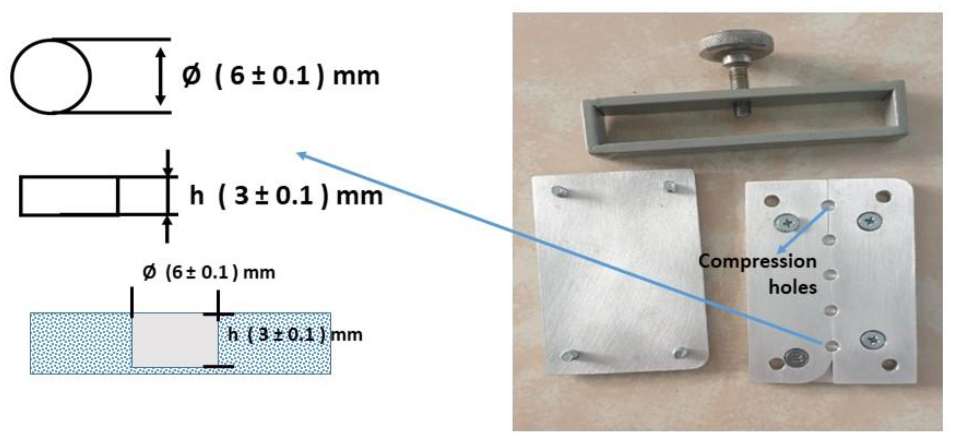
2.3. Preparation of Nanocomposite

Two scenarios of the MMA composite were prepared, each using SiO2/ZrO2 or ZrO2-SiO2 mixed filler with two diverse silane coupling agents, for instance, MPTS or TMSPMA. The sample code names were SiO2/ZrO2/MPTS, SiO2/ZrO2/TMSPMA, ZrO2-SiO2 mixed/MPTS, and ZrO2-SiO2 mixed/TMSPMA, as presented in Table 1, and compared with pristine MMA composite. The SiO2/ZrO2/MPTS composite was prepared by mixing MMA with MPTS under a magnetic stirrer and subsequently mixed with SiO2/ZrO2 filler using a wt.% ratio, as per Table 1. The mixture was kept under a magnetic stirrer for 1 h to obtain homogeneous suspension and well-dispersed SiO2/ZrO2 filler in the MMA matrix.
Before the preparation of the MMA composite, two types of molds were designed, each for DTS, as shown in Figure 1, and Flexural and ME, as shown in Figure 2. The mold was coated with Cold Mold Seal (CMS) before inserting the paste into the hole-shaped design. The polymerization was made by introducing an initiator of Benzoyl Peroxide into the suspension while keeping the suspension heated at 90 °C for 10 min until the solution turned into a paste. The paste was immediately poured into the peg mold and pressed to form the desired shape. The molding was placed in boiling water at 90 °C for 1 h, according to Wang et al. [35]. The other samples of SiO2/ZrO2/TMSPMA, ZrO2-SiO2 mixed/MPTS, and ZrO2-SiO2 mixed/TMSPMA were prepared with similar methods with different fillers and silanes. All composite samples were analyzed in terms of chemical bonding, crystallinity, and mechanical properties (DTS, Flexural, and ME) correspondingly using FTIR spectrometry (iD5 ATR, Nicolet iS5, Thermo Scientific, Waltham, MA, USA) in a wave number range of 4000–400 cm−1 at room temperature, X-ray Diffraction (XRD, Bruker D8 Advance with Cu Kα with = 1.54060 Å), and LLOYD Instrument (Type: LRX Plus, Ametek Company, Segenworth East Fareham, Hants, UK).

2.4. Toxicity Test

The dye exclusion test was used to determine the number of live cells present in the cell suspension. The toxicity test using the Doubling Time Trypan Blue protocol allows for light microscopic quantitation of cell viability. First, cells were suspended in PBS containing trypan blue and placed in the fibroblast cell line 3T3-L1 well plate (number of initial cell implantation = 250,000 cells/well). It is based on the principle that living cells have intact cell membranes that exclude certain dyes, such as Trypan Blue, Eosin, or Propidium, whereas dead cells do not. In this test, a cell suspension is mixed with a dye and visually examined to determine whether the cells are taking up or secreting the dye. In the protocol presented here, viable cells will have clear cytoplasm, whereas nonviable cells will have blue cytoplasm [36]. Cells suspended in PBS (Phosphate-Buffered Saline) containing Trypan Blue were then examined to determine the percentage of cells with clear cytoplasm (viable cells) versus cells with blue cytoplasm (nonviable cells) [36].

3. Results and Discussion

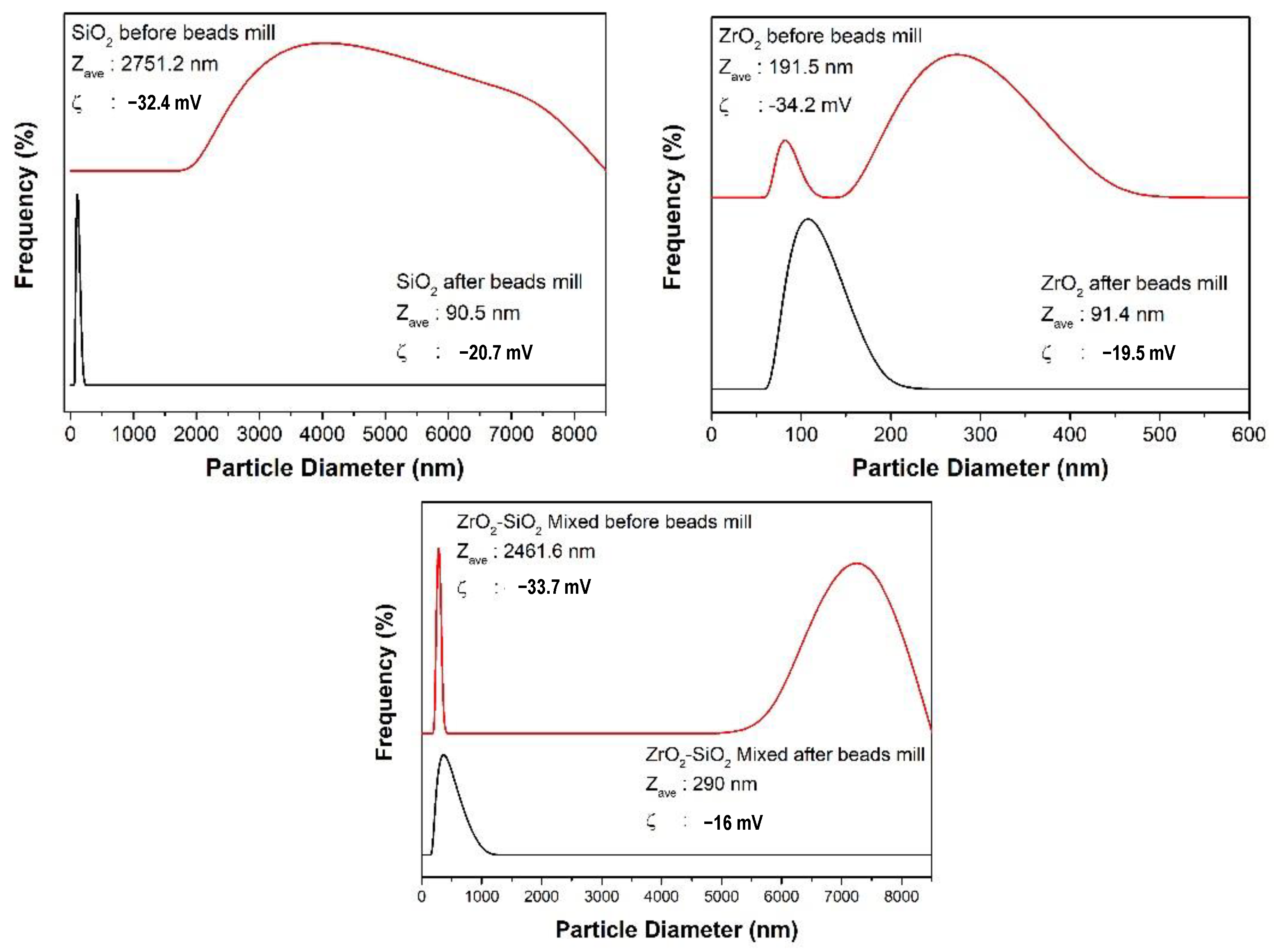
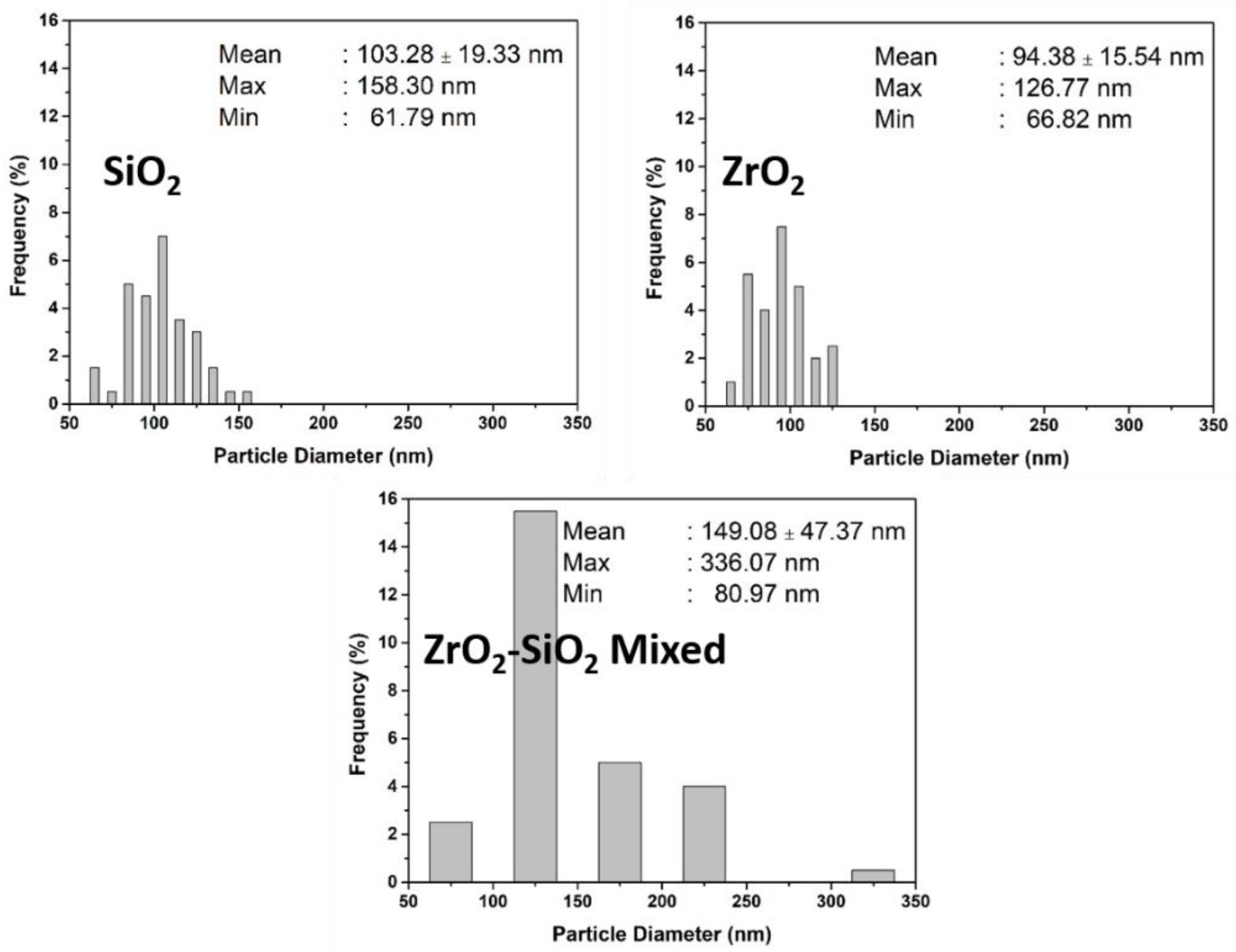
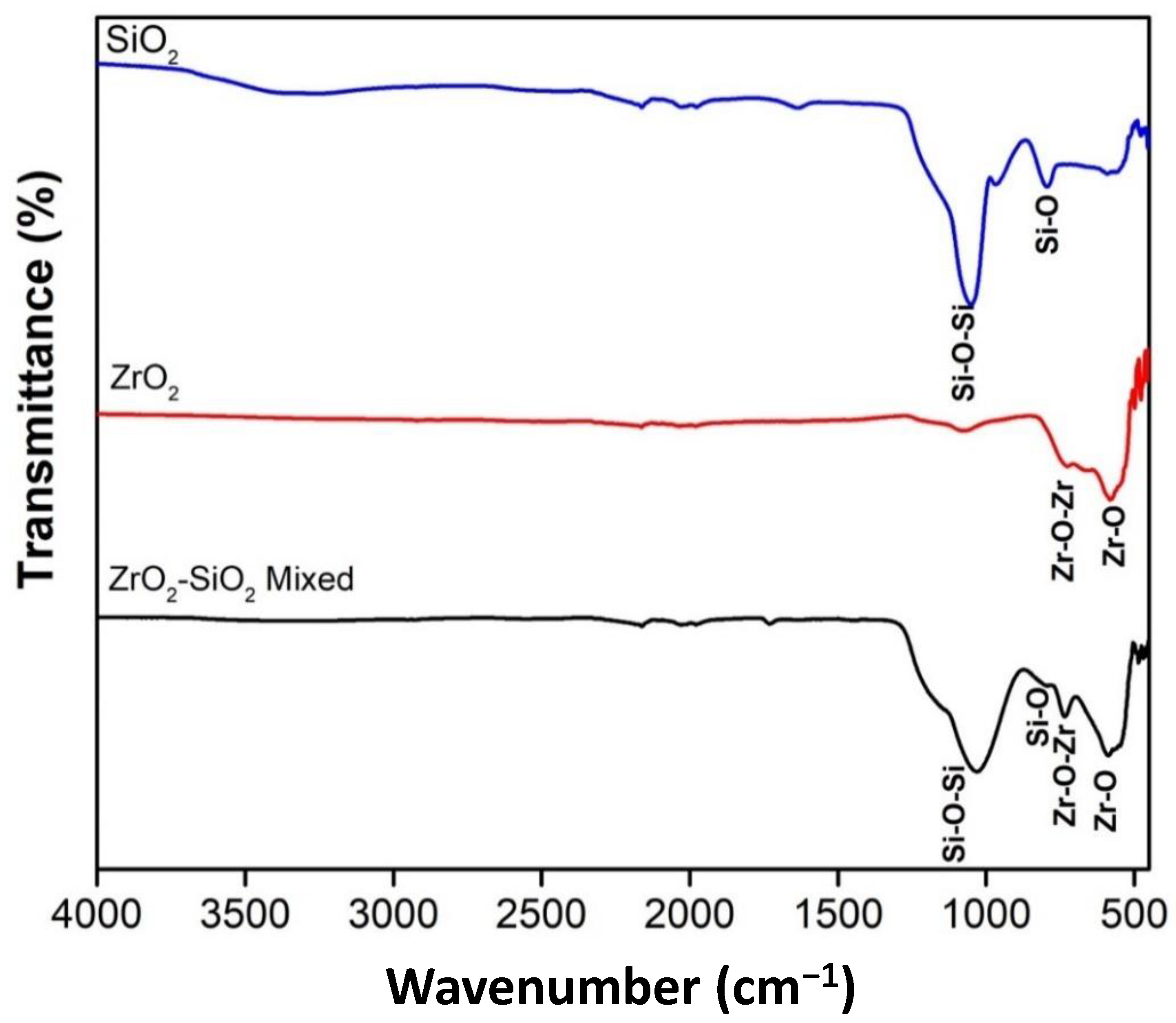
3.1. Characteristics of Filler before and after Beads Mill

The size distribution of the synthesized particles before and after bead milling is presented in Figure 3. Particles appeared to agglomerate before bead milling, but agglomerated particles could be disintegrated after bead milling. In contrast, the nano-sized SiO2 and ZrO2 were obtained after bead milling, correspondingly, 90.5 and 91.4 nm. The ZrO2-SiO2 mixed particle was submicron sized (290 nm) after the bead milling process. The well-dispersed suspension was obtained for SiO2 nanoparticles due to the high zeta potential −20.7 mv to prevent deagglomeration of particles during the bead milling process. The suspension of ZrO2 and ZrO2-SiO2 mixed particles received lower zeta potentials, causing some particles to deagglomerate and form larger sizes during the bead milling process [37]. Consequently, the higher size distribution of both suspensions was obtained compared to the SiO2 suspension.
The SEM images were obtained to establish the morphology and also the corresponding size distribution of fillers after the bead milling, as shown in Figure 4 and Figure 5. The morphology of SiO2 and ZrO2 was spherical in shape, while the ZrO2-SiO2 mixed was relatively irregular. However, the corresponding size and size distribution of SiO2 and ZrO2 were higher than their suspension based on the PSA observation (Figure 3). Therefore, the spherical morphology of SiO2 and ZrO2 nanoparticles agrees with our previous study [38].
Based on the elemental analysis of XRF, the purity of SiO2 and ZrO2, and ZrO2-SiO2 mixed was, correspondingly, 98 and 83.7, as presented in Table 2. Meanwhile, the ZrO2-SiO2 mixed particles contained 31.7% mass of SiO2 and 62.7% mass of ZrO2. Several other impurities of minerals appeared, with the highest content being the Mg element.
Figure 6 shows the FTIR SiO2, ZrO2, and ZrO2-SiO2 mixed after the bead milling process. The FTIR of SiO2 fine powder has two prominent characteristic peaks, observed at about 793 cm−1 and 1050 cm−1, namely Si-O bending vibration band and asymmetric stretching vibration siloxane bonds (Si-O-Si) [38]. This result confirmed the formation of SiO2 and its functional groups via sol–gel synthesis. SiO2 nanopowder was produced after undergoing the sol–gel process with typical reactions of hydrolysis and condensation of precursors. The TEOS precursor was first hydrolyzed to silicic acid during the sol–gel process. Then, the condensation reaction led to the formation of Si-O-Si bonds. The high deformability of the formation allows the inorganic gel to accommodate significant shrinkage, which prevents cracking and pulverization of the powder during drying and calcination.
The FTIR of ZrO2 shows a distinct peak attributed to Zr-O-Zr and Zr-O bonds. Similarly, the formation of zirconia was confirmed by the FTIR observation, indicating a successful precipitation process. In addition, several Si and Zr atoms appear in the FTIR of the ZrO2-SiO2 mixed sample, attributed to Si-O-Si, Si-O, and Zr-O bonds. This spectrum indicated the presence of the formation of zirconia and silica in the mixture.
Figure 7 shows the XRD spectrum of the SiO2, ZrO2, and ZrO2-SiO2 mixed samples compared with the reference spectrum based on Crystallography Open Database from Match version 3.6.2.121 software Crystal Impact, Bonn, Germany. The crystal structure of the SiO2 sample appeared to be triclinic (anorthic) Tridymite based on COD 96-901-3394, while O2 appeared to be a monoclinic Baddeleyite based on COD 96-900-7486. Meanwhile, the crystal structure of ZrO2-SiO2 mixed appeared to be a combination of crystalline tetragonal SiO2 Stishovite (COD 96-900-7155), monoclinic ZrO2 Baddeleyite (COD 96-900-7486), and orthorhombic ZrO2 (COD 96-900-9920), with ratios of 46%, 37.3%, and 16.7%, respectively.

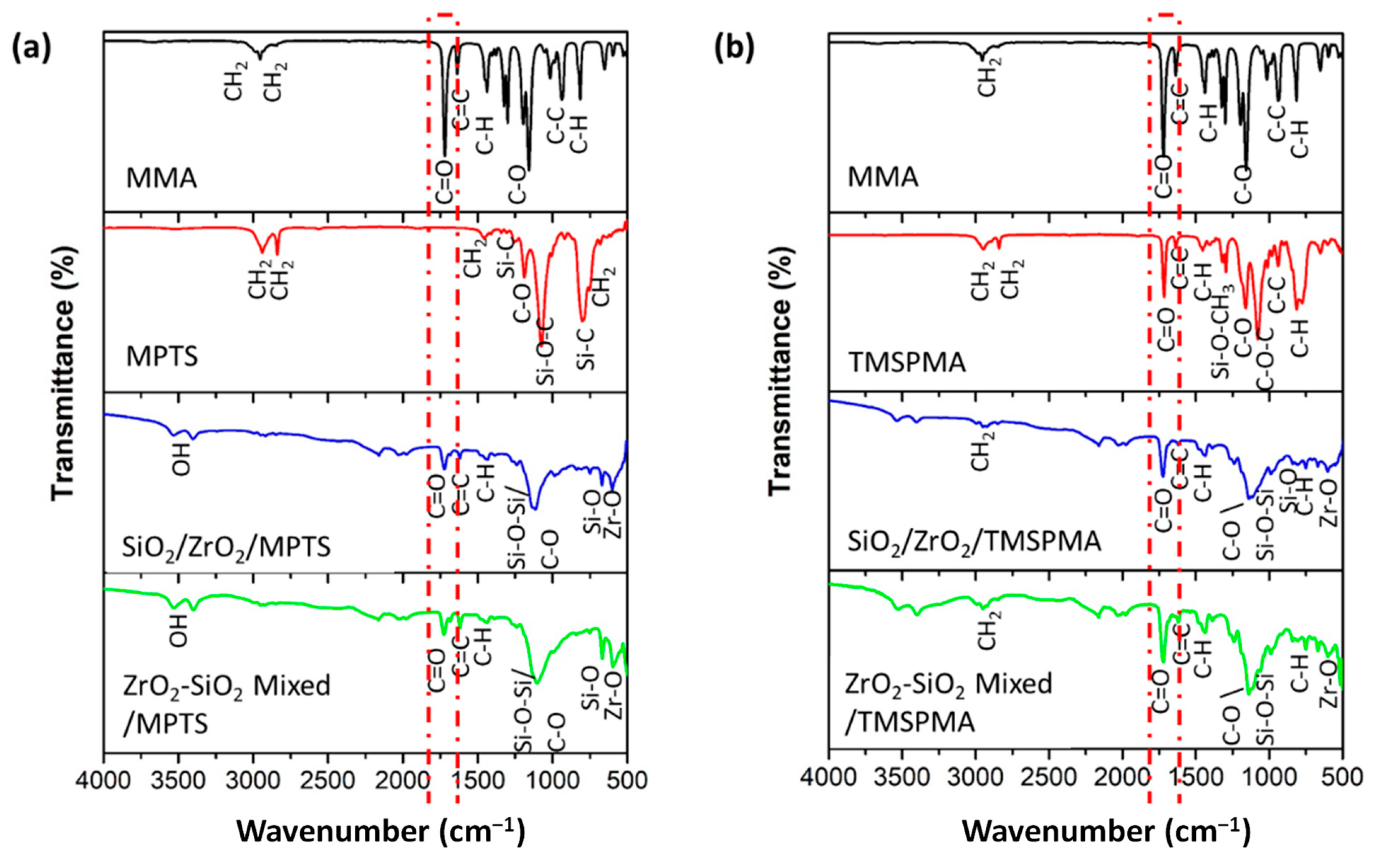
3.2. Characteristics of Composite with Various Types of Fillers

Two scenarios of the MMA composite were prepared and subjected to XRD observation where each composite used SiO2/ZrO2 or ZrO2-SiO2 mixed filler, with two diverse silane coupling agents, MPTS or TMSPMA. Figure 8 shows the XRD results of an endodontic implant composite with the addition of MPTS and TMSPMA silanes, which were analyzed using the Match version 3.6.2.121 software Crystal Impact, Bonn, Germany. In Figure 8a, the SiO2/ZrO2/MPTS sample has high peaks compared to ZrO2-SiO2 mixed/MPTS. The XRD pattern of both scenarios with high composite crystallinity indicates that the presence of fillers is well distributed in the polymer matrix.
Figure 9a shows that the SiO2/ZrO2 composite with the MPTS surfactant did not show a strong bond, as shown by the weak peaks of the MPTS functional groups in the composite. The peaks represent that the TMSPMA surfactant had a stronger bond with the MMA matrix than MPTS for both composites with different fillers (SiO2/ZrO2 and ZrO2-SiO2 mixed). The strong C = O bond at 1723.03 cm−1 (dashed red box) for the composite with the TMSPMA surfactant is considered to be responsible for a strong filler–MMA matrix. The C = O asymmetric stretch is a characteristic of methacrylate formed in both composites. Other functional groups at 1205.44 cm−1 were assigned to Si-O-CH3 (Si-O bending). The wavenumber range for common functional groups in Figure 9 is summarized in Table 3.

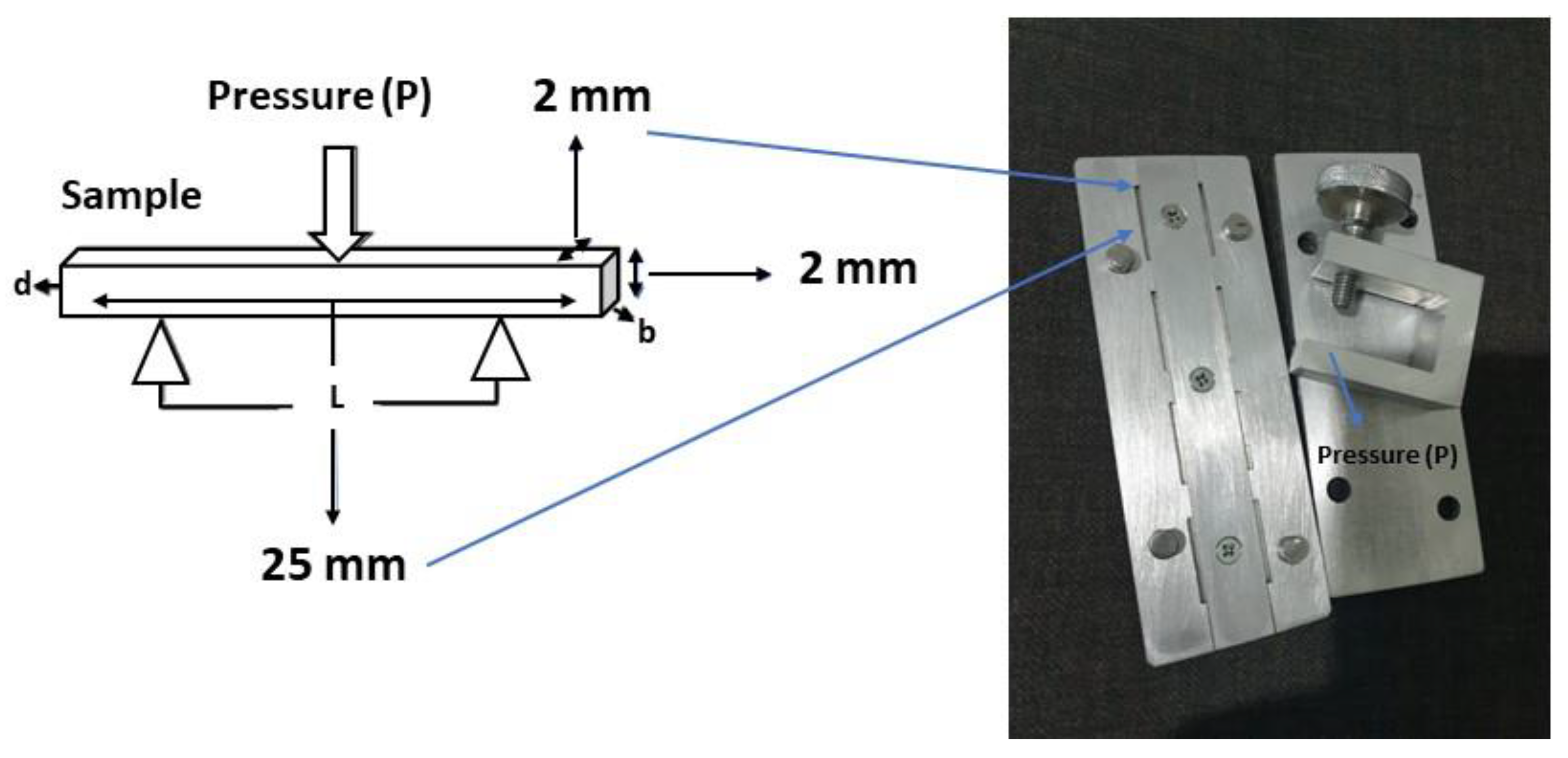
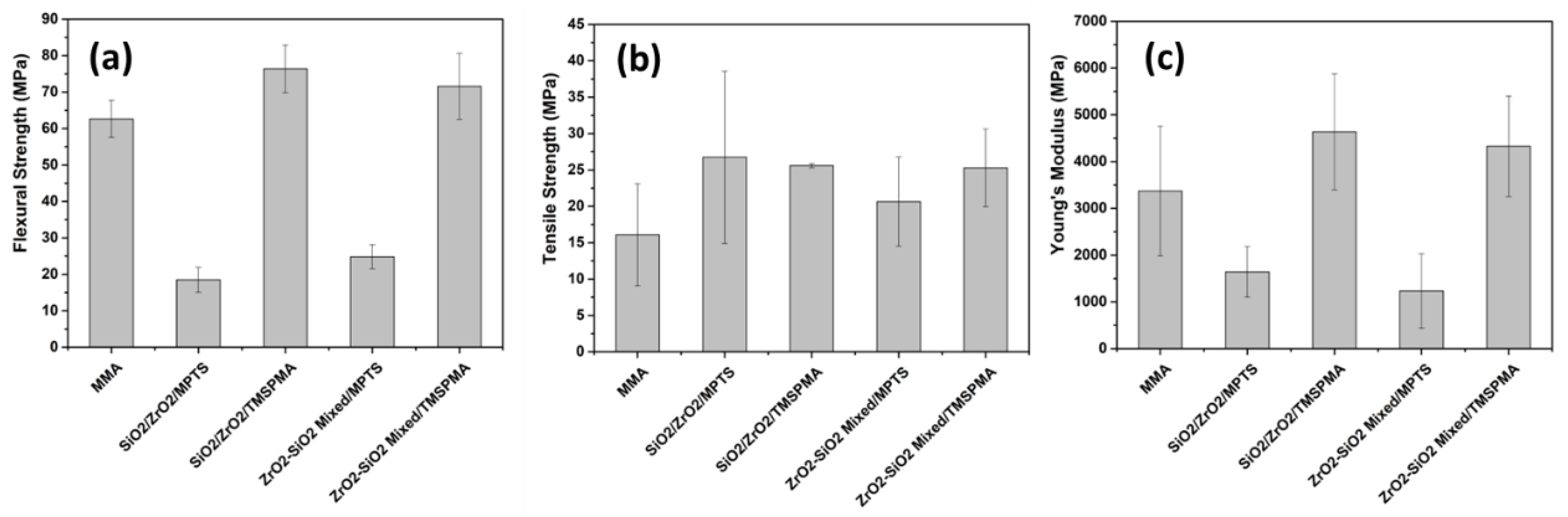
3.3. Mechanical Properties of Composite with Various Types of Fillers

Each sample was measured five times for flexural, DTS, and modulus of elasticity. Based on the results of mechanical tests on several samples, SiO2/ZrO2/ TMSPMA was better than the other samples seen from the flexural, DTS, and ME tests, with flexural values of 152.7 ± 13.0 MPa, DTS with values of 51.2 ± 0.6 MPa and ME of 9272.8 ± 2481.4 MPa (Figure 10). However, the mechanical properties of the composites obtained from MMA with MPTS were even lower than those of the MMA matrix without filler. This shows that MPTS is unsuitable as the MMA surfactant in the two fillers used. In contrast, the composite with TMSPMA showed enhanced mechanical properties compared to the composite with MPTS in both types of fillers (SiO2/ZrO2 and ZrO2-SiO2 mixed). It was highlighted that the composite with TMSPMA using SiO2/ZrO2 filler possessed higher mechanical properties than ZrO2-SiO2 mixed. The size and size distribution of SiO2/ZrO2 particles were lower compared to ZrO2-SiO2 mixed and so considered responsible for the strong bonding between the filler and matrix, causing stronger mechanical properties.
The SiO2/ZrO2/TMSPMA sample has a higher flexural value and a reasonably large DTS value than the other samples. In contrast, the SiO2/ZrO2/TMSPMA composite produced the highest modulus elastic properties. The mechanical properties of the composite are considered to be an endodontic implant when the mechanical properties are close to the dentin properties in terms of modulus elasticity and flexural strength. The modulus elasticity of general dentin is (17.5 ± 3.8) GPa [13], while the modulus elasticity of dentin in the anterior teeth is (5.3 ± 1.6) to (6.1 ± 1.6) MPa [39]. The flexural strength of dentin is 212.9 ± 41.9 MPa [13], while the flexural dentin in teeth after root canal treatment with saline irrigation after 30 days is 203.26 ± 16.76 [14]. The flexural dentin after root canal irrigation with NaOCl 5% is (135.72 ± 7.83)–(200 ± 8.66) MPa [14]. The as-prepared best composite with PMMA-SiO2/ZrO2/TMSPMA resulted in higher flexural strength than only PMMA with a Bis GMA base (83.5 ± 10.7 MPa), with an elastic modulus (5.0 ± 2.9) MPa [6]. Further, the flexural strength of the as-prepared SiO2/ZrO2/TMSPMA composite was higher compared to another study on a PMMA–silica nanofiber dental composite (132.74 ± 20.70) MPa [39]. In contrast, the as-prepared SiO2/ZrO2/TMSPMA composite showed excellent DTS compared to other reported PMMA composites using silica filler, with and without fiberglass, correspondingly (38.74 ± 3.05) and (28.39 ± 3.21) MPa [40]. It was emphasized that, based on the flexural strength, elastic modulus, and DTS, the PMMA composite with SiO2/ZrO2/TMSPMA is considered acceptable for an endodontic implant.

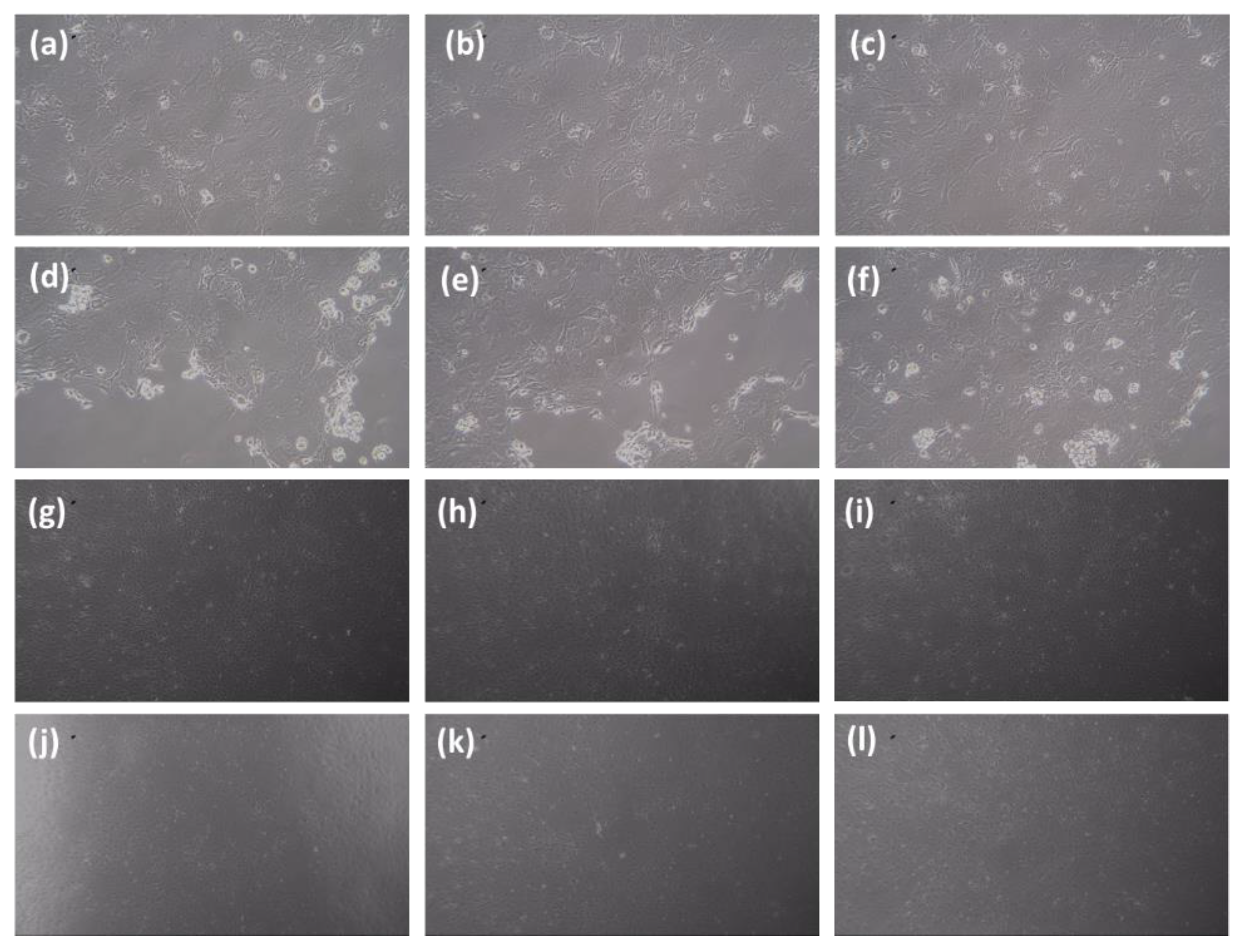
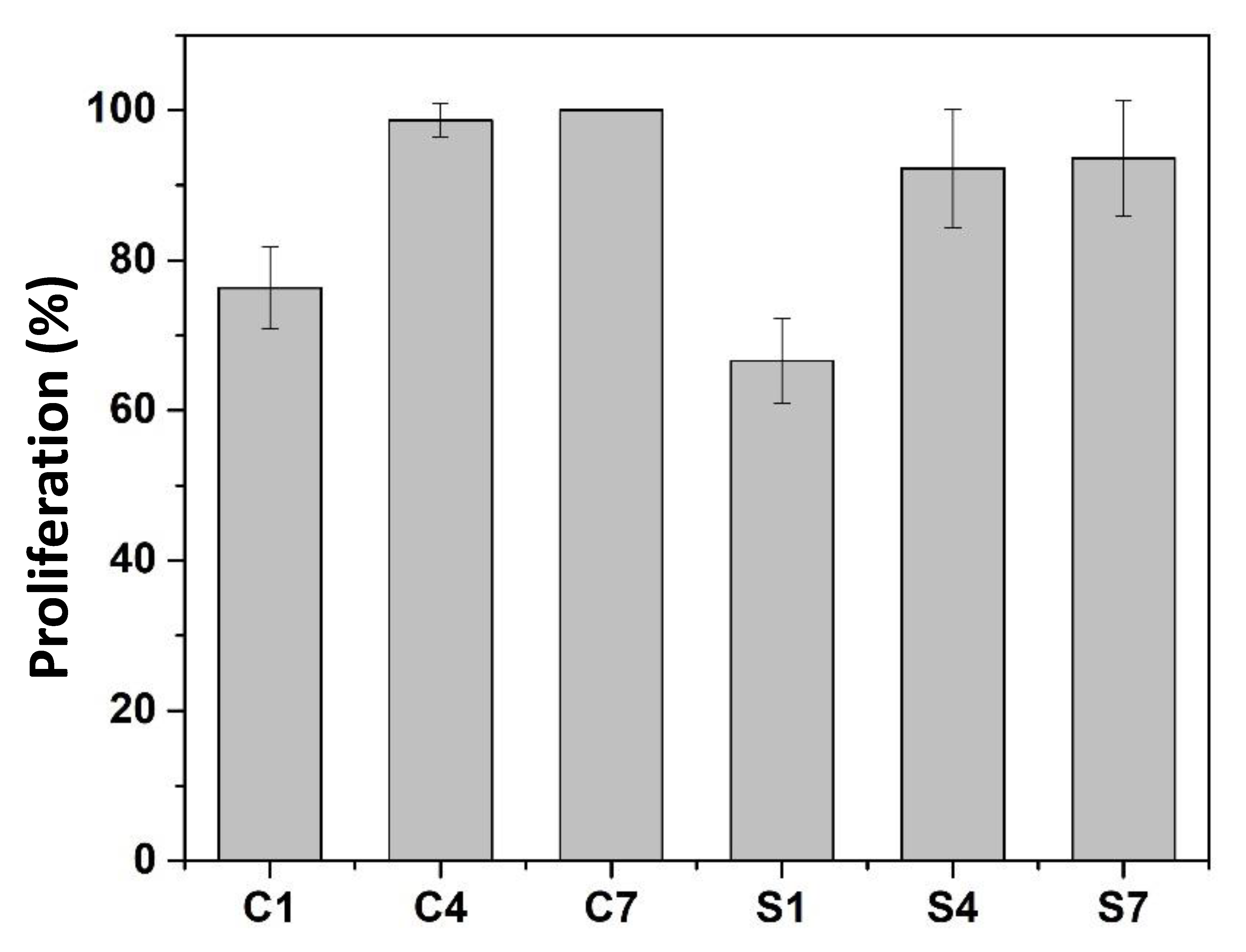
3.4. Toxicity Test of Selected Composite with Enhanced Mechanical Properties

The selected composite of SiO2/ZrO2/ TMSPMA with enhanced mechanical properties was subjected to a toxicity test as one of the essential composite criteria as an endodontic post or implant. Figure 11 shows a microscopic image of 3T3-L1 Fibroblast cells with control (a, b, c) and SiO2/ZrO2/TMSPMA post composite (d, e, f), incubation period of 1 day (g, h, i), incubation period of 4 days, and (j, k, l) 7 days incubation period. Based on the microscopic images, the viability of the living cell can be determined when the cell nucleus absorbs the blue color from trypan blue, according to the procedure carried out by Strober W, 2015 [36]. The cell is considered a living cell when it does not absorb the trypan blue color, the cell periphery glows, and the shape is perfectly round. On the other hand, the cell is considered to be dead when it is not perfectly round, and the cell wall is destroyed.
Figure 12 shows that the percentage of live cells (treated with control, C1, C4, and C7) on the first day of incubation (S1) was 87.06%, on the fourth day of incubation (S4) was 93.38%, and on the seventh day of incubation (S7) was 93.61%. The cytotoxicity level of each concentration of antibacterial agent was calculated as a percentage of cell viability, including absorbance values obtained for each system. According to ISO 10993-5, the percentage of cell viability was above 80%. Thus, it is considered non-cytotoxic based on the criteria that within 60–80% is weak, 40–60% moderate cytotoxicity, and below 40% severe cytotoxicity [41,42].
The endodontic implant obtained from the PMMA-ZrO2/SiO2-TMSPMA composite could improve its mechanical property close to dentin with good biocompatibility. Thus, this composite offers the possibility for clinical application since it can technically be produced in large quantities at an affordable cost. Further, theoretically, the presence of SiO2 and ZrO2 layers makes the composite non-corrosive and good at osteointegration with bone. Although further research is needed, particularly on the long-term biocompatibility properties, the recently existing properties meet the requirements for implants and endodontic posts.

4. Conclusions

The selected composite of SiO2/ZrO2/TMSPMA was considered acceptable as an endodontic implant in terms of the flexural strength, DTS, and elastic modulus, correspondingly (152.7 ± 13.0) MPa, (51.2 ± 0.6) MPa and (9272.8 ± 2481.4) MPa. The selected composite was also considered non-cytotoxic according to ISO 10993-5, with a percentage of cell viability above 80%. It was emphasized that the application of ZrO2/SiO2 nanofillers with a suitable and proper amount of silane coupling agent of TMSPMA enhanced the mechanical properties of PMMA to meet the criteria of the composite as an endodontic implant. However, the limitation of the present work is the biocompatibility, observed for only 14 days, so more extended periods are necessary. In addition, this result enabled an extended clinical test study to determine the healing rate of inflammation after implant placement, even observing the healing process during the induction of intermittent hypobaric hypoxia.

Author Contributions

Conceptualization, P.W. and I.M.J.; methodology, P.W.; software, P.W.; validation, P.W., W.M., N.D. and I.M.J.; formal analysis, P.W.; investigation, P.W.; resources, P.W.; data curation, P.W.; writing—original draft preparation, P.W.; writing—review and editing, I.M.J.; supervision, I.M.J.; funding acquisition, P.W. All authors have read and agreed to the published version of the manuscript.

Funding

This research was funded by the Ministry of the Defence, Republic of Indonesia, grant number 1216/X/2019, and the APC was funded by Academic Leadership Grant, Padjadjaran University, with contract number 2203/UN6.3.1/PT.00/2022.

Conflicts of Interest

The authors declare no conflict of interest.

References

  1. Vaishya, R.; Chauhan, M.; Vaish, A. Bone cement. J. Clin. Orthop. Trauma. 2013, 4, 157–163. [Google Scholar] [CrossRef] [Green Version]
  2. Tanuja, B. A Complete Review of Dental Implant Materials. Int. J. Recent Sci. Res. 2018, 9, 29665–29669. [Google Scholar]
  3. Frazer, R.Q.; Byron, R.T.; Osborne, P.B.; West, K.P. PMMA: An Essential Material in Medicine and Dentistry. J. Long-Term Eff. Med. Implants 2005, 15, 629–639. [Google Scholar] [CrossRef] [Green Version]
  4. Punet, X.; Mauchauffe, R.; Rodríguez-Cabello, J.C.; Alonso, M.; Engel, E.; Mateos-Timoneda, M.A. Biomolecular Functionalization for Enhanced Cell–Material Interactions of Poly (Methyl Methacrylate) Surfaces. Regen. Biomater. 2015, 2, 167–175. [Google Scholar] [CrossRef] [Green Version]
  5. Salehuddin, S.M.F.; Wahit, M.U.; Kadir, M.R.A.; Sulaiman, E.; Kasim, N.H.A. Mechanical and Morphology Properties of Feather Fiber Composite for Dental Post Application. Malays. J. Anal. Sci. 2014, 18, 368–375. [Google Scholar]
  6. Djustiana, N.; Faza, Y.; Cahyanto, A. Flexural Properties of Electrospun Polymethyl Methacrylate Microfiber-Reinforced BisGMA for Dental Post Prefabrication. Padjadjaran J. Dent. 2021, 33, 264–270. [Google Scholar] [CrossRef]
  7. Alla, R.K.; Raghavendra, S.K.N.; Vyas, R.; Tiruveedula, N.P.B.; Raju, A.M.K. Physical and Mechanical Properties of Heat activated Acrylic Denture Base Resin Materials. Res. J. Pharm. Technol. 2018, 11, 2258–2262. [Google Scholar] [CrossRef]
  8. Muneera, R.A.G.; Mattoo, K.A.; Youseef, A.M. A Novel Approach to Determine the Aesthetic Inclination of Cast Post Core–Case Report. J. Int. Dent. Med. Res. 2017, 3, DE14–DE17. [Google Scholar]
  9. Jain, S.; Jain, P. Fiber Post, Composite Core, All-Ceramic Crown and Resin Cement–An Aesthetic Cocktail. Int. J. Med. Rev. Case Rep. 2020, 4, 23–25. [Google Scholar] [CrossRef]
  10. Sevimli, G.; Cengiz, S.; Selcukoruc, M. Endocrowns Review. J. Istanb. Univ. Fac. Dent. 2015, 49, 57–63. [Google Scholar] [CrossRef] [PubMed] [Green Version]
  11. Ferrari, M.; Scotti, R. Fiber Posts. Characteristics and Clinical Applications, 1st ed.; Masson: Milan, Italy, 2002. [Google Scholar]
  12. Prisco, D.; De Santis, R.; Mollica, F.; Ambrosio, L.; Rengo, S.; Nicolais, L. Fiber Post Adhesion to Resin Luting Cements in The Restoration of Endodontically-Treated Teeth. Oper. Dent. 2003, 28, 515–521. [Google Scholar] [PubMed]
  13. Plotino, G.; Grande, N.M.; Bedini, R.; Pameijer, C.H.; Somma, F. Flexural Properties of Endodontic Posts and Human Root Dentin. Dent. Mater. 2007, 23, 1129–1135. [Google Scholar] [CrossRef]
  14. Diatri, N.R. The Effect of Irrigation Solution on Flexural Dentin. J. Dent. Indones. 2009, 16, 133–140. [Google Scholar]
  15. Goracci, C.; Ferrari, M. Current Perspectives on Post Systems: A Literature Review Department of Dental Materials and Fixed Prosthodontics of Siena. Aust. Dent. J. 2011, 56, 77–83. [Google Scholar] [CrossRef] [PubMed]
  16. Saruta, J.; Ozawa, R.; Hamajima, K.; Saita, M.; Sato, N.; Ishijima, M.; Kitajima, H.; Ogawa, T. Prolonged Post-Polymerization Biocompatibility of Polymethylmethacrylate-Tri-nButylborane (PMMA-TBB) Bone Cement. Materials 2021, 14, 1289. [Google Scholar] [CrossRef]
  17. Lamichhane, A.; Xu, C.; Zhang, F.Q. Dental fiber-post resin base material: A review. J. Adv. Prosthodont. 2014, 6, 60–65. [Google Scholar] [CrossRef] [Green Version]
  18. Lacerda-Santos, R.; Lima, A.B.L.; Penha, E.S.; Santos, A.; Carvalho, F.G.; Pithon, M.M.; Dantas, A.F.M. In Vivo Biocompatibility of Silicon Dioxide Nanofilm Used as Antimicrobial Agent on Acrylic Surface. An. Acad. Bras. Ciências 2020, 92, e20181120. [Google Scholar] [CrossRef]
  19. Harianawala, H.; Kheur, M.; Kheur, S.; Sethi, T.; Bal, A.; Burhanpurwala, M.; Sayed, F. Biocompatibility of Zirconia. J. Adv. Med. Dent. Sci. Res. 2016, 4, 35–39. [Google Scholar]
  20. Salih, S.; Oleiwi, J.K.; Hamad, Q.A. Comparative Study the Flexural Properties and Impact Strength for PMMA Reinforced by Particles and Fibers for Prosthetic Complete Denture Base. Iraqi J. Mech. Mater. Eng. 2015, 15, 288–307. [Google Scholar]
  21. Baltatu, M.S.; Vizureanu, P.; Sandu, A.V. Microstructural Analysis and Tribological Behavior of Ti-Based Alloys with a Ceramic Layer Using the Thermal Spray Method. Coatings 2020, 10, 1216. [Google Scholar] [CrossRef]
  22. Tan, G.; Zhou, L.; Ning, C.; Tan, Y.; Ni, G.; Liao, J.; Yu, P.; Chen, X. Biomimetically-mineralized composite coatings on titanium functionalized with gelatin methacrylate hydrogels. Appl. Surf. Sci. 2013, 279, 293–299. [Google Scholar] [CrossRef]
  23. Focsaneanu, S.C.; Vizureanu, P.; Sandu, A.V.; Ciobanu, G. Experimental Study on the Influence of Zirconia Surface Preparation on Deposition of Hydroxyapatite. Rev. Chim. 2019, 70, 2273–2275. [Google Scholar] [CrossRef]
  24. Honga, G.; Liaoa, M.; Wu, T.; Zhou, Q. Improving osteogenic activity of Y-TZP (Yttria-stabilized tetragonal zirconia polycrystal) surfaces by grafting of silanes with different end groups. Appl. Surf. Sci. 2021, 570, 1–11. [Google Scholar] [CrossRef]
  25. Nakonieczny, D.; Radco, T.; Drewniak, S. ZrO2-CeO2 ceramic powders obtained from a sol-gel process using acetylacetone as a chelating agent for potential application in prosthetic dentistry. Acta Bioeng. Biomech. 2016, 18, 54–60. [Google Scholar] [CrossRef]
  26. Nakonieczny, D.S.; Kern, F.; Dufner, L.; Dubiel, A. Effect of Calcination Temperature on the Phase Composition, Morphology, and Thermal Properties of ZrO2 and Al2O3 Modified with APTES (3-aminopropyltriethoxysilane). Materials 2021, 14, 6651. [Google Scholar] [CrossRef] [PubMed]
  27. Nafsin, N.; Hasan, M.M.; Dey, S.; Castro, R.H.R. Effect of ammonia on the agglomeration of zirconia nanoparticles during synthesis and sintering by spark plasma sintering. Mater. Lett. 2016, 183, 143–146. [Google Scholar] [CrossRef] [Green Version]
  28. Joni, I.M.; Purwanto, A.; Iskandar, F.; Okuyama, K. Dispersion stability enhancement of titania nanoparticles in organic solvent using a bead mill process. Ind. Eng. Chem. Res. 2009, 48, 6916–6922. [Google Scholar] [CrossRef]
  29. Chung, J.J.; Fujita, Y.; Li, S.; Stevens, M.M.; Kasuga, T.; Georgiou, T.K.; Jones, J.R. Biodegradable inorganic-organic hybrids of methacrylate star polymers for bone regeneration. Acta Biomater. 2017, 54, 411–418. [Google Scholar] [CrossRef]
  30. Hirai, K.; Furusho, H.; Hirota, K.; Sasaki, H. Activation of hipoksia-inducible factor 1 attenuates periapical inflammation and bone loss. Int. J. Oral Sci. 2018, 10, 12. [Google Scholar] [CrossRef] [Green Version]
  31. Sasaki, H.; Furusho, H.; Rider, D.B.; Dobeck, J.M.; Kuo, W.P.; Fujimura, A.; Yoganathan, S.; Hirai, K. Endodontik Infection-induced Inflammation Resembling Osteomyelitis of the Jaws in Toll-like Receptor 2/Interleukin 10 Double-knockout Mice. J. Endod. 2019, 45, 181–188. [Google Scholar] [CrossRef]
  32. Dubey, R.S.; Rajesh, Y.B.R.D.; More, M.A. Synthesis and Characterization of SiO2 Nanoparticles via Sol-Gel, Method for Industrial Applications. Mater. Today Proc. 2015, 2, 3575–3579. [Google Scholar] [CrossRef]
  33. Rudzani, A.; Mavundla, S.E.; Moloto, N.; Mokrani, T. Synthesis of Zirconia-Based Solid Acid Nanoparticles for Fuel Cell Application. J. Energy S. Afr. 2016, 27, 60–67. [Google Scholar]
  34. Elsandika, G.; Putri, A.D.C.; Musyarofah, M.; Pratapa, S. Synthesis of ZrSiO4 Powders by Sol-Gel Method with Varied Calcination Temperatures. IOP Conf. Ser. Mater. Sci. Eng. 2019, 496, 012047. [Google Scholar] [CrossRef]
  35. Wang, H.; Zhong, W.; Shen, L.; Xu, P.; Du, Q. Transparent Poly (Methyl Methacrylate)/Silica/Zirconia Nanocomposites with Excellent Thermal Stabilities. Polym. Degrad. Stab. 2004, 87, 319–327. [Google Scholar] [CrossRef]
  36. Strober, W. Trypan Blue Exclusion Test of Cell Viability. Curr. Protoc. Immunol. 2015, 111, A3.B.1–A3.B.3. [Google Scholar] [CrossRef]
  37. Faza, Y.; Cahyanto, A.; Djustiana, N.; Joni, I.M. Synthesis and Characterization of Mullite-Zirconia Prepared Through Solid Sintering Of 3Al2O3-2SiO2 Xerogel and ZrO2 Xerogel as A Dental Implant Material. AIP Conf. Proc. 2020, 2219, 080005. [Google Scholar]
  38. Joni, I.M.; Nulhakim, L.; Vanitha, M.; Panatarani, C. Characteristics of Crystalline Silica (SiO2) Particles Prepared by Simple Solution Method Using Sodium Silicate (Na2SiO3) Precursor. J. Phys. Conf. Ser. 2018, 1080, 012006. [Google Scholar] [CrossRef]
  39. Chun, K.; Choi, K.; Lee, J.Y. Comparison of mechanical property and role between enamel and dentin in the human teeth. J. Dent. Biomech. 2014, 5, 1758736014520809. [Google Scholar] [CrossRef]
  40. Djustiana, N.; Faza, Y.; Sudiyasari, N.; Firdaus, A.T.; Usri, K.; Cahyanto, A. Performance of Electrospun PMMA-Silica Nanofiber as Reinforced Material. Experimental article. J. Int. Dent. Med. Res. 2020, 13, 975–978. [Google Scholar]
  41. Karlina, E.; Ramadhani, E.; Veni Takarini, Y.; Djustiana, N. Diametral Tensile Strength on Restorative Dental Composite: Contrasting Results from the Addition of PMMA Fiber Filler. AIP Conf. Proc. 2020, 2219, 080008. [Google Scholar]
  42. ISO 10993-5:2009; Biological Evaluation of Medical Devices. Part 5: Tests for In Vitro Cytotoxicity. ISO Copyright Office Case Postale 56 • CH-1211 Geneva 20. IHS: Geneva, Switzerland, 2009.
Figure 1. The compression molding of nanocomposite sample for DTS test.
Dentistry 11 00057 g001
Figure 2. The compression molding of nanocomposite sample for flexural test and ME.
Dentistry 11 00057 g002
Figure 3. Particle size and stability of SiO2, ZrO2, and ZrO2-SiO2 mixed filler solutions.
Dentistry 11 00057 g003
Figure 4. The SEM images of SiO2, ZrO2, and ZrO2-SiO2 mixed fillers.
Dentistry 11 00057 g004
Figure 5. The Size Distribution of SiO2, ZrO2, and ZrO2-SiO2 mixed fillers.
Dentistry 11 00057 g005
Figure 6. FTIR Filler SiO2, ZrO2, and ZrO2-SiO2 mixed.
Dentistry 11 00057 g006
Figure 7. XRD Filler SiO2, ZrO2, and ZrO2-SiO2 mixed. XRD spectra references are SiO2 Tridymite/COD 96-900-7155 (solid square), SiO2 Stishovite/COD 96-900-7155 (hollow square), ZrO2 orthorhombic/COD 96-900-9920 (solid circle) and ZrO2 Baddeleyite/COD 96-900-7486 (hollow circle).
Dentistry 11 00057 g007
Figure 8. XRD composite post with silane addition: (a) MPTS and (b) TMSPMA. XRD spectra references are SiO2 Tridymite/COD 96-900-7155 (solid square) and ZrO2 Baddeleyite/COD 96-900-7486 (solid circle).
Dentistry 11 00057 g008
Figure 9. FTIR composite post with silane addition: (a) MPTS and (b) TMSPMA.
Dentistry 11 00057 g009
Figure 10. (a) Flexural, (b) DTS, and (c) Modulus of Elasticity.
Dentistry 11 00057 g010
Figure 11. Microscopic image of 3T3-L1 Fibroblast cells with control (ac) and SiO2/ZrO2/TMSPMA post composite (df) incubation period of 1 day, (gi) incubation period of 4 days, and (jl) 7-day incubation period.
Dentistry 11 00057 g011
Figure 12. The positive cell growth curves at incubation periods of 1, 4, and 7 days, respectively, control 1 day (C1), control 4 days (C4), control 7 days (C7), sample SiO2/ZrO2/TMSPMA 1 day (S1), SiO2/ZrO2/TMSPMA 4 days (S4), and SiO2/ZrO2/TMSPMA 7 days (S7).
Dentistry 11 00057 g012
Table 1. Experimental setup of the composite samples in comparison with only MMA.
NoSamples CodeMatrix
MMA
(wt.%)
Coupling Agent Silane (wt.%)Initiator
BP
(wt.%)
Filler (wt.%)
MPTSTMSPMAZrO2SiO2ZrO2-SiO2 Mixed
1MMA98.5--1.5---
2SiO2/ZrO2/MPTS830.75-1.253.7511.25-
3SiO2/ZrO2/ TMSPMA83-0.751.253.7511.25-
4ZrO2-SiO2 mixed/MPTS830.75-1.25--15
5ZrO2-SiO2 mixed/TMSPMA83-0.751.25--15
MMA = Methyl Methacrylate; MPTS = 3-(Mercaptopropyl) trimethoxysilane; TMSPMA = 3-(trimethoxysilyl) propyl Methacrylate; BP = Benzoyl peroxide.
Table 2. XRF Filler SiO2, ZrO2, and ZrO2-SiO2 mixed.
ComponentSiO2ZrO2ZrO2-SiO2 Mixed
(mass%)(mass%)(mass%)
AlND3.3901.670
Au0.001NDND
Bi0.0100.012ND
Ca0.0280.0120.038
CoND0.026ND
Cr0.0000.0010.002
Cu0.0010.115ND
Fe0.0050.0330.005
GaND0.121ND
K0.028ND0.018
Mg1.82012.6003.830
PbND0.012ND
Pt0.0010.018ND
S0.002NDND
Sn0.0010.0120.012
Ti0.004ND0.003
Zn0.014ND0.025
SiO298ND31.7
ZrO2 0.09083.762.7
Table 3. The wavenumber range for common functional groups.
Samples CodeWavenumber (cm−1)AssignmentFunctional Group NameSamples CodeWavenumber (cm−1)AssignmentFunctional Group Name
MMA2982.59CH2AlkaneSiO2/ZrO2/MPTS3530.39OHhydroxyl
2953.85CH2Alkane1722.46C = OCarbonyl
1719.98C = OCarbonyl1684.25C = CAlkene
1637.01C = CAlkene1434.77C-HAlkane
1437.75C-HMetyl1114.06Si-O-SiSiloxane
1157.50C-OEster1114.06C-OEster
938.66C-CAlkene669.30Si-OSilanol
814.86C-HAlkene600.88Zr-OZirconia
MPTS2939.65CH2AlkaneZrO2-SiO2 mixed/MPTS3530.38OHHydroxyl
2838.98CH2Alkane1725.70C = OCarbonyl
1455.22CH2Alkane1618.71C = CAlkene
1257.78SiCSilicon carbide1435.17C-HAlkane
1188.16C-OEster1103.81Si-O-SiSiloxane
1074.31Si-O-CSiloxane1103.81C-OEster
800.71Si-CSilicon carbide667.32Si-OSilanol
753.42CH2Alkene596.10Zr-OZirconia
TMSPMA2945.83CH2AlkaneSiO2/ZrO2/ TMSPMA2949.29CH2Alkane
2840.60CH2Alkane1723.03C = OCarbonyl
1716.56C = OCarbonyl1619.85C = CAlkene
1636.83C = CAlkene1435.15C-HAlkane
1295.44Si-O-CH3Methoxysilyl1138.27Si-O-SiSiloxane
1160.59C-OEster1113.74C-OEster
1077.23C-O-CEter750.19Si-OSilanol
939.16C-CAlkene669.00C-HAlkene
813.12C-HAlkene601.48Zr-OZirconia
ZrO2-SiO2 mixed/TMSPMA2949.80CH2Alkane
1721.10C = OCarbonyl
1619.54C = CAlkene
1435.26C-HAlkane
1139.12C-OEster
1139.12Si-O-SiSiloxane
668.51C-HAlkene
600.75Zr-OZirconia
Disclaimer/Publisher’s Note: The statements, opinions and data contained in all publications are solely those of the individual author(s) and contributor(s) and not of MDPI and/or the editor(s). MDPI and/or the editor(s) disclaim responsibility for any injury to people or property resulting from any ideas, methods, instructions or products referred to in the content.

