Communication

Development of Multiple Fano-Resonance-Based All-Dielectric Metastructure for High-Contrast Biomedical Applications

1
School of Physics Science and Information Engineering, Liaocheng University, Liaocheng 252000, China
2
Institute of Semiconductors, Chinese Academy of Sciences, Beijing 100083, China
3
Shandong Provincial Key Laboratory of Optical Communication Science and Technology, Liaocheng University, Liaocheng 252000, China
4
Liaocheng Key Laboratory of Industrial-Internet Research and Application, Liaocheng 252000, China
*
Authors to whom correspondence should be addressed.
Photonics 2023, 10(6), 616; https://doi.org/10.3390/photonics10060616
Submission received: 4 May 2023 / Revised: 23 May 2023 / Accepted: 24 May 2023 / Published: 26 May 2023
(This article belongs to the Special Issue Optically Active Nanomaterials for Sensing Applications)

Abstract

:
In this paper, an all-dielectric metastructure-based high-contrast refractive index sensor is proposed. This structure can be utilized to detect various concentrations of glycerol-water mixtures by evaluating transmission spectral lines and resonant wavelength shifts related with liquid concentration detection. The experimental and calculated results of the developed sensor structure are able to excite three resonance peaks, demonstrating that the structure is capable of reaching excellent sensing capabilities. It has been established that this work has the potential to be useful in medical and biological detection; this is of great scientific and practical significance.

1. Introduction

The metastructures are an ultrathin two-dimensional planar device composed of arrays of sub-wavelength structures, that can be classified as metal or dielectric [1,2]. The metal metastructures are affected by the inherent strong ohmic loss and saturation effect and the figure of merit (FOM) for metallic materials sensors is limited [3,4,5]. The dielectric metastructures make it possible for Fano resonances with high Q-factors to be achieved more easily due to the avoidance of ohmic losses [6,7,8]. In the past decade, extensive research has been done on subwavelength periodic structures and their applications, especially on their periodicity perpendicular to the incident direction of light. A case in point is the ultra-thin high-refractive-index contrast 1D dielectric grating, called the high-contrast grating (HCG), and the 2D variation of this structure, now is summarized as the high contrast metastructures (HCM). Among them, the high contrast all-dielectric metastructures (HCAM) are widely used as one of the dielectric metastructures sensors owing to the combination of narrow linewidth and high sensitivity without the need for separate coupling [9,10].
These structures using guided mode resonance (GMR) have been demonstrated to be optical sensors, which are typically designed to support Fano resonance applying the strictly electromagnetic methods [11,12,13]. The Fano resonance can be considered as a classical analogue of electromagnetically induced transparency under certain conditions and is a scattering resonance phenomenon that produces asymmetric spectral lines [14,15,16]. Destructive interference between discrete states (bright mode) and continuous states (dark mode) with broad spectral lines can obstruct the absorption of light at particular optical wavelengths, so that it leads to an asymmetric line shape [17,18]. The all-dielectric metastructures with Fano resonances can take advantage of the electromagnetic field enhancement generated at resonance wavelengths Meanwhile, this type of sensor has attracted a lot of attention by virtue of its easy integration and no separate coupling is required [19,20,21,22]. For instance, Hu et al., designed an optical sensor based on a composite resonant waveguide grating to support the Fano effect [23]. Sulabh et al., proposed an optical sensing device based on Fano resonance with a periodic alignment of nanodiscs and rectangular grating [24]. Kilic et al., proposed and numerically analyzed a high-performance and tunable two-dimensional silicon strip photonic crystal cavity for a biosensor [25]. In addition, the investigation results based on such structures show that it is difficult to achieve multi-Fano resonance responses and that some of the Fano resonance lines are wide. The resolution is determined by the linewidth, and the spectral line width used for detection is narrower, which implies higher resolution can be achieved in the case of low concentration or small molecule detection.
In this paper, the HCAM is designed and fabricated by adopting inductively coupled plasma (ICP) etching and electron beam lithography (EBL). This metastructure makes it easy to realize multi-Fano resonance with a high Q-factor, in which the modulation depth of nearly 100% and maximum Q-factor of over 103 can be obtained through simulation. The experimental test results show that modulation depths of up to 60% and the maximum sensitivity obtained at P2 of up to 306 nm/RIU and the FOM of up to 124. The proposed structure can promote the future development of sensing, laser, and nonlinear optics.

2. Design Method and Simulation

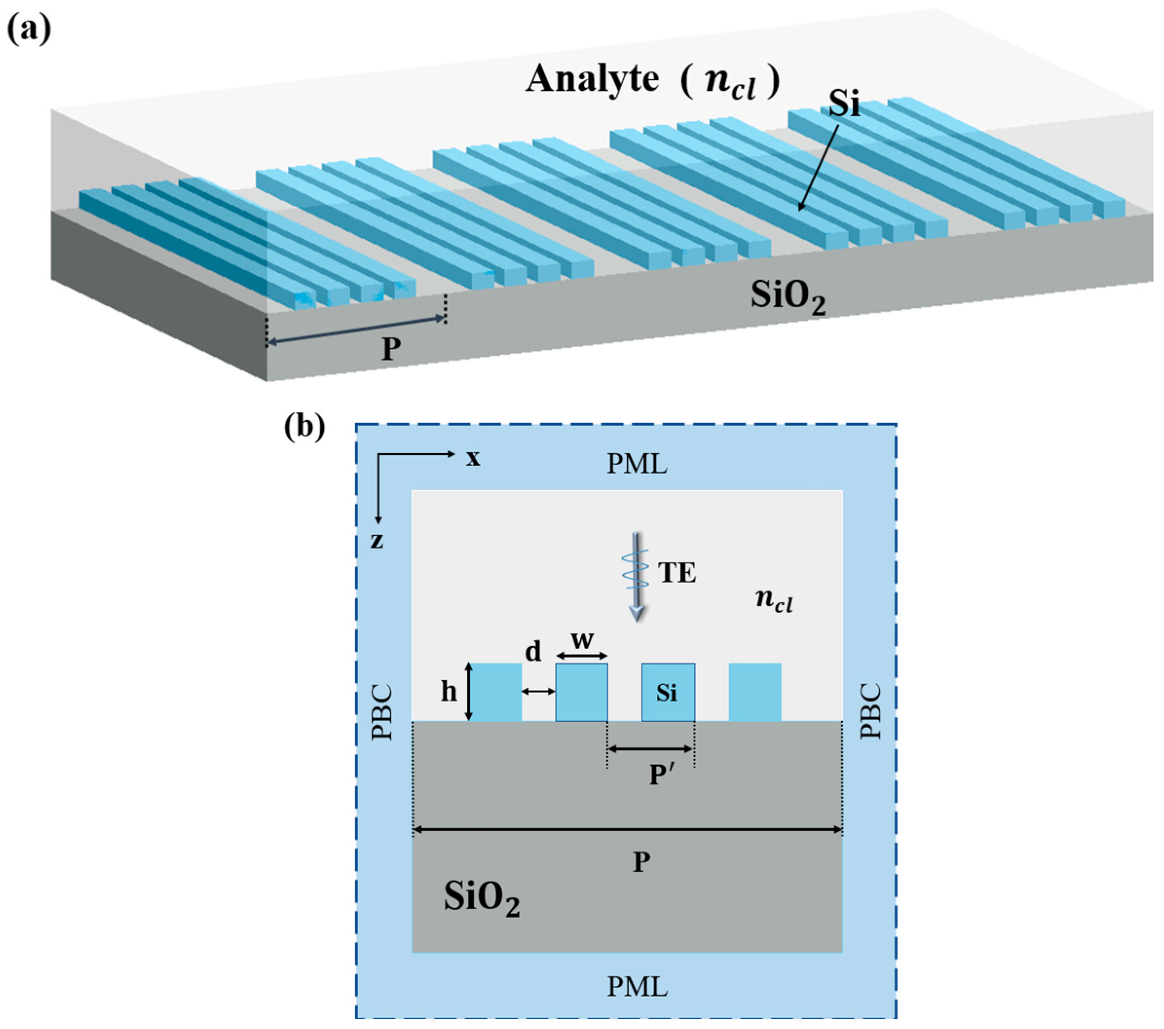
2.1. Simulation Model and Materials

The stereoscopic structure of the HCAM is illustrated in Figure 1a and the cross-section view of the proposed sensor can be viewed in Figure 1b. The HCAM has been covered in a liquid to be measured of refractive index n c l . Set the light as a TE polarised plane wave incident perpendicularly. The structure is infinite in the y-direction and the periodic boundary condition (PBC) is set along the x-direction. In order to absorb the outgoing electromagnetic field, a perfectly matched layer (PML) is assumed at the z boundary. When the TE wave reaches the PML layer, it is completely accepted and not reflected by the layer.
The ideal HCAM can be fully characterized with four parameters: period P = 895 nm, silicon grating height h = 270 nm, width w = 140 nm and distance between grating d = 65 nm. Refractive indexes for both Si and SiO2 materials can be found in the Palik Refractive Index database values [26]. It is fundamental to be capable of achieving sharp Fano resonances while efficiently performing the detection and allowing the analyte to obtain local field enhancement. In order to better validate the sensor performance, the parameters are improved based on previous studies [27] and the improved structure is prepared and experimentally tested.

2.2. Methods and Formula

In this paper, the temporal coupled-mode theory (TCMT) and the eigenmode theory are adopted to establish the theoretical model for analyzing the Fano resonance effect, and the multiple Fano resonance optical sensor HCAM-based is designed. The optical properties of the sensor have been analysed by using finite element method (FEM)-based COMSOL Multiphysics. FEM simulation is used to calculate the eigenvalues of the HCAM as well as through the TCMT to compute the line-shape function. The proposed superstructure can be regarded as a symmetric single-mode two-port system based on the time-coupled mode theory. In the TCMT method, the coupled mode equation can be written as follows:
d α d t = j ω 0 1 τ α + k 1   k 2 W 1 + W 2 +
W 1 W 2 = C W 1 + W 2 + + k 1 k 2 α
where α is the amplitude of the resonant mode, ω 0 and τ are the central frequency and the total decay rate of the resonance, respectively. W 1 + and W 2 + are the amplitude of the incident wave; W 1   and W 2 are the amplitudes of the outgoing waves; k1 and k2 are the coupling coefficients at two ports. The matrix C must be an arbitrary scattering matrix, and if the coupling constant is fixed, the phase of the coupling constant can be determined from the scattering matrix C of the direct process. The scattering matrix C is expressed as follows:
C = e i Φ r j t j t r
where Φ is the phase factor; r and t are amplitude reflection coefficient and transmission coefficient respectively.
Based on the above discussion the transmission spectra of the HCAM are calculated using the formula below [28]:
T = 1 r 2 ω ω 0 2 + t 2 N i m a g 2 2 r t ω ω 0 N i m a g ω ω 0 2 + N i m a g 2
The eigenvalue is X = X r e a l i X i m a g , where X r e a l and X i m a g are the real part and the imaginary part of the X , respectively; r and t are the reflection and transmission coefficients of the scattering matrix, ω is the resonance frequency.

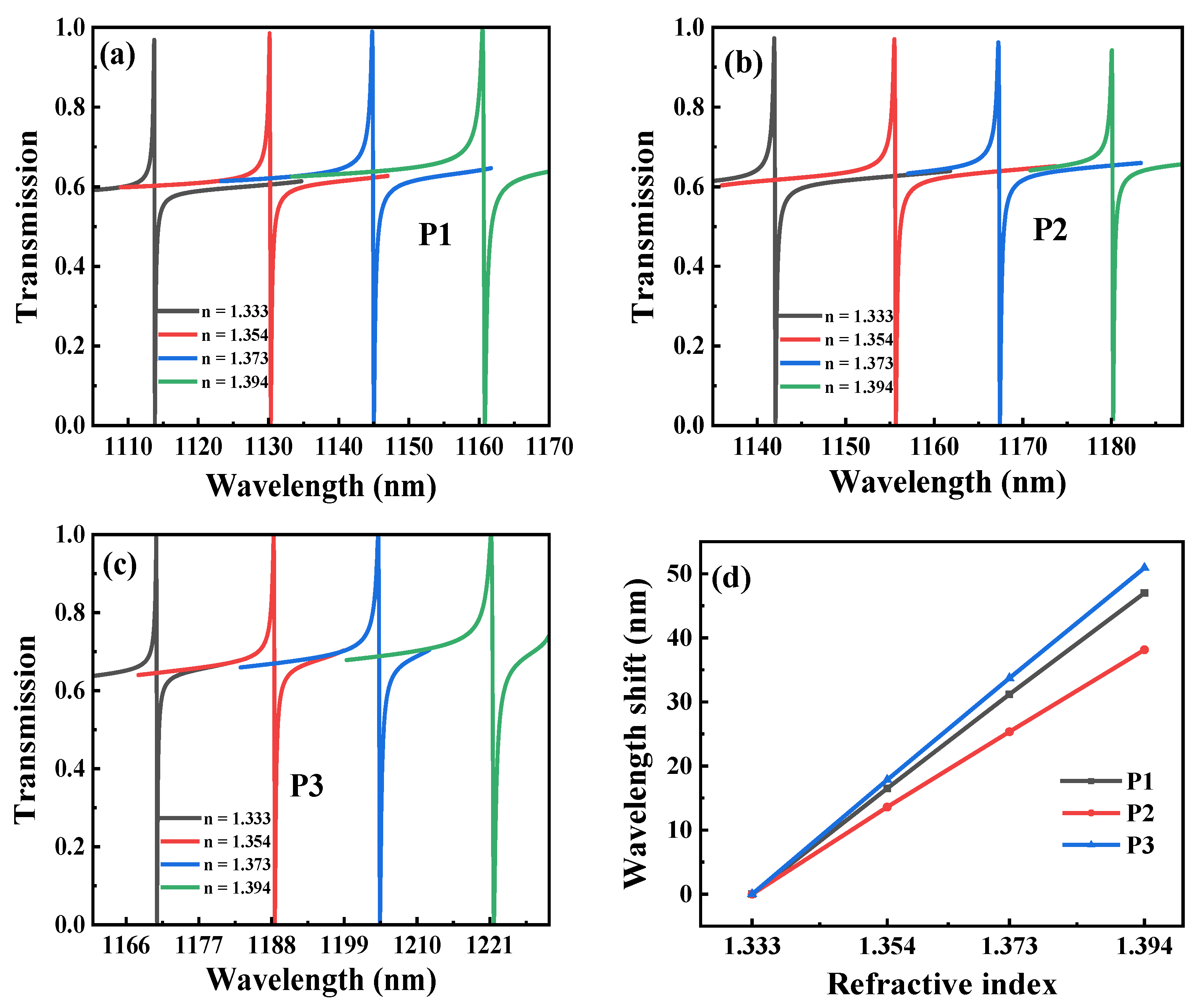
2.3. Simulation Results and Discussions

The refractive indexes for glycerol concentrations of 0%, 17%, 32% and 47% correspond to 1.333, 1.354, 1.373 and 1.394, respectively. The transmission spectra of HCAM are plotted for different refractive indexes (corresponding to different concentrations in the experiment), as shown in Figure 2a–c. There are three sharp Fano resonance peaks with modulation depths close to 100% in the transmission spectra. Modulation depth T = T peak T dip , i.e., is the difference in transmittance between the Fano peaks and the Fano dips. A distinct change in the location of these peaks can be seen, although the change in the index is small. The main performance parameters of the optical resonance sensor are the sensitivity (S) and FOM. Herein, S = Δ λ / Δ n , in which Δ n represents the amount of change in refractive index and Δ λ represents the shift of the resonance peak as the refractive index changes. FOM = S / FWHM , in which the S divided by the full width at half maximum (FWHM). The optimal sensitivity of P1, P2 and P3 peaks are calculated to be 785.6, 647.2 and 849.3, corresponding to FOM of 1671.4, 1966.5 and 2573.5, respectively. The maximum Q-factor obtained at P2 can exceed 3.6 × 10³.

3. Experimental Section
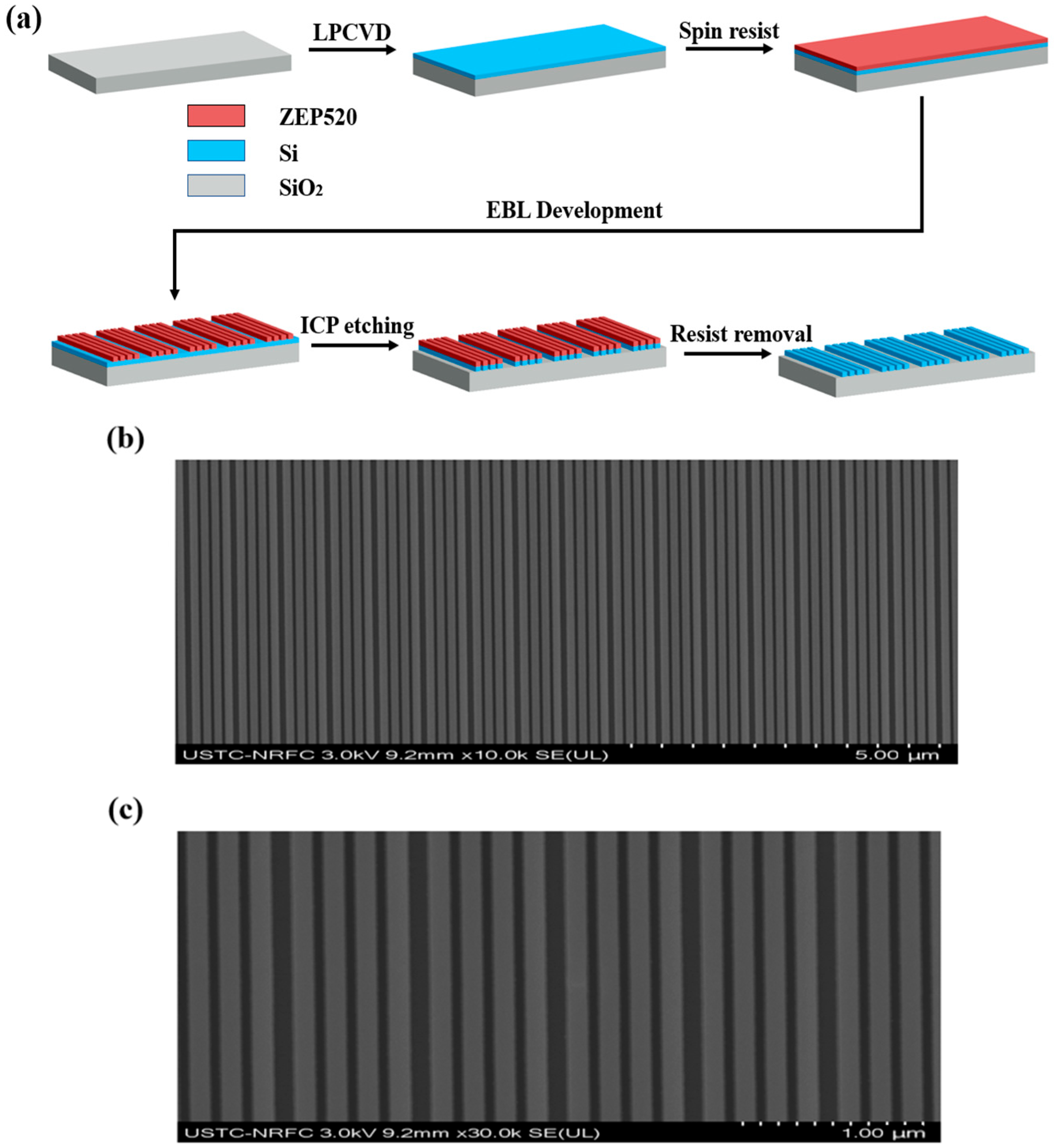
3.1. Fabrication

A schematic diagram illustrating the manufacturing procedure of the HCAM structures can be seen in Figure 3a. First, Si films are deposited on SiO2 substrates by using the method of low-pressure physical vapor deposition (LPCVD). ZEP520A photoresist is spin-coated and baked onto a Si plane. Next, using the EBL and development techniques. Then, the grating structures can be obtained by ICP etching. Eventually, the photoresist is removed away with deionized water. The entire process is simple and can be applied to the actual structure of sensor at low cost. Figure 3b,c show the scanning electron microscope (SEM) images from the HCAM in top view. There is no obvious unevenness in the edge of etched grating strips.

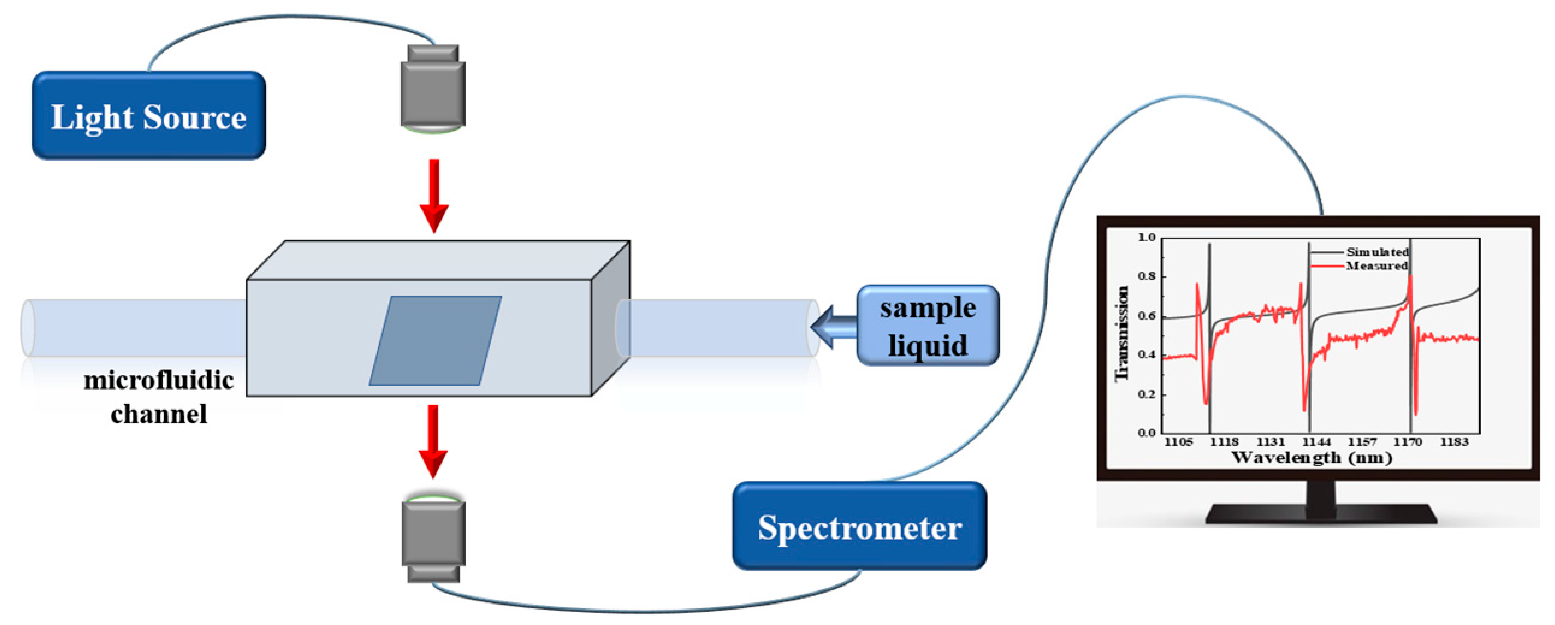
3.2. Testing Process

The test device for evaluating the sensing properties of metastructure is illustrated in Figure 4. The wide-spectrum (470–2400 nm) lasers are adopted for the light source and the full-spectrum power of the laser can be controlled by computer software. A pair of fiber collimators are used to realize light emission and space light collection. The fabricated sensor is immersed in different refractive index solutions and TE polarized light is incident vertically upon the device. The transmission spectrum is recorded using an optic spectrometer when the refractive index of the measured solution changes. In this paper, glycerol solutions diluted to different concentrations by purified water are employed as source solutions to be tested.

4. Experimental Results and Discussion

4.1. Comparison of Experimental and Simulation Data

A comparison of the simulated and experimental transmission profiles of HCAM in liquid is illustrated in Figure 5. The HCAM etches the grating on top of the waveguide layer, and it is the presence of the diffraction grating that gives rise to the GMR. Because the diffraction gratings are thought of as the planar waveguide with periodic modulation, leakage in the grating waveguide is caused by the periodic modulation of the grating and consequent redistribution of the wave energy leakage, which forms the guided-mode resonance. Multiple leakage modes can be stimulated for a given abrupt diffraction order, so that multiple resonance peaks are excited. Therefore, when the liquid is applied to the surface of the structure, the simulation and experiment can produce three sharp Fano resonance peaks. As sensing technology has evolved, research related to optical Fano resonance has expanded from single Fano resonance to multi-Fano resonance. Multi-Fano resonances can be widely used in multi-wavelength surface enhancement spectroscopy, multi-channel biosensors and wavelength slow light devices. However, few studies have achieved three or more high Q-factor Fano resonances simultaneously. Due to the irregular linear shape of the Fano resonances and the ability of small changes in refractive index to cause significant linear shifts in the Fano peaks, multiple Fano resonances can enable multiple detection points in practical applications, making the results more reliable and offering good potential for applications in micro and nano refractive index sensing. The corresponding simulated spectra are also shown in Figure 5, indicated by the red curve. The measured and simulated outcomes agree with each other at peak and dip positions.

4.2. Experimental Results

To observe the sensing performance of the HCAM, the metastructure is immersed in a glycerol-water mixture at 0%, 17%, 32% and 47%, respectively. The transmission spectra at different concentrations are observed and recorded, as shown in Figure 6. Results of experimental tests have shown the optimal sensitivity obtained at P2 can reach 306 nm/RIU and FOM is up to 124. The proposed sensor has a high standard of performance compared to previously studied plasma sensors and other all-dielectric sensors.

4.3. Causes of Experimental Error

It is probable that dimensional errors arising from the fabrication of the samples lead to the introduction of scattering losses and influence the coherence of the sensor. Moreover, the extra losses in Si caused by the creation of surface states in the reactive ion etching process results in a slight increase of uptake compared to theory in the array. It is worth noting that there will be some errors in the structure preparation process and it can be difficult to achieve a rectangular grating with a regular contour like a simulated structure diagram. Hence, there are inevitable deviations in the thickness of the gratings as well as the gaps among the gratings. As a result, the obtained curves are offset from the simulated transmission spectra in the experiment, the modulation depth is slightly smaller, however these deviations are acceptable. It can improve during packaging and commercialization. Our proposed technology is well applicable to medical and biological detection.

4.4. Comparison with Existing Sensors

Table 1 compares the sensitivity, figure of merit of sensors comprised of diverse materials or structures. These sensors have disadvantages such as complex preparation, high losses and low sensing indexes. The abbreviations for experiments and simulations in the table are Exp. and Sim., respectively. Sensors based on metal materials approach suffers from the two main limitations. First, complex and expensive equipments required to couple and monitor light hinders its application in the fields of various concentrations of glycerol water mixtures testing and sensing. Second, their decay lengths are the order of half of the resonance wavelength (few hundred nanometers), meaning that it is not quite suitable for monitoring the local RI changes at the nanometer level caused by small analytes.
The proposed structure in this paper uses all-dielectric materials and owns the characteristics of low losses, high confinements and compatibility with CMOS technology, whilst supporting ultra-high quality (Q) factor Fano resonance and strongly local field enhancements. To increase the FOM of a sensor, we have reduced the full width at half maximum (FWHM) and increased its sensitivity. Thus, more accurate detection of glycerol-water mixtures can be achieved.

5. Conclusions

In this work, we have designed and fabricated an all-dielectric metastructure for application in the sensing area through the use of intelligent design and nanotechnology experimentally. The sensing properties of the all-dielectric metastructure can be significantly enhanced where high Q-factor and FOM can be achieved owing to the benefits such as the lower absorption losses, narrow linewidth, and local field enhancement. Therefore, we calculate the maximum Q-factor of exceeding 3.6 × 10³ by simulation, and the experimental results show that modulation depths of up to 60%, with an optimum sensitivity of 306 and FOM of 124, respectively. The HCAM can be produced simply and inexpensively, which makes it possible to integrate with CMOS easily. It has a broad application potential in sensing fields such as label-free chemistry and biosensing.

Author Contributions

Writing—original draft preparation, L.B.; writing—review and editing, X.F., S.C., Y.Y., C.L., H.Z., X.W., W.F. and S.K.; project administration, funding acquisition—X.F., W.F., H.N., C.B. and S.K. All authors have read and agreed to the published version of the manuscript.

Funding

This work is supported by the Cultivation Plan for Young Scholars in Universities of Shandong Province; the Natural Foundation of Shandong Province (ZR2021MF053, ZR2022MF253, ZR2021MF070 and ZR2022MF305); the Open Fund of the Key State Laboratory (BUPT, IPOC) (IPOC2021B07); the Double-Hundred Talent Plan of Shandong Province, China.

Institutional Review Board Statement

Not applicable.

Informed Consent Statement

Not applicable.

Data Availability Statement

Data will be made available on request.

Conflicts of Interest

The authors declare no conflict of interest.

References

  1. Zhu, L.; Kapraun, J.; Ferrara, J.; Chang-Hasnain, C. Flexible photonic metastructures for tunable coloration. Optica 2015, 3, 255. [Google Scholar] [CrossRef]
  2. Zhu, L.; Yang, W.; Chang-Hasnain, C. Very high efficiency optical coupler for silicon nanophotonic waveguide and single mode optical fiber. Opt. Express 2017, 25, 18462–18473. [Google Scholar] [CrossRef] [PubMed]
  3. Liu, J.; Hu, H.; Shao, S. Polarization-insensitive ultra-narrow plasmon-induced transparency and short-range surface plasmon polariton bloch wave in ultra-thin metallic film nanostructures. Plasmonics 2019, 14, 139. [Google Scholar] [CrossRef]
  4. Han, C.; Wang, Z.; Lv, X.; Dai, J. Phase and Wavelength Sensitivities of Optical Refractive Index Sensor Based on Fano Resonance. IEEE Sens. J. 2022, 22, 21628–21634. [Google Scholar] [CrossRef]
  5. Chu, S.; Wang, Q.; Yu, L.; Gao, H.; Liang, Y.; Peng, W. Numerical investigation on multiple resonant modes of double-layer plasmonic grooves for sensing application. Nanomaterials 2020, 10, 308. [Google Scholar] [CrossRef]
  6. Luo, X.; Li, X.; Lang, T.; Jing, X.; Hong, Z. Excitation of high q toroidal dipole resonance in an all-dielectric metasurface. Opt. Mater. Express 2019, 10, 358. [Google Scholar] [CrossRef]
  7. Chen, Y.; Zhou, X.; Zhang, M.; Xiao, C.; Zhou, J. Fano resonance sensing based on coupled sub-wavelength dielectric grating and periodic photonic crystal. Phys. Lett. A 2020, 384, 126877. [Google Scholar] [CrossRef]
  8. Lim, W.; Han, S.; Gupta, M.; Macdonald, K.; Singh, R. Near-infrared linewidth narrowing in plasmonic fano-resonant metamaterials via tuning of multipole contributions. Appl. Phys. Lett. 2017, 111, 061104. [Google Scholar] [CrossRef]
  9. Hu, J.; Huang, Y.; Ren, X.; Duan, X.; Li, Y.; Wang, Q.; Zhang, X.; Wang, J. Modeling of fano resonance in high-contrast resonant grating structures. Chin. Phys. Lett. 2014, 31, 064205. [Google Scholar] [CrossRef]
  10. Qiao, P.; Yang, W.; Chang-Hasnain, C. Recent advances in high-contrast metastructures, metasurfaces, and photonic crystals. Adv. Opt. Photonics 2018, 10, 180. [Google Scholar] [CrossRef]
  11. Qian, L.; Wang, K.; Zhu, W.; Han, C.; Yan, C. Enhanced sensing ability in a single-layer guided-mode resonant optical biosensor with deep grating. Opt. Commun. 2019, 452, 273–280. [Google Scholar] [CrossRef]
  12. Luk’yanchuk, B.; Zheludev, N.; Maier, S.; Halas, N.; Nordlander, P.; Giessen, H.; Chong, C. The Fano resonance in plasmonic nanostructures and metamaterials. Nat. Mater. 2010, 9, 707–715. [Google Scholar] [CrossRef]
  13. Yang, Z.; Hao, Z.; Lin, H.; Wang, Q. Plasmonic Fano resonances in metallic nanorod complexes. Nanoscale 2014, 6, 4985. [Google Scholar] [CrossRef]
  14. Brunetti, G.; Conteduca, D.; Armenise, M.N.; Ciminelli, C. Novel micro-nano optoelectronic biosensor for label-free real-time biofilm monitoring. Biosensors 2021, 11, 361. [Google Scholar] [CrossRef]
  15. Maleki, M.; Mehran, M. Guided-mode resonance sensors: Different schemes for different applications. JOSA B 2022, 39, 1634–1643. [Google Scholar] [CrossRef]
  16. Liu, Z.; Liu, Z.; Li, J.; Li, W.; Li, J.; Gu, C.; Li, Z. 3D conductive coupling for efficient generation of prominent Fano resonances in metamaterials. Sci. Rep. 2016, 6, 27817. [Google Scholar] [CrossRef]
  17. Liu, B.; Yao, X.; Zhang, L.; Lin, H.; Chen, S.; Zhong, J.; Ren, B. An efficient platform for flexible engineering of superradiant, fano-type, and subradiant resonances. ACS Photonics 2015, 2, 1725–1731. [Google Scholar] [CrossRef]
  18. Bakhti, S.; Bonod, N.; Dhuey, S.; Schuck, P.; Destouches, N. Fano-like resonance emerging from magnetic and electric plasmon mode coupling in small arrays of gold particles. Sci. Rep. 2016, 6, 32061. [Google Scholar] [CrossRef]
  19. Wang, W.; Zheng, L.; Xiong, L.; Qi, J.; Li, B. High q-factor multiple fano resonances for high-sensitivity sensing in all-dielectric metamaterials. OSA Continuum 2019, 2, 2818. [Google Scholar] [CrossRef]
  20. Wang, H.; Jiang, L.; Xiang, P. Improving the durability of the optical fiber sensor based on strain transfer analysis. Opt. Fiber Technol. 2018, 42, 97–104. [Google Scholar] [CrossRef]
  21. Zhang, X.; Wang, L.; Tang, S.; Cui, H.; Xie, X.; Wu, H.; Liu, X.; Yang, D.; Wang, H.; Xiang, P. Investigations on the shearing performance of ballastless CRTS II slab based on quasi-distributed optical fiber sensing. Opt. Fiber Technol. 2023, 75, 103129. [Google Scholar]
  22. Wang, H.; Xiang, P.; Jiang, L. Optical fiber sensing technology for full-scale condition monitoring of pavement layers. Road Mater. Pavement Des. 2020, 21, 1258–1273. [Google Scholar] [CrossRef]
  23. Hu, J.; Liu, X.; Zhao, J.; Zou, J. Investigation of Fano resonance in compound resonant waveguide gratings for optical sensing. Chin. Opt. Lett. 2017, 15, 030502. [Google Scholar]
  24. Singh, L.; Jain, S.; Kumar, M. Nanophotonic device based on fano resonance in engineered slot waveguide for optical detection of viral infections. IEEE Sens. J. 2021, 21, 2805–2812. [Google Scholar]
  25. Kilic, S.; Kocaman, S. Highly sensitive and tunable fano-like rod-type silicon photonic crystal refractive index sensor. IEEE Sens. J. 2021, 21, 7551–7557. [Google Scholar] [CrossRef]
  26. Edwards, D. Silicon (Si). In Handbook of Optical Constants of Solids, 2nd ed.; Palik, E.D., Ed.; Elsevier: Amsterdam, The Netherlands, 1985. [Google Scholar]
  27. Bi, L.; Fan, X.; Zhao, H.; Liu, L.; Wei, X.; Niu, H.; Li, C.; Bai, C.; Fang, W. Enhanced sensing ability in multiple Fano resonance optical biosensor with high-contrast metastructures. Results Opt. 2022, 9, 100276. [Google Scholar] [CrossRef]
  28. Fan, S.; Suh, W.; Joannopoulos, J. Temporal coupled-mode theory for the Fano resonance in optical resonators. JOSA A 2003, 20, 569–572. [Google Scholar] [CrossRef]
  29. Liu, G.; Zhai, X.; Wang, L.; Lin, Q.; Xia, S.; Luo, X.; Zhao, C. A high-performance refractive index sensor based on fano resonance in si split-ring metasurface. Plasmonics 2018, 13, 15–19. [Google Scholar] [CrossRef]
  30. Yang, Y.; Kravchenko, I.; Briggs, D.; Valentine, J. All-dielectric metasurface analogue of electromagnetically induced transparency. Nat. Commun. 2014, 5, 1–7. [Google Scholar] [CrossRef]
Figure 1. (a) HCAM structure schematic. (b) Computational model of the HCAM structure.
Photonics 10 00616 g001
Figure 2. Three resonance peaks in transmission spectra at different refractive indexes (a) P1. (b) P2. (c) P3. (d) The wavelength shifts of the resonance peaks.
Photonics 10 00616 g002
Figure 3. HCAM. (a) Schematic displaying the fabrication procedure of HCAM. (b,c) Top-view SEM images of the HCAM.
Photonics 10 00616 g003
Figure 4. Experimental apparatus for evaluating sensing performance of metastructures.
Photonics 10 00616 g004
Figure 5. Transmission spectra of the HCAM in liquid. The measured and simulated results are shown as black solid line and red dotted line, respectively.
Photonics 10 00616 g005
Figure 6. Transmission spectra of the three resonance peaks measured at different concentrations of glycerol-water mixed liquids. (a) P1, (b) P2, (c) P3.
Photonics 10 00616 g006
Table 1. Performance comparison of proposed sensor with existing sensors.
Sensor TypeSim.: SFOMExp.: SFOMRef.
Metal mushroom arraysn.r. an.r. a525 nm/RIU38[1]
Single-layer guided mode resonance structure241.7 nm/RIU690229.43 nm/RIU31.52[11]
Si split-ring452 nm/RIU56.5n.r. an.r. a[29]
Silicon rods and ringsn.r. an.r. a289 nm/RIU103[30]
All-dielectric metastructure849.3 nm/RIU2573.5306 nm/RIU124This work
a not reported.
Disclaimer/Publisher’s Note: The statements, opinions and data contained in all publications are solely those of the individual author(s) and contributor(s) and not of MDPI and/or the editor(s). MDPI and/or the editor(s) disclaim responsibility for any injury to people or property resulting from any ideas, methods, instructions or products referred to in the content.

