Article

Application of ZnO/WO3 Composite Nanofiber Photocatalysts in Textile Wastewater Treatment

1
College of Urban and Environmental Sciences, Xuchang University, Xuchang 461000, China
2
College of Chemical and Materials Engineering, Xuchang University, Xuchang 461000, China
*
Author to whom correspondence should be addressed.
Separations 2023, 10(6), 339; https://doi.org/10.3390/separations10060339
Submission received: 12 April 2023 / Revised: 8 May 2023 / Accepted: 11 May 2023 / Published: 31 May 2023
(This article belongs to the Section Environmental Separations)

Abstract

:
Semiconductor photocatalysis technology is an environmentally friendly and efficient emerging technology. This method can use sunlight as a driving force to quickly decompose organic pollutants in water bodies. Zinc oxide (ZnO) and tungsten oxide (WO3) photocatalysts can absorb sunlight and participate in photocatalytic degradation reactions due to their relatively narrow band gap. Highly photosensitive WO3 nanofibers and ZnO/WO3 composite nanofibers were fabricated via the electrospinning method. When 100 mg/L of rhodamine B (Rh B) solution was used as the degradation substrate, the degradation efficiencies of WO3 and ZnO/WO3 for Rh B dye were 70% and 90%, respectively, after a photocatalytic reaction of 120 min. The surface morphology, crystal structure, and optical properties of ZnO/WO3 composite nanofibers and WO3 nanofibers were characterized by SEM, XRD, XPS, and UV-vis absorption spectra, and the experimental results were analyzed and explained using different mechanisms. The results show that ZnO/WO3 composite nanofibers have better UV-visible light absorption performance, and the sample has a higher UV-visible light utilization rate. This was mainly due to the fact that a P-N heterojunction was formed in the semiconductor composite, and the electron–hole pair could realize rapid separation under the drive of a built-in electric field force, which promoted the migration of carrier. Therefore, the photocatalytic activity of the ZnO/WO3 catalyst was significantly higher than that of the WO3 catalyst, which promoted rapid improvement of the photocatalytic degradation efficiency of the Rh B dye.

Graphical Abstract

1. Introduction

Water occupies an indispensable position in human production activities and is the source of human survival [1]. However, with the development of science and technology, a large amount of sewage is discharged into nature, polluting the water environment. The aggravation of water pollution leads to the shortage of water resources. Therefore, the development of wastewater treatment and reuse technology is urgently required in the future [2]. At present, biological treatment, adsorption, chemical oxidation treatment, physical chemical treatment, and other methods are mainly used for wastewater treatment [1,3]. In recent years, with the rapid development of industry and the widespread use of various drugs, more and more new pollutants have been detected in the water environment [4,5,6,7]. These pollutants are widely distributed and of various kinds. When traditional treatment methods are adopted, there will be many problems, such as difficulty in degrading pollutants thoroughly and secondary pollution caused by by-products after water treatment. An increasing number of researchers pay attention to the research of water pollution control technology, and develop low-cost green photocatalytic degradation technology [8,9,10,11]. The core of photocatalytic technology is photocatalytic materials, most of which are semiconductor materials. The semiconductor photocatalyst is simple and efficient to degrade pollutants, which can completely mineralize and remove pollutants without secondary pollution, making it a research hotspot of current sewage treatment technology [12].
Since the discovery of TiO2, it has been widely studied as a classical photocatalyst. At present, TiO2 preparation, crystal control, morphology control, and other aspects of technology have become increasing perfect [13,14,15]. However, a TiO2 wide band [1] gap leads to a low visible light response rate, and cannot fully and effectively use visible light for photocatalysis, restricting its widespread industrial utilization [16]. In order to solve this problem, researchers began to modify TiO2, hoping to broaden its visible light absorption range, so as to modify TiO2 using semiconductor doping elements, semiconductor surface modification, and the construction of heterojunction. Park B.G. et al. [17] prepared TiO2 photocatalysts doped with metal ions (Fe, Ag, and Ni) and non-metallic N using the sol–gel method, and explored its photocatalytic performance under visible light. The results showed that the doped elements successfully expanded the light absorption range of TiO2 to a visible light region. The photoluminescence intensity of N/Ni/TiO2 was the highest among many doping schemes, and the photodegradation of formaldehyde and methylene blue was the best. Akpan U.G. et al. [18] researched the TiO2 doping of various sol–gels and sol–gel-related systems, and by doping rare earth metal ions, transition metal ions, alkaline earth metal ions, and non-metallic ions, they finally concluded that TiO2 co-doping generally improved the photocatalytic efficiency of the catalysts. However, doping transition metals can reduce the photocatalytic efficiency of the catalyst. TiO2 modified by these methods can indeed improve its visible light absorption performance to a certain extent, but the photocatalytic activity of the material still has plenty of room for improvement. Therefore, it is a very valuable research mission to search for other new photocatalytic materials with stable properties, suitable band structures, and visible light responses. At present, common photocatalytic materials include TiO2, WO3, ZnO, Fe2O3, Ag3PO4, etc. [19,20,21,22]. Among them, WO3 is a kind of metal oxide with photocatalytic activity, a large surface area, stable properties, good adsorption, and excellent physical and chemical properties, and is an important N-type semiconductor. At the same time, WO3 is non-toxic, cheap, and easy to prepare; its band gap is relatively small, and its band structure is suitable and can respond to visible light [23,24,25]. These advantages eventually make it a crucial material in the field of photocatalysis. Li et al. [26] synthesized MnWO4/WO3 nanofibers to degrade methylene blue (MB) dye in a short and rapid time, and proposed a new low-cost and efficient way to degrade organic pollutants.
The band gap width of WO3 is 2.4–2.7 eV, which is lower than that of TiO2 (3.2 eV). Therefore, theoretically, WO3 has a higher utilization rate of sunlight. Under the excitation of visible light, the electrons in the valence band of WO3 undergo a semiconductor transition to the conduction band, which stimulates both electrons and holes to participate in the photocatalytic degradation reaction [27]. In addition, compared with TiO2, the price of WO3 is relatively low. Hence, it is cost-effective, with practical production and application conditions.
ZnO is one of the most commonly used photocatalytic materials in the third generation of wide-band-gap semiconductors, and its band gap is about 3.2 eV [28,29]. It has a conductivity of about 10−7–10−3 S/cm [30]. Under the excitation of near-ultraviolet (UV) light, ZnO can show excellent photocatalytic activity in degrading organic pollutants. However, the band gap width of ZnO is too large, so it is necessary to reduce its gap width, which can improve the absorption range of visible light [31]. The photocatalytic performance of ZnO can also be improved by reducing the recombination rate of photoelectrons and holes. At the same time, the morphology of nano-ZnO can be controlled and the specific surface area can be increased to improve the photocatalytic performance of ZnO. However, the undoped ZnO exhibits low photocatalytic efficiency due to the inadequate utilization of sunlight and the rapid recombination of charge carriers, thus affecting the catalysis rate of the toxic dye degradation.
Redouane et al. [32] synthesized the ZnO/Mt nanocomposite via a simple approach. In comparison with pure ZnO, the photocatalytic activity of ZnO/Mt nanocomposite towards the degradation of the organic pollutant Rhodamine B (Rh B) is greatly enhanced when exposed to UV light, and they have found the ZnO/Mt nanocomposite sample can effectively degrade hazardous organic pollutants under UV light and can have strong potential application in environmental management. Hence, it is necessary to reduce the band gap of the ZnO to harvest more sunlight for photocatalysis.
In this study, WO3 and ZnO/WO3 nanofibers were prepared via electrospinning, and their photocatalytic properties were tested. The ZnO/WO3 composite nanofibers can respond to both near-UV and visible light irradiations. This composite method can effectively broaden the spectrum response range of semiconductor materials. The relationship between the photocatalytic activity and degradation efficiency of Rh B solution with a concentration of 100 mg/L was studied. A 300 W xenon lamp was used as the light source to carry out photocatalytic degradation experiments in a photocatalytic protection chamber. The photocatalytic performance of WO3 nanofibers and ZnO/WO3 composite nanofibers in the degradation of Rh B was compared under the irradiation of UV-visible light. The experimental results show that the adsorption and photocatalytic degradation efficiencies of ZnO/WO3 composite nanofibers were significantly higher than those of WO3 nanofibers.

2. Materials and Methods

2.1. Materials

Ammonium metatungstate (AMT), rhodamine B(Rh B), zinc acetate (Zn (CH3COO)2), N, n-dimethylformamide (DMF), and absolute ethyl alcohol were purchased from Shanghai Aladdin Biochemical Technology Co., Ltd. (Shanghai, China). Polyvinylpyrrolidone (PVP) and acetone were purchased from Maclin Biochemical Technology Co., LTD (Shanghai, China). All the reagents used in the synthesis procedure were analytical pure-grade chemicals.

2.2. Preparation of WO3 and ZnO/WO3 Composite Nanofibers

Firstly, a certain amount of AMT and PVP was weighed using an electronic balance and dissolved into 6 mL of deionized water and 3 mL of DMF. Transparent WO3 precursor solution was formed after 10 h of constant rotation and agitation with magnetometer. Then, 6 mL of deionized water and 3 mL of DMF were added to a certain amount of AMT and zinc acetate in a beaker. Uniform transparent ZnO/WO3 precursor solution was obtained after the mixed solution was stirred at a uniform speed with a magnetic agitator for 10 h.
The configured WO3 and ZnO/WO3 precursor solutions were placed into syringes, respectively. Before electrostatic spinning, the voltage of the working high-voltage power supply was set to 14.5 kV, the needle of the syringe was connected to the positive pole of the DC power supply, and the aluminum foil metal plate was connected to the negative pole of the DC voltage source. The spinning needle was adjusted to a certain distance from the aluminum foil. Generally, the distance between the aluminum foil metal plate and the spinning needle was set to 10 cm. WO3 and WO3/ZnO precursor solutions were spun under the action of an electric field force and a double-channel injection pump at uniform speed, and the filamentous nanofibers were received by an aluminum foil metal plate. Then, the prepared fiber samples were kept under a constant temperature at 80 °C for 3 h in a drying oven. Finally, the two fiber samples were placed in a tubular furnace at a heating rate of 2 °C/min and calcined at 500 °C for 4 h to obtain WO3 and ZnO/WO3 composite nanofibers.

2.3. Photocatalytic Degradation

The photocatalytic degradation of Rh B using WO3 and ZnO/WO3 nanofiber samples prepared in this study was carried out in a special photocatalytic protective chamber. Firstly, 300 mL of 100 mg/L Rh B aqueous solution was measured and 0.20 g of WO3 and ZnO/WO3 fiber samples were weighed and added into an Rh B aqueous solution, respectively. In order to avoid the interference caused by the adsorption of the dye to the experimental data, the solution was first placed in a dark environment and stirred at a constant speed for 30 min, and then stopped after reaching the adsorption equilibrium condition. Then, a 300 W high-power xenon lamp was used as an UV light source, and the distance between the light source and the reactor was about 15 cm. The photocatalytic degradation experiment was carried out for 120 min under UV irradiation, and 6 mL samples were taken every 20 min. The concentration of Rh B was measured using UV–visible spectrophotometry (PerkinElmer, Lambda 35) at a maximum absorption wavelength of 550–554 nm. The degradation rate was calculated using Equation (1) [6]:
E = 1 C / C 0 × 100 %
where C is the Rh B concentration after photodegradation with UV–visible light and C0 is the initial concentration of Rh B.

2.4. Characterizations

The morphologies of WO3 and ZnO/WO3 nanofiber samples were characterized with a scanning electron microscope (SEM, Zeiss, Jena, Germany), manufactured by Hitachi, Japan (S-3400 model), with a working voltage of 20 KV. SEM involves the use of high-energy electron beams to bombard the surface of the sample and stimulate the physical information of the sample. Through information amplification, collection, and re-imaging, the purpose of material morphology characterization is achieved.
Cu-ray source X-ray diffraction (XRD) was used to determine the crystal structure, manufactured by Brock GMBH (D8advance model). XRD involves the use of X-ray incident beams to determine the crystallinity of a substance, according to the spatial arrangement of atoms and structural characteristics. In this study, XRD was used to test WO3 and ZnO/WO3 nanofiber samples’ crystal shape and growth direction. XRD test conditions were set to 30 kV and 20 mA Cu-Kα radiation to determine the crystal phase and crystal type.
X-ray photoelectron spectroscopy (XPS, Escalab 250Xi, Thermo Fisher Scientific, Waltham, MA, USA) was used to measure the surface ion valence states, elemental composition, and electron binding energy of the WO3 and ZnO/WO3 nanofiber samples. Moreover, XPS can be used to semi-quantitatively analyze the surface composition and determination of element valence. The wide application of XPS technology enables us to determine the valence state and surface electronic state of elements more accurately. This method has the characteristics of shallow analysis depth, small test area, and intact sample shape.
The ultraviolet–visible (UV-vis) absorbance/transmittance data were recorded with a UV-1800 spectrophotometer (Shimadzu Co., Kyoto, Japan) or a Varioskan LUX micro-plate reader (Thermo Fisher Scientific Inc., Waltham, MA, USA). UV-vis can be used to measure the absorption spectrum of UV-visible light with a wavelength range of 200–800 nm, which can be used to test the absorption band edge and band gap width of WO3 and ZnO/WO3 nanofiber samples.

3. Results and Discussion

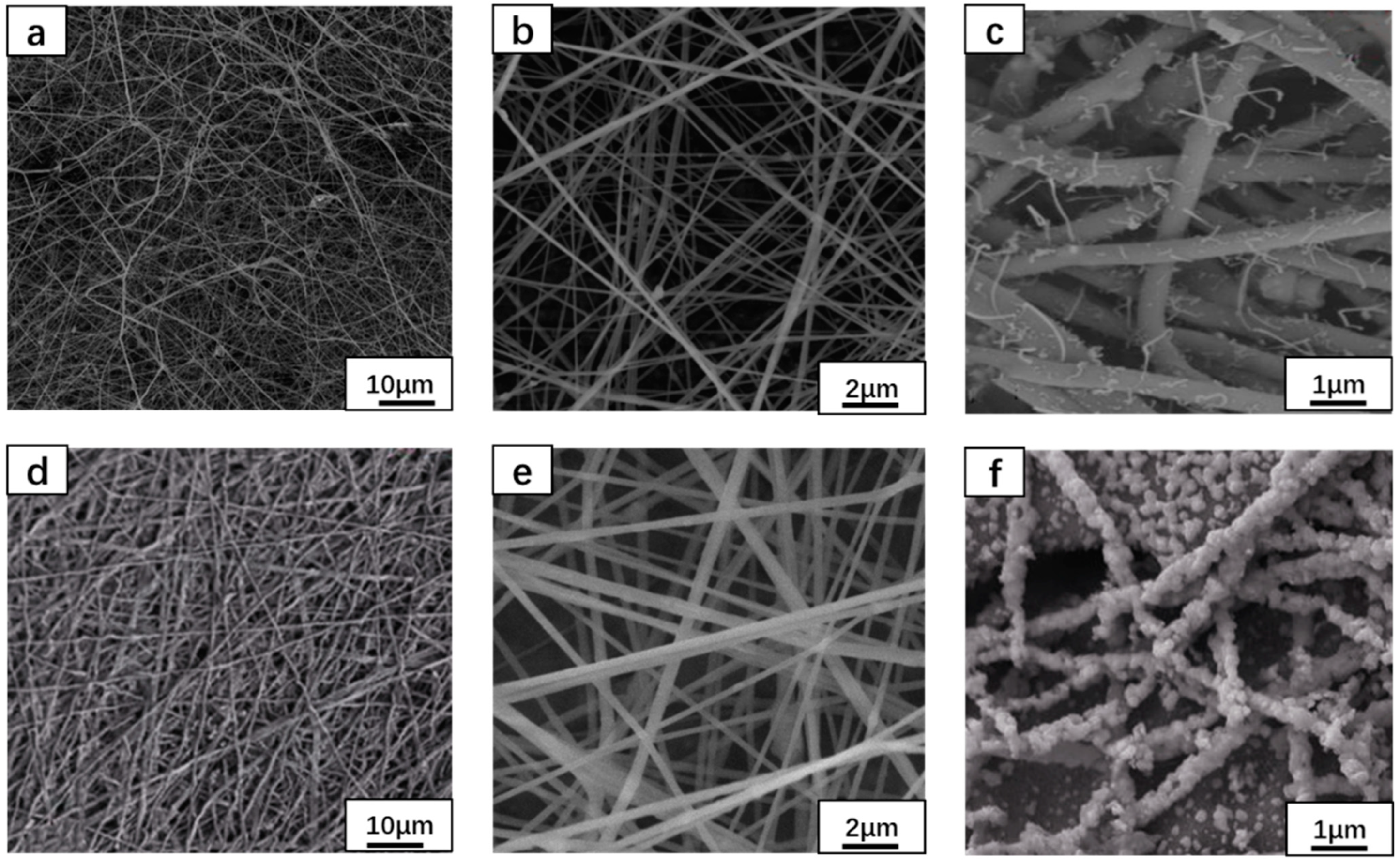
3.1. Morphological Analysis

The morphologies of the prepared WO3 and ZnO/WO3 nanofiber were observed using SEM, as shown in Figure 1. Figure 1a,b show the WO3 nanofibers and Figure 1c shows an SEM image of the WO3 nanofibers calcined at 500 °C. Figure 1d,e show the ZnO/WO3 composite nanofibers and Figure 1f shows the SEM image of the ZnO/WO3 nanofibers calcined at 500 °C. Figure 1 shows that the overall morphologies of WO3 and ZnO/WO3 composite nanofibers were uniformly distributed, with smooth surfaces and similar diameters, as well as an average diameter of about 2 μm. After the nanofibers were calcined at high temperatures, the crystal water was completely decomposed and volatilized, the overall morphology of the fiber changed significantly, the surface pores increased, and the fiber became rougher because the nanoparticles formed by crystallization were widely distributed on the surface of the fiber. After being calcined at high temperatures, ZnO/WO3 composite nanofibers promoted surface particle crystallization due to phase separation, the fiber diameter became thinner, and the surface was rough and had no directional distribution. The fibers thinning would significantly increase the specific surface area, which would help to improve the light absorption efficiency and provide more active sites for the photocatalytic degradation of organic dyes [33].

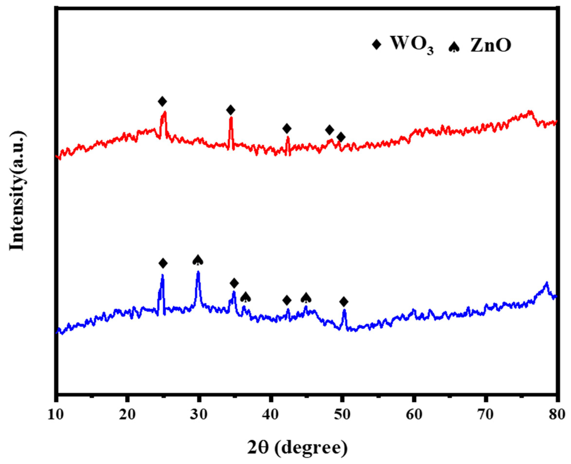
3.2. X-ray Diffraction Spectroscopy

In order to study the phase structure of WO3 nanofibers and ZnO/WO3 composite nanofibers, we analyzed them using a XRD characterization test. As shown in Figure 2, at diffraction angles of 2θ = 23.139°, 34.936°, 42.235°, 46.243°, and 49.362° peaks were observed, corresponding to monoclinic phase WO3. At diffraction angles of 2θ = 30.783°, 36.791°, and 45.764°, peaks were observed, corresponding to ZnO. The XRD pattern shows that the diffraction peaks of WO3 nanofibers and ZnO/WO3 composite nanofibers after high temperature calcination have great intensity and sharp shapes, which proves its high crystallinity. No diffraction peaks of other compounds were detected in the phase structure, indicating that the WO3 nanofibers and ZnO/WO3 composite nanofibers prepared by us are single pure substances.

3.3. Absorption Characteristics Analysis
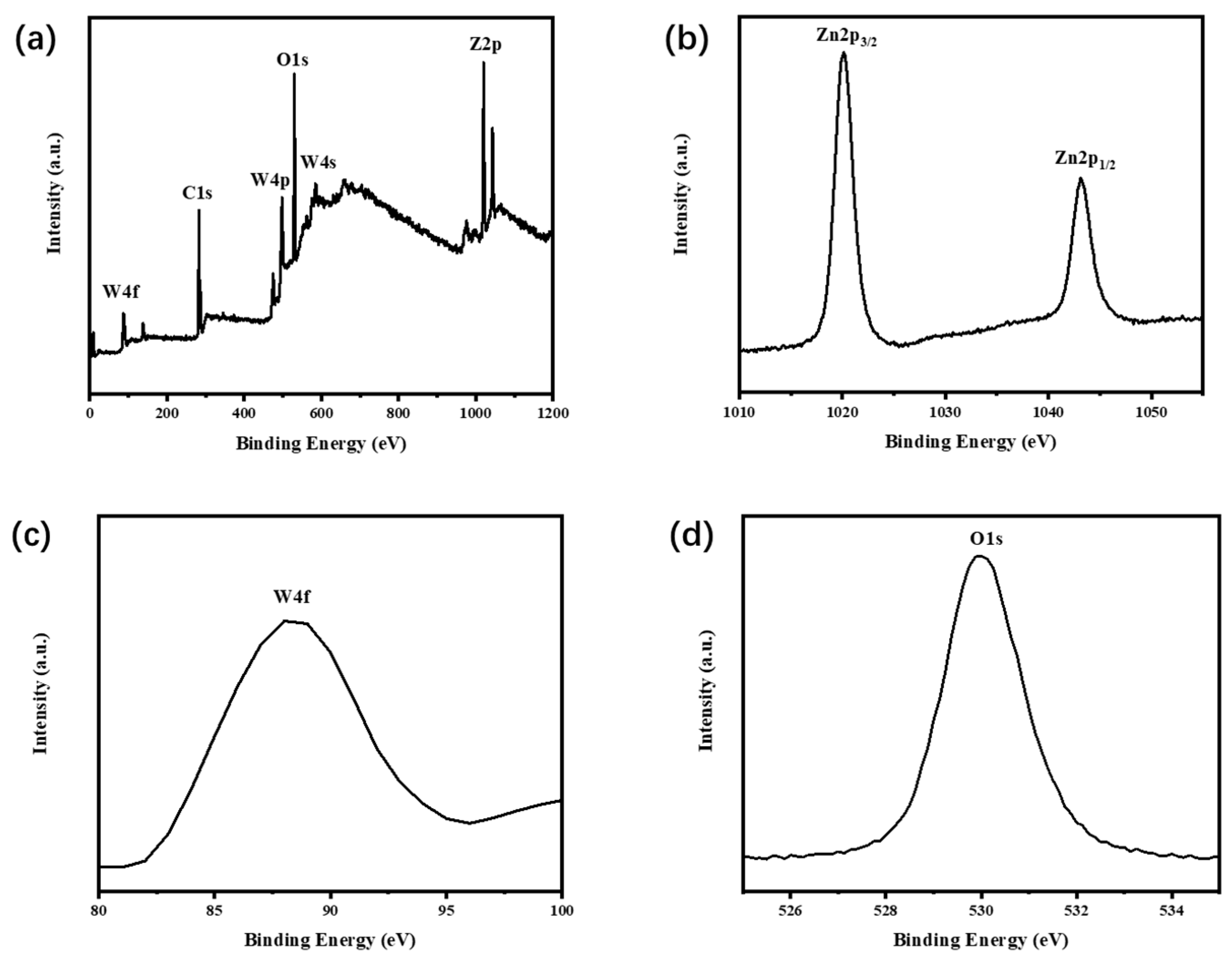
In order to study the optical absorption characteristics of WO3 nanofibers and ZnO/WO3 composite nanofibers, the UV absorption spectra of the fiber samples were tested and analyzed. Figure 3a,b, respectively, show the experimental results of the photocatalytic degradation of Rh B dye using WO3 nanofibers and ZnO/WO3 composite nanofibers from the initial time to the end of 120 min. With the progress of the photocatalytic reaction, the color of Rh B dye was gradually lightened due to continuous degradation, and the absorbance of the solution decreased rapidly, indicating that RhB was rapidly degraded, which was the result of the direct fracture of the conjugated chromophores [30]. In addition, the maximum absorption wavelength of Rh B was 553 nm, and the experimental results show that the strongest absorption peak was observed at a 542 nm wavelength and the maximum absorption wavelength was blue-shifted. The possible reason for this phenomenon is the aggregation phenomenon between molecules. A large number of nanoparticles gathered on the surface of the fiber, which made the surface of the nanofiber rough and increased the specific surface area in contact with the Rh B dye, so that more active sites could become involved in the reaction. Figure 3 also shows that during the degradation of Rh B dye, the degradation degree of ZnO/WO3 composite nanofibers was significantly more thorough than that of WO3 nanofibers. After 120 min of photocatalytic degradation, the absorption peak strength of ZnO/WO3 composite nanofibers became very weak, indicating that Rh B dye became almost completely degraded within 120 min, while WO3 nanofibers still had a high absorption peak strength. This indicates that Rh B dye was not completely degraded within 120 min. Therefore, ZnO/WO3 composite nanofibers have better photocatalytic degradation efficiency and will be suitable for textile wastewater treatment applications.

3.4. XPS Analysis

In order to study the elemental composition and chemical valence identification of ZnO/WO3 composite nanofiber samples, X-ray photoelectron spectroscopy was used to test and analyze the ZnO/WO3 nanofiber samples. Figure 4a shows the full XPS spectra of ZnO/WO3 samples. The figure shows that ZnO/WO3 indicating that the samples are composed of two compounds, ZnO and WO3, which is consistent with the test results of XRD. Figure 4b shows the two characteristic peaks corresponding to the Zn2p atomic states. The characteristic peaks at the electron binding energy of 1022.1 eV and 1044.9 eV corresponded to the Zn2p3/2 and Zn2p1/2 atomic states of Zn2+. Figure 4c shows the two characteristic peaks corresponding to W4f. The characteristic peak at the electron binding energy of 88.5 eV corresponded to the atomic state W4f. Figure 4d shows the characteristic peak corresponding to O1s, and the corresponding electron binding energy was 530.5 eV. The analysis results are consistent with the elemental composition detected by XRD.

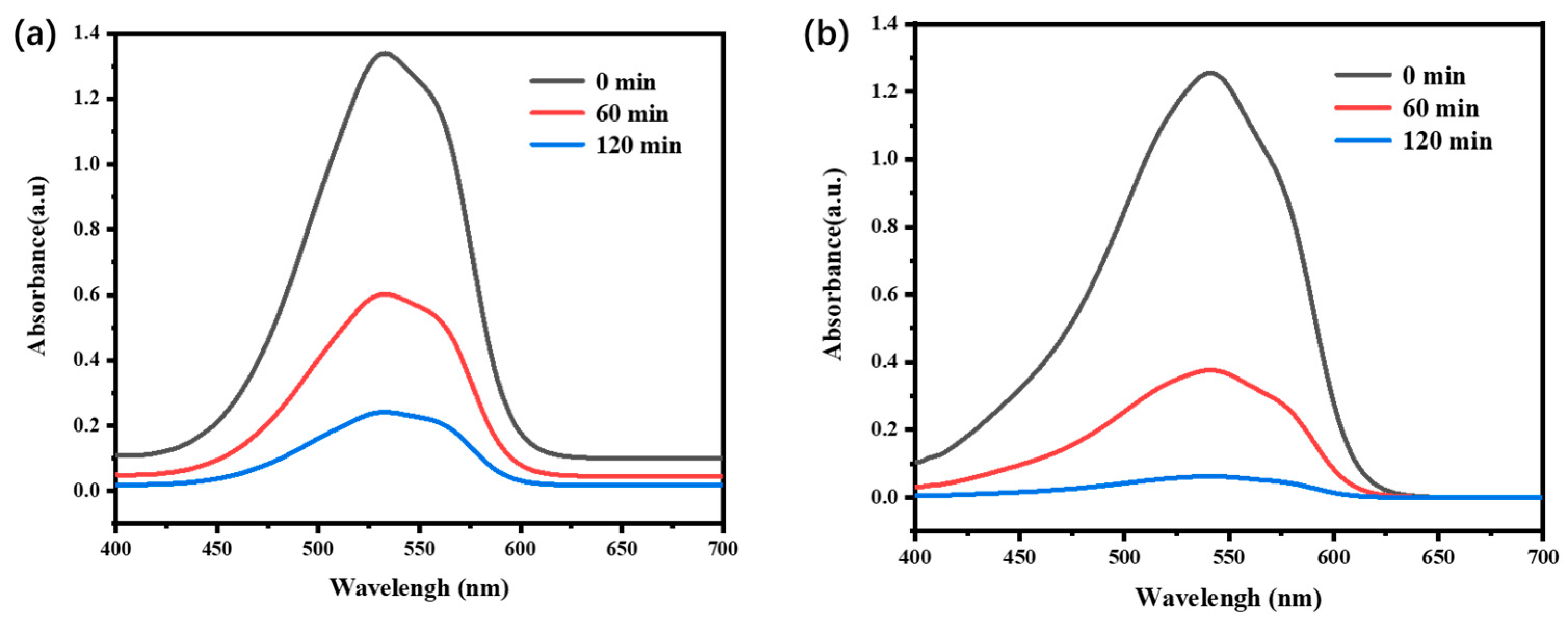
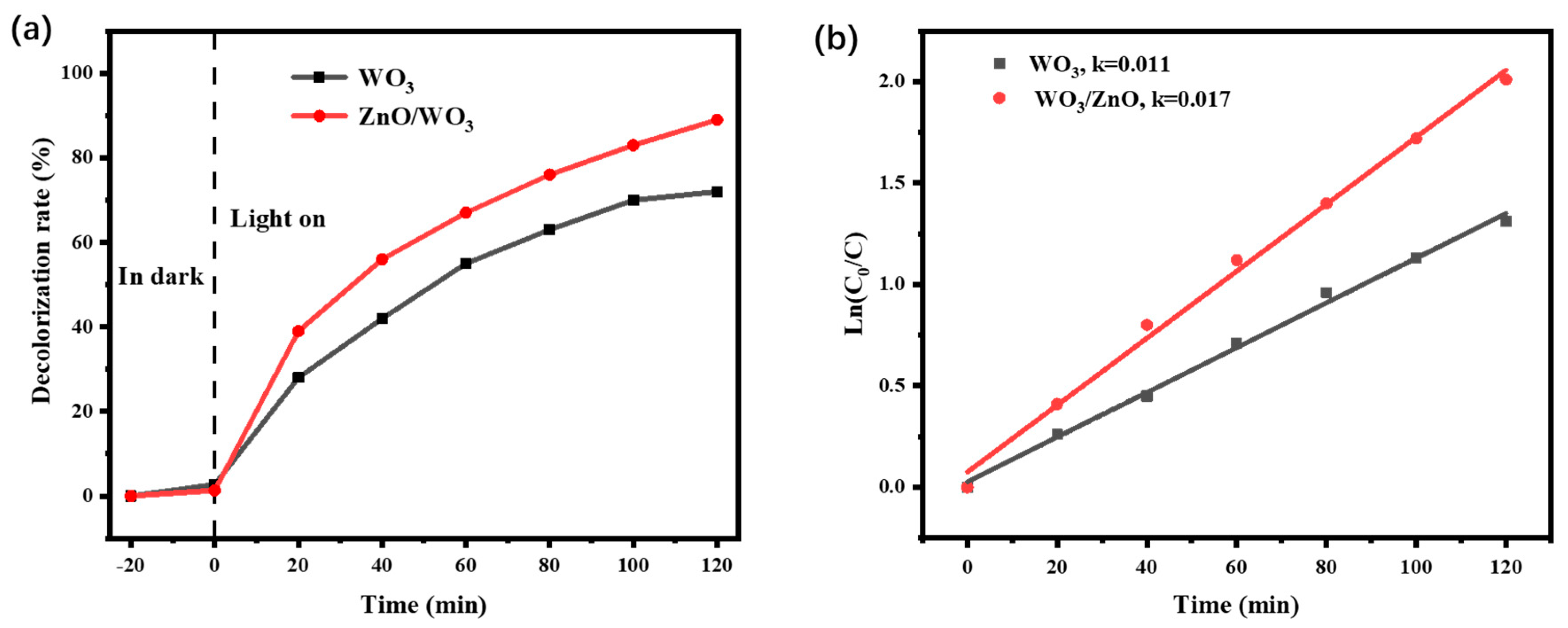
3.5. Photocatalytic Degradation Efficiency
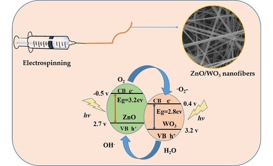
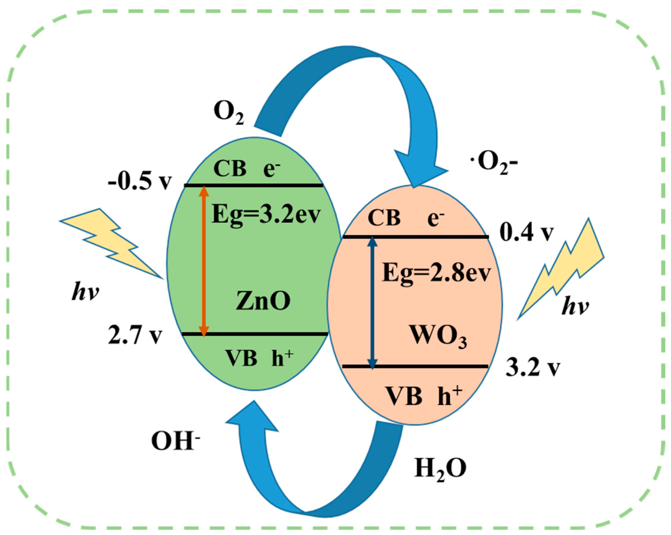
In order to study the photocatalytic degradation efficiency of Rh B in WO3 and ZnO/WO3 nanofiber samples, 100 mg/L of Rh B aqueous solution was selected as the target pollutant, and the photocatalytic degradation performance of WO3 and ZnO/WO3 nanofiber samples was tested. In order to avoid the error of degradation efficiency caused by the adsorption of the sample to Rh B solution, the adsorption–desorption experiment of the sample was first carried out in dark conditions, and then the degradation efficiency of the sample was measured under the irradiation of UV-visible light. Figure 5a shows the photocatalytic degradation efficiency of WO3 nanofibers and ZnO/WO3 composite nanofibers on Rh B solution after 120 min photocatalytic degradation experiment. Figure 5a shows that the degradation rate increased with the increase in reaction time. However, the degradation efficiency of a single WO3 nanofiber was low after 120 min of UV-visible light irradiation, and the degradation efficiency of WO3 on Rh B solution was measured to reach about 70%, indicating that WO3 nanofibers did not completely degrade Rh B. Compared to WO3 nanofibers, ZnO/WO3 composite nanofibers showed better photocatalytic performance. After 120 min of UV-visible light irradiation, the photocatalytic degradation efficiency of ZnO/WO3 composite nanofibers reached 90%, which indicates that the photocatalytic degradation efficiency of ZnO/WO3 composite nanofibers is significantly higher than that of WO3 nanofibers.
For pseudo-first-order reaction kinetics, Formula (2) was used for the fitting analysis of the photodegradation process of R hB. Time was used as the independent variable to process the test data and draw the kinetics fitting curve.
ln C 0 C t = k t  
where t represents the illumination time; t represents the concentration of R hB in solution when the reaction time is t. C0 represents the RhB concentration in the solution before photocatalytic treatment.
Figure 5b shows the fitting curve of degradation kinetics, pseudo-first-order kinetic model for the data fitting. The degradation rate constants (k) of both WO3 nanofibers and ZnO/WO3 composite nanofibers were 0.011 min−1 and 0.017 min−1, respectively. The k value of ZnO/WO3 composite nanofibers was 1.5 times that of WO3 nanofibers, and this was due to the fact that ZnO had provided adsorption sites, which enabled Rh B molecules in contact with the catalyst surface to be absorbed in large quantities, accelerating the degradation of Rh B [34].
Figure 6 shows the photocatalytic mechanism of ZnO/WO3 composite nanofibers on Rh B dye. When the energy generated by external visible light is greater than the band gap width (Eg) of ZnO (Eg = 3.2 eV) and WO3 (Eg = 2.7 eV), the photogenerated electrons in ZnO and WO3 valence bands would be excited and jump to the conduction band, and a large number of holes would be generated in ZnO and WO3 valence bands. Because the conduction potential of WO3 is higher than that of ZnO, and the valence potential of ZnO is lower than that of WO3, when ZnO and WO3 are in contact with each other, heterojunction is formed in the interface region of contact [35]. Under the action of potential gradients and the internal electric field, the excited photogenerated electrons transfer from the surface of ZnO to WO3 conduction band, and finally a large number of photogenerated electrons accumulate in the WO3 conduction band. Under the action of the built-in electric field, the holes in the WO3 valence band would migrate to the ZnO valence band with a lower electric potential, and eventually a large number of holes would gather in the ZnO valence band, thus realizing the effective separation of electron–hole pairs. A large number of photogenerated electrons gathered in the WO3 conduction band would be captured by O2 molecules dissolved in aqueous solution, forming superoxide radical ·O2-, while a large number of holes gathered in ZnO valence band would be captured by H2O molecules in aqueous solution, forming OH radicals. In a photocatalytic reaction system, both electrons and holes can participate in the degradation process, but in different ways. Electrons are mainly involved in the degradation of the chromophores, while holes are mainly involved in the process of deethylation [36]. The heterogeneous composite semiconductor can effectively separate the electron–hole pairs, enhance the photocatalytic activity of ZnO/WO3, and greatly improve the photocatalytic degradation efficiency of Rh B dye. The photocatalytic mechanism involved in the degradation of Rh B organic dye can be expressed as follows [37]:
  • ZnO/WO3+ hν → ZnO/WO3 + e + h+;
  • O2 + e− → ·O2;
  • ·O2 + e + 2H+ → H2O2;
  • H2O2 + e → ·OH + OH;
  • H2O + h+ → ·OH + h+;
  • ·O2 (or ·OH) + Rh B (dye) → degradation products;
  • h+ + Rh B(dye) → degradation products.

4. Conclusions

We successfully prepared WO3 nanofibers and ZnO/WO3 composite nanofibers using the electrospinning method, with a specific surface morphology and size of composite nanofibers. Compared with traditional synthesis methods, WO3 nanofibers and ZnO/WO3 composite nanofibers prepared using the electrospinning method were found to have smaller diameters and smoother surfaces, and a large number of nanoparticles were attached to the surface of the fibers to increase the contact area with the Rh B dye surface, thus effectively improving the photocatalytic activity. Therefore, nanostructured fiber materials prepared by electrospinning were found to have stronger photocatalytic activity. Compared with WO3 nanofibers, ZnO/WO3 composite nanofibers were found to have better UV-visible light absorption performance, and the sample has a higher UV-visible light utilization rate. This is mainly due to the formation of a P-N heterojunction in a semiconductor recombination model and the rapid separation of electron–hole pairs under the drive of a built-in electric field force, which promotes carrier migration. The photocatalytic degradation results show that the degradation efficiency of ZnO/WO3 composite nanofibers reached about 90% after 120 min photocatalytic degradation under the same conditions, which can almost achieve the complete degradation of Rh B dye. The semiconductor composite construction technology of heterojunction will promote the rapid development of semiconductor photocatalysis. The excellent photocatalytic degradation performance will expand the use of ZnO/WO3 composite nanofibers in the textile wastewater treatment field in the future.

Author Contributions

Conceptualization, Y.X. and H.Y.; methodology, H.Y.; software, Y.X.; validation, H.Y. and T.C.; formal analysis, Y.X.; investigation, Y.X.; resources, H.Y.; data curation, Y.X.; writing—original draft preparation, H.Y.; writing—review and editing, Y.X.; visualization, T.C.; supervision, T.C.; project administration, Y.X.; funding acquisition, Y.X. All authors have read and agreed to the published version of the manuscript.

Funding

This research received no external funding.

Data Availability Statement

Data available on request from the authors.

Acknowledgments

This work was supported by National Natural Science Foundation of China (no. 42173062) and the Natural Science Foundation of Key Scientific Research Projects of Colleges and Universities in Henan Province (no. 23B610014).

Conflicts of Interest

The authors declare no conflict of interest.

References

  1. Evgenidou, E.N.; Konstantinou, I.K.; Lambropoulou, D.A. Occurrence and removal of transformation products of PPCPs and illicit drugs in wastewaters: A review. Sci. Total Environ. 2015, 505, 905–926. [Google Scholar] [CrossRef] [PubMed]
  2. Bethi, B.; Sonawane, S.H.; Bhanvase, B.A.; Gumfekar, S.P. Nanomaterials-based advanced oxidation processes for wastewater treatment: A review. Chem. Eng. Process. Process Intensif. 2016, 109, 178–189. [Google Scholar] [CrossRef]
  3. Aworanti, O.A.; Agbede, O.O.; Agarry, S.E.; Ajani, A.O.; Ogunkunle, O.; Laseinde, O.T.; Rahman, S.M.A.; Fattah, I.M.R. Decoding Anaerobic Digestion: A Holistic Analysis of Biomass Waste Technology, Process Kinetics, and Operational Variables. Energies 2023, 16, 3378. [Google Scholar] [CrossRef]
  4. Almulhem, N.K.; Awada, C.; Shaalan, N.M. Study of Phenol Red Photocatalytic Decomposition on KBrO3-Supported TiO2 Nanoparticles for Wastewater Treatment. Separations 2023, 10, 162. [Google Scholar] [CrossRef]
  5. Boutafda, A.; Hafidi, M.; Ouhdouch, Y.; Pinelli, E.; Jemo, M.; El Fels, L. Fungal Strain as Biological Tool to Remove Genotoxicity Effect of Phenolic Compounds from Olive Mill Wastewater. Sustainability 2023, 15, 6510. [Google Scholar] [CrossRef]
  6. Mansoor, B.; Ashraf, S.; Rehman, U.; Ullah, Z.; Sheikh, Z. Integration of Microalgae-Microbial Fuel Cell with Microbial Electrolysis Cell for Wastewater Treatment and Energy Production. Environ. Sci. Proc. 2023, 25, 72. [Google Scholar] [CrossRef]
  7. Slimani, Y.; Almessiere, M.A.; Mohamed, M.J.S.; Hannachi, E.; Caliskan, S.; Akhtar, S.; Baykal, A.; Gondal, M.A. Synthesis of Ce and Sm Co-Doped TiO2 Nanoparticles with Enhanced Photocatalytic Activity for Rhodamine B Dye Degradation. Catalysts 2023, 13, 668. [Google Scholar] [CrossRef]
  8. Ge, M.; Li, Z. All-Solid-State Z-Scheme Photocatalytic Systems Based on Silver-Containing Semiconductor Materials. Prog. Chem. 2017, 29, 846–858. [Google Scholar]
  9. Zango, Z.U.; Rozaini, M.N.; Abu Bakar, N.H.H.; Zango, M.U.; Haruna, M.A.; Dennis, J.O.; Alsadig, A.; Ibnaouf, K.H.; Aldaghri, O.A.; Wadi, I.A. Advancements in Clay Materials for Trace Level Determination and Remediation of Phenols from Wastewater: A Review. Separations 2023, 10, 125. [Google Scholar] [CrossRef]
  10. Chuaicham, C.; Trakulmututa, J.; Shu, K.; Shenoy, S.; Srikhaow, A.; Zhang, L.; Mohan, S.; Sekar, K.; Sasaki, K. Recent Clay-Based Photocatalysts for Wastewater Treatment. Separations 2023, 10, 77. [Google Scholar] [CrossRef]
  11. El Batouti, M.; Alharby, N.F.; Elewa, M.M. Review of New Approaches for Fouling Mitigation in Membrane Separation Processes in Water Treatment Applications. Separations 2021, 9, 1. [Google Scholar] [CrossRef]
  12. Sun, Y.; Wang, T. Application of Biochar, Adsorbent and Nanomaterials in Wastewater Treatment. Water 2023, 15, 1320. [Google Scholar] [CrossRef]
  13. Jing, H.-X.; Gao, Y.-L.; Li, L.-X.; Wang, X.; Pei, W.J.; Yang, X.F. Synthesis of a Novel Double Z-Scheme TiO2/Bi2O3-g-C3N4 Photocatalyst with Enhanced Photocatalytic Performance to Rhodamine B Under Sunlight. J. Clust. Sci. 2022, 34, 1347–1354. [Google Scholar] [CrossRef]
  14. Li, M.; Dong, B.; Chang, Z.; Dang, H.; Ma, S.; Li, W. Synthesis of TiO2/g-C3N4 Photocatalyst with Recovered TiO2 from Spent SCR Catalyst for Photodegrading Rhodamine B. Waste Biomass Valorization 2023, 14, 687–701. [Google Scholar] [CrossRef]
  15. Yang, J.; Wang, J.; Zhao, W.; Wang, G.; Wang, K.; Wu, X.; Li, J. 0D/1D Cu2−xS/TiO2 S-scheme heterojunction with enhanced photocatalytic CO2 reduction performance via surface plasmon resonance induced photothermal effects. Appl. Surf. Sci. 2023, 613, 156083. [Google Scholar] [CrossRef]
  16. Zheng, X. Application of nano-TiO2 photocatalyst in marine pollution control. Desalin. Water Treat. 2022, 268, 303–312. [Google Scholar] [CrossRef]
  17. Park, B.-G. Photocatalytic Activity of TiO2-Doped Fe, Ag, and Ni with N under Visible Light Irradiation. Gels 2022, 8, 14. [Google Scholar] [CrossRef] [PubMed]
  18. Akpan, U.G.; Hameed, B.H. The advancements in sol-gel method of doped-TiO2 photocatalysts. Appl. Catal. A-Gen. 2010, 375, 1–11. [Google Scholar] [CrossRef]
  19. Kunyapat, T.; Xu, F.; Neate, N.; Wang, N.; De Sanctis, A.; Russo, S.; Zhang, S.; Xia, Y.; Zhu, Y. Ce-Doped bundled ultrafine diameter tungsten oxide nanowires with enhanced electrochromic performance. Nanoscale 2018, 10, 4718–4726. [Google Scholar] [CrossRef]
  20. Lin, R.; Wan, J.; Xiong, Y.; Wu, K.; Cheong, W.-C.; Zhou, G.; Wang, D.; Peng, Q.; Chen, C.; Li, Y. Quantitative Study of Charge Carrier Dynamics in Well-Defined WO3 Nanowires and Nanosheets: Insight into the Crystal Facet Effect in Photocatalysis. J. Am. Chem. Soc. 2018, 140, 9078–9082. [Google Scholar] [CrossRef]
  21. Zhang, S.; Yi, J.; Chen, J.; Yin, Z.; Tang, T.; Wei, W.; Cao, S.; Xu, H. Spatially confined Fe2O3 in hierarchical SiO2@TiO2 hollow sphere exhibiting superior photocatalytic efficiency for degrading antibiotics. Chem. Eng. J. 2020, 380, 122583. [Google Scholar] [CrossRef]
  22. Wang, M.; Han, J.; Yuan, G.; Guo, R. Carbon/TiO2/Fe2O3 hybrid shells as efficient visible light photocatalysts. New J. Chem. 2019, 43, 11282–11287. [Google Scholar] [CrossRef]
  23. Hakimi-Tehrani, M.J.; Hassanzadeh-Tabrizi, S.A.; Koupaei, N.; Saffar, A.; Rafiei, M. Synthesis of Z-scheme g-C3N4/WO3 nano-photocatalyst with superior antibacterial characteristics for wastewater treatment. J. Sol-Gel Sci. Technol. 2023, 105, 212–219. [Google Scholar] [CrossRef]
  24. Murillo-Sierra, J.C.; Hernandez-Ramirez, A.; Pino-Sandoval, D.A.; Ruiz-Ruiz, E.; Martínez-Hernández, A. Promoting multielectron CO2 reduction using a direct Z-scheme WO3/ZnS photocatalyst. J. CO2 Util. 2022, 63, 102122. [Google Scholar] [CrossRef]
  25. Zhang, L.; Shen, Q.; Huang, F.; Jiang, L.; Liu, J.; Sheng, J.; Li, Y.; Yang, H. Electrospinning directly synthesis of 0D/1D CuBi2O4@WO3 nanofiber photocatalyst with S-scheme heterojunction. Appl. Surf. Sci. 2023, 608, 155064. [Google Scholar] [CrossRef]
  26. Li, F.; Ruan, S.; Yin, Y.; Zhang, N.; Zhang, H.; Li, C.; Chen, Y. Facile synthesis of MnWO4/WO3 electrospun nanofibers as high performance visible-light driven photocatalysts. Mater. Lett. 2018, 229, 98–102. [Google Scholar] [CrossRef]
  27. Zhou, X.; Zhang, C.; Li, Y.; Xiong, X.; Wang, Y.; Rong, S. Promoted microbial denitrification and carbon dioxide fixation via photogenerated electrons stored in novel core/shell memory photocatalysts in darkness. Chemosphere 2022, 303, 135259. [Google Scholar] [CrossRef]
  28. Xu, L.; Su, J.; Zheng, G.; Zhang, L. Enhanced photocatalytic performance of porous ZnO thin films by CuO nanoparticles surface modification. Mater. Sci. Eng. B Adv. Funct. Solid-State Mater. 2019, 248, 114405. [Google Scholar] [CrossRef]
  29. Xu, M.; Wang, H.; Wang, G.; Zhang, L.; Liu, G.; Zeng, Z.; Ren, T.; Zhao, W.; Wu, X.; Xue, Q. Study of synergistic effect of cellulose on the enhancement of photocatalytic activity of ZnO. J. Mater. Sci. 2017, 52, 8472–8484. [Google Scholar] [CrossRef]
  30. Zhang, Y.; Zhou, J.; Cai, W.; Zhou, J.; Li, Z. Enhanced photocatalytic performance and degradation pathway of Rhodamine B over hierarchical double-shelled zinc nickel oxide hollow sphere heterojunction. Appl. Surf. Sci. 2018, 430, 549–560. [Google Scholar] [CrossRef]
  31. Molla, M.A.I.; Ahmed, A.Z.; Kaneco, S. Chapter 3—Reaction mechanism for photocatalytic degradation of organic pollutants. In Nanostructured Photocatalysts; Nguyen, V.-H., Vo, D.-V.N., Nanda, S., Eds.; Elsevier: Amsterdam, The Netherlands, 2021; pp. 63–84. [Google Scholar]
  32. Molla, M.A.I.; Furukawa, M.; Tateishi, I.; Katsumata, H.; Kaneco, S. Fabrication of Ag-doped ZnO by mechanochemical combustion method and their application into photocatalytic Famotidine degradation. J. Environ. Sci. Health Part A-Toxic/Hazard. Subst. Environ. Eng. 2019, 54, 914–923. [Google Scholar] [CrossRef]
  33. Ahmed, A.Z.; Islam, M.M.; Islam, M.M.U.; Masum, S.M.; Islam, R.; Molla, M.A.I. Fabrication and characterization of B/Sn-doped ZnO nanoparticles via mechanochemical method for photocatalytic degradation of rhodamine B. Inorg. Nano-Met. Chem. 2020, 51, 1369–1378. [Google Scholar] [CrossRef]
  34. Haounati, R.; Ighnih, H.; Malekshah, R.E.; Alahiane, S.; Alakhras, F.; Alabbad, E.; Alghamdi, H.; Ouachtak, H.; Addi, A.A.; Jada, A. Exploring ZnO/Montmorillonite photocatalysts for the removal of hazardous RhB Dye: A combined study using molecular dynamics simulations and experiments. Mater. Today Commun. 2023, 35, 105915. [Google Scholar] [CrossRef]
  35. Largo, F.; Haounati, R.; Ouachtak, H.; Hafid, N.; Jada, A.; Addi, A.A. Design of organically modified sepiolite and its use as adsorbent for hazardous Malachite Green dye removal from water. Water Air Soil Pollut. 2023, 234, 183. [Google Scholar] [CrossRef]
  36. Nguyen Thi Thanh, T.; Dao Sy, D.; Doan Van, T.; Al Tahtamouni, T.; Pham, T.D.; Hanh, N.T.; Tran, D.T.; Nguyen, M.V.; Dang, N.M.; Le Chi, N.T.P.; et al. The advanced photocatalytic degradation of atrazine by direct Z-scheme Cu doped ZnO/g-C3N4. Appl. Surf. Sci. 2019, 489, 875–882. [Google Scholar]
  37. Kumar, S.G.; Rao, K.K. Comparison of modification strategies towards enhanced charge carrier separation and photocatalytic degradation activity of metal oxide semiconductors (TiO2, WO3 and ZnO). Appl. Surf. Sci. 2017, 391, 124–148. [Google Scholar] [CrossRef]
Figure 1. (ac) SEM images of WO3 nanofibers, (c) after heat treatment; (df) SEM images of ZnO/WO3 composite nanofibers, (f) after heat treatment.
Separations 10 00339 g001
Figure 2. XRD patterns of WO3 nanofibers and ZnO/WO3 composite nanofibers after heat treatment.
Separations 10 00339 g002
Figure 3. UV–visible absorption spectra of (a) WO3 nanofibers and (b) ZnO/WO3 composite nanofibers from the start to 120 min.
Separations 10 00339 g003
Figure 4. (a) XPS spectra of ZnO/WO3 nanofiber samples, (b) XPS spectra of Zn2p, (c) XPS spectra of W4f, and (d) XPS spectra of O1s.
Separations 10 00339 g004
Figure 5. (a) Degradation efficiency and (b) fitting curve of degradation kinetics of WO3 nanofibers and ZnO/WO3 composite nanofibers.
Separations 10 00339 g005
Figure 6. Degradation mechanism of Rh B under visible light using ZnO/WO3 composite nanofibers.
Separations 10 00339 g006
Disclaimer/Publisher’s Note: The statements, opinions and data contained in all publications are solely those of the individual author(s) and contributor(s) and not of MDPI and/or the editor(s). MDPI and/or the editor(s) disclaim responsibility for any injury to people or property resulting from any ideas, methods, instructions or products referred to in the content.

