Article

Flavonol-Glycoside and Rare Triterpenoid Derivatives Isolated from Leaves of Combretum glutinosum Perr. Ex Dc. with In Vitro Cytotoxic Activity

by
Sherouk Hussein Sweilam
1,2,*,
Maha B. O. Ebrahim
3,
Mehnaz Kamal
4,
El-Sayed Khafagy
5,6,
Ashraf N. Abdalla
7,
Mohamed E. Elzubier
8 and
Ehssan H. Moglad
5,9
1
Department of Pharmacognosy, College of Pharmacy, Prince Sattam bin Abdulaziz University, Al-Kharj 11942, Saudi Arabia
2
Department of Pharmacognosy, Faculty of Pharmacy, Egyptian Russian University, Cairo-Suez Road, Badr City, Cairo 11829, Egypt
3
Department of Chemistry, Faculty of Science and Technology, Alneelain University, Khartoum P.O. Box 12702, Sudan
4
Department of Pharmaceutical Chemistry, College of Pharmacy, Prince Sattam bin Abdulaziz University, Al-Kharj 11942, Saudi Arabia
5
Department of Pharmaceutics, College of Pharmacy, Prince Sattam bin Abdulaziz University, P.O. Box 173, Alkharj 11942, Saudi Arabia
6
Department of Pharmaceutics and Industrial Pharmacy, Faculty of Pharmacy, Suez Canal University, Ismailia 41522, Egypt
7
Department of Pharmacology and Toxicology, Faculty of Pharmacy, Umm Al Qura University, Makkah 21955, Saudi Arabia
8
Department of Biochemistry, Faculty of Medicine, Umm Al-Qura University, Makkah 21955, Saudi Arabia
9
Department of Microbiology and Parasitology, Medicinal and Aromatic Plants Research Institute, National Center for Research, Khartoum P.O. Box 2404, Sudan
*
Author to whom correspondence should be addressed.
Separations 2023, 10(3), 209; https://doi.org/10.3390/separations10030209
Submission received: 18 February 2023 / Revised: 11 March 2023 / Accepted: 13 March 2023 / Published: 17 March 2023

Abstract

:
Combretaceae plants are used traditionally by many cultures, especially in Sudanese patients for the treatment of diverse ailments such as anti-inflammatory, antimicrobial, antitumor, and antioxidant disorders. Of these plants, the genus Combretum are traditional medicinal plants. Thus, they are formed from the non-polar or polar extracts of many isolated phytochemicals. Of these necessities, the use of Combretum extracts for their medicinal properties can be found in the earliest of myths and traditions used to document the plants’ ability to treat diseases. Combretum glutinosum Perr. Ex Dc. is a common shrub native to the African continent, especially Sudan. Currently, there are no published data regarding its cytotoxic activity. Additionally, there are few chemical and biological reports of C. glutinosum. Therefore, the current study aimed to isolate the chemical bioactive compounds (16) from the ethyl acetate (EtOAc) extract of C. glutinosum. A new flavonoid compound, namely, glutosinumoside (4), was afforded, and five known compounds were obtained: three oleanane-glycosides (13) and two phenolic acids (5,6). The structures of the six compounds were determined by spectroscopic analysis, including one- and two-dimensional (1D and 2D) NMR, mass spectrometry, and chromatographic analysis. Moreover, an in vitro cytotoxic evaluation of the successive extracts and the bioactive EtOAc fractions of C. glutinosum against MCF7 (breast), HT29 (colon), HepG2 (liver), and MRC5 (normal lung) cell lines was performed. The isolated compounds showed comparable cytotoxic activities with the crude EtOH extract and doxorubicin against the tested cell lines. Compounds (1) and (6) exhibited the highest cytotoxicity against MCF7 (1.37 ± 0.21 and 1.48 ± 0.34 µg/mL, respectively) and HepG2 (3.30 ± 0.02 and 2.10 ± 0.22 µg/mL, respectively) in the MTT assay. In addition, compounds (1) and (3) demonstrated a significant upregulation of cancer’s two important hallmarks (caspase 3 and bax genes) by inducing apoptosis and perturbing the MCF7 cell cycle.

1. Introduction

Plants of the family Combretaceae are used significantly for therapeutic applications because they contain a distinct and wide range of secondary metabolites. The family consists of approximately twenty genera with six hundred species. The genus Combretum is considered one of the largest genera, having 370 species, and is distributed in Africa and Brazil (tropical and subtropical areas) [1]. Traditionally, in African and Asian countries, the Combretum species are extensively used for herbal remedies for the treatment of different ailments, including abortifacient, antibacterial, anticancer, antifungal, antimicrobial, antischistosomal, antitussive, antiviral, and molluscicidal activities [2,3].
Phytochemicals of everal species of the genus Combretum have been reported, including alkaloids, triterpenes, flavonoids, lignans, phenanthrenes, saponins, and stilbenoids. Considerable attention has been given to the screening of the plants’ extracts for present and possible relationships with their reported biological activities, such as anticancer, antihyperglycemic, anti-inflammatory, antimalarial, and antioxidant activities [1].
Combretum glutinosum Perr. ex DC. is one of approximately 140 species belonging to the largest genus within the Combretaceae family, “Combretum” [3]. It is a small tree or bushy shrub distributed in tropical Africa and is popularly named “Habil” [1,4]. A systematic phytochemical screening of the C. glutinosum leaves revealed the presence of alkaloids [5], benzene dicarboxylic acids [6], flavonoids [4,7], phytosterols such as β-sitosterol and β-sitosterol-glycoside [8], polyphenols [4,5], stilbenes, tannins [4], and triterpenoids such as lupeol, oleanolic acid, betulinic acid, β-amyrin, and 3 β-acetoxydammar-21(22)-ene [8,9]. Among these, oleanane glycosides have a unique structure, and their wild distribution in the genus demonstrated relationships between chemical structure and cytotoxic activity [10]. In previous in vitro biological investigations, C. glutinosum’s antibacterial, antifungal, antimalarial, antischistosomal, and molluscicidal activities were significantly demonstrated due to the presence of several bioactive components, such as polyphenols (gallic and ellagic acids) [4,5], flavonoid glycosides (such as quercitrin and rutin) [4,5,11], and tannins such as combreglutinin, 2,3-(S)-hexahydroxydiphenoyl-D-glucose, punicalagin, and punicalin [4]. Likewise, β-amyrin, isolated from the stem bark of C. glutinosum, was used as an Ayurvedic formula for the treatment of flu and cold symptoms [9]. The phenolic compound 2,4-di-tert-butylphenol (DTBP) was used as a raw drug for the manufacture of antioxidants [9]. In most African savannahs, the leaves or stembark of the C. glutinosum are used to treat microbial infections, anemia, anorexia, diarrhea, fever, malaria, liver failure, respiratory disorders such as bronchitis and cough, as antitussives, and for wound healing [11,12].
One of the major public health diseases is cancer, which causes global death. There are 277 different types of cancer. Breast and prostate cancer are considered the major distributed types in women and men, respectively, in addition to colon, rectum, lung, urinary, and bladder cancer, and cancer of the uterine corpus in one of the sexes [13,14].
To the best of the authors’ knowledge, there is no study that focused on the anticancer properties of C. glutinosum leaves. This study aimed to isolate, elucidate compounds, and perform an in vitro cytotoxic evaluation of C. glutinosum. The chemical investigation was assessed using different chromatographic techniques, and the in vitro cytotoxic activity against MCF7 (breast), HT29 (colon), and HepG2 (liver) cell lines and its selectivity were assessed using MRC5 (normal lung) cell lines in an MTT assay.

2. Materials and Methods

2.1. Plant Material and Extraction

C. glutinosum (leaves) were obtained from their natural habit in Aldbibate Town (12°29′55.2″ N 29°47′48.8″ E), South Kordofan state, Sudan, in March 2018. The plant material was identified by a taxonomist, Dr. Yahya Sulman Mohamed, in the herbarium of Medicinal and Aromatic Plant Research Institute (MAPRI), National Research Center (NCR), Khartoum, Sudan. A voucher specimen (No. MAPRI/2018/03) was deposited in the MAPRI herbarium. The leaves were dried in the shade and ground manually at room temperature. The powdered leaves of C. glutinosum (1923 g) were extracted at room temperature, as described by [15], using 96% ethanol until complete exhaustion of the plant material. The EtOH yield (212.9 g) was partitioned between dichloromethane (DCM) and H2O (1:1, v/v). The resulting DCM-soluble fraction (120 g) was subsequently partitioned between MeOH-H2O, 8:2, and n-hexane, according to the NCI protocol, to provide the corresponding EtOH (212.9 g), n-hexane (7.9 g), 80% MeOH (8 g), DCM (120 g), and remaining aqueous (8 g) extracts. The aqueous-soluble fraction (25.3 g) was partitioned with ethyl acetate to produce EtOAc extract (1.8 g) (Flowchart S1).

2.2. General Chemistry

An NMR (1D and 2D, Bruker UltraShield Plus 500 MHz) spectrometer was utilized at the College of Pharmacy, Prince Sattam bin Abdulaziz University, KSA(Bruker, Fällanden, Switzerland, using deuterated CDCl3, pyridine, and CD3OD at room temperature (Cambridge Isotope Laboratories, Andover, MA, USA). HR-ESI-MS data were acquired by direct injection with electrospray ionization in negative or positive ion modes on an UPLC RS Ultimate 3000—Q Exactive (Thermo Fisher Scientific, Waltham, MA, USA) hybrid quadrupole-Orbitrap mass spectrometer combined with high-performance quadrupole precursor selection with high resolution and accurate mass Orbitrap™ detection. Open column chromatography was performed using Sephadex LH-20 (Amersham Biosciences, Uppsala, Sweden) and silica gel 60/230–400 mesh (EM Science). The thin layer chromatography (TLC) analysis was performed on Keisel gel 60 F254 plates (Merck, Darmstadt, Germany). The reagents and solvents were purchased from Sigma-Aldrich, Loba Chemie Pvt. Ltd. (St. Louis, MO, USA), and SD Fine Chem. Ltd. (Mumbai, India).

2.3. Isolation and Purification of Compounds from the Ethyl Acetate Fraction

The EtOAc fraction (1.8 g) was chromatographed on a Sephadex LH-20 CC and eluted with CHCl3 (100%), a mixture of CHCl3-MeOH (1% increments of methanol in CHCl3 until 10%) to achieve 8 fractions (A-H). The fractions were monitored by TLC on Si gel GF245 plates using a spraying reagent, 10% vanillin-sulphuric acid, for visualization. Fraction B (631 mg) was further separated by CC on a silica gel and eluted with a mixture of CHCl3-MeOH (1% to 10%) to furnish 10 subfractions (B.1-B.10). Subfraction B.5 (60.9 mg) was subjected to Si gel CC with isocratic elution (n-Hex-EtOAc, 22%) to afford sub-subfraction (SB.5-2, 34.6 mg). The latter (34.6 mg) was purified on a Sephadex LH-20 CC and eluted with a mixture of CHCl3-MeOH (0% to 3%) to obtain compound (1) (8 mg). The subfraction B.8 (41 mg) purified by using a Sephadex LH-20 CC, eluted with CHCl3-MeOH (0% to 7%) to afford compounds (4) (3 mg) and (2) (3.8 mg), while (3) (10 mg) was recrystallized from subfraction B.9 (CHCl3-MeOH 92:8, 34 mg). After subfraction B.4 (13.4 mg) was repeatedly chromatographed on Si gel CC, using a step gradient elution with n-Hex-EtOAc (5% to 25%), compounds (5) (3 mg) and (6) (3.1 mg) were obtained (Flowchart S2).

2.4. In Vitro Cytotoxicity Assay

2.4.1. Cell Lines

The cytotoxic activity was evaluated against MCF7 (breast), HT29 (colon), and HepG2 (liver) cell lines. In addition to MRC5 (a normal fibroblast), all were obtained from the ATCC, USA. The three cancer cells were sub-cultured in RPMI-1640 media (10% FBS), while MRC5 was maintained in Eagle’s minimum essential medium (EMEM, 10% FBS), all at 37 °C and 5% CO2 for a maximum of 5–10 passages [16,17].

2.4.2. MTT Assay

Five extracts, six compounds (16, Figure 1), and doxorubicin were subjected to an MTT cytotoxicity assay, according to a previous report [18]. Each cell line was separately cultured in a 96-well plate (3 × 10³/well) and incubated with either the extract at final concentrations of 0–100 μg/mL or each of the compounds at a final concentration of 0–50 μM at 37 °C overnight (DMSO 0.1%; n = 3, three independent experiments). After three days of incubation, the cytotoxicity of each extract or compound was evaluated by MTT (0.5 mg/mL added/well for 3 h) assay. Next, the supernatant was removed, the DMSO was added, and the absorbance values were measured using a multi-plate reader (BIORAD, PR 4100, Hercules, CA, USA). The selectivity index (S.I.) of the EtOH extract, each of the six compounds, and doxorubicin was calculated according to a previously described method [19] by dividing the IC50 of the corresponding extract/compound against MRC-5 cells/ IC50 of the same component against either MCF7, HT29, or HepG2 cells.

2.4.3. Quantitative Real-Time PCR

The quantification of apoptosis-related (caspase 3, bax, or surviving) or cell cycle-related (PCNA and CCND1) gene expression was performed in MCF7 cells using a real-time PCR (Applied Biosystems 7500 Fast Real-Time PCR System, Waltham, MA, USA) for two compounds (1,3), according to their resulting yield, though the yield of compound (1) was not high enough for the RT-PCR analysis. Briefly, MCF cells (1 × 10⁶) were cultivated in 6-well plates. Treatments: vehicle control (0.1% DMSO), doxorubicin (0.1 μM), compound (1) (0.1 μM), and compound (3) (2 μM) for 24 h. The manufacturer’s instructions were used to isolate the total RNA using an isolation kit and the SYBR Green Master mix (Thermo Fisher Scientific, Waltham, MA, USA). The synthesis of the complementary DNA (cDNA) using the isolated RNA (RevertAid First Strand cDNA Synthesis Kit, Thermo Fisher Scientific, Waltham, MA, USA) was performed. The mixture of cDNA, 2X SYBR Green I Mastermix, PCR-grade water, and forward and reversed human primers of the selective genes were used for the RT-PCR (Table 1) [18].

2.4.4. Statistics

An ANOVA (one-way analysis of variance) and Tukey’s post-hoc were employed to determine statistical differences using Graphpad Prism software (version 9).

3. Results and Discussion

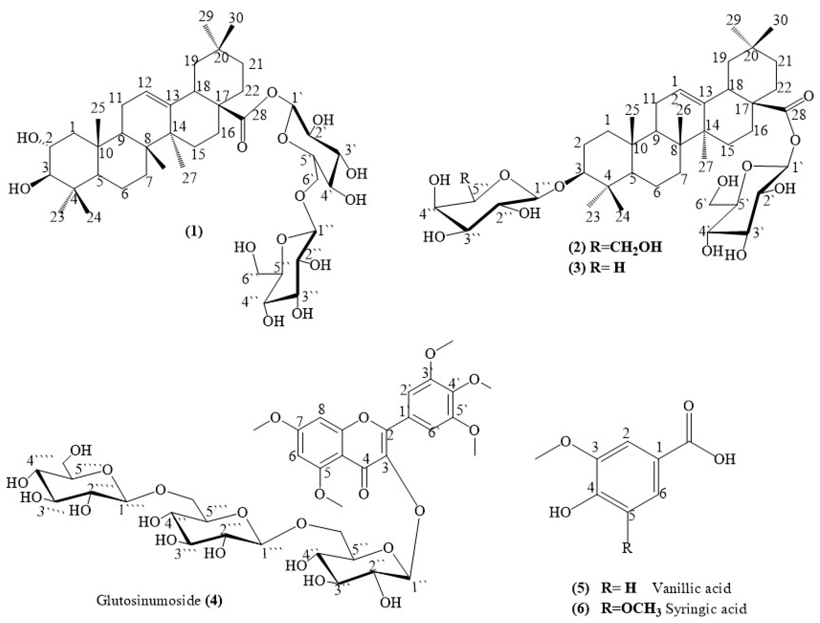
3.1. Identification of the Isolated Compounds

The phytochemical investigation of the C. glutinosum leaves showed the isolation of six compounds. The spectral data of the isolated compounds are recorded in Table 2, Table 3 and Tables S1–S4 and Figures S1–S43 using 1D-, 2D-NMR, and HR-ESI-MS data. The isolated compounds included three oleanane glycosides (13), one flavonol-glycoside (4), and two known phenolic compounds (5,6). It is worth noting that this is the first report on the isolation and identification of compound (4) from nature, although the four other compounds were isolated for the first time from the genus [viz. (13), and (6)], and compound (5) was previously identified from C. glutinosum by HPLC [20].

3.1.1. Identification of Compound (1)

From the HR-ESI-MS data in the negative mode, the pseudo-molecular formula of compound (1) was determined to be C43H70O15, m/z = 843.4384 [M + HCOOH, 31%, calcd. 843.4697. The molecular formula of (1) was dedicated as C42H67O13, m/z = 795.4777 [M–H+, 10%, calcd. 795.4531], and the most abundant peaks were found at m/z = 727.3915 [M–H+– 4OH, 100%, calcd. 727.4421] and 304.2999 [M– C22H36O122•, 100%, calcd. 304.2402] in positive mode (Table S1, Figures S11 and S12), identified as yellow amorphous. The 1H-APT, DEPT-NMR, and HSQC spectral data of compound (1) (Table 2, Figures S1–S7) were consistent with the basic skeleton of oleanane triterpenes with the presence of the hydroxyl groups of the A-ring and sugar moieties attached to the 28-carboxylic group. The 1H-NMR spectrum of (1) displayed seven tertiary methyl groups (each s) at δH: 1.35, 1.24 (2-CH3), 0.98, 0.92, and 0.91(2-CH3), and a broad olefinic proton doublet for H-12 at δH: 5.31 (d, J = 8.3 Hz); and two broad singlets for H-3 and H-18 at δH: 3.25 (1H, s) and 3.03 (1H, s), respectively. The two characteristic downfield protons of two methine groups at δH: 4.34 (br.s) and 3.24 (br.s) and the two anomeric signals at δH: 5.31 (d, J = 8.3 Hz) which were correlated in the HSQC spectrum (Figures S6 and S7) with the carbon signals at δC 69.3, 82.9, and 96.4, respectively, suggest that this compound has characteristic two hydroxy groups of a glycosidic-triterpenoid type. The 13C-NMR data showed thirty-three carbon signals categorized as seven methyls, ten methylenes, five methines, and seven quaternary carbon atoms. In addition, the data from the C-2, C-3, and C-28 were downfield at δC 69.3, 82.9, and 179.1, respectively, indicating that the aglycone of (1) is maslinic [21,22]. The COSY spectrum data (Figure S8) established the triterpene skeleton as an oleanane type (maslinic acid), and the sugars are two moieties of D-glucose (Glc) [23,24]. The HMBC spectrum data (Figures S9 and S10) establish the attachment of the sugar moieties with the triterpene skeleton at the C-28 position [25]. In the HMBC spectrum of (1), long-range couplings were observed between H-1′ of the Glc moiety (δH 5.31) and C-28 of an oleanolic acid moiety at δC 179.1 and between H-5′ (δH 3.31) of Glc and C-1″ of the terminal Glc moiety at δC 96.4. The anomeric configuration of the Glc moieties was established to be β-based on the relatively large 3JH-6′-H-1″ values (8.3 Hz). Thus, (1) was assigned as 28-O-[β-D-glucopyranosyl]-(1″→6′)-[β-D-glucopyranosyl]-maslinic acid (Figure 1 and Figure 2) [21,22,23,24,25].
Table 2. NMR Spectroscopic data for the compounds (1) in deuterated methanol.
PositionType HSQCCOSY HMBC (H→C)Maslinic acid a/b
* δH (J in Hz) ** δC (1H-1H)J2J3J4δH (J in Hz) a/bδC a/b
Aglycone
1CH21.74, 1.46, m 41.6C2 C25 0.91, 2.0046.3
2CH4.34, br.s69.3C1 3.70, m68.9
3CH3.24, br.s82.9 3.01, d (9.5)83.2
4C 36.4 39.4
5CH1.25, m49.8 C6, C10 0.85, bd (11.7)55.4
6CH20.98, br.s, 1.74, m29.8C7 1.40, ddd, 1.55, m18.4
7CH21.25, 1.73, m33.7C6C6, C8 1.32, dt, 1.46, ddd 32.8
8C 40.5 39.3
9CH1.81, m50.1C11 1.62, m47.7
10C 39.1 38.0
11CH21.23, 1.96, m25.1C9, C12 C13 1.90, 1.95, ddd23.5
12CH5.31, d (8.3)125.4C11 5.30, t (3.65)121.7
13C 144.2 C11 144.4
14C 43.6 41.7
15CH20.98, br.s, 1.69, m29.8C16C14 1.09, dt, 1.71, ddd27.8
16CH22.29, 1.67, m28.9C15 1.62, m, 2.00, dt23.3
17C 47.6 46.2
18CH3.03, br.s45.4 2.83, dd41.5
19CH21.84, 0.85, m50.3 1.63, m, 1.67, ddd46.0
20C 39.1 30.5
21CH21.26, 1.70, m33.7C22C22 1.22, m, 1.35, ddd33.8
22CH21.75, 0.98, m29.9C21 1.59, m, 1.78, td32.7
23CH30.91, s29.1 C4C3, C24 1.03, s 28.9
24CH30.92, s25.6 C4C3, C23 0.83, s 17.1
25CH31.35, s19.3 C9, C10 0.99, s16.4
26CH30.98, s19.1 C8C9, C14 0.82, s17.2
27CH31.24, s25.4 C14C8, C13 1.14, s25.7
28C 179.1 179.0
29CH30.91, s29.1 C19, C30 0.91, s32.8
30CH31.24, s25.4 C20C29C220.93, s23.3
β-D-Glucose c–e/f
1′CH5.31, d (8.3)96.4C2′C28 6.31, d (8.0)96.0
5.37, d (8.1)95.7
2′CH3.29, br.s74.4C1′C1′, C3′ 4.2374.2
3.33, m73.9
3′CH3.36, d (8.8)78.7 C4′C5′ 4.3079.1
3.35, d (8.5)78.2
4′CH3.32, br.s71.6 C1″4.3471.3
3.39, m71.1
5′CH3.31, br.s79.1C6′C4′C1″ 4.0479.5
3.42, m78.5
6′CH23.54, d (11.1);
3.41, d (11.1)
66.4C5′ C4′ 4.97 dd (1.9; 11.5)
4.70 dd (5.5; 11.5)
67.1
1″CH5.31, d (8.3)96.4C2″ 6.31, d (8.0)96.0
5.37, d (8.1)95.7
2″CH3.29, br.s74.4C1″C1″C5″ 4.2374.2
3.33, m73.9
3″CH3.27, br.s78.7C4″C2″C1″ 4.3079.1
3.35, d (8.5)78.2
4″CH3.71, br.s70.1C3″, C5″ 4.3471.3
3.39, m71.1
5″CH3.26, br.s78.7C4″C1″ 4.0479.5
3.42, m78.5
6″CH23.78, d (11.8);
3.65, d (11.7)
62.8 C5″ 4.43, 4.48, d (11.8)62.4
3.80; 3.71 m62.5
* Measured at 500 MHz, ** Measured at 125 MHz, a/b [21,22], c–e [23,24,25], f [26]. Assignments were based on the HMBC, HSQC, COSY, and DEPT experiments.

3.1.2. Identification of Compound (2)

The molecular formula of Compound (2) [yellow amorphous] was determined to be C42H68O13, m/z 780.4891 ([M], calculated 780.4660). It had a pseudo-molecular peak, C43H70O15 at m/z 826.4355 [M+ HCOOH, 1%, calculated 826.4715], the most abundant peak at m/z 353.0881 [M–H+ –C21H32O72 –2CH3, 100%, calculated 353.1964], and showed a characteristic fragment at m/z 701.3679 [M–2H+ –4CH3 –OH, 80%, calculated 701.3537] and 421.1637 [M–H+ –2sugars, 10%, calculated 421.3470] based on HR-ESI-MS data (Figure S20) in the negative mode. Compound (2) bore a close similarity to (1) in the 13C-NMR and 1H-NMR spectral data (Figures S13 and S14) of the aglycone and the oleanane skeleton [27] except for the absence of a 2-hydroxyl group attached to the ring A and the type/position of one of the sugar moieties. However, except for the lack of signal from the terminal glucose sugar unit, a comparison of the NMR (1D, 2D) spectral data of (2) with those of (1) indicated the close attachment of the aglycone with the β-Glu- at C-28. The presence of the galactose sugar unit in the significant downfield shift of C-3 (δC 82.9) and the coupling patterns of the anomeric protons confirmed the presence of β-D-glucose and β-D-galactose at C-28 and C-3, respectively (Table S3). The characteristic key HMBC correlations (Figures S18 and S19) of δH 3.24 (1H, d, J = 3.5 Hz, H-3) with δC 96.3 (galactose C-1″) and δH 5.33 (1H, d, J = 8.3 Hz, H-1′) with δC 179.0 (aglycone C-28), assigned in the structure of (2) as 3β-O-β-D-galactopyranosylolean-12-en-28-oic acid 28-O-β-D-glucopyranosyl ester, (Figure 1) was confirmed by the 2D-NMR and mass spectral data (Figures S13–S20) [24,25,26,28]. This is the first report of the isolation of the compound (2) from the Combretaceae family by the SciFinder database; it was isolated before from the leaves of Aralia elata [29].

3.1.3. Identification of Compound (3)

Compound (3) was identified as a white amorphous compound. Its molecular formula was determined to be C41H66O12, m/z 750.4072 ([M], 4%, calcd. 750.4554), its most abundant peak was at m/z 711.3965 ([M–3H]+ –2H2O, 100%, calcd. 711.4108), and it showed a characteristic fragment at m/z 713.4022 ([M–H]+ –2H2O, 10%, calcd. 713.4265) based on HR-ESI-MS data (Figure S29) in the negative mode. The comparison of the 1D- and 2D-NMR spectral data of (3) with those of (2) indicated their close relationship with the aglycone, the β-Glu- at C-28, and also in the significant downfield shift of C-3 (δC 81.7). The assignments of the spectral data analysis of the sugar moieties, which were established by 1H–1H COSY and HMBC correlations (Figures S27 and S28), suggest that the glycosidic sequence of (3) does not correspond to sequence of (2). The 1H- and 13C-NMR (Figures S21–S23) demonstrated signals for one terminal β-D-glucopyranosyl (Glc) and α-arabinose (Ara) units [δH 6.37, d (8.0), H-1′, δC 96.6, 74.9, 79.7, 71.5, 80.1 (each CH), and 62.8 (CH2) of Glc; and δH 6.06, d (5.5), H-1″, δC 96.6, 71.5, 74.9, 69.6 (each CH), and 67.1 (CH2) of Ara]. In the HMBC spectrum of (3), long-range correlations were observed between H-1′ of Glc and C-28 of the aglycone at δC 178.1 and between H-1″ of Ara with C-3 of the aglycone at δC 81.7. Thus, (3) was assigned as 3β-O-β-L-arabinopyranosylolean-12-en-28-oic acid, 28-O-β-D-glucopyranosyl ester (Table S3 and Figure 1 and Figures S21–S28) [30]. Compound (3) is a rare triterpenoid saponin that is being reported for the first time in the Combretaceae family. In accordance with the SciFinder database, this is the second report of (3) in nature. It is noteworthy that compound (3) was firstly isolated by Geizi et al. from the leaves of Lafoensia glyptocarpa [30].

3.1.4. Identification of Compound (4)

Compound (4) displayed as yellow amorphous. Its chemical formula was determined as C38H50O23, Mol. Wt. 874.7950. Its most abundant peak is m/z 575.2101 [M—7OH –C9H8O42−, 100%, calcd. 575.2422] in positive mode (Figures S38 and S44, Table S2), and it showed characteristic fragments at m/z 263.1319 [M–2H—3 sugar moieties –CH3 –3CH3O, 50%, calcd. 263.0344] and 587.1901 [M–2H –6OH –CH3O –C8H8O32CH3O, 2%, calcd. 587.1765] based on the HR-ESI-MS data (Figures S39 and S45, Table S2) in negative mode. The results of the spectral data analysis revealed the presence of a flavonol skeleton substituted with penta-methoxy groups, which was identified as a myricetin pentamethyl ether [24,31,32,33], based on the 1H, 13C, and DEPT-NMR spectra (Table 3, Figures S30–S33). These spectra demonstrated the presence of four aromatic protons, including two meta-coupled protons at δH 6.38 (overlapped, H-6) and 6.52 (br.s, H-8) of the A-ring, δH 6.69 (t, H-2′) and 6.70 (t, H-6′) of the B-ring of the flavonol moiety, and five methoxy groups at δH 3.80 (br.s, CH3OCH3O), 3.76 (s, CH3O), 3.75 (br.s, CH3OCH3O), 3.70 (s, CH3OCH3O), and 3.27 (s, CH3O) which were correlated with the five quaternary carbons at δC 57.0, 56.8, 57.2, 57.3, and 60.4, respectively. The HSQC (Figure S34) confirmed the presence of 5,7,3′,4′,5′-pentamethyl ether [6,12,24,31,32,33]. The NMR spectral data of compound (4) (Table 3 and Figures S30–S37) showed the possible presence of the three sugar moieties; this was confirmed by the presence of three anomeric proton signals at δH 6.37, (d, J = 8.4 Hz), 4.17, (d, J = 9.0 Hz), and 3.95, (d, J = 7.6 Hz), which were correlated with the three anomeric carbons at δC 107.3, 105.5, and 105.2, respectively, from the HSQC (Figure S34) [24,27,31,32,33] and from the presence of a characteristic fragment at m/z 517.1487 [M–H—2 glucose units—CH3, 5%, calcd. 517.1346], which was coincident with C25H25O12 (Table S2, Figures S39 and S45) in a negative mode. The anomeric configuration of the three Glu was established to be β-based on the relatively large 3JH-1-H-6 values (7.6–9.0 Hz) (Figures S30–S37). The presence of a flavonol substitutional part was confirmed by the presence of a characteristic peak at m/z 371.15604 [M–3glucose parts, 1%, calcd. 371.1131], coincident with C20H19O7 (Table S2, Figure S38 and S45) in positive mode. In addition, the presence of 3-O-β-glucosyl substitutions was evident from the HMBC correlation between the proton signal at δH 6.37 (d, J = 8.4 Hz, H-1″) and at C-2 and C-3 (δC 149.4 and 139.3, respectively) (Figure 2, Figures S36 and S37). Thus, the structure of (4) was confirmed to be 3-[β-O-D-glucopyranosyl]-(1‴→6″)-O-β-D-glucopyranosyl-(1⁗→6‴)-O-β-D-glucopyranosyl-5,7,3′,4′,5′-pentamethoxy flavonol, named glutosinumoside (Figure 1 and Figure 2). When compared with the SciFinder database, no reports on the occurrence of (4) in the herbal plants have been found in the literature.
Table 3. NMR spectral data of compound (4) in CD3OD (500 MHz) for 1H-NMR and (125 MHz) for 13C-NMR.
PositionType HSQCCOSYHMBCMyricetin *
δH (J in Hz) δC 1H-1H (H→C) δH (J in Hz) δC
Aglycone
2C 149.4 157.2
3C 139.3 134.3
4C 178.5 178.1
5C 149.5 161.4
6CH 6.37 (overlapped)108.2 C5, C7, C9, C106.67, br.s98.6
7C 149.1 165.8
8CH 6.52, br.s108.2 C7, C9, C106.36, br.s92.8
9C 149.5 157.0
10C 139.2 105.6
1′C 123.6 122.1
2′CH 6.69, t116.3 C2, C3, C1′, C2′, C3′7.55, br.s116.4
3′C 149.3 146.5
4′C 148.0 150.6
5′C 147.6 7.04 (d, J = 8.7 Hz) 111.9
6′CH 6.70, t114.6 C2, C3, C1′, C4′, C5′7.73 (br.d, J = 8.7 Hz)123.0
β-Glucose
1″CH 6.37, d (8.4)107.4 C2, C34.4, d (7.9)105.6
5.69, d (7.7)104.9
2″CH 3.12–3.19, m75.3 C3″, C4″, C5″3.17, t (8.5)75.5
4.0875.7
3″CH 3.23, br.s78.4 C2″, C4″, C1‴3.3378.3
4.2778.2
4″CH 3.27, m71.7 C3″, C5″3.2971.5
4.1571.4
5″CH 3.12–3.19, m75.3C6″C2″, C4″3.2777.7
4.0476.2
6″CH2 3.76, 3.80, br.s67.5C5″C4″3.67, dd (11.9, 4.9)
3.83, dd (11.9, 2.1)
62.7
4.35; 4.65, brd (10.7)69.3
β-Glucose
1‴CH 4.17, dd (2.6, 9.0)105.5C2‴C1″, C4″4.4, d (7.9)105.6
2‴CH 3.12–3.19, m75.3C1‴C1‴, C3‴, C4‴, C5‴3.17, t (8.5)75.5
3‴CH 3.31, br.s78.5 C2‴3.3378.3
4‴CH 3.31, m71.7 C2‴3.2971.5
5‴CH 3.27, br.s78.5 C2‴3.2777.7
6‴CH2 3.75, 3.80, br.s67.5 C1⁗, C1‴, C4‴3.67, dd (11.9, 4.9)
3.83, dd (11.9, 2.1)
62.7
4.35; 4.65, brd (10.7)69.3
β-Glucose
1⁗CH 3.95, d (7.6)105.2C2⁗ 4.4, d (7.9)105.6
2⁗CH 3.22, br.d75.4C1⁗C3⁗, C4⁗, C5⁗3.17, t (8.5)75.5
3⁗CH 3.31, br.s78.5 C2⁗3.3378.3
4⁗CH 3.31, m71.7 C2⁗3.2971.5
5⁗CH 3.27, br.s78.4 C2⁗3.2777.7
6⁗CH2 3.70, 3.74, br.s65.5 C1⁗, C4⁗3.67, dd (11.9, 4.9)
3.83, dd (11.9, 2.1)
62.7
5-O-CH33.75, br.s57.2 C53.86, s56.7
7-O-CH33.80, br.s57.0 C73.85, s56.2
3′-O-CH33.70, s57.3 C3′3.86, s56.7
4′-O-CH33.27, s60.4 C4′, C5′3.86, s56.7
5′-O-CH33.76, s56.8 C5′3.86, s56.7
* [24,27,31,32,33].

3.1.5. Identification of Compound (5)

The 1D-NMR data (Figures S40 and S41) identified the structure of compound (5) as white, amorphous vanillic acid, which was in accordance with the results of an authentic sample and reported data in the literature [34,35]. This compound was previously identified from C. glutinosum by HPLC analysis [20]. 1H-NMR spectrum data (CD3OD, 500 MHz) are as follows: δH: 6.76 (1H, d, J = 8.0 Hz, H-5), 7.50 (1H, s, H-2), 7.48(1H, br.d, J = 9.0 Hz, H-6), 3.85 (3H, s, CH3O−); 13C-NMR spectrum data (CD3OD, 125 MHz) δC: 169.2 (COOH), 116.2 (C-2), 149.1 (C-3), 152.8 (C-4), 114.3 (C-5), 125.7 (C-6), and 56.8 (CH3O) [34,35].

3.1.6. Identification of Compound (6)

Compound (6) was yellow and amorphous, identified by 1H and 13C-NMR (CD3OD, 500 and 125 MHz) spectral data (Figures S42 and S43), as follows: δH: 7.27 (2H, s, H-2/H-6) and 3.82 (3H, s, CH3O−); δC: 171.0 (COOH), 123.0 (C-3/C-5), 142.0 (C-4), 149.3 (C-1), 108.7 (C-2/C-6), 57.2 (CH3O−) [36]. In addition, co-chromatography with an authentic sample revealed that this compound (6) is syringic acid. This compound was isolated from the genus Combretum for the first time.

3.2. Biological Activities of Main Fractions and Isolates from C. glutinosum

3.2.1. In Vitro Cytotoxic Activity

Based on the biological, bio-guided procedure, the anticancer activity of the five extracts of C. glutinosum was determined to select the most significant extract(s) for further phytochemical investigations (Table 4). The anticancer activity of DCM and the aqueous extracts against MCF7 and HT29 demonstrated a growth inhibition of 62%-93% compared to the control after treatment with 50 µg/mL, indicating that the activity is associated with the natural compounds within the DCM extract. Therefore, the DCM extract was kept for future investigations. The growth inhibition % of the crude EtOH was superior to that of the DCM, reaching 93% against MCF7 (Table 4). Thus, further investigations were carried out on the crude EtOH extract. Table 5 shows the cytotoxic activity of the crude EtOH extract in addition to the six isolated compounds and doxorubicin. The most sensitive cell line was MCF7; the most active compounds were (1), followed by (5), (6), and (4) (Table 5).

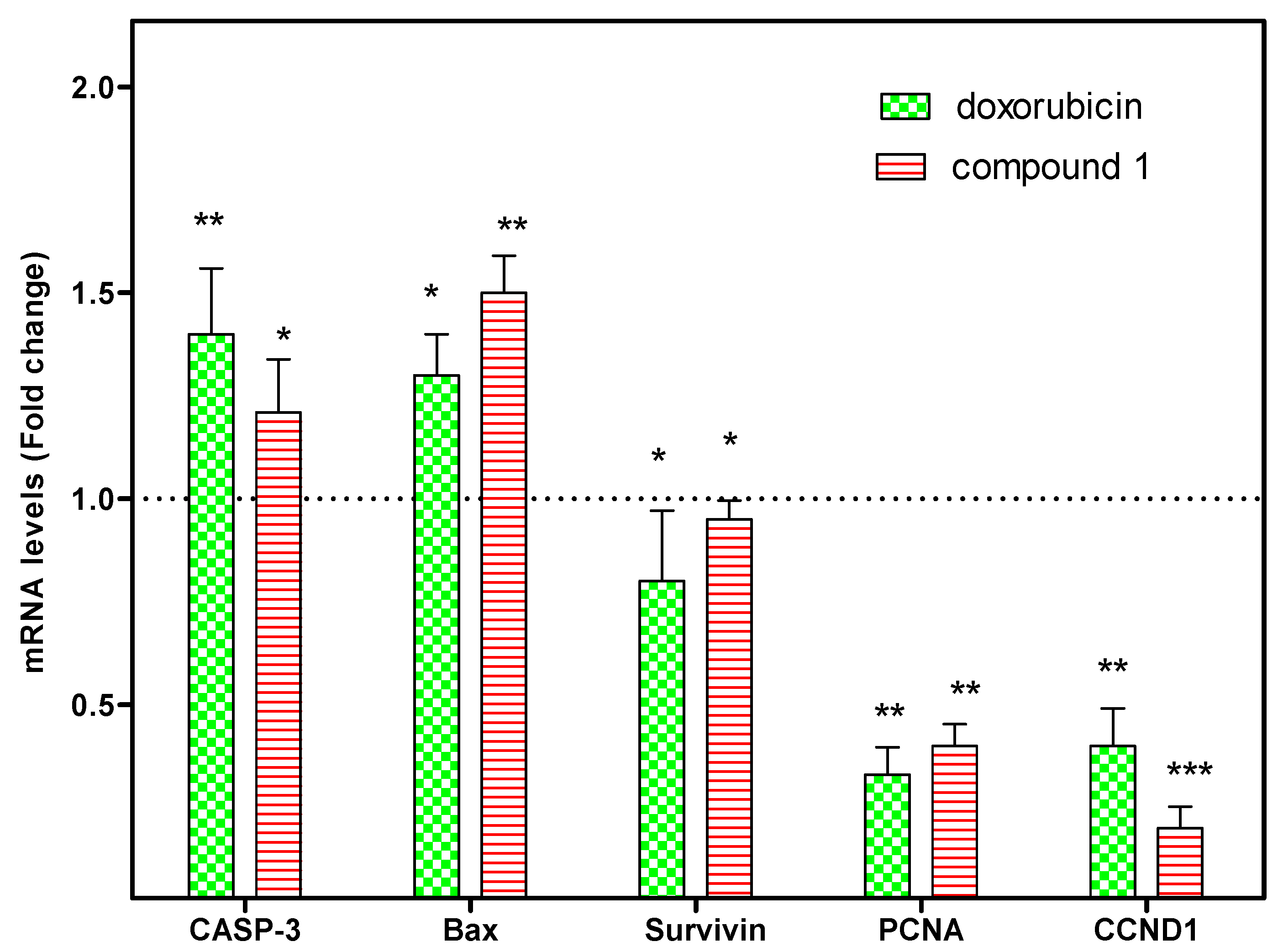
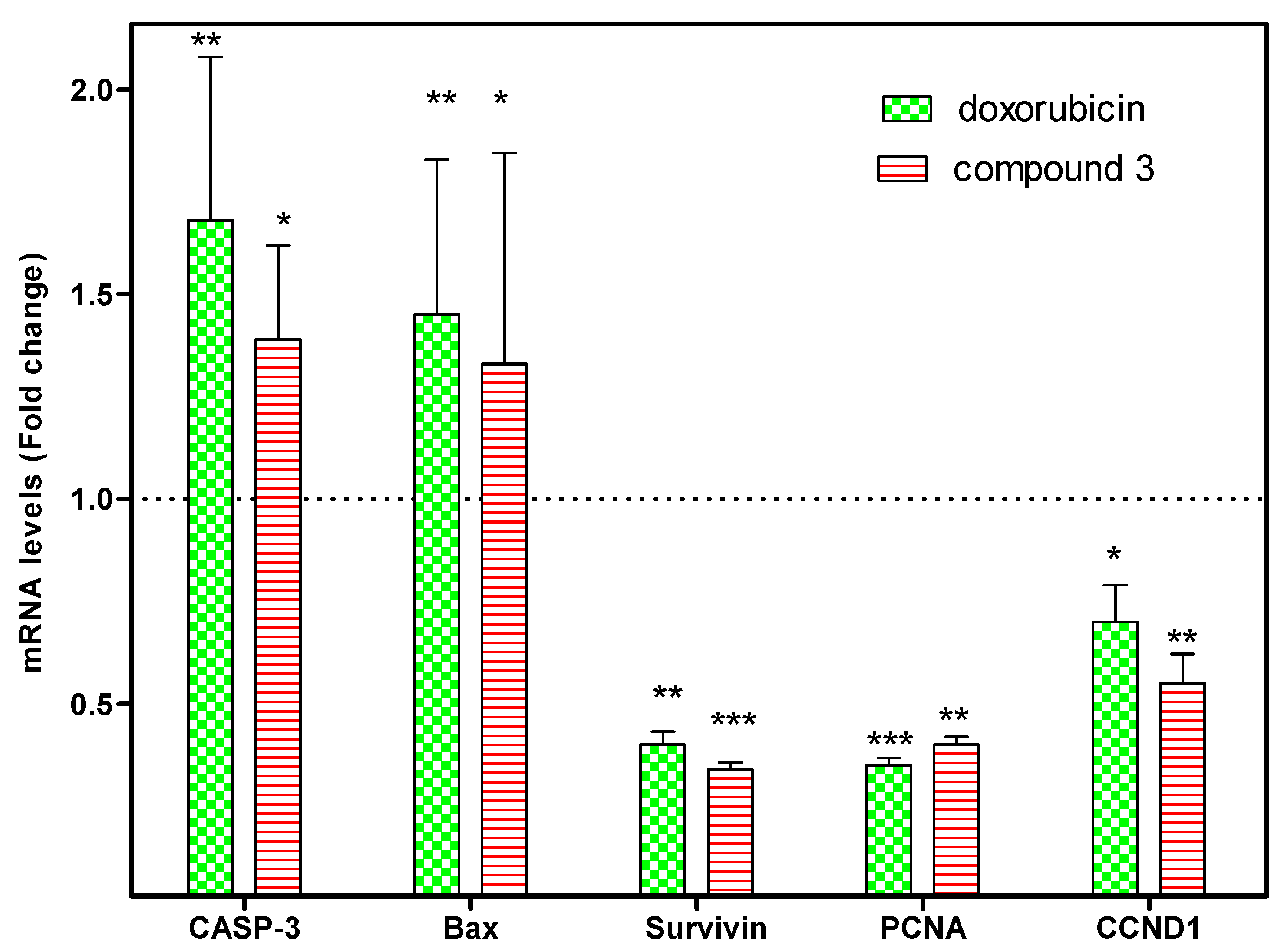
3.2.2. Effect of Compounds (1) and (3) on Gene Expression

Compared to the vehicle control, both doxorubicin and compound (1) caused a significant upregulation of the pro-apoptotic caspase 3 and bax gene expression and the downregulation of survivin in MCF7 cells. The effect of doxorubicin was more significant compared to compound (1) with respect to Caspase 3 and survivin, while compound (1) showed a greater bax upregulatory effect than doxorubicin, as shown in Figure 3. Moreover, both compounds showed significant cell-cycle effects through the downregulation of PCNA and CCND1 gene expression, while compounds (1) and (3) surpassed the effect of doxorubicin with respect to CCND1. The effect of doxorubicin was more remarkable compared to compound (3) with respect to the caspase 3 and bax genes, while compound (3) surpassed doxorubicin in downregulating survivin, as shown in Figure 4. Thus, compounds (1) and (3) were shown to affect cancer’s critical hallmarks by inducing apoptosis and perturbing the MCF7 cell cycle.

4. Conclusions

This study was conducted to evaluate the cytotoxic activity of the polar fraction (ethyl acetate) of the Sudanian plant, C. glutinosum, to identify new phytochemicals for effective therapy. The study reported herein the isolation and identification of six bioactive components, including three triterpenoid saponins [28-O-[β-D-glucopyranosyl-(1″→6′)-[β-D-glucopyranosyl]-maslinic acid (1), 3β-O-β-D-galactopyranosylolean-12-en-28-oic acid, 28-O-β-D-glucopyranosyl ester (2), and 3β-O-β-L-arabinopyranosylolean-12-en-28-oic acid, 28-O-β-D-glucopyranosyl ester (3)], a flavonol-glycoside derivative (4), and two phenolic acids (5,6). One compound of isolates was identified for the first time from nature, viz., glutosinumoside (4) [flavonol-glycoside]. This research also showed that four bioactive phytochemicals [(13) and (6)] had not before been isolated from C. glutinosum nor the Combretaceae family. The biological results of this study demonstrated that the plant has significant cytotoxic activity.

Supplementary Materials

The following supporting information can be downloaded at: https://www.mdpi.com/article/10.3390/separations10030209/s1, Tables S1 and S2: HR-ESI-MS spectral data for the compounds (1,4); Table S3: NMR spectral data for the compounds (2,3); Table S4: NMR spectral data for the compounds (5,6); Figures S1–S43: Spectra of the compounds (16); Figures S44 and S45: Mass fragmentations of the compound (4); Flowchart S1: Extraction and Fractionation procedure of the C. glutinosum leaves; Flowchart S2: Fractionation and purification of the ethyl acetate extract of C. glutinosum leaves. References [22,23,24,31,36] are cited in the Supplementary Material.

Author Contributions

Conceptualization, A.N.A. and E.H.M.; methodology, S.H.S.; compound analysis and validation, S.H.S.; writing—original draft, S.H.S.; writing—review & editing, S.H.S. and A.N.A.; final manuscript revision, A.N.A., S.H.S., M.K. and E.-S.K.; in vitro biological investigation, A.N.A., E.H.M., M.E.E. and M.B.O.E.; final revisions, A.N.A., S.H.S., M.K. and E.-S.K. All authors have read and agreed to the published version of the manuscript.

Funding

This study is supported via funding from Prince Sattam bin Abdulaziz University, project number (PSAU/2023/R/1444).

Institutional Review Board Statement

Not applicable.

Informed Consent Statement

Not applicable.

Data Availability Statement

The data presented in this study are available in the Supplementary Materials.

Acknowledgments

The authors demonstrate their gratitude to the members of the Pharmacognosy Department, College of Pharmacy, Prince Sattam bin Abdulaziz University, for their kind help and the use of lab facilities.

Conflicts of Interest

The authors declare no conflict of interest.

References

  1. Bekele, B.; Lemma, B. Bioactive compounds from ten species of the genus Combretum: Review. Int. J. Chem. Sci. 2021, 5, 18–30. [Google Scholar]
  2. Lima, G.R.D.M.; Sales, I.R.D.; Filho, M.R.C.; Jesus, N.Z.D.; Falcão, H.D.S.; Barbosa-Filho, J.M.; Cabral, A.G.; Souto, A.L.; Tavares, J.F.; Batista, L.M. Bioactivities of the genus Combretum (Combretaceae): A review. Molecules 2012, 17, 9142–9206. [Google Scholar] [CrossRef] [Green Version]
  3. Bisoli, E.; Freire, T.V.; Yoshida, N.C.; Garcez, W.S.; Queiróz, L.M.M.; Matos, M.F.C.; Perdomo, R.T.; Garcez, F.R. Cytotoxic Phenanthrene, Dihydrophenanthrene, and Dihydrostilbene Derivatives and Other Aromatic Compounds from Combretum laxum. Molecules 2020, 25, 3154. [Google Scholar] [CrossRef]
  4. Géorcelin, A.; Olounlade, P.A.; Azando, E.V.B.; Dedehou, V.F.G.N.; Daga, F.D.; Adote, M.S.H. A review of Bridelia ferruginea, Combretum glutinosum and Mitragina inermis plants used in zootherapeutic remedies in West Africa: Historical origins, current uses and implications for conservation. J. Appl. Biosci. 2015, 87, 8003. [Google Scholar] [CrossRef] [Green Version]
  5. Niass, O.; Diop, A.; Mariko, M.; Géye, R.; Thiam, K.; Sarr, O.; Ndiaye, B.; Diop, Y.M. Comparative Study of the Composition of Aqueous Extracts of Green Tea (Camellia Sinensis) in Total Alkaloids, Total Flavonoids, Total Polyphenols and Antioxidant Activity with the Leaves of Combretum glutinosum, Combretum Micranthum and the Red Pulps of Hibiscus Sabdariffa. Int. J. Progress. Sci. Technol. 2017, 5, 71–75. [Google Scholar]
  6. Dawe, A.; Pierre, S.; Tsala, D.E.; Habtemariam, S. Phytochemical Constituents of Combretum Loefl. (Combretaceae). Pharm. Crop. 2013, 4, 38–59. [Google Scholar] [CrossRef]
  7. Aderogba, M.A.; Kgatle, D.T.; McGaw, L.J.; Eloff, J.N. Isolation of antioxidant constituents from Combretum apiculatum subsp. apiculatum. S. Afr. J. Bot. 2012, 79, 125–131. [Google Scholar] [CrossRef] [Green Version]
  8. Toklo, P.M.; Ladekan, E.Y.; Linden, A.; Hounzangbe-Adote, S.; Kouam, S.F.; Gbenou, J.D. Anthelmintic flavonoids and other compounds from Combretum glutinosum Perr. ex DC (Combretaceae) leaves. Acta Cryst. Sect. C 2021, 77, 505–512. [Google Scholar] [CrossRef]
  9. Amako, N.F.; Amupitan, J.O. A dammarane triterpenoid ester from Combretum glutinosum Perr. Ex. Dc., stem bark. J. Chem. Soc. Nigeria 2017, 40, 1–5. [Google Scholar]
  10. Morgan, A.M.A.; Mohamed, A.E.; Saophea, C.; Park, S.U.; Kim, Y.H. Pentacyclic triterpenes from the stem bark of Combretum hartmannianum Schweinf. Biochem. Syst. Ecol. 2018, 77, 48–50. [Google Scholar] [CrossRef]
  11. Harouna, S.; Adama, H.; Djifaby, S.P.A.E.; Moussa, C.; Roland, M.; Jeanne, M.; Germaine, N.O. Phytochemistry and Biological Activities of Extracts from Two Combretaceae Found in Burkina Faso: Anogeissus leiocarpus (DC) Guill. and Perr. and Combretum glutinosum Perr. Ex DC. Univers. J. Environ. Res. Technol. 2012, 2, 383–392. [Google Scholar]
  12. Roy, S.; Gorai, D.; Acharya, R.; Roy, R. COMBRETUM (COMBRETACEAE): BIOLOGICAL ACTIVITY AND PHYTOCHEMISTRY. Indo Am. J. Pharm. Res. 2014, 4, 5266–5299. [Google Scholar]
  13. Hassanpour, S.H.; Mohammadamin, D. Review of cancer from perspective of molecular. J. Cancer Res. Pract. 2017, 4, 127–129. [Google Scholar] [CrossRef]
  14. Sweilam, S.H.; Abdel Bar, F.M.; Foudah, A.I.; Alqarni, M.H.; Elattal, N.A.; El-Gindi, O.D.; El-Sherei, M.M.; Abdel-Sattar, E. Phytochemical, Antimicrobial, Antioxidant, and In Vitro Cytotoxicity Evaluation of Echinops erinaceus Kit Tan. Separations 2022, 9, 447. [Google Scholar] [CrossRef]
  15. Handa, S.S.; Khanuja, S.P.S.; Longo, G.; Rakesh, D.D. Extraction Technologies for Medicinal and Aromatic Plants; United Nations Industrial Development Organization and the International Center for Science and High Technology: Trieste, Italy, 2008; p. 116. [Google Scholar]
  16. Abdalla, A.N.; Stefano, M.D.; Poli, G.; Tuccinardi, T.; Bader, A.; Vassallo, A.; Abdallah, M.E.; El-Readi, M.Z.; Refaat, B.; Algarni, A.S.; et al. Co-Inhibition of P-gp and Hsp90 by an Isatin-Derived Compound Contributes to the Increase of the Chemosensitivity of MCF7/ADR-Resistant Cells to Doxorubicin. Molecules 2021, 27, 90. [Google Scholar] [CrossRef] [PubMed]
  17. Abdallah, Q.; Al-Deeb, I.; Bader, A.; Hamam, F.; Saleh, K.; Abdulmajid, A. Anti-angiogenic activity of Middle East medicinal plants of the Lamiaceae family. Mol. Med. Rep. 2018, 18, 2441–2448. [Google Scholar] [CrossRef]
  18. Abdalla, A.N.; Abdallah, M.E.; Aslam, A.; Bader, A.; Vassallo, A.; Tommasi, N.; Malki, W.H.; Gouda, A.M.; Mukhtar, M.H.; El-Readi, M.Z.; et al. Synergistic Anti Leukemia Effect of a Novel Hsp90 and a Pan Cyclin Dependent Kinase Inhibitors. Molecules 2020, 25, 2220. [Google Scholar] [CrossRef]
  19. Abdalla, A.N.; Malki, W.H.; Qattan, A.; Shahid, I.; Hossain, M.A.; Ahmed, M. Chemosensitization of HT29 and HT29-5FU Cell Lines by a Combination of a Multi-Tyrosine Kinase Inhibitor and 5FU Downregulates ABCC1 and Inhibits PIK3CA in Light of Their Importance in Saudi Colorectal Cancer. Molecules 2021, 26, 334. [Google Scholar] [CrossRef]
  20. Muhammad, B.Y.; Shaban, N.Z.; Elrashidy, F.H.; Ghareeb, D.A. Antioxidant, Anti-inflammatory, Antiproliferative and Antimicrobial Activities of Combretum glutinosum and Gardenia aqualla Extracts in vitro. Free Radic. Antioxid. 2020, 9, 66–72. [Google Scholar] [CrossRef]
  21. Woo, K.W.; Han, J.Y.; Choi, S.U.; Kim, K.H.; Lee, K.R. Triterpenes from Perilla frutescens var. acuta and Their Cytotoxic Activity. Nat. Prod. Sci. 2014, 20, 71–75. [Google Scholar]
  22. Dais, P.; Plessel, R.; Williamson, K.; Hatzakis, E. Complete 1H and 13C NMR assignment and 31P NMR determination of pentacyclic triterpenic acids. Anal. Methods 2017, 9, 949–957. [Google Scholar] [CrossRef]
  23. Kim, B.; Han, J.W.; Ngo, M.T.; Dang, Q.L.; Kim, J.; Kim, H.; Choi, G.J. Identification of novel compounds, oleanane- and ursane-type triterpene glycosides, from Trevesia palmata: Their biocontrol activity against phytopathogenic fungi. Sci. Rep. 2018, 8, 14522. [Google Scholar] [CrossRef] [PubMed] [Green Version]
  24. Agrawal, P.K. NMR spectroscopy in the structural elucidation of oligosaccharides and glycosides. Phytochemistry 1992, 31, 3307–3330. [Google Scholar] [CrossRef] [PubMed]
  25. Yang, H.; Kim, H.W.; Kim, Y.C.; Sung, S.H. Cytotoxic activities of naturally occurring oleanane-, ursane-, and lupane-type triterpenes on HepG2 and AGS cells. Pharmacogn. Mag. 2017, 13, 118–122. [Google Scholar] [CrossRef] [Green Version]
  26. Silchenko, A.S.; Kalinovsky, A.I.; Avilov, S.A.; Andrijaschenko, P.V.; Popov, R.S.; Chingizova, E.A.; Kalinin, V.I.; Dmitrenok, P.S. Triterpene Glycosides from the Far Eastern Sea Cucumber Psolus chitonoides: Chemical Structures and Cytotoxicities of Chitonoidosides E1, F, G, and H. Mar. Drugs 2021, 19, 696. [Google Scholar] [CrossRef]
  27. Augustin, J.M.; Drok, S.; Shinoda, T.; Sanmiya, K.; Nielsen, J.K.; Khakimov, B.; Olsen, C.E.; Hansen, E.H.; Kuzina, V.; Ekstrøm, C.T.; et al. UDP-Glycosyltransferases from the UGT73C Subfamily in Barbarea vulgaris Catalyze Sapogenin 3-O-Glucosylation in Saponin-Mediated Insect Resistance1[W][OA]. Plant Physiol. 2012, 160, 1881–1895. [Google Scholar] [CrossRef] [Green Version]
  28. Yao, H.; Wang, J.; Yin, J.; Nie, S.; Xie, M. A review of NMR analysis in polysaccharide structure and conformation: Progress, challenge and perspective. Food Res. Int. 2021, 143, 110290. [Google Scholar] [CrossRef]
  29. Guohong, Y.; Zhou, J.Z.; Changchun, S.; Xingquan, M.; Junjie, W. Chemical study on glycosides from the leaves of Japanese Aralia (Aralia elata). Zhongcaoyao 1995, 26, 514–517. [Google Scholar]
  30. Carvalho, G.J.A.; Carvalho, M.G.; Braz-Filhob, R. A triterpenoid saponin isolated from Lafoensia glyptocarpa. Phytochemistry 1999, 52, 1617–1619. [Google Scholar] [CrossRef]
  31. Ünlü, A.; Teralı, K.; Aydın, Z.Ü.; Dönmez, A.; Yusufoglu, H.; Calis, I. Isolation, Characterization and In Silico Studies of Secondary Metabolites from the Whole Plant of Polygala inexpectata Peşmen & Erik. Molecules 2022, 27, 684. [Google Scholar] [CrossRef]
  32. Taketa, A.T.C.; Breitmaiera, E.; Schenkel, E.P. Triterpenes and Triterpenoidal Glycosides from the Fruits of Ilex paraguariensis (Maté). J. Braz. Chem. Soc. 2004, 15, 205–211. [Google Scholar] [CrossRef] [Green Version]
  33. Note, O.P.; Jihu, D.; Antheaume, C.; Guillaume, D.; Pegnyemb, D.E.; Kilhoffer, M.C.; Lobstein, A. Triterpenoid saponins from Albizia boromoensis Aubrev. & Pellegr. Phytochem. Lett. 2015, 11, 37–42. [Google Scholar]
  34. Chang, S.; Kim, K.; Kyun, L.; Choi, S.; Yong, R.S.; Ro, L. Phytochemical constituents of Bistorta manshuriensis. Nat. Prod. Sci. 2009, 15, 234–240. [Google Scholar]
  35. Shi, G.; Xu, M.; Duan, W.; Fang, L.; Liu, W.; Wang, X.; Zhang, Y. Chemical Constituents from Trichosanthis pericarpium. Asian J. Chem. 2014, 26, 4626–4630. [Google Scholar] [CrossRef]
  36. Bun-Sung, J.; Young-Je, C. Inhibitory Activity against Helicobacter pylori of Isolated Compounds from Pinus koraiensis Siebold et Zucc Leaves. J. Appl. Biol. Chem. 2016, 59, 19–23. [Google Scholar] [CrossRef] [Green Version]
Figure 1. Structures of isolated compounds from Combretum glutinosum (16).
Separations 10 00209 g001
Figure 2. COSY and HMBC correlations of isolated compounds (1,4).
Separations 10 00209 g002
Figure 3. The effect of vehicle control, doxorubicin (0.1 μM), and compound (1) (1 μM) on the expression of caspase 3, Bax, survivin, PCNA, and CCND1 in MCF7 cells (24 h). RT-PCR was used for the estimation of the proteins (mean ± S.D., fold change = 1 dashed line, n = 2 × 2 independent experiments). p < 0.05 (*), p < 0.01 (**), and p < 0.001 (***) were considered significant.
Separations 10 00209 g003
Figure 4. The effect of vehicle control, doxorubicin (0.1 μM), and compound (3) (2 μM) on the expression of caspase 3, Bax, survivin, PCNA, and CCND1 in MCF7 cells (24 h) RT-PCR was used for the estimation of the proteins (mean ± S.D., fold change = 1 dashed line, n = 2 × 2 independent experiments). p < 0.05 (*), p < 0.01 (**), and p < 0.001 (***) were considered significant.
Separations 10 00209 g004
Table 1. Sequence of primers used in the RT-PCR.
GeneSequence
Caspase 3F: ACATGGAAGCGAATCAATGGACTC
R: AAGGACTCAAATTCTGTTGCCACC
Bax F: GCCCTTTTGCTTCAGGGTTT
R: TCCAATGTCCAGCCCATGAT
SurvivinF: TTGCTCCTGCACCCCAGAGC
R: AGGCTCAGCGTAAGGCAGCC
PCNAF: CACCAAGGAGGGTGTCAAGT
R: GATCTTGGGGTGCCAGATAA
CCND1F: GGATGCTGGAGGTCTGCGA
R: AGAGGCCACGAACATGCAAG
GAPDHF: AGGTCGGTGTGAACGGATTTG
R: TGTAGACCATGTAGTTGAGGTCA
Table 4. Percent (%) of the growth inhibition compared to control after treatment with each of the five crude extracts of C. glutinosum (50 µg/mL, n = 3, 72 h).
Extract Growth Inhibition% ± SD
MCF-7HT-29MRC-5
Crude EtOH 93 ± 2.4090 ± 1.2485 ± 3.21
DCM 89 ± 293 ± 194.2 ± 0.6
MeOH 80% 55 ± 335 ± 547 ± 4
n-Hex 31 ± 525 ± 448.4 ± 5.5
Re. Aq 81 ± 162 ± 249 ± 3
EtOH = ethanol; DCM = dichloromethane; MeOH = methanol; n-Hex = n-hexane; Re. Aq = remaining aqueous.
Table 5. Cytotoxic activity (IC50 µM ± SD, n = 3, 72 h) of the isolated compounds from C. glutinosum and doxorubicin against three cell lines and normal fibroblasts.
Extract */
Compound
MCF7HT29HepG2Average ** IC50MRC5
EtOH Ext.3.50 ± 0.7412.09 ± 2.222.99 ± 0.566.1913.84 ± 1.57
11.37 ± 0.214.54 ± 1.483.30 ± 0.023.0710.22 ± 1.46
22.40 ± 0.2218.48 ± 0.783.37 ± 0.028.0814.76 ± 0.32
32.27 ± 0.196.24 ± 0.932.42 ± 0.213.643.54 ± 1.35
41.72 ± 0.193.99 ± 1.033.30 ± 0.933.0015.68 ± 3.02
51.48 ± 0.026.62 ± 0.673.82 ± 0.503.977.04 ± 0.61
61.48 ± 0.344.06 ± 0.482.10 ± 0.222.5413.85 ± 2.00
Doxo0.29 ± 0.030.13 ± 0.032.33 ± 0.190.913.71 ± 0.02
* IC50 of extract in µg/mL, ** average cytotoxicity (IC50) of each extract/ compound against the three cancer cells.
Disclaimer/Publisher’s Note: The statements, opinions and data contained in all publications are solely those of the individual author(s) and contributor(s) and not of MDPI and/or the editor(s). MDPI and/or the editor(s) disclaim responsibility for any injury to people or property resulting from any ideas, methods, instructions or products referred to in the content.

