Article

Development and Validation of a Confirmatory Method for the Determination of 12 Coccidiostat Residues in Eggs and Muscle by Means of Liquid Chromatography Coupled to Hybrid High Resolution Mass Spectrometry

by
Federica Castellani
,
Matteo Ricci
,
Maria Novella Colagrande
,
Giampiero Scortichini
and
Giorgio Saluti
*
Istituto Zooprofilattico Sperimentale dell’Abruzzo e del Molise “G. Caporale”, Via Campo Boario, 64100 Teramo, Italy
*
Author to whom correspondence should be addressed.
Separations 2023, 10(3), 202; https://doi.org/10.3390/separations10030202
Submission received: 22 February 2023 / Revised: 8 March 2023 / Accepted: 10 March 2023 / Published: 14 March 2023
(This article belongs to the Special Issue Novel Applications of Separation Technology)

Abstract

:
A confirmatory, highly selective multi-residue method based on liquid chromatography coupled to hybrid high resolution mass spectrometry (LC-Q-Orbitrap) was developed and validated for the determination of 12 regulated coccidiostats in eggs and muscle. Particularly, ionophore antibiotics (lasalocid, maduramicin, monensin, narasin, salinomycin and semduramicin) and synthetic coccidiostats (diclazuril, halofuginone, nicarbazin as 4,4′-dinitrocarbanilide fraction, robenidine and toltrazuril as toltrazuril-sulphone) were included in the method. The sample preparation consisted in the extraction of the analytes from the matrix with acetonitrile, followed by a clean-up step with Oasis® PRiME HLB SPE and a defatting procedure with n-hexane. Validation was successfully performed according to Commission Implementing Regulation (EU) 2021/808, starting from 1 µg kg−1. The procedure was verified through the analysis of a certified reference material (CRM) and the occurrence of the residues was assessed in the context of the Italian National Residue Control Plan (NRCP).

Graphical Abstract

1. Introduction

Coccidiosis is a disease caused by parasites of genus Eimeria and Isospora that may be prevalent in warm humid conditions. It can result in lesions to the intestinal tract, poor weight gain, diarrhoea, scarce feed conversion and, sometimes, death. This infection is very contagious, and so, it may result in economic losses especially for intensive farms. Coccidiostats encompass the category of naturally occurring polyether ionophores such as lasalocid, maduramicin, monensin, narasin, salinomycin and semduramicin and as synthetic (or chemical) coccidiostats such as diclazuril, halofuginone, nicarbazin, robenidine and toltrazuril. These drugs are authorized by the European Union (EU) as feed additives and are, nowadays, widely used in intensive farms posing the possibility to develop resistance of the parasites overall due to the unavoidable carry-over phenomenon from target to non-target feed that potentially occur during feed production, but also during storage and transport. This aspect can obviously affect the health of animals and humans for the presence of residues in feed and foodstuffs [1]. As a matter of fact, the EU set fixed maximum limits for edible tissue and eggs, and it is noteworthy that some of them are banned and others are regulated in a range between 2 and 4000 µg kg−1 [2,3,4,5,6,7,8,9,10,11,12].The development of multi-residue methods able to determine all the regulated compounds is still quite challenging, owing to their very different physicochemical properties and the low limits of detection (LODs) that have to be reached. Liquid chromatography coupled to low resolution tandem mass spectrometry (LC-MS/MS) is currently the most used technique for the determination of coccidiostats in feed and food [13,14,15,16,17,18,19,20]. To the best of our knowledge, in only three cases, these analytes were determined by means of high resolution mass spectrometry (HRMS); particularly, in 2013, Kaklamanos and collaborators [21] developed and validated a method for the determination of coccidiostats and other veterinary drugs in feed using liquid chromatography coupled to Orbitrap mass spectrometry operating in full scan acquisition. Later, Matus et al. [22] developed a method in animal tissues with liquid chromatography-quadrupole-time of flight mass spectrometry (full scan acquisition mode) and finally, in 2019, Rusko et al. [23] developed a multi-residue method based on LC-Q-Orbitrap (full scan/dd-MS2 acquisition) for the determination of anticoccidials in poultry and eggs.
The present work was carried out to obtain a fit-for-purpose confirmatory method that provided low LODs and the highest possible selectivity for the determination of coccidiostat residues in eggs and muscle with LC-Q-Orbitrap, performing for the first time parallel reaction monitoring (PRM) acquisition. Moreover, a full validation study, adopting a flexible and simple plan, was carried out according to the Commission Implementing Regulation (EU) 2021/808 [24].

2. Materials and Methods

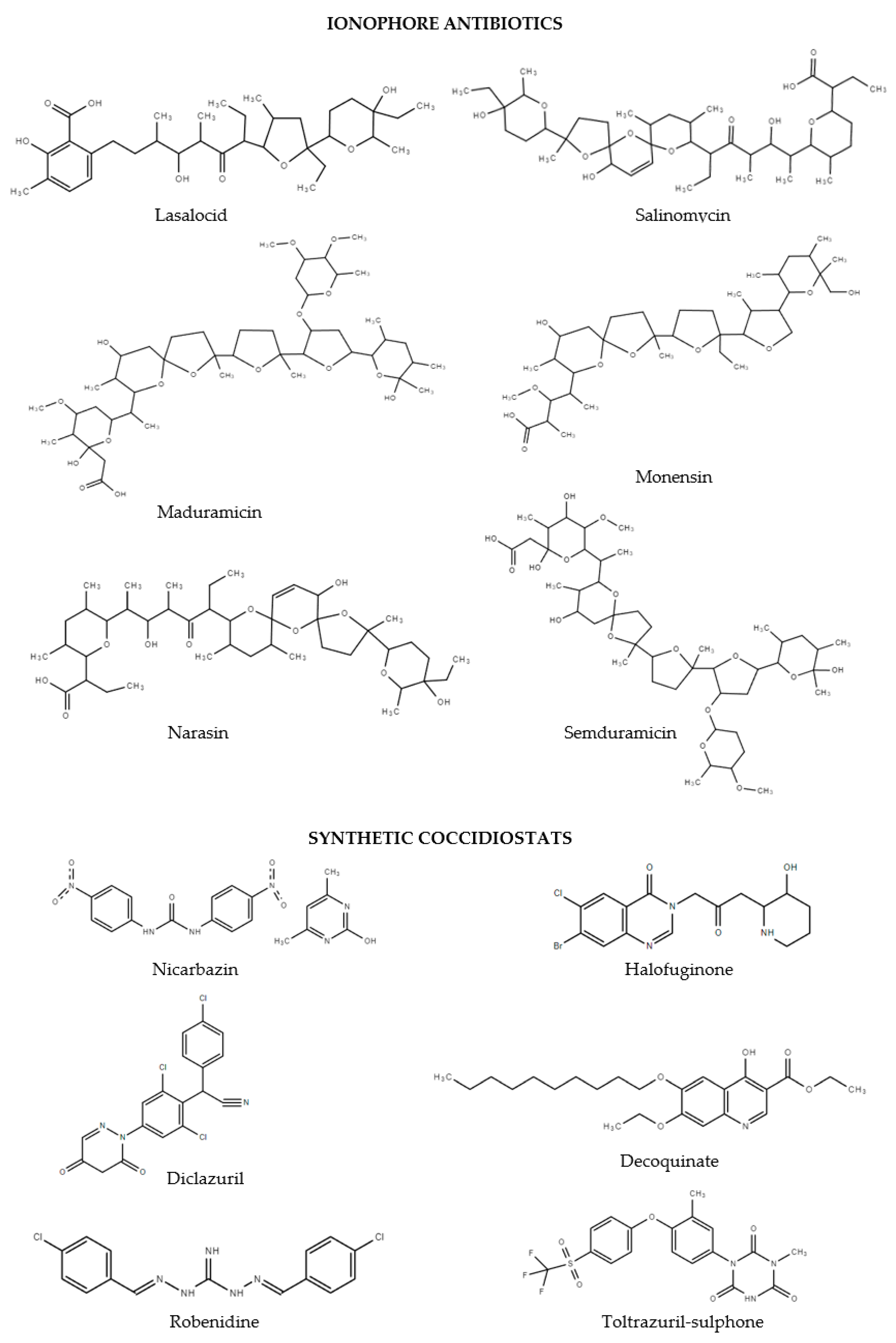
2.1. Chemicals, Reagents, Stock and Intermediates Solutions

LC-MS-grade acetonitrile (ACN), LC-MS-grade methanol (MeOH), dimethylsulfoxide (DMSO) and n-hexane were provided by Merck KGaA (Darmstadt, Germany). LC-MS-grade deionized water and formic acid ≥ 98% were supplied by Biosolve Chimie (Dieuze, France). The Oasis PRiME HLB (60 mg, 3 mL and 150 mg, 3 mL) Solid-Phase Extraction (SPE) cartridges were obtained from Waters Corporation (Milford, MA, USA) and Bond Elut EMR-Lipid from Agilent Technologies (Waldbronn, Germany). Decoquinate, diclazuril, lasalocid sodium, diclazuril-methyl, monensin sodium, nicarbazin and salinomycin sodium were provided by Dr. Ehrenstrofer (Augsburg, Germany). Robenidine hydrochloride, semduramicin sodium (100 µg mL−1 acetonitrile solution), toltrazuril-d3, toltrazuril-sulphone were bought from HPC Standards GmbH (Cunnersdorf, Germany), while maduramicin ammonium, narasin and nigericin sodium were bought from Merck KGaA. Halofuginone-13C6 hydrobromide, decoquinate-d5, 4,4′-dinitrocarbanilide-d8 (DNC-d8), robenidine-d8 hydrochloride were purchased from Witega (Berlin, Germany) and finally, halofuginone hydrobromide from TRC Inc. (Toronto, ON, Canada).
Coccidiostat stock solutions (1 mg mL−1) were prepared by weighing 5 mg of reference material and dissolving in 5 mL of solvent. The ionophore antibiotics were solubilized in ACN, while the chemical coccidiostats (Figure 1) were dissolved in various solvents: DMSO for diclazuril, diclazuril-methyl, nicarbazin (intended as DNC), DNC-d8, robenidine hydrochloride and robenidine-d8 hydrochloride. Again, nigericin sodium, toltrazuril-sulphone and toltrazuril-d3 were prepared in ACN, halofuginone hydrobromide and halofuginone-13C6 hydrobromide in ACN/water 50/50 (v/v), and finally, decoquinate and decoquinate-d5 were solubilized in ACN/formic acid 50/50 (v/v).
The intermediate solutions of analytes at 10 and 0.1 µg mL−1 in ACN were stable for 12 months and 3 days, respectively, when stored in refrigerator and freezer. The intermediate ISs solutions at 10 and 1 µg mL−1 were stored in refrigerator (stability of 12 months) and freezer (3 days), respectively.

2.2. Chromatographic Conditions

Chromatography was performed on a Thermo Ultimate 3000 High Performance Liquid Chromatography system (San Jose, CA, USA). The analytes were separated on a Synergi Fusion column (150 × 2.0 mm, 80 Å; 4 μm; Phenomenex, Torrance, CA, USA), connected to a Fusion RP guard column (4 × 2.0 mm). HPLC mobile phase A was an aqueous solution containing 0.1% (v/v) formic acid and eluent B was acetonitrile including 0.1% (v/v) formic acid. The gradient was started with 15% eluent B for 2 min, continued with increase to 25% B in 1 min, then with further linear increment to 95% in 12.5 min and maintained in this condition for 11.5 min. The system returned to 15% B in 1 min and was equilibrated for 4 min for a total run time of 32 min. The column compartment and the sample temperature were kept at 40 °C and 16 °C, respectively. The flow rate was 0.25 mL min−1 and the injection volume was 10 μL.

2.3. MS Conditions

A Q-Orbitrap mass spectrometer (Thermo Scientific, San Jose, CA, USA) was equipped with a heated electrospray ionization (HESI-II) source. The HESI-II and capillary temperatures were set at 320 and 300 °C, respectively, and the electrospray voltage at 3.20 kV (positive and negative ionization mode). Sheath and auxiliary gas were 35 and 15 arbitrary units, respectively. The mass spectrometer was controlled by Xcalibur 3.0 software (Thermo Fisher Scientific, San Jose, CA, USA). The exact mass of the compounds was calculated using Freestyle in Xcalibur 3.0. Instrument calibration was performed for every analytical batch with a direct infusion of LTQ Velos ESI Positive Ion Calibration Solution (Pierce Biotechnology Inc., Rockford, IL, USA). The individual compounds were infused with a syringe through a T union connected to an LC system with a mobile phase flow rate of 0.1 mL min−1 (50% eluent A). The product ions were found by increasing the collision energy (CE) using Q Exactive Tune 2.3 software (Thermo Fisher Scientific, Waltham, MA, USA). After choosing the more intense product ions, fragmentation energies were optimized with spiked samples of eggs and muscles at 1 µg kg−1 using the optimized gradient program. Quantitative analysis was performed with QuanBrowser in Xcalibur 3.0. All Q Exactive parameters as resolution, automatic gain control (AGC) and injection time (IT) were optimized to improve instrumental signals and selectivity. MS acquisition was performed combining parallel reaction monitoring (PRM) experiments in positive and negative ionization: the adduct ion was filtered with an isolation window of m/z 0.9, the resolution set at 35,000 FWHM (m/z 200). The AGC was fixed at 1 × 10⁶ ions for a maximum injection time of 140 ms. The monitored adduct and product ions such as the CE are shown in Table 1.

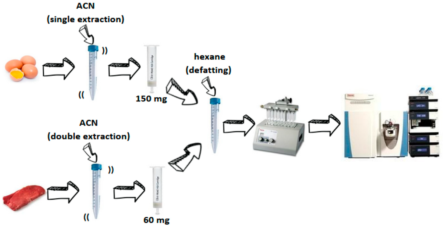
2.4. Sample Preparation

Two-half gram of homogenized sample was weighed in a Falcon tube and then, was spiked with internal standards (ISs), specifically 25 µL of a solution containing the ISs at 1 µg mL−1. The protocols for eggs and muscle were slightly different; the latter was extracted twice with 5 mL of ACN through shaking and sonicating while the analytes were extracted from eggs due to a single extraction (10 mL of ACN). The Oasis® PRiME HLB SPE cartridge (150 mg and 60 mg for eggs an muscle, respectively) was conditioned with 3 mL of ACN and loaded with the sample extract, collecting the eluate that was defatted twice with 3 mL of hexane. After that, the extract was evaporated to dryness under gentle nitrogen stream (40 °C). Finally, the dry residue was redissolved in 1 mL of MeOH. After centrifugation, the sample was injected. The muscle final extract was freshly prepared while the egg extract was stable for 48 h in the autosampler.

2.5. Method Validation

An in-house validation plan, based on the Commission Implementing Regulation (EU) 2021/808 [24], was applied to perform a confirmation method, following an alternative experimental plan, as indicated by Section 2.2.3 of the Regulation. This choice was justified by the wide range of established MRLs and legal limits (LLs) [2,3,4,5,6,7,8,9,10,11,12]. Briefly, the analytes were validated at the spiking levels encompassing 1–100 µg kg−1; for lasalocid and nicarbazine, the range was 1–1000 µg kg−1 (eggs). Regarding the muscle, for robenidine and toltrazuril-sulphone, the validation levels included 1–333 µg kg−1; for decoquinate and diclazuril, the range was 1–3333 µg kg−1 and finally, it was 1–6000 µg kg−1 for nicarbazine (Table 2).
The selectivity was tested for both of the matrices analysing different lots of eggs and muscles belonging to the main animal species (i.e., poultry eggs, poultry, bovine and swine muscle). The linearity was evaluated in neat solvent (MeOH) and in matrix. The matrix-matched standards (MMSs) were prepared adding the analytes immediately prior to the LC injection. The curves were constructed applying the regression model at 6 concentration levels (0 was included), encompassing 2.5–83.3 ng mL−1 (solvent) and 1–33.3 µg kg−1 (matrix), plotting the analyte area against its concentration except for halofuginone, robenidine, DNC and decoquinate (analyte/IS area ratio against concentration). The precision, recovery (trueness), decision limit (CCα), uncertainty, limit of detection (LOD) and limit of quantitation (LOQ) were evaluated following the plan described in Table 1. A blank and an MMS were analysed for each analytical batch and each validation level experiment was repeated for three occasions varying operator, time and calibration status of the MS system. Moreover, the relative matrix effect (matrix factor) and ruggedness (major changes) were studied.

2.6. Real Samples Analysis

The validated method was accredited on June 2022 and applied at samples of Italian NRCP. Each sample was located in a plastic container and stored at −20 °C after the homogenization. Particularly, 2 poultry eggs and 16 muscles belonging to ovine, poultry and turkey species were analysed. Finally, internal and external quality control (IQC and EQC) activities were implemented.

3. Results and Discussion

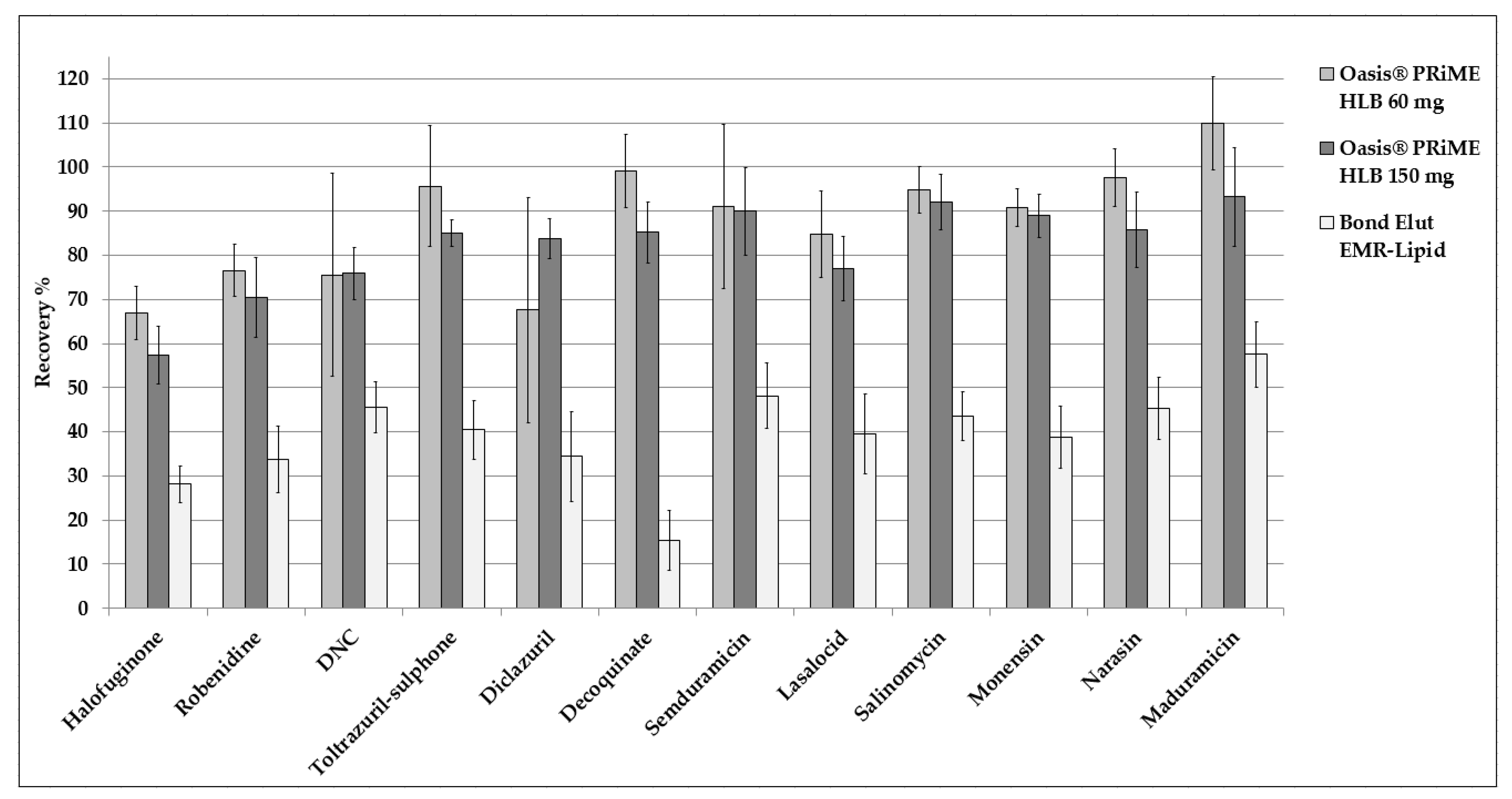
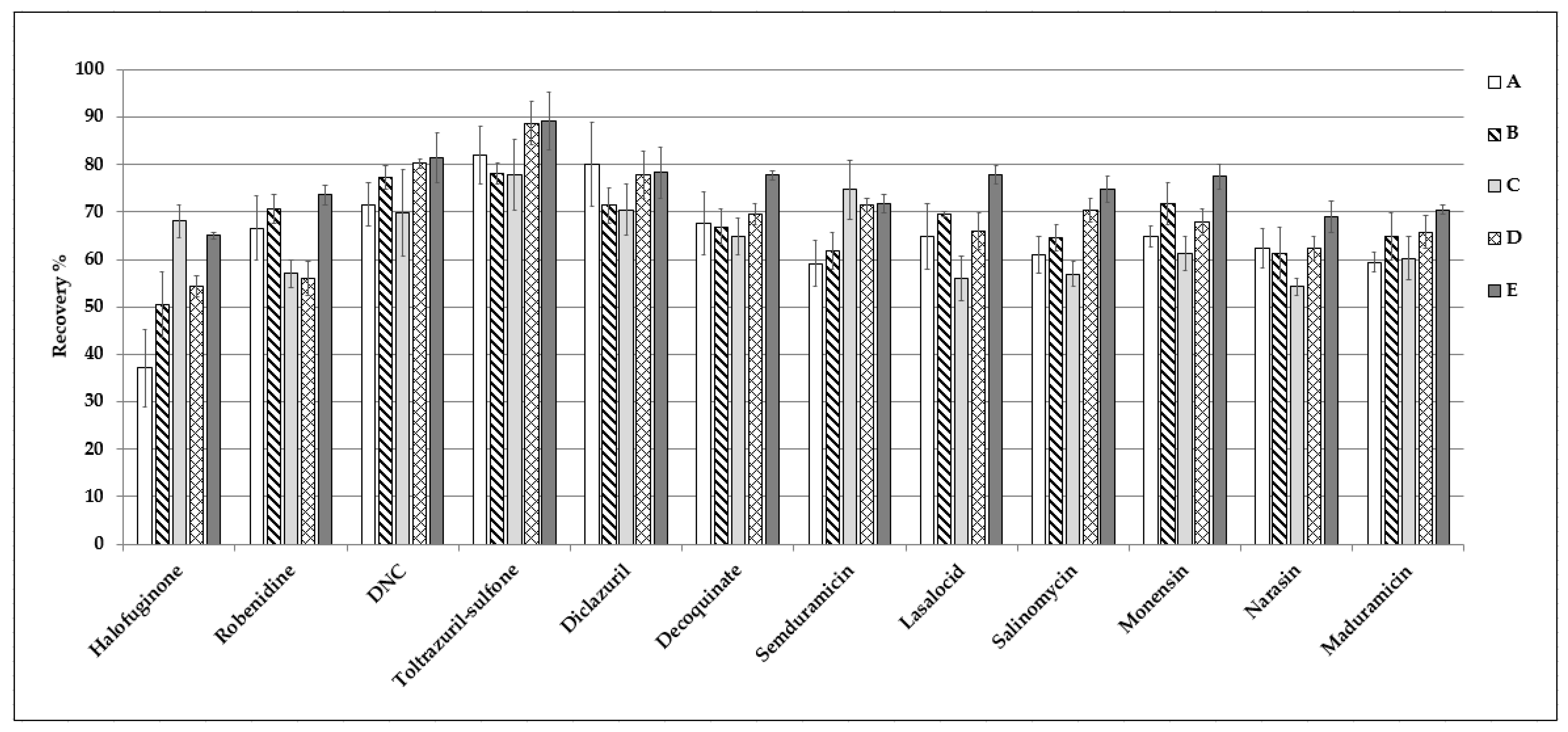
3.1. Sample Preparation

Eggs and muscle are complex matrices, rich in lipids and proteins, and so, it is often necessary to perform more than one clean-up step to obtain a “clean” final extract. Before the purification step, acetonitrile or its mixtures were more frequently used for the extraction of analytes [13,19,23,25]. Recently, Martins and collaborators [26] reported the clean-up methodologies, among other things, present in the literature from 2011 to 2020 related to food matrices, concluding that SPE were very often carried out. By way of example, in 2011, Olejnik et al. [27] used Alumina-N and Oasis® HLB cartridges for the determination of 12 coccidiostats in eggs and liver. Later, Ha and collaborators [25] provided the clean-up step by means of a graphitized non-porous carbon based SPE and in 2019, in a study by Dasenaki and Thomaidis [16], 16 coccidiostat residues were determined in eggs and muscle with a purification by dispersive SPE with a C18 sorbent.
The developed sample preparation of eggs and muscle was based on a solid–liquid extraction of the molecules from the matrix with ACN, followed by a SPE clean-up step and a defatting. During the development of the protocol for eggs, although various SPE cartridges were tested [16,25,27], identifying of state of the art SPE cartridges to remove proteins, phospholipids and other matrix interferences was the intent, considering the very different physico-chemical properties of the analytes. Oasis® PRiME HLB (60 and 150 mg) SPE and Bond Elut EMR-Lipid dispersive SPE were tested in this context and Figure 2 shows the recoveries provided by the experiments. The Bond Elut EMR-Lipid determined a very high retention of the analytes contrary to Oasis® PRiME HLB (150 mg) that provided good recoveries and better precision than the 60 mg format, probably due to the cleaner final extract. The defatting step was also investigated and Figure 3 demonstrates the better precision using n-hexane, hypothetically for the same, latter reason.
It is noteworthy that 150 mg format of Oasis® PRiME HLB was tested for the sample preparation of muscle determining worse performances (i.e., recovery and precision) than eggs protocol, especially for halofuginone. Its recovery enhanced due to the addition of EDTA, probably for higher polarity of the extraction mixture; however, the recoveries of other analytes were lower. Moreover, the effect of a double extraction and the effect of 60 mg format of SPE were studied and the experiments are summarized in Figure 4.

3.2. Optimization of LC-HRMS/MS Conditions

The chromatographic gradient was based on Galarini et al. [15] with some modifications. Briefly, the run was shortened from 40 min to 32 min and sodium acetate was not added to the aqueous mobile phase for limiting the ion suppression phenomenon. The development of MS settings was performed, taking into account that most of the analytes had very low MRLs or are banned and the clean-up steps can not be selective. Thus, the PRM acquisition was carried out in order to obtain the highest possible selectivity. In PRM, a predefined precursor ion was filtered by the quadrupole and transferred via the C-trap to the high collision dissociation (HCD) cell for the fragmentation. The C-trap can fill with ions for long times, increasing signal-to-noise ratio of the ions measured in the Orbitrap detector. From the HCD cell, fragment ions were moved back to the C-trap and eventually injected and analysed in the detector providing a product ion spectrum [28]. Initially, the adduct ion to be fragmented was chosen on the basis of its intensity through full scan acquisitions experiments. As shown in Table 1, generally, [M+H]+ and [M-H] adducts were acquired; the exceptions were the ionophore coccidiostats (i.e., [M+Na]+). The fairly long gradient permitted a good separation of the analytes, providing the possibility to switch from positive to negative polarity within a delimited scan time (i.e., 11–14 min, see Table 3) and work with a resolution as much as possible.
Toltrazuril-sulphone and toltrazuril-d3 produced fragment ions below m/z 50 [14], but unfortunately, the LC-Q-Orbitrap system was able to acquire product ion spectra above this m/z. Therefore, the adduct ion was monitored administrating the lowest values of CE for PRM acquisition (i.e., 10 eV) for both of them and, consequently, toltrazuril-sulphone was only detected, according the qualitative confirmation criteria defined by the Commission Implementing Regulation (EU) 2021/808 [24]. During the method development, the linearity studies showed a good linearity for all the compounds in the range from 1 to 250 ng mL−1 except for DNC ([M-H], m/z 301.0578). Investigations with full scan experiments revealed the presence of various adducts likely attributable to DNC dimers such as [2M-H] (m/z 603.1230), [2M+Cl] (m/z 639.0986) and [2M+HCOO] (m/z 649.1284) (Figure 5).
Its formation in ESI source was dependent on the concentration, which indicated a non-covalent association and provided an explanation for the limited linearity range [29]. Applying in-source collision-induced dissociation (IS-CID), the relative abundance of the dimer species was reduced, improving linearity range (Figure 6). However, the IS-CID was not administered in the developed method for the insufficient intensity signal of toltrazuril-sulphone (coelution of DNC and toltrazuril-sulphone). Consequently, the “operative” linearity range was fixed in the range encompassing 2.5 and 83.3 ng mL−1 for DNC and other analytes.

3.3. Method Validation

The selectivity was evaluated analysing more than 20 blank samples per matrix and no peaks were found in the region of interest where the target analyte was expected to elute. Particularly, poultry eggs and bovine, poultry and swine muscles were analysed. The relative matrix effect (ME) was also investigated on the same samples, calculating the ratio % between the peak area of the analyte in MMS and in neat solvent for the coccidiostats quantified by means of external calibration, while for the other ones, using the following equation: MEanalyte/MEIS % [24]. The Commission Implementing Regulation (EU) 2021/808 did not fix the coefficient of variation (CV) tolerances when no IS was used during the validation study. However, the CVME tolerance of ≤20% (normalised for an IS) was satisfied in both cases (Table 4). Good linearity was observed for all the molecules (deviations of back-calculated concentration ≤20% [30]). The trueness results reported in Table 4 were obtained applying a mixed quantitation approach with MMS (internal and external calibration). Particularly, chemical coccidiostats were determined with internal calibration, except for toltrazuril-sulphone and ionophore coccidiostats.
CV were not affected by the analyte spiking level; therefore, the CV could be pooled into a single data set (Table 4). Repeatability and within-laboratory reproducibility encompassed the range of 3.5% (robenidine)–7.1% (monensin) (CVr) and 5.7% (robenidine) and 13% (salinomycin) (CVwR) for eggs, respectively. The same parameters for muscle were within 4.7% (halofuginone) 9.3% (maduramicin) (CVr) and 5.4% (halofuginone) and 13% (maduramicin) (CVwR), respectively. The CCα values were obtained following Method 2 of paragraph 2.6 of the Commission Implementing Regulation (EU) 2021/808 [24] (Equation (1)) after the calculation of combined standard measurement uncertainty (uc) [31]:
CCα = MRL (or LL) + 1.64 × uc
uc was derived following the top-down approach, or rather, combining random and systematic error uncertainty components [31,32]. It is noteworthy that uc was calculated on the basis of CVwR pooled on the entire range of investigated concentrations instead of on at MRL or LL.
The LODs and LOQs were experimentally defined based on the trueness observed at the first validation level for both of the matrices (i.e., 1 µg kg−1 for all the analytes). Figures S1 and S2 show the chromatograms of an egg and a poultry muscle sample spiked at this level.
Ruggedness experiments were successfully verified by analysing bovine and swine muscles spiked at 10 µg kg−1 (four replicates in two different days for a total of eight observations for species) (Table 5).
It is, finally, noteworthy that the Commission Implementing Regulation (EU) 2021/808 established a minimum of four identification points (IPs) needed for the confirmation of the identity of a permitted substance. The very high selectivity was obtained by the application of PRM acquisition that provided one (indirect) IP for the selection of the precursor ion filtered by the quadrupole and five (2.5 + 2.5) IPs for the product ions (accurate mass measurements) for a total of seven IPs, considering also the LC separation (i.e., one IP). So, in summary: an analyte was considered as positively identified in a sample when (i) the relative retention time (RRT) (RTanalyte/RTIS) in the sample and in a spiked sample/MMS or a standard solution was within ± 1% tolerance, (ii) both the fragment ions were detected with a mass deviation < 5 ppm (or <1 mDa in case of m/z 200) with respect to a spiked sample/MMS or a standard solution and iii) the % ratio of their intensities matched with ±40% tolerance.

3.4. Real Samples Analysis and QC Activities

The validated and accredited method was applied for the determination of coccidiostat residues in 18 official samples of Abruzzo and Molise Regions (NRCP). An IQC was implemented for the analytical batches by the addition of a solution containing six ISs to each sample before the extraction. Halofuginone-13C6, robenidine-d8, DNC-d8, decoquinate-d5 were used with quantitative purposes, contrary to toltrazuril-d3 and nigericin that were employed only to determine the yield of the process for toltrazuril-sulphone and synthetic coccidiostats, respectively. Moreover, a blank and a spiked eggs/muscle sample at 1 µg kg−1 were analysed to verify the absence of false positive/negative results. Lastly, a MMS was prepared by the addition of the analytes immediately prior to LC injection. Suspected samples were newly carried out by twice performing ad hoc spiked and MMS samples taking also into account of the dilution factor to be applied.
Despite a few analysed samples, a coccidiostat residue was detected in an egg sample: lasalocid was found at 1.2 µg kg−1 (Figure 7).
Finally, accuracy data obtained from the analysis of certified reference material (CRM) M1406/CM lyophilised turkey muscle (Test Veritas, Progetto Trieste, Italy) represented a good agreement of the detected concentrations with the reference values (Table 6). Interestingly, the exception was represented for DNC for which the provider reported an assigned concentration with a not negligible uncertainty. The calculated accuracy expressed as a percentage ratio of the measured concentration and the consensus value was in the range of 89–105%, excluding DNC.

4. Conclusions

A highly selective, sensitive and accurate confirmatory LC-Q-Orbitrap method was developed for the determination of 12 coccidiostat residues in eggs and muscles starting from 1 µg kg−1. The main advantage of the method was the quite quick sample preparation associated with a very high selectivity, making sure that the procedure can be applied for the official control analysis and EQC activities. Moreover, the method was validated for the first time according to the Commission Implementing Regulation (EU) 2021/808 [24].

Supplementary Materials

The following supporting information can be downloaded at: https://www.mdpi.com/article/10.3390/separations10030202/s1, Figure S1. Chromatogram of an egg sample spiked at 1 µg kg−1. Figure S2. Chromatogram of a poultry muscle sample spiked at 1 µg kg−1.

Author Contributions

F.C., methodology, validation and investigation; M.R., validation, investigation; M.N.C., investigation; G.S. (Giampiero Scortichini), conceptualization, methodology, supervision. G.S. (Giorgio Saluti), conceptualization, formal analysis, methodology, supervision, writing—original draft preparation, writing—review and editing. All authors have read and agreed to the published version of the manuscript.

Funding

This research received no external funding.

Institutional Review Board Statement

The study did not require ethical approval.

Informed Consent Statement

Informed consent was obtained from all subjects involved in the study.

Acknowledgments

The authors thankfully acknowledge Alessia Manucci (Istituto Zooprofilattico Sperimentale dell’Abruzzo e del Molise “G. Caporale”) for the graphic support.

Conflicts of Interest

The authors declare no conflict of interest.

Abbreviations

ACN = acetonitrile; AGC = automatic gain control; CCα = decision limit; CE = collision energy; CRM = certified reference material; CVr = coefficient of variation in repeatability conditions; CVwR = coefficient of variation in within-laboratory reproducibility conditions; DMSO = dimethylsulfoxide; DNC = 4-4′-dinitrocarboanilide; EDTA = ethylenediaminetetraacetic acid; HCD = high collision dissociation; IP = identification point; IQC = internal quality control; EQC = external quality control; IS-CID = in-source collision-induced dissociation; IT = injection time; LC-HRMS/MS = liquid chromatography coupled to high resolution mass spectrometry; LC-MS/MS = liquid chromatography coupled to low resolution tandem mass spectrometry; LL = legal limit; LOD = limit of detection; LOQ = limit of quantitation; ME = relative matrix effect; MeOH = methanol; MMS = matrix-matched standard; MRL = maximum residue limit; MS = mass spectrometry; NRCP = National Residue Control Plan; PRM = parallel reaction monitoring; R = recovery; RT = retention time; RRT = relative retention time; SPE = solid-phase extraction; uc = combined uncertainty.

References

  1. Clarke, L.; Fodey, T.L.; Crooks, S.R.H.; Moloney, M.; O’Mahony, J.; Delahaut, P.; O’Kennedy, R.; Danaher, M. A Review of Coccidiostats and the Analysis of Their Residues in Meat and Other Food. Meat Sci. 2014, 97, 358–374. [Google Scholar] [CrossRef] [PubMed]
  2. The Commission of the European Communities. Commission Regulation (EU) No 37/2010 of 22 December 2009 on Pharmacologically Active Substances and Their Classification Regarding Maximum Residue Limits in Foodstuffs of Animal Origin. Off. J. Eur. Communities 2009. Available online: http://data.europa.eu/eli/reg/2010/37(1)/2022-05-09 (accessed on 9 March 2023).
  3. Commission Regulation. Reg 388/2011. 2006, Volume 1881, pp. 1–5. Available online: http://data.europa.eu/eli/reg_impl/2011/388/2013-11-12 (accessed on 9 March 2023).
  4. Union, T.; Journal, O.; Union, E. Reg 124/2009. Ec 2074/2005 2017. Volume 10, pp. 1–21. Available online: http://data.europa.eu/eli/reg/2009/124/2020-04-27 (accessed on 9 March 2023).
  5. The Commission of the European Communities. Reg 495/2011. 2011, Volume 9. Available online: http://data.europa.eu/eli/reg_impl/2011/495/oj (accessed on 9 March 2023).
  6. European Commission. Reg 2094/2021. 2018, Volume 2016, pp. 48–119. Available online: http://data.europa.eu/eli/reg_impl/2021/2094/oj (accessed on 9 March 2023).
  7. The Commission of the European Communities. Reg 1417/2015. 2015, Volume 13, pp. 20–22. Available online: http://data.europa.eu/eli/reg_impl/2015/1417/oj (accessed on 9 March 2023).
  8. The Commission of the European Communities. Reg 118/2012. 2012. No. 118. pp. 36–39. Available online: http://data.europa.eu/eli/reg_impl/2012/118/oj (accessed on 9 March 2023).
  9. Commission Regulation. Reg 532/2011. 2006, Volume 1881, pp. 1–5. Available online: http://data.europa.eu/eli/reg_impl/2011/532/2013-11-12 (accessed on 9 March 2023).
  10. The Commission of the European Communities. Reg 875/2010. 2010, Volume 8, pp. 4–6. Available online: http://data.europa.eu/eli/reg/2010/875/oj (accessed on 9 March 2023).
  11. Union, T.; Journal, O.; Union, E. Reg 885/2010. Ec 2074/2005 2017. Volume 10, pp. 1–21. Available online: http://data.europa.eu/eli/reg/2010/885/oj (accessed on 9 March 2023).
  12. The Commission of the European Communities. Scientific Opinion on the Safety and Efficacy of Koffogran (Nicarbazin) as a Feed Additive for Chickens for Fattening. EFSA J. 2010, 8, 2011–2013. [Google Scholar] [CrossRef]
  13. Barreto, F.; Ribeiro, C.; Hoff, R.B.; Costa, T.D. A Simple and High-Throughput Method for Determination and Confirmation of 14 Coccidiostats in Poultry Muscle and Eggs Using Liquid Chromatography–Quadrupole Linear Ion Trap-Tandem Mass Spectrometry (HPLC–QqLIT-MS/MS): Validation According to European. Talanta 2017, 168, 43–51. [Google Scholar] [CrossRef] [PubMed]
  14. Buiarelli, F.; Di Filippo, P.; Riccardi, C.; Pomata, D.; Giannetti, L.; Neri, B.; Rago, D. Liquid Chromatography Tandem Mass Spectrometry Analysis of Synthetic Coccidiostats in Eggs. Separations 2017, 4, 15. [Google Scholar] [CrossRef]
  15. Galarini, R.; Fioroni, L.; Moretti, S.; Pettinacci, L.; Dusi, G. Development and Validation of a Multi-Residue Liquid Chromatography-Tandem Mass Spectrometry Confirmatory Method for Eleven Coccidiostats in Eggs. Anal. Chim. Acta 2011, 700, 167–176. [Google Scholar] [CrossRef] [PubMed]
  16. Dasenaki, M.E.; Thomaidis, N.S. Multi-Residue Methodology for the Determination of 16 Coccidiostats in Animal Tissues and Eggs by Hydrophilic Interaction Liquid Chromatography–Tandem Mass Spectrometry. Food Chem. 2019, 275, 668–680. [Google Scholar] [CrossRef] [PubMed]
  17. Shao, B.; Wu, X.; Zhang, J.; Duan, H.; Chu, X.; Wu, Y. Development of a Rapid LC-MS-MS Method for Multi-Class Determination of 14 Coccidiostat Residues in Eggs and Chicken. Chromatographia 2009, 69, 1083–1088. [Google Scholar] [CrossRef]
  18. Clarke, L.; Moloney, M.; O’Mahony, J.; O’Kennedy, R.; Danaher, M. Determination of 20 Coccidiostats in Milk, Duck Muscle and Non-Avian Muscle Tissue Using UHPLC-MS/MS. Food Addit. Contam.-Part A 2013, 30, 958–969. [Google Scholar] [CrossRef] [PubMed]
  19. Chang, S.H.; Lai, Y.H.; Huang, C.N.; Peng, G.J.; Liao, C.D.; Kao, Y.M.; Tseng, S.H.; Wang, D.Y. Multi-Residue Analysis Using Liquid Chromatography Tandem Mass Spectrometry for Detection of 20 Coccidiostats in Poultry, Livestock, and Aquatic Tissues. J. Food Drug Anal. 2019, 27, 703–716. [Google Scholar] [CrossRef] [PubMed] [Green Version]
  20. Wang, B.; Liu, J.; Zhao, X.; Xie, K.; Diao, Z.; Zhang, G.; Zhang, T.; Dai, G. Determination of Eight Coccidiostats in Eggs by Liquid-Liquid Extraction-Solid-Phase Extraction and Liquid Chromatography-Tandem Mass Spectrometry. Molecules 2020, 25, 987. [Google Scholar] [CrossRef] [PubMed] [Green Version]
  21. George, K.; Vincent, U.; von Holst, C. Analysis of Antimicrobial Agents in Pig Feed by Liquid Chromatography Coupled to Orbitrap Mass Spectrometry. J. Chromatogr. A 2013, 1293, 60–74. [Google Scholar] [CrossRef] [PubMed]
  22. Matus, J.L.; Boison, J.O. A Multi-Residue Method for 17 Anticoccidial Drugs and Ractopamine in Animal Tissues by Liquid Chromatography-Tandem Mass Spectrometry and Time-of-Flight Mass Spectrometry. Drug Test. Anal. 2016, 8, 465–476. [Google Scholar] [CrossRef] [PubMed] [Green Version]
  23. Rusko, J.; Jansons, M.; Pugajeva, I.; Zacs, D.; Bartkevics, V. Development and Optimization of Confirmatory Liquid Chromatography—Orbitrap Mass Spectrometry Method for the Determination of 17 Anticoccidials in Poultry and Eggs. J. Pharm. Biomed. Anal. 2019, 164, 402–412. [Google Scholar] [CrossRef] [PubMed]
  24. Commission, E. Commission Implementing Regulation (EU) 2021/808 of 22 March 2021 on the Performance of Analytical Methods for Residues of Pharmacologically Active Substances Used in Food-Producing Animals and on the Interpretation of Results as Well as on the Methods To. Off. J. Eur. Union 2021, 180, 84–109. [Google Scholar]
  25. Ha, J.; Song, G.; Ai, L.F.; Li, J.C. Determination of Six Polyether Antibiotic Residues in Foods of Animal Origin by Solid Phase Extraction Combined with Liquid Chromatography-Tandem Mass Spectrometry. J. Chromatogr. B Anal. Technol. Biomed. Life Sci. 2016, 1017–1018, 187–194. [Google Scholar] [CrossRef] [PubMed]
  26. Martins, R.R.; Silva, L.J.G.; Pereira, A.M.P.T.; Esteves, A.; Duarte, S.C.; Pena, A. Coccidiostats and Poultry: A Comprehensive Review and Current Legislation. Foods 2022, 11, 2738. [Google Scholar] [CrossRef] [PubMed]
  27. Olejnik, M.; Szprengier-Juszkiewicz, T.; Jedziniak, P.; Śledzińska, E.; Szymanek-Bany, I.; Korycińska, B.; Pietruk, K.; Zmudzki, J. Residue Control of Coccidiostats in Food of Animal Origin in Poland during 2007–2010. Food Addit. Contam. Part B Surveill. 2011, 4, 259–267. [Google Scholar] [CrossRef] [PubMed]
  28. Rauniyar, N. Parallel Reaction Monitoring: A Targeted Experiment Performed Using High Resolution and High Mass Accuracy Mass Spectrometry. Int. J. Mol. Sci. 2015, 16, 28566–28581. [Google Scholar] [CrossRef] [PubMed] [Green Version]
  29. Pan, H. A Non-Covalent Dimer Formed in Electrospray Ionisation Mass Spectrometry Behaving as a Precursor for Fragmentations. Rapid Commun. Mass Spectrom. 2008. [Google Scholar] [CrossRef] [PubMed]
  30. European Comission. Document N0 SANTE 11312/2021. Analytical Quality Control and Method Validation Procedures for Pesticide Residues Analysis in Food and Feed. 2021, pp. 1–57. Available online: https://www.Eurl-Pesticides.Eu/Docs/Public/Tmplt_article.Asp?CntID=727 (accessed on 9 March 2023).
  31. Galarini, R.; Moretti, S.; Saluti, G. Quality Assurance and Validation General Considerations and Trends. In Chromatogr. Anal. Environ. Mass Spectrom. Based Approaches, 4th ed.; CRC Press: Boca Raton, FL, USA, 2017; p. 45. [Google Scholar]
  32. Magnusson, B.; Naikki, T.; Hovind, H.; Krysell, M. Handbook for Calculation of Measurement Uncertainty in Environmental Laboratories. NT TR 537, ed. 3.1. 2012. Available online: http://www.nordtest.info (accessed on 9 March 2023).
Figure 1. Molecular structures of the investigated coccidiostats.
Separations 10 00202 g001
Figure 2. Recoveries derived by different clean-up approaches in egg samples (n = 4 per experiment) spiked at 33.3 µg kg−1.
Separations 10 00202 g002
Figure 3. Recoveries derived by the use of n-hexane (defatting) in egg samples (n = 8 per experiment) spiked at 33.3 µg kg−1.
Separations 10 00202 g003
Figure 4. Recovery factors (n = 4 muscle spiked at 10 µg kg−1 per experiment) carrying out the eggs sample preparation A, using Oasis® PRiME HLB (60 mg) B, adding 300 µL of 0.1 M disodium EDTA during the extraction C, extracting twice with ACN (5 + 5 mL) D and combining B and D (i.e., chosen protocol) E.
Separations 10 00202 g004
Figure 5. Chromatogram of [M-H] adduct of DNC (a) and the full scan spectrum (b) of a standard solution at 250 ng mL−1.
Separations 10 00202 g005
Figure 6. Calibration curve in MeOH of [M-H] adduct of DNC without (a) and with (b) IS-CID of 20 eV.
Separations 10 00202 g006
Figure 7. Chromatograms (left) and product ion spectra (right) of lasalocid in an egg sample at 1.2 µg kg−1 (a) and in a spiked sample at 1 µg kg−1 (b).
Separations 10 00202 g007
Table 1. LC-Q-Orbitrap parameters of the investigated coccidiostats.
AnalyteRT
(min)
RRTMolecular FormulaAdductMonoisotopic
Exact Mass
(m/z)
CE
(eV)
Fragment 1 Accurate Mass 1
(m/z)
Fragment 2 Accurate Mass
(m/z)
Halofuginone-13C6 (IS)5.77-13C6C10H17BrClN3O3[M+H]+420.041625100.0757-
Halofuginone5.771.00C16H17BrClN3O3[M+H]+414.041625100.0757120.0808
Robenidine-d8 (IS)8.36-C15H5D8Cl2N5[M+H]+342.112325159.0622-
Robenidine8.381.00C15H13Cl2N5[M+H]+334.062125138.0105155.0372
DNC-d8 (IS)12.09-C13H2D8N4O5[M-H]309.108045141.0603-
DNC12.131.00C13H10N4O5[M-H]301.057845137.0352107.0369
Toltrazuril-sulphone12.180.92C18H14F3N3O6S[M-H]456.048410456.0484 2-
Diclazuril12.721.02C17H9Cl3N4O2[M-H]404.971835333.9713298.9785
Diclazuril-methyl (IS)12.97-C18H11Cl3N4O2[M-H]418.987435320.9760-
Toltrazuril-d3 (IS)13.19-C18D3H11F3N3O4S[M-H]427.077210427.0772 2-
Decoquinate-d5 (IS)14.90-C24H30D5NO5[M+H]+423.290240255.1020-
Decoquinate14.991.01C24H35NO5[M+H]+418.258840250.0709390.2275
Semduramicin17.480.83C45H76O16[M+Na]+895.502665833.5019705.4189
Lasalocid17.650.84C34H54O8[M+Na]+613.371130377.2659613.3711
Salinomycin18.850.90C42H70O11[M+Na]+773.481045431.2401531.3294
Monensin19.010.91C36H62O11[M+Na]+693.418470461.2876501.3186
Narasin19.830.94C43H72O11[M+Na]+787.496750431.2409531.3301
Maduramicin20.160.96C47H80O17[M+Na]+939.528875877.5291719.4343
Nigericin (IS)21.00-C40H67O11[M+H]+747.465475237.1093-
1 Ion used for quantitative purposes. 2 Adduct ion monitored.
Table 2. Validation plan.
Eggs
Spiking Level
(µg kg−1)
Number
of Spiked
Samples Day−1
Concentration
of Analyte
Solution
(µg mL−1)
Added Volume
of Analyte
Solution
(µL)
IS Spiking Level
(µg kg−1)
Concentration
of IS Solution
(µg mL−1)
Added Volume
of IS Solution
(µL)
Dilution
Factor
140.125101250.4
240.150101250.4
3.3340.183.3101250.4
104125101250.4
33.34183.3101250.4
1004102510010254
333 *41083.310010254
1000 *41025010001025040
Muscle
140.125101250.4
240.150101250.4
3.3340.183.3101250.4
104125101250.4
33.34183.3101250.4
1004102510010254
333 *41083.310010254
1000 *41025010001025040
3333 *410083.310001025040
6000 *410015010000100250200
* Levels validated for a part of the analytes/ISs.
Table 3. PRM acquisition experiments.
Time
(min)
PolarityAnalytes in the Inclusion List
3–11+halofuginone, halofuginone-13C6, robenidine, robenidine-d8
11–14-DNC, DNC-d8, toltrazuril-sulphone, toltrazuril-d3, diclazuril, diclazuril-methyl
14–24+decoquinate, decoquinate-d5, semduramicin, lasalocid, salinomycin, monensin, narasin, maduramicin, nigericin
Table 4. Validation performances of the analytes for poultry eggs and muscle sorted by elution order.
AnalyteMean Recovery
(%)
CVr
(%)
CVwR
(%)
uc
(%)
MRL or LL a
(µg kg−1)
CCα
(µg kg−1)
LOD
(µg kg−1)
LOQ
(µg kg−1)
ME
(%)
CVME
(%)
Eggs
Halofuginone933.65.89.666.91110011
Robenidine1003.55.79.62529119817
DNC1006.37.0103003491110010
Toltrazuril-sulphone b844.76.59.8Banned-1-9010
Diclazuril1025.16.49.822.31111111
Decoquinate1004.14.69.32023119811
Semduramicin835.68.91122.4119615
Lasalocid717.99.611150177118015
Salinomycin705.4131333.6116315
Monensin757.1121222.4117120
Narasin685.57.71022.3118119
Maduramicin846.612121214119218
Muscle
Halofuginone954.75.49.5Banned11110516
Robenidine1026.07.0102002331110216
DNC1006.58.210400046821110917
Toltrazuril-sulphone896.31011100-1-8412
Diclazuril997.08.0105005841110414
Decoquinate1026.68.21050058511993
Semduramicin818.6101122.41110817
Lasalocid767.08.81160701110614
Salinomycin777.18.811151811908
Monensin807.11112810119517
Narasin747.49.6115059118911
Maduramicin849.313133036119013
a Values established for poultry. b Performance characteristics calculated only for information (qualitative determination). Abbreviations: CVr, CVwR = coefficient of variation in repeatability and within-laboratory reproducibility conditions, respectively; uc = combined uncertainty; MRL = maximum residue limit; LL = legal limit; ME = relative matrix effect; CVME = coefficient of variation of ME.
Table 5. Recovery factors and standard deviations in bovine and swine muscle samples at 10 µg kg−1.
AnalyteBovine
R ± SD (%)
Swine
R ± SD (%)
Halofuginone99 ± 1110 ± 7
Robenidine104 ± 1101 ± 6
DNC98 ± 994 ± 7
Toltrazuril-sulphone79 ± 488 ± 7
Diclazuril97 ± 4101 ± 7
Decoquinate98 ± 199 ± 7
Semduramicin80 ± 383 ± 6
Lasalocid74 ± 177 ± 6
Salinomycin76 ± 282 ± 6
Monensin79 ± 482 ± 5
Narasin75 ± 477 ± 7
Maduramicin77 ± 384 ± 10
Table 6. Analysis of CRM M1406/CM turkey muscle (Test Veritas).
AnalyteObtained Value ± SD (n = 2)
(µg kg−1)
Assigned Value ± σ
(µg kg−1)
Satisfactory Range
(µg kg−1)
Accuracy
(%)
Nicarbazin (as DNC fraction) a95.72 ± 7.32 a67.28 ± 17.4332.41–102.14142
Salinomycin21.28 ± 0.6420.19 ± 5.818.57–31.82105
Monensin14.35 ± 0.3516.18 ± 4.826.55–25.8189
Diclazuril22.35 ± 1.1220.54 ± 5.908.75–32.34109
a The uncertainty is considered not negligible.
Disclaimer/Publisher’s Note: The statements, opinions and data contained in all publications are solely those of the individual author(s) and contributor(s) and not of MDPI and/or the editor(s). MDPI and/or the editor(s) disclaim responsibility for any injury to people or property resulting from any ideas, methods, instructions or products referred to in the content.

