

Dermatopathology, Volume 9, Issue 4 (December 2022) – 10 articles
Cover Story (view full-size image):
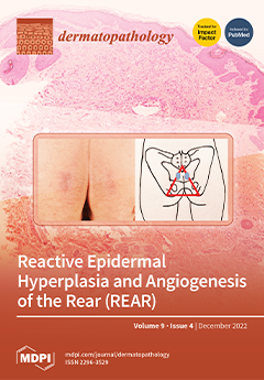
Senile gluteal dermatosis (SGD) is a common but seldom recognized condition. It is characterized clinically by unilateral or bilateral hyperkeratotic, lichenified plaques on the gluteal area, being attributed to prolonged sitting, particularly in the elderly. SGD also encompasses the recently proposed entity of prurigiform angiomatosis. Histologically, there are features of lichenification such as epidermal hyperplasia and a preserved granular layer, with prominent dermal angioproliferation. We report four cases of this condition as well as novel findings of variably increased mast cells and superficial lymphatic vessels in addition to proliferation of dermal blood vessels. We propose a unifying name of Reactive Epidermal hyperplasia and Angiogenesis of the Rear (REAR) to encapsulate the characteristic clinical and histological features of this distinct entity. View this paper
- Issues are regarded as officially published after their release is announced to the table of contents alert mailing list.
- You may sign up for e-mail alerts to receive table of contents of newly released issues.
- PDF is the official format for papers published in both, html and pdf forms. To view the papers in pdf format, click on the "PDF Full-text" link, and use the free Adobe Reader to open them.
Previous Issue
Next Issue