Next Issue
Volume 10, June
Previous Issue
Volume 9, December


Dermatopathology, Volume 10, Issue 1 (March 2023) – 18 articles

Cover Story (view full-size image): 
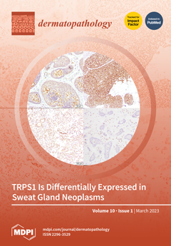
We analyzed TRPS1 expression in a spectrum of cutaneous sweat gland tumors. We stained five microcystic adnexal carcinomas (MACs), three eccrine adenocarcinomas, two syringoid eccrine carcinomas, four hidradenocarcinomas, six porocarcinomas, one eccrine carcinoma-NOS, eleven hidradenomas, nine poromas, seven cylindromas, three spiradenomas, and ten syringomas with TRPS1 antibodies. Our study demonstrates a very high (86%) expression of TRPS1 in malignant and benign adnexal tumors that are mainly composed of islands or nodules with polygonal cells, e.g., hidradenomas. On the other hand, tumors with small ducts or strands of cells, such as MACs, appear to be completely negative. This differential staining among types of sweat gland tumors may represent either differential cells of origin or divergent differentiation and has the potential to be used as a diagnostic tool in the future. View this paper
  • Issues are regarded as officially published after their release is announced to the table of contents alert mailing list.
  • You may sign up for e-mail alerts to receive table of contents of newly released issues.
  • PDF is the official format for papers published in both, html and pdf forms. To view the papers in pdf format, click on the "PDF Full-text" link, and use the free Adobe Reader to open them.
Order results
Result details
Section
Select all
Export citation of selected articles as:
8 pages, 4248 KiB  
Review
Is It Time to Supersede the Diagnostic Term “Melanoma In Situ with Regression?” A Narrative Review
by Anna Colagrande, Giuseppe Ingravallo and Gerardo Cazzato
Dermatopathology 2023, 10(1), 120-127; https://doi.org/10.3390/dermatopathology10010018 - 22 Mar 2023
Cited by 1 | Viewed by 3030
Abstract
Traditionally, the term melanoma in situ (MIS) is used to designate a horizontal (radial) growth phase of malignant melanoma (MM) in which there is no histological evidence of any invasion (or microinvasion) of neoplastic melanocytic cells into the superficial or papillary dermis. In [...] Read more.
Traditionally, the term melanoma in situ (MIS) is used to designate a horizontal (radial) growth phase of malignant melanoma (MM) in which there is no histological evidence of any invasion (or microinvasion) of neoplastic melanocytic cells into the superficial or papillary dermis. In daily dermatopathological practice, we are faced with misleading definitions, such as “melanoma in situ with regression,” which risk affecting homogeneity for comparison purposes of pathological reports of malignant melanoma. The authors conducted a literature review using PubMed and Web of Science (WoS) as the main databases and using the following keywords: “Malignant Melanoma in situ” or “Melanoma in situ” and “regression” and/or “radial growth phase regression.” A total of 213 articles from both analyzed databases were retrieved; finally, only eight articles in English were considered suitable for the chosen inclusion criteria. In consideration of the absence of studies with large case series, of reviews with meta-analyses, and, therefore, of a broad scientific consensus, expressions including “melanoma in situ with regression” should be avoided in the histopathological report. Instead, they should be replaced with clearer and more exhaustive definitions. Full article
Show Figures

Figure 1

8 pages, 44553 KiB  
Case Report
Cutaneous Epithelioid Angiomatous Nodule: Report of a New Case and Literature Review
by Margaux Dubus and Jean Kanitakis
Dermatopathology 2023, 10(1), 112-119; https://doi.org/10.3390/dermatopathology10010017 - 13 Mar 2023
Viewed by 2238
Abstract
Cutaneous epithelioid angiomatous nodule is a rare benign vascular tumour of the skin with characteristic microscopic features, of which 65 cases have so far been reported after the initial description of this entity in 2004. We present here a new typical case of [...] Read more.
Cutaneous epithelioid angiomatous nodule is a rare benign vascular tumour of the skin with characteristic microscopic features, of which 65 cases have so far been reported after the initial description of this entity in 2004. We present here a new typical case of this rare lesion and provide a comprehensive review of all the previously published cases, delineating the salient clinicopathological features of this rare tumour. Full article
(This article belongs to the Section Clinico-Pathological Correlation in Dermatopathology)
Show Figures

Figure 1

8 pages, 1259 KiB  
Case Report
Synthesis and Physiological Remodeling of CD34 Cells in the Skin following the Reversal of Fibrosis through Intensive Treatment for Lower Limb Lymphedema: A Case Report
by Jose Maria Pereira de Godoy, Ana Carolina Pereira de Godoy, Maria de Fatima Guerreiro Godoy and Dalisio de Santi Neto
Dermatopathology 2023, 10(1), 104-111; https://doi.org/10.3390/dermatopathology10010016 - 09 Mar 2023
Viewed by 1736
Abstract
A novel type of cell underwent identification between 2005 and 2008 and was denominated the “telocyte” in 2010. In 2012, transmission electron microscopy revealed the presence of telocytes in the dermis. The aim of the present study was to report important changes in [...] Read more.
A novel type of cell underwent identification between 2005 and 2008 and was denominated the “telocyte” in 2010. In 2012, transmission electron microscopy revealed the presence of telocytes in the dermis. The aim of the present study was to report important changes in immunostained CD34 cells following the treatment of lower limb lymphedema using a specific lymphatic therapy technique. A clinical trial involving the evaluation of changes in immunostained CD34 cells in the epidermis and dermis (10 randomly selected histological fields) of a patient before and after intensive treatment for clinical stage II lymphedema was conducted using the Godoy Method, which was adapted to the treatment of skin fibrosis. The evaluation involved the use of the Weibel multi-point morphometric method. Comparisons were performed using the t-test with a 95% significance level. An important increase in CD34 cells was found with redistribution occurring following treatment. The treatment of primary lymphedema of the lower limbs resulted in the clinical reversal of fibrosis and an increase in the number of immunomarked CD34 cells. Full article
(This article belongs to the Section Clinico-Pathological Correlation in Dermatopathology)
Show Figures

Figure 1

9 pages, 6029 KiB  
Article
Histopathologic Aspects of Malignancy-Associated Granuloma Annulare: A Single Institution Experience
by Buket Bagci, Cansu Karakas, Harsimran Kaur and Bruce R. Smoller
Dermatopathology 2023, 10(1), 95-103; https://doi.org/10.3390/dermatopathology10010015 - 04 Mar 2023
Cited by 1 | Viewed by 3788
Abstract
Granuloma annulare (GA) is a benign, self-limiting granulomatous inflammatory disease that exhibits different histologic patterns. Infrequently, granuloma annulare can be associated with malignancy, the so-called malignancy-associated granuloma annulare (MGA). In this study, we aimed to compare the clinical and histopathological differences between GA [...] Read more.
Granuloma annulare (GA) is a benign, self-limiting granulomatous inflammatory disease that exhibits different histologic patterns. Infrequently, granuloma annulare can be associated with malignancy, the so-called malignancy-associated granuloma annulare (MGA). In this study, we aimed to compare the clinical and histopathological differences between GA and MGA. We retrospectively reviewed patient charts and identified 35 patients diagnosed with GA and concurrent hematological or solid organ malignancies as a case group. Additionally, we identified 33 patients without any known solid organ or hematological malignancy as a control group. MGA is commonly seen in the seventh decade of life, while GA affects the younger population. MGA is most commonly presented in the extremities of the body. The most common malignancy associated with MGA was chronic lymphocytic leukemia. Prostate cancer was the most common type of solid organ malignancy that was associated with MGA. The most common histopathological pattern seen in MGA was interstitial, comprising half of the cases. Multinucleated giant cells were present in half of the MGA cases and in most of the control group. In the literature, there are no established features that distinguish MGA from GA. Although MGA and GA have overlapping features, in our series, we found that the interstitial pattern was more common in MGA, while the necrobiotic pattern was more common in GA. Full article
Show Figures

Figure 1

2 pages, 196 KiB  
Editorial
Artificial Intelligence in Dermatopathology: An Analysis of Its Practical Application
by Marina Kristy Ibraheim, Rohit Gupta, Jerad M. Gardner and Ashley Elsensohn
Dermatopathology 2023, 10(1), 93-94; https://doi.org/10.3390/dermatopathology10010014 - 16 Feb 2023
Cited by 4 | Viewed by 2337
Abstract
In recent years, researchers have explored potential uses for artificial intelligence (AI) in medical practice [...] Full article
(This article belongs to the Special Issue Artificial Intelligence in Dermatopathology)
2 pages, 184 KiB  
Editorial
Histopathological Diagnosis of Malignant Melanoma at the Dawn of 2023: Knowledge Gained and New Challenges
by Gerardo Cazzato
Dermatopathology 2023, 10(1), 91-92; https://doi.org/10.3390/dermatopathology10010013 - 13 Feb 2023
Cited by 3 | Viewed by 2059
Abstract
Year after year, the incidence and prevalence rates of cutaneous Malignant Melanoma (MM) show a continuous increase and, according to the most up-to-date American Cancer Society (ACS) projections, it is estimated that 97,610 new melanomas will be diagnosed in 2023 (about 58,120 in [...] Read more.
Year after year, the incidence and prevalence rates of cutaneous Malignant Melanoma (MM) show a continuous increase and, according to the most up-to-date American Cancer Society (ACS) projections, it is estimated that 97,610 new melanomas will be diagnosed in 2023 (about 58,120 in men and 39,490 in women) and approximately 7990 people are expected to die of melanoma (about 5420 men and 2570 women) [...] Full article
5 pages, 1565 KiB  
Case Report
Post-Pemphigus Acanthomata Presenting as an Isolated, Hyperkeratotic Plaque
by Rachita Pandya, Yanjia Zhou, Mansee Desai, Nancy Anderson and Ashley Elsensohn
Dermatopathology 2023, 10(1), 86-90; https://doi.org/10.3390/dermatopathology10010012 - 13 Feb 2023
Viewed by 1869
Abstract
Post-pemphigus acanthomas have been rarely discussed in the literature. A prior case series identified 47 cases of pemphigus vulgaris and 5 cases of pemphigus foliaceus, out of which 13 developed acanthomata as a part of the healing process. Additionally, a case report by [...] Read more.
Post-pemphigus acanthomas have been rarely discussed in the literature. A prior case series identified 47 cases of pemphigus vulgaris and 5 cases of pemphigus foliaceus, out of which 13 developed acanthomata as a part of the healing process. Additionally, a case report by Ohashi et al. reported similar recalcitrant lesions on the trunk of a patient with pemphigus foliaceus being treated with prednisolone, IVIG, plasma exchange, and cyclosporine. Some view post-pemphigus acanthomas as variants of hypertrophic pemphigus vulgaris, being difficult to diagnose when they present as only single lesions, with a clinical differential of an inflamed seborrheic keratosis or squamous cell carcinoma. Here, we present a case of a 52-year-old female with a history of pemphigus vulgaris and four months of only topical therapy (fluocinonide 0.05%) who presented with a painful, hyperkeratotic plaque on the right mid-back that was found to be a post-pemphigus acanthoma. Full article
Show Figures

Figure 1

11 pages, 21749 KiB  
Article
TRPS1 Is Differentially Expressed in a Variety of Malignant and Benign Cutaneous Sweat Gland Neoplasms
by Hatice B. Zengin, Chau M. Bui, Kristin Rybski, Tatsiana Pukhalskaya, Bahadir Yildiz and Bruce R. Smoller
Dermatopathology 2023, 10(1), 75-85; https://doi.org/10.3390/dermatopathology10010011 - 02 Feb 2023
Cited by 7 | Viewed by 2539
Abstract
Neoplasms of sweat glands and the breast may be morphologically and immunophenotypically similar. A recent study showed that TRPS1 staining is a highly sensitive and specific marker for breast carcinoma. In this study, we analyzed TRPS1 expression in a spectrum of cutaneous sweat [...] Read more.
Neoplasms of sweat glands and the breast may be morphologically and immunophenotypically similar. A recent study showed that TRPS1 staining is a highly sensitive and specific marker for breast carcinoma. In this study, we analyzed TRPS1 expression in a spectrum of cutaneous sweat gland tumors. We stained five microcystic adnexal carcinomas (MACs), three eccrine adenocarcinomas, two syringoid eccrine carcinomas, four hidradenocarcinomas, six porocarcinomas, one eccrine carcinoma-NOS, 11 hidradenomas, nine poromas, seven cylindromas, three spiradenomas, and 10 syringomas with TRPS1 antibodies. All of the MACs and syringomas were negative. Every cylindroma and two of the three spiradenomas demonstrated intense staining in cells lining the ductular spaces, with negative to relatively weak expression in surrounding cells. Of the 16 remaining malignant entities, 13 were intermediate to high positive, one was low positive, and two were negative. From the 20 hidradenomas and poromas, intermediate to high positivity was revealed in 14 cases, low positivity in three cases, and negative staining in three cases. Our study demonstrates a very high (86%) expression of TRPS1 in malignant and benign adnexal tumors that are mainly composed of islands or nodules with polygonal cells, e.g., hidradenomas. On the other hand, tumors with small ducts or strands of cells, such as MACs, appear to be completely negative. This differential staining among types of sweat gland tumors may represent either differential cells of origin or divergent differentiation and has the potential to be used as a diagnostic tool in the future. Full article
Show Figures

Figure 1

5 pages, 1394 KiB  
Case Report
Loss of CD34 Expression within an Interstitial Dermal Lymphoid Cell Infiltrate Is a Helpful Clue to the Diagnosis of Morphea
by Maged Daruish, Anoud Zidan, Danielle T. Greenblatt and Catherine M. Stefanato
Dermatopathology 2023, 10(1), 70-74; https://doi.org/10.3390/dermatopathology10010010 - 02 Feb 2023
Viewed by 1653
Abstract
A dermal interstitial lymphocytic infiltrate may represent a diagnostic challenge, particularly if the clinical history is not provided. We present three cases within the histological spectrum of morphea in which the immunohistochemical marker CD34 was helpful in confirming the diagnosis. Full article
(This article belongs to the Section Clinico-Pathological Correlation in Dermatopathology)
Show Figures

Figure 1

7 pages, 3745 KiB  
Case Report
An Underrecognized Histologic Clue to the Diagnosis of Mucous Membrane Pemphigoid: A Case Report and Review of Diagnostic Guidelines
by Jason R. McFadden, Advaita S. Chaudhari, Debra Birenbaum, Lynette Margesson, Jorge Gonzalez and Aravindhan Sriharan
Dermatopathology 2023, 10(1), 63-69; https://doi.org/10.3390/dermatopathology10010009 - 02 Feb 2023
Viewed by 2592
Abstract
Mucous membrane pemphigoid (MMP), also known as cicatricial pemphigoid (CP), is a heterogeneous group of subepidermal blistering diseases that affect the mucous membranes, most frequently in the eye and oral cavity. MMP is often unrecognized or misdiagnosed in its early stages due to [...] Read more.
Mucous membrane pemphigoid (MMP), also known as cicatricial pemphigoid (CP), is a heterogeneous group of subepidermal blistering diseases that affect the mucous membranes, most frequently in the eye and oral cavity. MMP is often unrecognized or misdiagnosed in its early stages due to its rarity and nonspecific presentation. We present the case of a 69-year-old female in which MMP of the vulva was not initially suspected. The first biopsy, from lesional tissue for routine histology, revealed fibrosis, late-stage granulation tissue, and nonspecific findings. A second biopsy, from perilesional tissue for direct immunofluorescence (DIF), revealed DIF findings typical of MMP. Scrutiny of both the first and second biopsies revealed a subtle but telling histologic feature: subepithelial clefts along adnexae in the context of a scarring process with neutrophils and eosinophils, which can be an important clue to MMP. This histologic clue has been previously described; reinforcing its importance may prove useful for future cases, especially those for which DIF is not feasible. Our case demonstrates the protean presentations of MMP, the need for persistence in sampling unusual cases, and the relevance of inconspicuous histologic features. The report highlights this underrecognized yet potentially decisive histologic clue to MMP, reviews current biopsy guidelines when MMP is suspected, and delineates the clinical and morphological features of vulvar MMP. Full article
Show Figures

Figure 1

9 pages, 5783 KiB  
Review
Rare Variants of Dermatofibrosarcoma Protuberans: Clinical, Histologic, and Molecular Features and Diagnostic Pitfalls
by Celestine M. Trinidad, Sintawat Wangsiricharoen, Victor G. Prieto and Phyu P. Aung
Dermatopathology 2023, 10(1), 54-62; https://doi.org/10.3390/dermatopathology10010008 - 29 Jan 2023
Cited by 3 | Viewed by 4029
Abstract
Dermatofibrosarcoma protuberans (DFSP) is a dermal malignant mesenchymal tumor. Most variants are associated with a high risk of local recurrence and a low risk of metastasis. The classic histomorphology of this tumor is made up of uniform, spindle-shaped cells, arranged in a storiform [...] Read more.
Dermatofibrosarcoma protuberans (DFSP) is a dermal malignant mesenchymal tumor. Most variants are associated with a high risk of local recurrence and a low risk of metastasis. The classic histomorphology of this tumor is made up of uniform, spindle-shaped cells, arranged in a storiform pattern. Tumor cells characteristically infiltrate the underlying subcutis in a honeycomb pattern. Less common variants of DFSP have been identified: myxoid, pigmented, myoid, granular cell, sclerosing, atrophic, and fibrosarcomatous. Only the fibrosarcomatous variant has been shown to differ significantly from classic DFSP in terms of clinical outcome; fibrosarcomatous DFSP has been shown to be associated with a greater risk of local recurrence and metastatic potential than classic DFSP. However, the other variants may pose diagnostic difficulty as they resemble other types of spindle cell neoplasms, especially in small biopsy specimens. This article reviews the clinical, histologic, and molecular features of DFSP variants, as well as possible pitfalls in their diagnosis and how to resolve them. Full article
(This article belongs to the Special Issue Dermatopathology in Asia)
Show Figures

Figure 1

2 pages, 172 KiB  
Editorial
Acknowledgment to the Reviewers of Dermatopathology in 2022
by Dermatopathology Editorial Office
Dermatopathology 2023, 10(1), 52-53; https://doi.org/10.3390/dermatopathology10010007 - 19 Jan 2023
Viewed by 1275
Abstract
High-quality academic publishing is built on rigorous peer review [...] Full article
6 pages, 1945 KiB  
Case Report
Widespread and Long-Enduring Hyperkeratosis Lenticularis Perstans (Flegel’s Disease): Clinico-Pathological and Dermoscopic Features of a Rare Presentation
by Giorgio Stabile, Giovanni Paolino, Nathalie Rizzo and Franco Rongioletti
Dermatopathology 2023, 10(1), 46-51; https://doi.org/10.3390/dermatopathology10010006 - 13 Jan 2023
Viewed by 3043
Abstract
Hyperkeratosis lenticularis perstans, also known as Flegel’s disease (FD), is a rare cutaneous disorder affecting mainly the lower extremities of middle-aged people. Due to its rarity, this disease is usually not recognized by physicians resulting in a delay in diagnosis, especially in those [...] Read more.
Hyperkeratosis lenticularis perstans, also known as Flegel’s disease (FD), is a rare cutaneous disorder affecting mainly the lower extremities of middle-aged people. Due to its rarity, this disease is usually not recognized by physicians resulting in a delay in diagnosis, especially in those cases with atypical cutaneous involvement. Herein, we present a 72-year-old woman who developed FD characterized by a generalized distribution, involving, in addition to the lower limbs, the trunk and the upper limbs as well. We performed a description of the dermoscopic and pathologic features of this rare entity, also carrying out a brief reappraisal of the cases of FD with a diffuse, atypical and generalized distribution that have been described in the literature. Histopathology with clinical correlation is the cornerstone of the diagnosis, even and especially in atypical cases. This patient with a disease duration of 58 years also represents the longest-lasting case of FD reported in the literature. Full article
Show Figures

Figure 1

5 pages, 18846 KiB  
Case Report
Multiply Recurrent Composite Hemangioendothelioma of Penis with Histologic Progression to High-Grade Features
by Chau M. Bui and Bonnie Balzer
Dermatopathology 2023, 10(1), 41-45; https://doi.org/10.3390/dermatopathology10010005 - 11 Jan 2023
Cited by 1 | Viewed by 1663
Abstract
Composite hemangioendothelioma (CHE) is a very rare low-grade malignant vascular neoplasm. Here, we present the first case of it occurring on a penis with two local recurrences over a 9 year span and its progression to a high-grade morphology. Full article
Show Figures

Figure 1

11 pages, 249 KiB  
Article
Concordance of Clinical, Histologic and Direct Immunofluorescence Findings in Patients with Autoimmune Bullous Dermatoses in Vietnam
by Giang Huong Tran, Nhan Thi Ai Le, Minh Hoang Dang, Thao Thi Phuong Doan and Thuy L. Phung
Dermatopathology 2023, 10(1), 30-40; https://doi.org/10.3390/dermatopathology10010004 - 10 Jan 2023
Viewed by 1898
Abstract
Introduction: Autoimmune bullous dermatoses (ABD) represent a heterogeneous group of blistering disorders that may be debilitating with high morbidity. Clinical, histological, and direct immunofluorescence (DIF) studies are essential in establishing an accurate diagnosis of ABD, which is essential for its clinical management. [...] Read more.
Introduction: Autoimmune bullous dermatoses (ABD) represent a heterogeneous group of blistering disorders that may be debilitating with high morbidity. Clinical, histological, and direct immunofluorescence (DIF) studies are essential in establishing an accurate diagnosis of ABD, which is essential for its clinical management. Our study objective was to perform a systematic evaluation of ABD cases in a patient population at an academic medical center in Ho Chi Minh City, Vietnam, and determine the degree of concordance of clinical, histological, and DIF findings in ABD. Methodology: A systematic retrospective cross-sectional study was performed on 92 patients diagnosed with ABD by clinical, histological, and DIF studies at the University of Medicine and Pharmacy in Ho Chi Minh City, Vietnam, between September 2019 and September 2021. The clinical histories, H and E stained tissue sections, and DIF stains were evaluated by pathologists at the University of Medicine and Pharmacy. Results: ABD was evaluated as a whole and subdivided into an intraepidermal blister subgroup and a subepidermal blister subgroup. The analysis of paired diagnostic methods (clinical, histological, and DIF) for concordance with the final diagnosis was performed and showed that there were no statistically significant differences between the paired methods (McNemar’s test, p > 0.05). There was moderate concordance between the clinical, histological, and DIF diagnoses among all ABD cases (Brennan-Prediger coefficient Kappa test, κBP = 0.522, CI = 0.95). In the intraepidermal blister subgroup, the diagnostic accuracies of the histology and DIF stains were comparable to each other, and both were more accurate than a clinical diagnosis alone. In the subepidermal blister subgroup, there was no statistically significant difference in each pair of the three diagnostic methods (clinical, histological, and DIF) (McNemar’s test, p > 0.05). The concordance between the clinical, histological, and DIF diagnoses was high for the intraepidermal blister subgroup (Kappa test, κBP = 0.758, CI = 0.95). However, the concordance between the clinical, histological, and DIF diagnoses was slight for the subepidermal blister subgroup (Kappa test, κBP = 0.171, CI = 0.95). Conclusion: Histological evaluation is highly accurate in the diagnosis of the intraepidermal blister subgroup, but it is not as accurate in the diagnosis of the subepidermal blister subgroup in the Vietnamese patient cohort in which clinical, histological, and DIF studies were performed. DIF stains are a crucial diagnostic tool for ABD in this patient population. Full article
(This article belongs to the Special Issue Dermatopathology in Asia)
5 pages, 1831 KiB  
Case Report
Epithelioid Fibrous Histiocytoma with CARS-ALK Fusion: First Case Report
by Léo-Paul Secco, Louis Libbrecht, Elsa Seijnhaeve, Silke Eggers, Anne-France Dekairelle and An-Katrien De Roo
Dermatopathology 2023, 10(1), 25-29; https://doi.org/10.3390/dermatopathology10010003 - 03 Jan 2023
Cited by 2 | Viewed by 1930
Abstract
Epithelioid fibrous histiocytoma (EFH) is a type of uncommon skin tumor mostly harboring Anaplastic Lymphoma Kinase (ALK) gene rearrangement, with different fusion partners reported. Whether this tumor is a separate entity or has a relationship with conventional fibrous histiocytomas is still [...] Read more.
Epithelioid fibrous histiocytoma (EFH) is a type of uncommon skin tumor mostly harboring Anaplastic Lymphoma Kinase (ALK) gene rearrangement, with different fusion partners reported. Whether this tumor is a separate entity or has a relationship with conventional fibrous histiocytomas is still a matter of debate. Benign course is the rule after complete surgical excision. A rare subtype of EFH with fusiform cells has been described, with specific fusion partners. Inflammatory myofibroblastic tumor (IMT) is a type of soft tissue tumor rarer than EFH, and it can display distant metastases. Some cases of primary cutaneous IMT included two with Cysteinyl-tRNA Synthetase 1 (CARS)-ALK rearrangement. IMT can have the same fusion partners as EFH, such as DCTN1, TMP3 or EML4 genes. We report the case of a 42-year-old woman presenting EFH with fusiform morphology harboring CARS-ALK fusion and discuss similarities and differences with IMT. Full article
(This article belongs to the Section Molecular Dermatopathology)
Show Figures

Figure 1

5 pages, 730 KiB  
Case Report
Amyloidosis Cutis Dyschromica in a 16-Year-Old Filipino Girl: A Case Report
by Fendi EJ R. Bautista, Marcia Marie S. Marte-Jimenez and Maria Jasmin J. Jamora
Dermatopathology 2023, 10(1), 20-24; https://doi.org/10.3390/dermatopathology10010002 - 29 Dec 2022
Viewed by 2365
Abstract
Amyloidosis cutis dyschromica is a rare variant of primary cutaneous amyloidosis characterized by hyper- and hypopigmented macules. In this paper, we reported a case of a 16-year-old Filipino girl with hyper- and hypopigmented to depigmented macules on the upper and lower extremities, which [...] Read more.
Amyloidosis cutis dyschromica is a rare variant of primary cutaneous amyloidosis characterized by hyper- and hypopigmented macules. In this paper, we reported a case of a 16-year-old Filipino girl with hyper- and hypopigmented to depigmented macules on the upper and lower extremities, which started when she was 9 years of age. Full article
(This article belongs to the Special Issue Dermatopathology in Asia)
Show Figures

Figure 1

19 pages, 14737 KiB  
Review
Histopathological Variants of Cutaneous Neurofibroma: A Compendious Review
by Neha S. Nagrani and Jag Bhawan
Dermatopathology 2023, 10(1), 1-19; https://doi.org/10.3390/dermatopathology10010001 - 26 Dec 2022
Cited by 4 | Viewed by 6565
Abstract
The first description of histopathological variants of neurofibroma dates back to 1994. Over the years, many individual case reports elucidating unusual histologic features in neurofibroma have been added to the literature, some of which have defined criteria, with the others falling under the [...] Read more.
The first description of histopathological variants of neurofibroma dates back to 1994. Over the years, many individual case reports elucidating unusual histologic features in neurofibroma have been added to the literature, some of which have defined criteria, with the others falling under the roof of benign neural neoplasms. These unusual features, which sometimes may lead to pauses in identifying a common benign tumor such as neurofibroma. Awareness of these variants may help dermatopathologists avoid misinterpretation. Thus, this review aims to summarize all novel and unusual histopathological variants of cutaneous neurofibroma reported to date, in addition to any unusual variants that we encountered in our practice. Full article
(This article belongs to the Section Clinico-Pathological Correlation in Dermatopathology)
Show Figures

Figure 1

Previous Issue
Next Issue
Back to TopTop