
Current Trends in Toxicity Assessment of Herbal Medicines: A Narrative Review


 , , ,
, , ,
Abstract
:1. Introduction
2. The Importance of Standardization for Safety and Toxicity Profiling of Herbal Medicine—Assuring the Herbs’ Authenticity
2.1. Morphological Identification
2.2. The Metabolomics Approach in Herbal Medicinal Products Identification and Standardization-Chemical Fingerprinting
2.3. DNA-Based Techniques for Herbal Products Authentication
3. Intrinsic Toxicity Evaluation
3.1. Acute/Sub-Acute/Chronic Toxicity Evaluation
3.2. Genotoxicity and Carcinogenicity Evaluation
3.3. Omics-Based Toxicology
3.3.1. Toxico-Transcriptomics
3.3.2. Toxico-Proteomics
3.3.3. Toxico-Metabonomics
4. Evaluation of Toxicity Arising from Extrinsic Sources
4.1. Mycotoxins Control
4.2. Pesticides and Fumigation Agent Residues Control
4.3. Heavy Metals Control
5. Conclusions
Author Contributions
Funding
Data Availability Statement
Conflicts of Interest
References
- Woo, C.S.J.; Lau, J.S.H.; El-Nezami, H. Herbal medicine: Toxicity and recent trends in assessing their potential toxic effects. In Advances in Botanical Research; Shyur, L.-F., Lau, A.S.Y., Eds.; Elsevier Academic Press: Cambridge, MA, USA, 2012; Volume 62, pp. 365–384. [Google Scholar]
- Islam, S.U.; Dar, T.U.; Khuroo, A.A.; Bhat, B.A.; Mangral, Z.A.; Tariq, L.; Tantray, W.W.; Malik, A.H. DNA barcoding aids in identification of adulterants of Trillium govanianum Wall. ex D. Don. J. Appl. Res. Med. Aromat. Plants 2021, 23, 100305. [Google Scholar] [CrossRef]
- Aware, C.B.; Patil, D.N.; Suryawanshi, S.S.; Mali, P.R.; Rane, M.R.; Gurav, R.G.; Jadhav, J.P. Natural bioactive products as promising therapeutics: A review of natural product-based drug development. S. Afr. J. Bot. 2022, 151, 512–528. [Google Scholar] [CrossRef]
- Tnah, L.; Lee, S.; Tan, A.; Lee, C.; Ng, K.; Ng, C.; Farhanah, Z.N. DNA barcode database of common herbal plants in the tropics: A resource for herbal product authentication. Food Control 2019, 95, 318–326. [Google Scholar] [CrossRef]
- Jordan, S.A.; Cunningham, D.G.; Marles, R.J. Assessment of herbal medicinal products: Challenges, and opportunities to increase the knowledge base for safety assessment. Toxicol. Appl. Pharmacol. 2010, 243, 198–216. [Google Scholar] [CrossRef]
- Newmaster, S.G.; Grguric, M.; Shanmughanandhan, D.; Ramalingam, S.; Ragupathy, S. DNA barcoding detects contamination and substitution in North American herbal products. BMC Med. 2013, 11, 222. [Google Scholar] [CrossRef] [Green Version]
- Wallace, L.J.; Boilard, S.M.; Eagle, S.H.; Spall, J.L.; Shokralla, S.; Hajibabaei, M. DNA barcodes for everyday life: Routine authentication of Natural Health Products. Food Res. Int. 2012, 49, 446–452. [Google Scholar] [CrossRef]
- Lo, Y.T.; Shaw, P.C. Application of next-generation sequencing for the identification of herbal products. Biotechnol. Adv. 2019, 37, 107450. [Google Scholar] [CrossRef]
- Tankeu, S.; Vermaak, I.; Chen, W.; Sandasi, M.; Viljoen, A. Differentiation between two “fang ji” herbal medicines, Stephania tetrandra and the nephrotoxic Aristolochia fangchi, using hyperspectral imaging. Phytochemistry 2016, 122, 213–222. [Google Scholar] [CrossRef]
- Kerchner, A.; Farkas, Á. Worldwide poisoning potential of Brugmansia and Datura. Forensic Toxicol. 2020, 38, 30–41. [Google Scholar] [CrossRef] [Green Version]
- Han, J.; Li, M.; Luo, K.; Liu, M.; Chen, X.; Chen, S. Identification of Daturae flos and its adulterants based on DNA barcoding technique. Yao Xue Xue Bao 2011, 46, 1408–1412. [Google Scholar]
- Pandey, R.; Tiwari, R.K.; Shukla, S.S. Omics: A newer technique in herbal drug standardization & quantification. J. Young Pharm. 2016, 8, 76. [Google Scholar]
- Liu, M.; Li, X.-W.; Liao, B.-S.; Luo, L.; Ren, Y.-Y. Species identification of poisonous medicinal plant using DNA barcoding. Chin. J. Nat. Med. 2019, 17, 585–590. [Google Scholar] [CrossRef]
- Gowthaman, N.; Lim, H.; Gopi, S.; Amalraj, A. Identification of toxicology biomarker and evaluation of toxicity of natural products by metabolomic applications. In Inflammation and Natural Products, 1st ed.; Gopi, S., Amalraj, A., Kunnumakkara, A., Thomas, S., Eds.; Elsevier Academic Press: Cambridge, MA, USA, 2021; pp. 407–436. [Google Scholar]
- Gajula, S.N.R.; Nanjappan, S. Metabolomics: A recent advanced omics technology in herbal medicine research. In Medicinal and Aromatic Plants; Aftab, T., Hakeem, K.R., Eds.; Elsevier Academic Press: Cambridge, MA, USA, 2021; pp. 97–117. [Google Scholar]
- Mukne, A.; Momin, M.; Betkar, P.; Joshi, V. Standardization of herbal biomolecules. In Herbal Biomolecules in Healthcare Applications, 1st ed.; Mandal, S.C., Nayak, A.K., Dhara, A.K., Eds.; Elsevier Academic Press: Cambridge, MA, USA, 2022; pp. 643–667. [Google Scholar]
- Putri, S.P.; Nakayama, Y.; Matsuda, F.; Uchikata, T.; Kobayashi, S.; Matsubara, A.; Fukusaki, E. Current metabolomics: Practical applications. J. Biosci. Bioeng. 2013, 115, 579–589. [Google Scholar] [CrossRef] [PubMed]
- Braconi, D.; Millucci, L.; Parisi, M.L.; Spiga, O.; Santucci, A. Omics-based technologies for food authentication and traceability. In Food Authentication and Traceability; Galanakis, C.M., Ed.; Elsevier Academic Press: Cambridge, MA, USA, 2021; pp. 215–245. [Google Scholar]
- Sánchez, M.; González-Burgos, E.; Divakar, P.K.; Gómez-Serranillos, M.P. DNA-based authentication and metabolomics analysis of medicinal plants samples by DNA barcoding and ultra-high-performance liquid chromatography/triple quadrupole mass spectrometry (UHPLC-MS). Plants 2020, 9, 1601. [Google Scholar] [CrossRef] [PubMed]
- Xiao, Q.; Mu, X.; Liu, J.; Li, B.; Liu, H.; Zhang, B.; Xiao, P. Plant metabolomics: A new strategy and tool for quality evaluation of Chinese medicinal materials. Chin. Med. 2022, 17, 45. [Google Scholar] [CrossRef]
- Pan, H.; Yao, C.; Yao, S.; Yang, W.; Wu, W.; Guo, D. A metabolomics strategy for authentication of plant medicines with multiple botanical origins, a case study of Uncariae Rammulus Cum Uncis. J. Sep. Sci. 2020, 43, 1043–1050. [Google Scholar] [CrossRef]
- Fukuda, E.; Baba, M.; Iwasaki, N.; Uesawa, Y.; Arifuku, K.; Kamo, O.; Tsubono, K.; Okada, Y. Identification of Glycyrrhiza species by direct analysis in real time mass spectrometry. Nat. Prod. Commun. 2010, 5, 1755–1758. [Google Scholar] [CrossRef] [Green Version]
- Bārzdiņa, A.; Paulausks, A.; Bandere, D.; Brangule, A. The Potential Use of Herbal Fingerprints by Means of HPLC and TLC for Characterization and Identification of Herbal Extracts and the Distinction of Latvian Native Medicinal Plants. Molecules 2022, 27, 2555. [Google Scholar] [CrossRef]
- Syed, M.; Khan, M.N.; Khadim, A.; Shadab, H.; Perveen, A.; El-Seedi, H.R.; Musharraf, S.G. Chemical fingerprinting of three Anemone species and an adulteration study to detect cross mixing of medicinal plants by HPLC-HR-ESI-MS/MS method. J. King Saud Univ. Sci. 2021, 33, 101461. [Google Scholar] [CrossRef]
- Soininen, T.H.; Jukarainen, N.; Auriola, S.O.; Julkunen-Tiitto, R.; Karjalainen, R.; Vepsäläinen, J.J. Quantitative metabolite profiling of edible onion species by NMR and HPLC–MS. Food Chem. 2014, 165, 499–505. [Google Scholar] [CrossRef]
- Zhang, J.; Wang, C.; Wu, W.; Jin, Q.; Wu, J.; Yang, L.; An, Y.; Yao, C.; Wei, W.; Song, J.; et al. Authentication of herbal medicines from multiple botanical origins with cross-validation mebabolomics, absolute quantification and support vector machine model, a case study of Rhizoma Alismatis. Arab. J. Chem. 2022, 15, 104118. [Google Scholar] [CrossRef]
- Rakhesh, K.V.; Ashalatha, S.N.; Mahima, K. In vitro regeneration and chromatographic fingerprint analysis of Aphanamixis polystachya (Wall.) Parker by HPTLC technique. S. Afr. J. Bot. 2022, 151, 259–265. [Google Scholar] [CrossRef]
- Jiménez-Sánchez, C.; Lozano-Sánchez, J.; Rodríguez-Pérez, C.; Segura-Carretero, A.; Fernández-Gutiérrez, A. Comprehensive, untargeted, and qualitative RP-HPLC-ESI-QTOF/MS2 metabolite profiling of green asparagus (Asparagus officinalis). J. Food Compos. Anal. 2016, 46, 78–87. [Google Scholar] [CrossRef]
- Dawane, V.; Pathak, B. Assessment of secondary metabolite profile and quantification method development for Lupeol and Caffeic acid by HPTLC in Avicennia marina pneumatophore roots. Biocatal. Agric. Biotechnol. 2020, 26, 101573. [Google Scholar] [CrossRef]
- Shah, A.P.; Travadi, T.; Sharma, S.; Pandit, R.; Joshi, C.; Joshi, M. Comprehensive analysis using DNA metabarcoding, PCR, and HPLC unveils the adulteration in Brahmi herbal products. bioRxiv 2022. [Google Scholar] [CrossRef]
- Ullah, I.; Subhan, F.; Rudd, J.A.; Rauf, K.; Alam, J.; Shahid, M.; Sewell, R.D. Attenuation of cisplatin-induced emetogenesis by standardized Bacopa monnieri extracts in the pigeon: Behavioral and neurochemical correlations. Planta Med. 2014, 80, 1569–1579. [Google Scholar]
- Ullah, I.; Subhan, F.; Lu, Z.; Chan, S.W.; Rudd, J.A. Action of Bacopa monnieri to antagonize cisplatin-induced emesis in Suncus murinus (house musk shrew). J. Pharmacol. Sci. 2017, 133, 232–239. [Google Scholar] [CrossRef]
- Wang, Y.; Harrington, P.d.B.; Chen, P. Metabolomic profiling and comparison of major cinnamon species using UHPLC–HRMS. Anal. Bioanal. Chem. 2020, 412, 7669–7681. [Google Scholar] [CrossRef]
- Petrakis, E.A.; Cagliani, L.R.; Polissiou, M.G.; Consonni, R. Evaluation of saffron (Crocus sativus L.) adulteration with plant adulterants by 1H NMR metabolite fingerprinting. Food Chem. 2015, 173, 890–896. [Google Scholar] [CrossRef]
- Windarsih, A.; Rohman, A.; Swasono, R.T. Application of H-NMR metabolite fingerprinting and chemometrics for the authentication of Curcuma longa adulterated with Curcuma manga. J. Appl. Pharm. Sci. 2018, 8, 075–081. [Google Scholar]
- Rohman, A.; Wijayanti, T.; Windarsih, A.; Riyanto, S. The authentication of Java turmeric (Curcuma xanthorrhiza) using thin layer chromatography and 1H-NMR based-metabolite fingerprinting coupled with multivariate analysis. Molecules 2020, 25, 3928. [Google Scholar] [CrossRef] [PubMed]
- Windarsih, A.; Rohman, A.; Swasono, R.T. Application of 1H-NMR based metabolite fingerprinting and chemometrics for authentication of Curcuma longa adulterated with C. heyneana. J. Appl. Res. Med. Aromat. Plants 2019, 13, 100203. [Google Scholar] [CrossRef]
- Gu, X.; Zhu, S.; Du, H.; Bai, C.; Duan, X.; Li, Y.; Hu, K. Comprehensive multi-component analysis for authentication and differentiation of 6 Dendrobium species by 2D NMR-based metabolomic profiling. Microchem. J. 2022, 176, 107225. [Google Scholar] [CrossRef]
- Ullrich, S.F.; Averesch, N.J.; Castellanos, L.; Choi, Y.H.; Rothauer, A.; Kayser, O. Discrimination of wild types and hybrids of Duboisia myoporoides and Duboisia leichhardtii at different growth stages using 1H NMR-based metabolite profiling and tropane alkaloids-targeted HPLC-MS analysis. Phytochemistry 2016, 131, 44–56. [Google Scholar] [CrossRef]
- Ibragic, S.; Barbini, S.; Oberlerchner, J.T.; Potthast, A.; Rosenau, T.; Böhmdorfer, S. Antioxidant properties and qualitative analysis of phenolic constituents in Ephedra spp. by HPTLC together with injection port derivatization GC–MS. J. Chromatogr. B 2021, 1180, 122877. [Google Scholar] [CrossRef]
- Kim, H.K.; Choi, Y.H.; Erkelens, C.; Lefeber, A.W.; Verpoorte, R. Metabolic fingerprinting of Ephedra species using 1H-NMR spectroscopy and principal component analysis. Chem. Pharm. Bull. 2005, 53, 105–109. [Google Scholar] [CrossRef] [Green Version]
- Singh, P.; Ranjan, R.; Tripathi, R.; Dixit, J.; Sinha, N.; Singh, A.K.; Tiwari, K.N. GC-MS and NMR spectroscopy based metabolite profiling of Panchvalkal kwath (polyherbal formulation). Nat. Prod. Res. 2021, 1–6. [Google Scholar] [CrossRef]
- Wang, Z.; Xie, H.; Ren, J.; Chen, Y.; Li, X.; Chen, X.; Chan, T.-W.D. Metabolomic approach for rapid differentiation of Fritillaria bulbs by matrix-assisted laser desorption/ionization mass spectrometry and multivariate statistical analysis. J. Pharm. Biomed. Anal. 2020, 185, 113177. [Google Scholar] [CrossRef]
- Xiao, J.-J.; Huang, H.; Lei, Y.-C.; Lin, T.-W.; Ma, Y.; Zhang, J.; Zhang, X.-G.; Zhang, D.-Q.; Lv, G.-H. Development of Tianma HPLC fingerprint and discriminant analysis. Zhongguo Zhong Yao Za Zhi 2017, 42, 2524–2531. [Google Scholar]
- Guo, J.; Wu, Y.; Jiang, M.; Wu, C.; Wang, G. An LC–MS-based metabolomic approach provides insights into the metabolite profiles of Ginkgo biloba L. at different developmental stages and in various organs. Food Res. Int. 2022, 159, 111644. [Google Scholar] [CrossRef]
- Farag, M.A.; Afifi, S.M.; Rasheed, D.M.; Khattab, A.R. Revealing compositional attributes of Glossostemon bruguieri Desf. root geographic origin and roasting impact via chemometric modeling of SPME-GC-MS and NMR metabolite profiles. J. Food Compos. Anal. 2021, 102, 104073. [Google Scholar] [CrossRef]
- Zhao, J.; Wang, M.; Adams, S.J.; Lee, J.; Chittiboyina, A.G.; Avula, B.; Ali, Z.; Raman, V.; Li, J.; Wu, C.; et al. Metabolite variation and discrimination of five licorice (Glycyrrhiza) species: HPTLC and NMR explorations. J. Pharm. Biomed. Anal. 2022, 220, 115012. [Google Scholar] [CrossRef] [PubMed]
- Farag, M.A.; Porzel, A.; Wessjohann, L.A. Comparative metabolite profiling and fingerprinting of medicinal licorice roots using a multiplex approach of GC–MS, LC–MS and 1D NMR techniques. Phytochemistry 2012, 76, 60–72. [Google Scholar] [CrossRef] [PubMed]
- Rizzato, G.; Scalabrin, E.; Radaelli, M.; Capodaglio, G.; Piccolo, O. A new exploration of licorice metabolome. Food Chem. 2017, 221, 959–968. [Google Scholar] [CrossRef] [PubMed]
- Frommenwiler, D.A.; Maire-Widmer, V.; Upton, R.; Nichols, J.; Heubl, G.; Reich, E. Qualitative and quantitative characterization of two licorice root species (Glycyrrhiza glabra L. and Glycyrrhiza uralensis Fisch.) by HPTLC, validated by HPLC and DNA sequencing. JPC-J. Planar Chromatogr.-Mod. TLC 2017, 30, 467–473. [Google Scholar] [CrossRef]
- Song, W.; Qiao, X.; Chen, K.; Wang, Y.; Ji, S.; Feng, J.; Li, K.; Lin, Y.; Ye, M. Biosynthesis-based quantitative analysis of 151 secondary metabolites of licorice to differentiate medicinal Glycyrrhiza species and their hybrids. Anal. Chem. 2017, 89, 3146–3153. [Google Scholar] [CrossRef]
- Shawky, E.; El Sohafy, S.M. Untargeted and targeted chemical profiling for efficacy-directed discrimination of Hedera helix L. subspecies using HPTLC-image analysis and HPTLC/MS. Ind. Crops Prod. 2020, 145, 111980. [Google Scholar] [CrossRef]
- Trute, A.; Nahrstedt, A. Identification and quantitative analysis of phenolic compounds from the dry extract of Hedera helix. Planta Med. 1997, 63, 177–179. [Google Scholar] [CrossRef]
- Xie, G.-Y.; Zhu, Y.; Shu, P.; Qin, X.-Y.; Wu, G.; Wang, Q.; Qin, M.-J. Phenolic metabolite profiles and antioxidants assay of three Iridaceae medicinal plants for traditional Chinese medicine “She-gan” by on-line HPLC–DAD coupled with chemiluminescence (CL) and ESI-Q-TOF-MS/MS. J. Pharm. Biomed. Anal. 2014, 98, 40–51. [Google Scholar] [CrossRef]
- Gao, W.; Yang, H.; Qi, L.-W.; Liu, E.-H.; Ren, M.-T.; Yan, Y.-T.; Chen, J.; Li, P. Unbiased metabolite profiling by liquid chromatography–quadrupole time-of-flight mass spectrometry and multivariate data analysis for herbal authentication: Classification of seven Lonicera species flower buds. J. Chromatogr. A 2012, 1245, 109–116. [Google Scholar] [CrossRef]
- Brahmi, F.; Nguyen, A.T.; Nacoulma, A.P.; Sheridan, H.; Wang, J.; Guendouze, N.; Madani, K.; Duez, P. Discrimination of Mentha species grown in different geographical areas of Algeria using 1H-NMR-based metabolomics. J. Pharm. Biomed. Anal. 2020, 189, 113430. [Google Scholar] [CrossRef] [PubMed]
- Kustiati, U.; Wihadmadyatami, H.; Kusindarta, D.L. Dataset of Phytochemical and secondary metabolite profiling of holy basil leaf (Ocimum sanctum Linn) ethanolic extract using spectrophotometry, thin layer chromatography, Fourier transform infrared spectroscopy, and nuclear magnetic resonance. Data Brief 2022, 40, 107774. [Google Scholar] [CrossRef] [PubMed]
- Nguyen, H.T.; Lee, D.-K.; Choi, Y.-G.; Min, J.-E.; Yoon, S.J.; Yu, Y.-H.; Lim, J.; Lee, J.; Kwon, S.W.; Park, J.H. A 1H NMR-based metabolomics approach to evaluate the geographical authenticity of herbal medicine and its application in building a model effectively assessing the mixing proportion of intentional admixtures: A case study of Panax ginseng: Metabolomics for the authenticity of herbal medicine. J. Pharm. Biomed. Anal. 2016, 124, 120–128. [Google Scholar] [PubMed]
- Santos, M.S.; Pereira-Filho, E.R.; Ferreira, A.G.; Boffo, E.F.; Figueira, G.M. Authenticity study of Phyllanthus species by NMR and FT-IR Techniques coupled with chemometric methods. Quím Nova 2012, 35, 2210–2217. [Google Scholar] [CrossRef] [Green Version]
- Ibrahim, R.S.; Fathy, H. Targeted and untargeted-metabolite profiling to track the compositional integrity of ginger during processing using digitally-enhanced HPTLC pattern recognition analysis. J. Chromatogr. B 2018, 1080, 59–63. [Google Scholar] [CrossRef]
- British Pharmacopoeia Commission. Deoxyribonucleic acid (DNA) based identification techniques for herbal drugs. In British Pharmacopoeia Appendix XI; TSO: London, UK, 2017; pp. 354–357. [Google Scholar]
- Wu, H.-Y.; Shaw, P.-C. Strategies for molecular authentication of herbal products: From experimental design to data analysis. Chin. Med. 2022, 17, 38. [Google Scholar] [CrossRef]
- Wong, T.-H.; But, G.W.-C.; Wu, H.-Y.; Tsang, S.S.-K.; Lau, D.T.-W.; Shaw, P.-C. Medicinal materials DNA barcode database (MMDBD) version 1.5—One-stop solution for storage, BLAST, alignment and primer design. Database 2018, 2018, bay112. [Google Scholar] [CrossRef]
- Kazi, T.; Hussain, N.; Bremner, P.; Slater, A.; Howard, C. The application of a DNA-based identification technique to over-the-counter herbal medicines. Fitoterapia 2013, 87, 27–30. [Google Scholar] [CrossRef]
- Mishra, P.; Kumar, A.; Nagireddy, A.; Mani, D.N.; Shukla, A.K.; Tiwari, R.; Sundaresan, V. DNA barcoding: An efficient tool to overcome authentication challenges in the herbal market. Plant Biotechnol. J. 2016, 14, 8–21. [Google Scholar] [CrossRef]
- Techen, N.; Parveen, I.; Pan, Z.; Khan, I.A. DNA barcoding of medicinal plant material for identification. Curr. Opin. Biotechnol. 2014, 25, 103–110. [Google Scholar] [CrossRef]
- Hebert, P.D.; Cywinska, A.; Ball, S.L.; DeWaard, J.R. Biological identifications through DNA barcodes. Proc. R. Soc. Lond. B Biol. Sci. 2003, 270, 313–321. [Google Scholar] [CrossRef] [Green Version]
- Lahaye, R.; Van der Bank, M.; Bogarin, D.; Warner, J.; Pupulin, F.; Gigot, G.; Maurin, O.; Duthoit, S.; Barraclough, T.G.; Savolainen, V. DNA barcoding the floras of biodiversity hotspots. Proc. Natl. Acad. Sci. USA 2008, 105, 2923–2928. [Google Scholar] [CrossRef] [PubMed] [Green Version]
- Chen, S.; Yao, H.; Han, J.; Liu, C.; Song, J.; Shi, L.; Zhu, Y.; Ma, X.; Gao, T.; Pang, X.; et al. Validation of the ITS2 region as a novel DNA barcode for identifying medicinal plant species. PLoS ONE 2010, 5, e8613. [Google Scholar] [CrossRef] [PubMed]
- Parveen, I.; Gafner, S.; Techen, N.; Murch, S.J.; Khan, I.A. DNA barcoding for the identification of botanicals in herbal medicine and dietary supplements: Strengths and limitations. Planta Med. 2016, 82, 1225–1235. [Google Scholar] [CrossRef] [PubMed] [Green Version]
- Gao, T.; Yao, H.; Song, J.; Liu, C.; Zhu, Y.; Ma, X.; Pang, X.; Xu, H.; Chen, S. Identification of medicinal plants in the family Fabaceae using a potential DNA barcode ITS2. J. Ethnopharmacol. 2010, 130, 116–121. [Google Scholar] [CrossRef]
- Sun, Z.; Chen, S. Identification of cortex herbs using the DNA barcode nrITS2. J. Nat. Med. 2013, 67, 296–302. [Google Scholar] [CrossRef]
- Pang, X.; Shi, L.; Song, J.; Chen, X.; Chen, S. Use of the potential DNA barcode ITS2 to identify herbal materials. J. Nat. Med. 2013, 67, 571–575. [Google Scholar] [CrossRef]
- Xu, B.; Zeng, X.-M.; Gao, X.-F.; Jin, D.-P.; Zhang, L.-B. ITS non-concerted evolution and rampant hybridization in the legume genus Lespedeza (Fabaceae). Sci. Rep. 2017, 7, 40057. [Google Scholar] [CrossRef] [Green Version]
- Costa, J.; Campos, B.; Amaral, J.S.; Nunes, M.E.; Oliveira, M.B.P.; Mafra, I. HRM analysis targeting ITS1 and matK loci as potential DNA mini-barcodes for the authentication of Hypericum perforatum and Hypericum androsaemum in herbal infusions. Food Control 2016, 61, 105–114. [Google Scholar] [CrossRef]
- Gao, Z.; Liu, Y.; Wang, X.; Wei, X.; Han, J. DNA mini-barcoding: A derived barcoding method for herbal molecular identification. Front. Plant Sci. 2019, 10, 987. [Google Scholar] [CrossRef]
- Pawar, R.S.; Handy, S.M.; Cheng, R.; Shyong, N.; Grundel, E. Assessment of the authenticity of herbal dietary supplements: Comparison of chemical and DNA barcoding methods. Planta Med. 2017, 83, 921–936. [Google Scholar] [CrossRef] [PubMed] [Green Version]
- Ming, S.; Gang-Qiang, D.; Zhang, Y.-Q.; Xia, L.; Wei, S. Identification of processed Chinese medicinal materials using DNA mini-barcoding. Chin. J. Nat. Med. 2017, 15, 481–486. [Google Scholar]
- Little, D.P. Authentication of Ginkgo biloba herbal dietary supplements using DNA barcoding. Genome 2014, 57, 513–516. [Google Scholar] [CrossRef] [PubMed] [Green Version]
- Frigerio, J.; Gorini, T.; Galimberti, A.; Bruni, I.; Tommasi, N.; Mezzasalma, V.; Labra, M. DNA barcoding to trace Medicinal and Aromatic Plants from the field to the food supplement. J. Appl. Bot. Food Qual. 2019, 92, 33–38. [Google Scholar]
- Zhang, W.; Yang, S.; Zhao, H.; Huang, L. Using the ITS2 sequence-structure as a DNA mini-barcode: A case study in authenticating the traditional medicine “Fang Feng”. Biochem. Syst. Ecol. 2016, 69, 188–194. [Google Scholar] [CrossRef]
- Raclariu, A.C.; Mocan, A.; Popa, M.O.; Vlase, L.; Ichim, M.C.; Crisan, G.; Brysting, A.K.; de Boer, H. Veronica officinalis product authentication using DNA metabarcoding and HPLC-MS reveals widespread adulteration with Veronica chamaedrys. Front. Pharmacol. 2017, 8, 378. [Google Scholar] [CrossRef] [Green Version]
- Ivanova, N.V.; Kuzmina, M.L.; Braukmann, T.W.; Borisenko, A.V.; Zakharov, E.V. Authentication of herbal supplements using next-generation sequencing. PLoS ONE 2016, 11, e0156426. [Google Scholar] [CrossRef]
- Raclariu, A.C.; Heinrich, M.; Ichim, M.C.; de Boer, H. Benefits and limitations of DNA barcoding and metabarcoding in herbal product authentication. Phytochem. Anal. 2018, 29, 123–128. [Google Scholar] [CrossRef]
- Raclariu, A.C.; Ţebrencu, C.E.; Ichim, M.C.; Ciupercǎ, O.T.; Brysting, A.K.; de Boer, H. What’s in the box? Authentication of Echinacea herbal products using DNA metabarcoding and HPTLC. Phytomedicine 2018, 44, 32–38. [Google Scholar] [CrossRef]
- Raclariu, A.C.; Paltinean, R.; Vlase, L.; Labarre, A.; Manzanilla, V.; Ichim, M.C.; Crisan, G.; Brysting, A.K.; de Boer, H. Comparative authentication of Hypericum perforatum herbal products using DNA metabarcoding, TLC and HPLC-MS. Sci. Rep. 2017, 7, 1291. [Google Scholar] [CrossRef] [Green Version]
- Coghlan, M.L.; Haile, J.; Houston, J.; Murray, D.C.; White, N.E.; Moolhuijzen, P.; Bellgard, M.I.; Bunce, M. Deep sequencing of plant and animal DNA contained within traditional Chinese medicines reveals legality issues and health safety concerns. PLoS Genet. 2012, 8, e1002657. [Google Scholar] [CrossRef] [PubMed] [Green Version]
- Arulandhu, A.J.; Staats, M.; Hagelaar, R.; Peelen, T.; Kok, E.J. The application of multi-locus DNA metabarcoding in traditional medicines. J. Food Compos. Anal. 2019, 79, 87–94. [Google Scholar] [CrossRef]
- Mahgoub, Y.A.; Shawky, E.; Eldakak, M.; Bahey-El-Din, M.; Darwish, F.A.; El Sebakhy, N.A.; El-Hawiet, A. Plant DNA barcoding and metabolomics for comprehensive discrimination of German Chamomile from its poisonous adulterants for food safety. Food Control 2022, 136, 108840. [Google Scholar] [CrossRef]
- Zhang, N.; Erickson, D.L.; Ramachandran, P.; Ottesen, A.R.; Timme, R.E.; Funk, V.A.; Luo, Y.; Handy, S.M. An analysis of Echinacea chloroplast genomes: Implications for future botanical identification. Sci. Rep. 2017, 7, 216. [Google Scholar] [CrossRef] [PubMed]
- Osathanunkul, M.; Suwannapoom, C.; Ounjai, S.; Rora, J.A.; Madesis, P.; De Boer, H. Refining DNA barcoding coupled high resolution melting for discrimination of 12 closely related croton species. PLoS ONE 2015, 10, e0138888. [Google Scholar] [CrossRef]
- Thongkhao, K.; Tungphatthong, C.; Phadungcharoen, T.; Sukrong, S. The use of plant DNA barcoding coupled with HRM analysis to differentiate edible vegetables from poisonous plants for food safety. Food Control 2020, 109, 106896. [Google Scholar] [CrossRef]
- Ounjai, S.; Osathanunkul, R.; Madesis, P.; Osathanunkul, M. Multi chloroplast genes for species identification in bar-HRM analysis of taxonomical complex medicinal plants group (Zingiberaceae). Chiang Mai J. Sci. 2017, 44, 1311–1321. [Google Scholar]
- Zhao, B.; Xiong, C.; Wu, L.; Xiang, L.; Shi, Y.; Sun, W.; Chen, S. DNA barcoding coupled with high resolution melting for rapid identification of Ardisia gigantifolia and its toxic adulterants. Biotechnol. Biotechnol. Equip. 2021, 35, 641–649. [Google Scholar] [CrossRef]
- Buddhachat, K.; Osathanunkul, M.; Madesis, P.; Chomdej, S.; Ongchai, S. Authenticity analyses of Phyllanthus amarus using barcoding coupled with HRM analysis to control its quality for medicinal plant product. Gene 2015, 573, 84–90. [Google Scholar] [CrossRef]
- Kalivas, A.; Ganopoulos, I.; Xanthopoulou, A.; Chatzopoulou, P.; Tsaftaris, A.; Madesis, P. DNA barcode ITS2 coupled with high resolution melting (HRM) analysis for taxonomic identification of Sideritis species growing in Greece. Mol. Biol. Rep. 2014, 41, 5147–5155. [Google Scholar] [CrossRef]
- Duan, B.-Z.; Wang, Y.-P.; Fang, H.-L.; Xiong, C.; Li, X.-W.; Wang, P.; Chen, S.-L. Authenticity analyses of Rhizoma Paridis using barcoding coupled with high resolution melting (Bar-HRM) analysis to control its quality for medicinal plant product. Chin. Med. 2018, 13, 8. [Google Scholar] [CrossRef] [PubMed] [Green Version]
- Liu, Y.-Y. Identification of Gentiana rhodantha and its adulterants by DNA barcoding coupled with high resolution melting analysis. Chin. Pharm. J. 2019, 24, 687–692. [Google Scholar]
- Xiong, C.; Zhi-Gang, H.; Yuan, T.; He-Gang, L.; Ping, W.; Ming-Ming, Z.; SHIi, Y.-H.; Lan, W.; Wei, S.; Shi-Lin, C. ITS2 barcoding DNA region combined with high resolution melting (HRM) analysis of Hyoscyami Semen, the mature seed of Hyoscyamus niger. Chin. J. Nat. Med. 2016, 14, 898–903. [Google Scholar] [CrossRef] [PubMed]
- Song, M.; Li, J.; Xiong, C.; Liu, H.; Liang, J. Applying high-resolution melting (HRM) technology to identify five commonly used Artemisia species. Sci. Rep. 2016, 6, 34133. [Google Scholar] [CrossRef] [Green Version]
- Osathanunkul, M.; Madesis, P.; De Boer, H. Bar-HRM for authentication of plant-based medicines: Evaluation of three medicinal products derived from Acanthaceae species. PLoS ONE 2015, 10, e0128476. [Google Scholar] [CrossRef] [PubMed] [Green Version]
- Liu, Y.; Xiang, L.; Zhang, Y.; Lai, X.; Xiong, C.; Li, J.; Su, Y.; Sun, W.; Chen, S. DNA barcoding based identification of Hippophae species and authentication of commercial products by high resolution melting analysis. Food Chem. 2018, 242, 62–67. [Google Scholar] [CrossRef]
- Grazina, L.; Amaral, J.S.; Costa, J.; Mafra, I. Towards authentication of Korean ginseng-containing foods: Differentiation of five Panax species by a novel diagnostic tool. LWT 2021, 151, 112211. [Google Scholar] [CrossRef]
- Fadzil, N.F.; Wagiran, A.; Mohd Salleh, F.; Abdullah, S.; Mohd Izham, N.H. Authenticity testing and detection of Eurycoma longifolia in commercial herbal products using bar-high resolution melting analysis. Genes 2018, 9, 408. [Google Scholar] [CrossRef] [Green Version]
- He, Y.; Hou, P.; Fan, G.; Song, Z.; Arain, S.; Shu, H.; Tang, C.; Yue, Q.; Zhang, Y. Authentication of Angelica anomala Avé-Lall cultivars through DNA barcodes. Mitochondrial DNA 2012, 23, 100–105. [Google Scholar] [CrossRef]
- Liu, J.; Shi, L.; Han, J.; Li, G.; Lu, H.; Hou, J.; Zhou, X.; Meng, F.; Downie, S.R. Identification of species in the angiosperm family Apiaceae using DNA barcodes. Mol. Ecol. Resour. 2014, 14, 1231–1238. [Google Scholar] [CrossRef]
- Lee, S.Y.; Ng, W.L.; Mahat, M.N.; Nazre, M.; Mohamed, R. DNA barcoding of the endangered Aquilaria (Thymelaeaceae) and its application in species authentication of agarwood products traded in the market. PLoS ONE 2016, 11, e0154631. [Google Scholar] [CrossRef] [PubMed] [Green Version]
- Xiao, W.-L.; Motley, T.J.; Unachukwu, U.J.; Lau, C.B.S.; Jiang, B.; Hong, F.; Leung, P.-C.; Wang, Q.-F.; Livingston, P.O.; Cassileth, B.R. Chemical and genetic assessment of variability in commercial Radix Astragali (Astragalus spp.) by ion trap LC− MS and nuclear ribosomal DNA barcoding sequence analyses. J. Agric. Food Chem. 2011, 59, 1548–1556. [Google Scholar] [CrossRef] [PubMed] [Green Version]
- Selvaraj, D.; Shanmughanandhan, D.; Sarma, R.K.; Joseph, J.C.; Srinivasan, R.V.; Ramalingam, S. DNA barcode ITS effectively distinguishes the medicinal plant Boerhavia diffusa from its adulterants. Genom. Proteom. Bioinform. 2012, 10, 364–367. [Google Scholar] [CrossRef] [PubMed] [Green Version]
- Saarela, J.M.; Sokoloff, P.C.; Gillespie, L.J.; Consaul, L.L.; Bull, R.D. DNA barcoding the Canadian Arctic flora: Core plastid barcodes (rbcL+ matK) for 490 vascular plant species. PLoS ONE 2013, 8, e77982. [Google Scholar] [CrossRef] [Green Version]
- Jamdade, R.; Mosa, K.A.; El-Keblawy, A.; Al Shaer, K.; Al Harthi, E.; Al Sallani, M.; Al Jasmi, M.; Gairola, S.; Shabana, H.; Mahmoud, T. DNA barcodes for accurate identification of selected medicinal plants (Caryophyllales): Toward barcoding flowering plants of the United Arab Emirates. Diversity 2022, 14, 262. [Google Scholar] [CrossRef]
- Nio, S.A.; Kolondam, B.J.; Tallei, T.E. Evaluation of matK and rbcL genes as markers in DNA barcoding of Codiaeum variegatum (L.) Blume. Biosci. Res. 2018, 15, 192–198. [Google Scholar]
- Gismondi, A.; Fanali, F.; Labarga, J.M.M.; Caiola, M.G.; Canini, A. Crocus sativus L. genomics and different DNA barcode applications. Plant Syst. Evol. 2013, 299, 1859–1863. [Google Scholar] [CrossRef]
- Parvathy, V.; Swetha, V.; Sheeja, T.; Sasikumar, B. Detection of plant-based adulterants in turmeric powder using DNA barcoding. Pharm. Biol. 2015, 53, 1774–1779. [Google Scholar] [CrossRef]
- Li, Q.; Wu, J.; Wang, Y.; Lian, X.; Wu, F.; Zhou, L.; Huang, Z.; Zhu, S. The phylogenetic analysis of Dalbergia (Fabaceae: Papilionaceae) based on different DNA barcodes. Holzforschung 2017, 71, 939–949. [Google Scholar] [CrossRef]
- Yu, M.; Liu, K.; Zhou, L.; Zhao, L.; Liu, S. Testing three proposed DNA barcodes for the wood identification of Dalbergia odorifera T. Chen and Dalbergia tonkinensis Prain. Holzforschung 2016, 70, 127–136. [Google Scholar] [CrossRef]
- Wong, K.-L.; But, P.-H.; Shaw, P.-C. Evaluation of seven DNA barcodes for differentiating closely related medicinal Gentiana species and their adulterants. Chin. Med. 2013, 8, 16. [Google Scholar] [CrossRef] [PubMed] [Green Version]
- Zhu, X.; Zhang, Y.; Liu, X.; Hou, D.; Gao, T. Authentication of commercial processed Glehniae Radix (Beishashen) by DNA barcodes. Chin. Med. 2015, 10, 35. [Google Scholar] [CrossRef] [PubMed] [Green Version]
- Poovitha, S.; Stalin, N.; Balaji, R.; Parani, M. Multi-locus DNA barcoding identifies matK as a suitable marker for species identification in Hibiscus L. Genome 2016, 59, 1150–1156. [Google Scholar] [CrossRef] [PubMed] [Green Version]
- Sun, Z.; Gao, T.; Yao, H.; Shi, L.; Zhu, Y.; Chen, S. Identification of Lonicera japonica and its related species using the DNA barcoding method. Planta Med. 2011, 77, 301–306. [Google Scholar] [CrossRef] [Green Version]
- Kumar, J.S.; Ramakrishan, M.; Seethapathy, G.; Krishna, V.; Shaanker, R.U.; Ravikanth, G. DNA barcoding of Momordica species and assessment of adulteration in Momordica herbal products, an anti-diabetic drug. Plant Gene 2020, 22, 100227. [Google Scholar] [CrossRef]
- Gogoi, B.; Bhau, B.S. DNA barcoding of the genus Nepenthes (Pitcher plant): A preliminary assessment towards its identification. BMC Plant Biol. 2018, 18, 153. [Google Scholar] [CrossRef]
- Chen, X.; Liao, B.; Song, J.; Pang, X.; Han, J.; Chen, S. A fast SNP identification and analysis of intraspecific variation in the medicinal Panax species based on DNA barcoding. Gene 2013, 530, 39–43. [Google Scholar] [CrossRef]
- Zuo, Y.; Chen, Z.; Kondo, K.; Funamoto, T.; Wen, J.; Zhou, S. DNA barcoding of Panax species. Planta Med. 2011, 77, 182–187. [Google Scholar] [CrossRef]
- Srirama, R.; Senthilkumar, U.; Sreejayan, N.; Ravikanth, G.; Gurumurthy, B.; Shivanna, M.; Sanjappa, M.; Ganeshaiah, K.; Shaanker, R.U. Assessing species admixtures in raw drug trade of Phyllanthus, a hepato-protective plant using molecular tools. J. Ethnopharmacol. 2010, 130, 208–215. [Google Scholar] [CrossRef]
- Sousa, A.I.; Ferreira, I.M.; Faria, M.A. Sensitive detection of Piper nigrum L. adulterants by a novel screening approach based on qPCR. Food Chem. 2019, 283, 596–603. [Google Scholar] [CrossRef]
- Parvathy, V.; Swetha, V.; Sheeja, T.; Leela, N.; Chempakam, B.; Sasikumar, B. DNA barcoding to detect chilli adulteration in traded black pepper powder. Food Biotechnol. 2014, 28, 25–40. [Google Scholar] [CrossRef]
- Zhang, J.-Q.; Meng, S.-Y.; Wen, J.; Rao, G.-Y. DNA barcoding of Rhodiola (Crassulaceae): A case study on a group of recently diversified medicinal plants from the Qinghai-Tibetan Plateau. PLoS ONE 2015, 10, e0119921. [Google Scholar] [CrossRef]
- Liu, Y.; Zhang, L.; Liu, Z.; Luo, K.; Chen, S.; Chen, K. Species identification of Rhododendron (Ericaceae) using the chloroplast deoxyribonucleic acid psbA-trnH genetic marker. Pharmacogn. Mag. 2012, 8, 29. [Google Scholar] [PubMed] [Green Version]
- Al-Qurainy, F.; Khan, S.; Tarroum, M.; Al-Hemaid, F.; Ali, M. Molecular authentication of the medicinal herb Ruta graveolens (Rutaceae) and an adulterant using nuclear and chloroplast DNA markers. Genet. Mol. Res. 2011, 10, 2806–2816. [Google Scholar] [CrossRef] [PubMed]
- Zhang, J.; Chen, M.; Dong, X.; Lin, R.; Fan, J.; Chen, Z. Evaluation of four commonly used DNA barcoding loci for Chinese medicinal plants of the family Schisandraceae. PLoS ONE 2015, 10, e0125574. [Google Scholar] [CrossRef] [PubMed]
- Pansa, M.; Runglawan, S.; Tawatchai, T.; Kowit, N.; Nat, B.; Arunrat, C. Species diversity, usages, molecular markers and barcode of medicinal Senna species (Fabaceae, Caesalpinioideae) in Thailand. J. Med. Plant Res. 2011, 5, 6173–6181. [Google Scholar]
- Vassou, S.L.; Kusuma, G.; Parani, M. DNA barcoding for species identification from dried and powdered plant parts: A case study with authentication of the raw drug market samples of Sida cordifolia. Gene 2015, 559, 86–93. [Google Scholar] [CrossRef]
- Wang, X.; Xue, J.; Zhang, Y.; Xie, H.; Wang, Y.; Weng, W.; Kang, Y.; Huang, J. DNA barcodes for the identification of Stephania (Menispermaceae) species. Mol. Biol. Rep. 2020, 47, 2197–2203. [Google Scholar] [CrossRef]
- Chen, Q.; Liu, Y.; Liu, Z.; Chen, S.; Chen, K. DNA bacording of Verbenaceae medicinal plant by using ITS2 and psbA-trnH region. Zhongguo Zhong Yao Za Zhi 2012, 37, 1107–1113. [Google Scholar]
- Thongkhao, K.; Prombutara, P.; Phadungcharoen, T.; Wiwatcharakornkul, W.; Tungphatthong, C.; Sukrong, M.; Sukrong, S. Integrative approaches for unmasking hidden species in herbal dietary supplement products: What is in the capsule? J. Food Compos. Anal. 2020, 93, 103616. [Google Scholar] [CrossRef]
- Efferth, T.; Greten, H.J. Potential of ‘Omics’ technologies for implementation in research on phytotherapeutical toxicology. In Advances in Botanical Research; Shyur, L.-F., Lau, A.S.Y., Eds.; Elsevier Academic Press: Cambridge, MA, USA, 2012; Volume 62, pp. 343–363. [Google Scholar]
- Wang, Y.-K.; Li, W.Q.; Xia, S.; Guo, L.; Miao, Y.; Zhang, B.-K. Metabolic activation of the toxic natural products from herbal and dietary supplements leading to toxicities. Front. Pharmacol. 2021, 12, 758468. [Google Scholar] [CrossRef] [PubMed]
- Boateng, I.D. A critical review of Ginkgolic acid in Ginkgo biloba leaves extract (EGb). Toxicity, technologies to remove the ginkgolic acids and its promising bioactivities. Food Funct. 2022, 13, 9226–9242. [Google Scholar] [CrossRef]
- Shang, M.-Y.; Tian, M.; Tanaka, H.; Li, X.-W.; Cai, S.-Q.; Shoyama, Y. Quality control of traditional chinese medicine by monoclonal antibody method. Curr. Drug Discov. Technol. 2011, 8, 60–65. [Google Scholar] [CrossRef]
- Loungratana, P.; Tanaka, H.; Shoyama, Y. Production of monoclonal antibody against ginkgolic acids in Ginkgo biloba Linn. Am. J. Chin. Med. 2004, 32, 33–48. [Google Scholar] [CrossRef] [PubMed]
- OECD. Test No. 407: Repeated Dose 28-day Oral Toxicity Study in Rodents. In OECD Guidelines for the Testing of Chemicals, Section 4; OECD Publishing: Paris, France, 2008. [Google Scholar] [CrossRef]
- OECD. Test No. 408: Repeated Dose 90-Day Oral Toxicity Study in Rodents. In OECD Guidelines for the Testing of Chemicals, Section 4; OECD Publishing: Paris, France, 2018. [Google Scholar] [CrossRef]
- OECD. Test No. 452: Chronic Toxicity Studies. In OECD Guidelines for the Testing of Chemicals, Section 4; OECD Publishing: Paris, France, 2018. [Google Scholar] [CrossRef]
- Toukourou, H.; Uwambayinema, F.; Yakoub, Y.; Mertens, B.; Laleye, A.; Lison, D.; Quetin-Leclercq, J.; Gbaguidi, F. In vitro and in vivo toxicity studies on Cymbopogon giganteus Chiov. leaves essential oil from Benin. J. Toxicol. 2020, 2020, 8261058. [Google Scholar] [CrossRef] [Green Version]
- Mishra, A.K.; Mishra, A.; Chattopadhyay, P. Screening of acute and sub-chronic dermal toxicity of Calendula officinalis L essential oil. Regul. Toxicol. Pharmacol. 2018, 98, 184–189. [Google Scholar] [CrossRef] [PubMed]
- Mekonnen, A.; Tesfaye, S.; Christos, S.G.; Dires, K.; Zenebe, T.; Zegeye, N.; Shiferaw, Y.; Lulekal, E. Evaluation of skin irritation and acute and subacute oral toxicity of Lavandula angustifolia essential oils in rabbit and mice. J. Toxicol. 2019, 2019, 5979546. [Google Scholar] [CrossRef] [PubMed] [Green Version]
- Teke, G.N.; Kuete, V. Acute and subacute toxicities of African medicinal plants. In Toxicological Survey of African Medicinal Plants; Kuete, V., Ed.; Elsevier Academic Press: Cambridge, MA, USA, 2014; pp. 63–98. [Google Scholar]
- Ahmed, M. Acute toxicity (lethal dose 50 calculation) of herbal drug somina in rats and mice. Pharm. Pharmacol. 2015, 6, 185. [Google Scholar] [CrossRef] [Green Version]
- Zhao, Y.-L.; Su, M.; Shang, J.-H.; Wang, X.; Njateng, G.S.S.; Bao, G.-L.; Ma, J.; Sun, Q.-D.; Yuan, F.; Wang, J.-K.; et al. Acute and chronic toxicity of indole alkaloids from leaves of Alstonia scholaris (L.) R. Br. In mice and rats. Nat. Prod. Bioprospect. 2020, 10, 77–88. [Google Scholar] [CrossRef]
- Ng’uni, T.; Klaasen, J.A.; Fielding, B.C. Acute toxicity studies of the South African medicinal plant Galenia africana. Toxicol. Rep. 2018, 5, 813–818. [Google Scholar] [CrossRef]
- Liyanagamage, D.S.N.K.; Jayasinghe, S.; Attanayake, A.P.; Karunaratne, V. Acute and subchronic toxicity profile of a polyherbal drug used in Sri Lankan traditional medicine. Evid.-Based Complement. Altern. Med. 2020, 2020, 2189189. [Google Scholar] [CrossRef] [PubMed]
- Alelign, T.; Chalchisa, D.; Fekadu, N.; Solomon, D.; Sisay, T.; Debella, A.; Petros, B. Evaluation of acute and sub-acute toxicity of selected traditional antiurolithiatic medicinal plant extracts in Wistar albino rats. Toxicol. Rep. 2020, 7, 1356–1365. [Google Scholar] [CrossRef] [PubMed]
- Maimaiti, A.; Jing-Jing, L.; Shi, L. Investigating the acute and sub-acute toxicity of medicinal Cuscuta chinensis Lam plant. J. Ethnopharmacol. 2021, 273, 114005. [Google Scholar] [CrossRef] [PubMed]
- Sung, J.H.; Wang, Y.I.; Narasimhan Sriram, N.; Jackson, M.; Long, C.; Hickman, J.J.; Shuler, M.L. Recent advances in body-on-a-chip systems. Anal. Chem. 2018, 91, 330–351. [Google Scholar] [CrossRef] [PubMed]
- Wu, Q.; Liu, J.; Wang, X.; Feng, L.; Wu, J.; Zhu, X.; Wen, W.; Gong, X. Organ-on-a-chip: Recent breakthroughs and future prospects. Biomed. Eng. Online 2020, 19, 9. [Google Scholar] [CrossRef] [Green Version]
- Badr-Eldin, S.M.; Aldawsari, H.M.; Kotta, S.; Deb, P.K.; Venugopala, K.N. Three-Dimensional In Vitro Cell Culture Models for Efficient Drug Discovery: Progress So Far and Future Prospects. Pharmaceuticals 2022, 15, 926. [Google Scholar] [CrossRef]
- Jackson Jr, G.R.; Maione, A.G.; Klausner, M.; Hayden, P.J. Prevalidation of an acute inhalation toxicity test using the EpiAirway in vitro human airway model. Appl. In Vitro Toxicol. 2018, 4, 149–158. [Google Scholar] [CrossRef] [Green Version]
- Jang, S.Y.; Park, M.K.; Im, J.M.; Park, H.S.; Seo, H.S.; Park, H.J.; Nah, S.S. In vitro acute inhalation toxicity for TiO2 (GST) using 3D human tissue model (EpiAirway™). Environ. Anal. Health Toxicol. 2021, 36, e2021015. [Google Scholar]
- Sharma, M.; Schoop, R.; Hudson, J. The efficacy of Echinacea in a 3-D tissue model of human airway epithelium. Phytother. Res. 2010, 24, 900–904. [Google Scholar] [CrossRef]
- Słoczyńska, K.; Popiół, J.; Gunia-Krzyżak, A.; Koczurkiewicz-Adamczyk, P.; Żmudzki, P.; Pękala, E. Evaluation of Two Novel Hydantoin Derivatives Using Reconstructed Human Skin Model EpiskinTM: Perspectives for Application as Potential Sunscreen Agents. Molecules 2022, 27, 1850. [Google Scholar] [CrossRef]
- Chang, S.-Y.; Weber, E.J.; Sidorenko, V.S.; Chapron, A.; Yeung, C.K.; Gao, C.; Mao, Q.; Shen, D.; Wang, J.; Rosenquist, T.A.; et al. Human liver-kidney model elucidates the mechanisms of aristolochic acid nephrotoxicity. JCI Insight 2017, 2, e95978. [Google Scholar]
- Oleaga, C.; Bernabini, C.; Smith, A.S.; Srinivasan, B.; Jackson, M.; McLamb, W.; Platt, V.; Bridges, R.; Cai, Y.; Santhanam, N.; et al. Multi-Organ toxicity demonstration in a functional human in vitro system composed of four organs. Sci. Rep. 2016, 6, 20030. [Google Scholar] [CrossRef] [PubMed]
- Modarresi Chahardehi, A.; Arsad, H.; Lim, V. Zebrafish as a successful animal model for screening toxicity of medicinal plants. Plants 2020, 9, 1345. [Google Scholar] [CrossRef] [PubMed]
- Gence, L.; Fernezelian, D.; Bringart, M.; Veeren, B.; Christophe, A.; Brion, F.; Meilhac, O.; Bascands, J.-L.; Diotel, N. Hypericum lanceolatum Lam. Medicinal Plant: Potential Toxicity and Therapeutic Effects Based on a Zebrafish Model. Front. Pharmacol. 2022, 13, 832928. [Google Scholar] [PubMed]
- Xavier, J.; Kripasana, K. Acute toxicity of Leaf extracts of Enydra fluctuans Lour in Zebrafish (Danio rerio Hamilton). Scientifica 2020, 2020, 3965376. [Google Scholar] [CrossRef] [Green Version]
- Zainol Abidin, I.Z.; Fazry, S.; Jamar, N.H.; Ediwar Dyari, H.R.; Zainal Ariffin, Z.; Johari, A.N.; Ashaari, N.S.; Johari, N.A.; Megat Abdul Wahab, R.; Zainal Ariffin, S.H. The effects of Piper sarmentosum aqueous extracts on zebrafish (Danio rerio) embryos and caudal fin tissue regeneration. Sci. Rep. 2020, 10, 14165. [Google Scholar] [CrossRef]
- Abidin, A.; Visepomaran, S.; Balan, S.; Bahari, H. Evaluation of Effect of Ethanol Extraction of Graptophyllum pictum on Zebrafish (Danio rerio) Embryo Model through Toxicity Assay Assessment. J. Toxicol. Risk Assess. 2021, 7, 040. [Google Scholar]
- Dinesh, D.; Nanjappa, D.P.; Babu, N.; Kalladka, K.; Chakraborty, G.; Chakraborty, A. Evaluation of Toxicity and Antiangiogenic Activity of Murraya koenigii Leaf Extracts in Zebrafish. J. Health Allied Sci. NU 2020, 10, 79–85. [Google Scholar]
- Purnomo, Y.; Aini, N.; Noerhayati, E. Acute Toxicity Level of Pulutan (Urena lobata) Leaf Extract on Zebrafish (Danio rerio) and its Analysis by In Silico Study. Res. J. Pharm. Technol. 2022, 15, 2477–2482. [Google Scholar]
- de Sá Hyacienth, B.M.; Sánchez-Ortiz, B.L.; Picanço, K.R.T.; Pereira, A.C.M.; de Sá Hyacienth, D.C.; de Souza, G.C.; Sarquis, R.d.S.F.R.; Aduanga, G.M.G.; Navarrete, A.; Carvalho, J.C.T. Endopleura uchi (Huber) Cuatrec.: A medicinal plant for gynecological treatments–A reproductive toxicity assessment in zebrafish (Danio rerio). J. Ethnopharmacol. 2020, 250, 112457. [Google Scholar] [CrossRef]
- Falcão, M.A.P.; de Souza, L.S.; Dolabella, S.S.; Guimarães, A.G.; Walker, C.I.B. Zebrafish as an alternative method for determining the embryo toxicity of plant products: A systematic review. Environ. Sci. Pollut. Res. 2018, 25, 35015–35026. [Google Scholar] [CrossRef]
- Mbarga, M.J.A.; Podoprigora, I.V.; Anyutoulou, K.L.D. Galleria mellonella (greater wax moth) as an eco-friendly in vivo approach for the assessment of the acute toxicity of medicinal plants: Application to some plants from Cameroon. Open Vet. J. 2021, 11, 651–661. [Google Scholar]
- ICH. Guidance on Genotoxicity Testing and Data Interpretation for Pharmaceuticals Intended for Human Use S2 (R1). 2011. Available online: https://database.ich.org/sites/default/files/S2_R1_Guideline.pdf (accessed on 10 November 2022).
- Baldrick, P. Genotoxicity test battery–An assessment of its utility in early drug development. Mutat. Res. Genet. Toxicol. Environ. Mutagen. 2021, 868, 503388. [Google Scholar] [CrossRef] [PubMed]
- da Silva Dantas, F.G.; de Castilho, P.F.; de Almeida-Apolonio, A.A.; de Araújo, R.P.; de Oliveira, K.M.P. Mutagenic potential of medicinal plants evaluated by the Ames Salmonella/microsome assay: A systematic review. Mutat. Res. Rev. Mutat. Res. 2020, 786, 108338. [Google Scholar] [CrossRef] [PubMed]
- Poivre, M.; Nachtergael, A.; Bunel, V.; Philippe, O.N.; Duez, P. Genotoxicity and carcinogenicity of herbal products. In Toxicology of Herbal Products; Pelkonen, O., Duez, P., Vuorela, P.M., Vuorela, H., Eds.; Springer: Cham, Switzerland, 2017; pp. 179–215. [Google Scholar]
- Kristanc, L.; Kreft, S. European medicinal and edible plants associated with subacute and chronic toxicity part I: Plants with carcinogenic, teratogenic and endocrine-disrupting effects. Food Chem. Toxicol. 2016, 92, 150–164. [Google Scholar] [CrossRef]
- European Medicines Agency. Guideline on the Assessment of Genotoxicity of Herbal Substances/Preparations. 2008. Available online: https://www.ema.europa.eu/en/documents/scientific-guideline/guideline-assessment-genotoxicity-herbal-substances/preparations_en.pdf (accessed on 10 November 2022).
- European Food Safety Authority. Scientific Opinion on genotoxicity testing strategies applicable to food and feed safety assessment. EFSA J. 2011, 9, 2379. [Google Scholar]
- Tsuboy, M.S.; Marcarini, J.C.; Ferreira, D.T.; Ferraz, E.R.A.; Chequer, F.M.D.; Oliveira, D.P.d.; Ribeiro, L.R.; Mantovani, M.S. Evaluation of extracts from Coccoloba mollis using the Salmonella/microsome system and in vivo tests. Genet. Mol. Biol. 2010, 33, 542–548. [Google Scholar] [CrossRef]
- Eren, Y.; Özata, A. Determination of mutagenic and cytotoxic effects of Limonium globuliferum aqueous extracts by Allium, Ames, and MTT tests. Rev. Bras. Farmacogn. 2014, 24, 51–59. [Google Scholar] [CrossRef] [Green Version]
- Florinsiah, L.; Farida Zuriana, M.; Nurshahirah, N.; Norfazlina, N.; Suziaina Zaila, C. Mutagenicity effect of Centella asiatica in aqueous extract by using Ames test. Open Conf. Proc. J. 2014, 4, 83–87. [Google Scholar] [CrossRef] [Green Version]
- Lee, M.-Y.; Seo, C.-S.; Ha, H.; Park, E.; Kim, J.-Y.; Shin, H.-K. The genotoxicity of an aqueous extract of Gyejibokryeong-hwan. BMC Complement. Altern. Med. 2018, 18, 21. [Google Scholar] [CrossRef] [Green Version]
- Saravanan, V.; Murugan, S.; Kumaravel, T. Genotoxicity studies with an ethanolic extract of Kalanchoe pinnata leaves. Mutat. Res. Genet. Toxicol. Environ. Mutagen. 2020, 856, 503229. [Google Scholar] [CrossRef] [PubMed]
- Schultz, F.; Osuji, O.F.; Nguyen, A.; Anywar, G.; Scheel, J.R.; Caljon, G.; Pieters, L.; Garbe, L.-A. Pharmacological assessment of the antiprotozoal activity, cytotoxicity and genotoxicity of medicinal plants used in the treatment of Malaria in the greater Mpigi Region in Uganda. Front. Pharmacol. 2021, 12, 678535. [Google Scholar]
- Santiago Quiles, M.R.; Oquendo-Jiménez, I.; Herreño-Saénz, D.; Antoun, M.D. Genotoxicity of alkaloid-rich extract from Lupinus termis seeds. Pharm. Crops 2010, 1, 18–23. [Google Scholar] [CrossRef] [Green Version]
- Njoya, E.M.; Moundipa, P.F.; Stopper, H. In vitro genotoxic and mutagenic evaluation of the aqueous extract of Codiaeum variegatum and its amoebicidal sub-fraction. J. Ethnopharmacol. 2014, 155, 823–829. [Google Scholar] [CrossRef] [PubMed]
- Liu, S.X.; Cao, J.; An, H. Molecular analysis of Tripterygium hypoglaucum (level) Hutch-induced mutations at the HPRT locus in human promyelocytic leukemia cells by multiplex polymerase chain reaction. Mutagenesis 2003, 18, 77–80. [Google Scholar] [CrossRef] [Green Version]
- Arora, S.; Brits, E.; Kaur, S.; Kaur, K.; Sohi, R.S.; Kumar, S.; Verschaeve, L. Evaluation of genotoxicity of medicinal plant extracts by the comet and VITOTOX® tests. J. Environ. Pathol. Toxicol. Oncol. 2005, 24, 193–200. [Google Scholar]
- Grujičić, D.; Marković, A.; Vukajlović, J.T.; Stanković, M.; Jakovljević, M.R.; Ćirić, A.; Djordjević, K.; Planojević, N.; Milutinović, M.; Milošević-Djordjević, O. Genotoxic and cytotoxic properties of two medical plants (Teucrium arduini L. and Teucrium flavum L.) in relation to their polyphenolic contents. Mutat. Res. Genet. Toxicol. Environ. Mutagen. 2020, 852, 503168. [Google Scholar] [CrossRef]
- de Quadros, A.P.O.; Mazzeo, D.E.C.; Marin-Morales, M.A.; Perazzo, F.F.; Rosa, P.C.P.; Maistro, E.L. Fruit extract of the medicinal plant Crataegus oxyacantha exerts genotoxic and mutagenic effects in cultured cells. J. Toxicol. Environ. Health A 2017, 80, 161–170. [Google Scholar] [CrossRef]
- Kahaliw, W.; Hellman, B.; Engidawork, E. Genotoxicity study of Ethiopian medicinal plant extracts on HepG2 cells. BMC Complement. Altern. Med. 2018, 18, 45. [Google Scholar] [CrossRef] [Green Version]
- Demma, J.; Hallberg, K.; Hellman, B. Genotoxicity of plumbagin and its effects on catechol and NQNO-induced DNA damage in mouse lymphoma cells. Toxicol. In Vitro 2009, 23, 266–271. [Google Scholar] [CrossRef]
- Ahmadi, A.; Gandomi, H.; Derakhshandeh, A.; Misaghi, A.; Noori, N. Phytochemical composition and in vitro safety evaluation of Ziziphora clinopodioides Lam. ethanolic extract: Cytotoxicity, genotoxicity and mutagenicity assessment. J. Ethnopharmacol. 2021, 266, 113428. [Google Scholar] [CrossRef] [PubMed]
- de Moura, D.F.; Rocha, T.A.; de Melo Barros, D.; da Silva, M.M.; de Lira, M.A.d.C.; dos Santos Souza, T.G.; da Silva, C.J.A.; de Aguiar Júnior, F.C.A.; Chagas, C.A.; da Silva Santos, N.P.; et al. Evaluation of the cytotoxicity, oral toxicity, genotoxicity, and mutagenicity of the latex extracted from Himatanthus drasticus (Mart.) Plumel (Apocynaceae). J. Ethnopharmacol. 2020, 253, 112567. [Google Scholar] [CrossRef]
- Mohamed, H.M.; Aly, M.S. Evaluation of genotoxicity of Euphorbia triaculeata Forssk. extract on mice bone marrow cells in vivo. Toxicol. Rep. 2018, 5, 625–631. [Google Scholar] [CrossRef] [PubMed]
- Hwang, Y.-H.; Kim, T.; Cho, W.-K.; Yang, H.J.; Kwak, D.H.; Ha, H.; Song, K.H.; Ma, J.Y. In vitro and in vivo genotoxicity assessment of Aristolochia manshuriensis Kom. Evid.-Based Complement. Altern. Med. 2012, 2012, 412736. [Google Scholar] [CrossRef] [PubMed] [Green Version]
- Soumaya, K.-J.; Dhekra, M.; Fadwa, C.; Zied, G.; Ilef, L.; Kamel, G.; Leila, C.-G. Pharmacological, antioxidant, genotoxic studies and modulation of rat splenocyte functions by Cyperus rotundus extracts. BMC Complement. Altern. Med. 2013, 13, 28. [Google Scholar] [CrossRef] [Green Version]
- Kulkarni, P.; Paul, R.; Ganesh, N. In vitro evaluation of genotoxicity of avocado (Persea americana) fruit and leaf extracts in human peripheral lymphocytes. J. Environ. Sci. Health C Environ. Carcinog. Ecotoxicol. Rev. 2010, 28, 172–187. [Google Scholar] [CrossRef]
- Harutyunyan, K.; Balayan, K.; Tadevosyan, G.; Hayrapetyan, M.; Musayelyan, R.; Grigoryan, R.; Khondkaryan, L.; Sarkisyan, N.; Babayan, N. Genotoxic potential of selected medicinal plant extracts in human whole blood cultures. J. Herbmed Pharmacol. 2019, 8, 160–162. [Google Scholar] [CrossRef]
- Asare, G.; Bugyei, K.; Sittie, A.; Yahaya, E.; Gyan, B.; Adjei, S.; Addo, P.; Wiredu, E.; Nyarko, A. Genotoxicity, cytotoxicity and toxicological evaluation of whole plant extracts of the medicinal plant Phyllanthus niruri (Phyllanthaceae). Genet. Mol. Res. 2012, 11, 100–111. [Google Scholar] [CrossRef]
- Asita, A.O.; Maga-ma, S.; Moahloli, T.M.; Baholo, S. Evaluation of extracts of wild Cannabis sativa L. for genotoxicity and phytochemical composition. Caryologia 2021, 74, 135–150. [Google Scholar] [CrossRef]
- Bruck de Souza, L.; Leitão Gindri, A.; de Andrade Fortes, T.; Felli Kubiça, T.; Enderle, J.; Roehrs, R.; Moura e Silva, S.; Manfredini, V.; Gasparotto Denardin, E.L. Phytochemical analysis, antioxidant activity, antimicrobial activity, and cytotoxicity of Chaptalia nutans leaves. Adv. Pharmacol. Sci. 2020, 2020, 3260745. [Google Scholar] [CrossRef]
- Paw, M.; Gogoi, R.; Sarma, N.; Pandey, S.K.; Borah, A.; Begum, T.; Lal, M. Study of anti-oxidant, anti-inflammatory, genotoxicity, and antimicrobial activities and analysis of different constituents found in rhizome essential oil of Curcuma caesia Roxb., collected from north east India. Curr. Pharm. Biotechnol. 2020, 21, 403–413. [Google Scholar] [CrossRef] [PubMed]
- Madić, V.; Stojanović-Radić, Z.; Jušković, M.; Jugović, D.; Popović, A.Ž.; Vasiljević, P. Genotoxic and antigenotoxic potential of herbal mixture and five medicinal plants used in ethnopharmacology. S. Afr. J. Bot. 2019, 125, 290–297. [Google Scholar] [CrossRef]
- Abdelmigid, H.M. New trends in genotoxicity testing of herbal medicinal plants. In New Insights into Toxicity and Drug Testing; InTech: Rijeka, Croatia, 2013; pp. 89–120. [Google Scholar]
- Li, H.-H.; Yauk, C.L.; Chen, R.; Hyduke, D.R.; Williams, A.; Frötschl, R.; Ellinger-Ziegelbauer, H.; Pettit, S.; Aubrecht, J.; Fornace Jr, A.J. TGx-DDI, a transcriptomic biomarker for genotoxicity hazard assessment of pharmaceuticals and environmental chemicals. Front. Big Data 2019, 2, 36. [Google Scholar] [CrossRef] [PubMed] [Green Version]
- Allemang, A.; De Abrew, K.N.; Shan, Y.K.; Krailler, J.M.; Pfuhler, S. A comparison of classical and 21st century genotoxicity tools: A proof of concept study of 18 chemicals comparing in vitro micronucleus, ToxTracker and genomics-based methods (TGx-DDI, whole genome clustering and connectivity mapping). Environ. Mol. Mutagen. 2021, 62, 92–107. [Google Scholar] [CrossRef]
- Kirkland, D.; Kasper, P.; Martus, H.-J.; Müller, L.; van Benthem, J.; Madia, F.; Corvi, R. Updated recommended lists of genotoxic and non-genotoxic chemicals for assessment of the performance of new or improved genotoxicity tests. Mutat. Res. Genet. Toxicol. Environ. Mutagen. 2016, 795, 7–30. [Google Scholar]
- Verschaeve, L. Investigations of plant-derived products with the in vitro comet assay. In Proceedings of the ICAW 2015—11th International Comet Assay Workshop, Antwerpen, Belgium, 1–4 September 2015. [Google Scholar]
- Ates, G.; Favyts, D.; Hendriks, G.; Derr, R.; Mertens, B.; Verschaeve, L.; Rogiers, V.; Doktorova, T.Y. The Vitotox and ToxTracker assays: A two-test combination for quick and reliable assessment of genotoxic hazards. Mutat. Res. Genet. Toxicol. Environ. Mutagen. 2016, 810, 13–21. [Google Scholar] [CrossRef]
- Edziri, H.; Mastouri, M.; Mahjoub, A.; Anthonissen, R.; Mertens, B.; Cammaerts, S.; Gevaert, L.; Verschaeve, L. Toxic and mutagenic properties of extracts from Tunisian traditional medicinal plants investigated by the neutral red uptake, VITOTOX and alkaline comet assays. S. Afr. J. Bot. 2011, 77, 703–710. [Google Scholar] [CrossRef] [Green Version]
- Chichioco-Hernandez, C.; Wudarski, J.; Gevaert, L.; Verschaeve, L. Evaluation of cytotoxicity and genotoxicity of some Philippine medicinal plants. Pharmacogn. Mag. 2011, 7, 171. [Google Scholar] [CrossRef] [Green Version]
- Elgorashi, E.E.; Taylor, J.L.; Maes, A.; van Staden, J.; De Kimpe, N.; Verschaeve, L. Screening of medicinal plants used in South African traditional medicine for genotoxic effects. Toxicol. Lett. 2003, 143, 195–207. [Google Scholar] [CrossRef]
- Buick, J.K.; Williams, A.; Meier, M.J.; Swartz, C.D.; Recio, L.; Gagné, R.; Ferguson, S.S.; Engelward, B.P.; Yauk, C.L. A modern genotoxicity testing paradigm: Integration of the high-throughput CometChip® and the TGx-DDI transcriptomic biomarker in human HepaRG™ cell cultures. Front. Public Health 2021, 9, 694834. [Google Scholar] [CrossRef]
- Verschaeve, L. Genotoxicity and antigenotoxicity studies of traditional medicinal plants: How informative and accurate are the results? Nat. Prod. Commun. 2015, 10, 1489–1493. [Google Scholar] [CrossRef] [PubMed]
- Kapadia, G.J.; Chung, E.; Ghosh, B.; Shukla, Y.; Basak, S.; Morton, J.; Pradhan, S. Carcinogenicity of some folk medicinal herbs in rats. J. Natl. Cancer Inst. 1978, 60, 683–686. [Google Scholar] [CrossRef] [PubMed]
- Kapadia, G.J.; Paul, B.; Chung, E.; Ghosh, B.; Pradhan, S. Carcinogenicity of Camellia sinensis (tea) and some tannin-containing folk medicinal herbs administered subcutaneously in rats. J. Natl. Cancer Inst. 1976, 57, 207–209. [Google Scholar] [CrossRef] [PubMed]
- Moreira, D.d.L.; Teixeira, S.S.; Monteiro, M.H.D.; De-Oliveira, A.C.A.; Paumgartten, F.J. Traditional use and safety of herbal medicines. Rev. Bras. Farmacogn. 2014, 24, 248–257. [Google Scholar] [CrossRef]
- Hoenerhoff, M.J.; Pandiri, A.R.; Snyder, S.A.; Hong, H.-H.L.; Ton, T.-V.; Peddada, S.; Shockley, K.; Witt, K.; Chan, P.; Rider, C.; et al. Hepatocellular carcinomas in B6C3F1 mice treated with Ginkgo biloba extract for two years differ from spontaneous liver tumors in cancer gene mutations and genomic pathways. Toxicol. Pathol. 2013, 41, 826–841. [Google Scholar] [CrossRef] [PubMed] [Green Version]
- Wang, H.; Wu, X.; Lezmi, S.; Li, Q.; Helferich, W.G.; Xu, Y.; Chen, H. Extract of Ginkgo biloba exacerbates liver metastasis in a mouse colon cancer Xenograft model. BMC Complement. Altern. Med. 2017, 17, 516. [Google Scholar] [CrossRef] [Green Version]
- Zhang, Z.; Chen, S.; Mei, H.; Xuan, J.; Guo, X.; Couch, L.; Dobrovolsky, V.N.; Guo, L.; Mei, N. Ginkgo biloba leaf extract induces DNA damage by inhibiting topoisomerase II activity in human hepatic cells. Sci. Rep. 2015, 5, 14633. [Google Scholar] [CrossRef] [Green Version]
- Program, N.T. Toxicology and carcinogenesis studies of pulegone (CAS No. 89-82-7) in F344/N rats and B6C3F1 mice (gavage studies). Natl. Toxicol. Program Tech. Rep. Ser. 2011, 563, 1–201. [Google Scholar]
- NTP. Toxicology and carcinogenesis studies of alpha, beta-thujone (CAS No. 76231-76-0) in F344/N rats and B6C3F1 mice (gavage studies). National Toxicology Program. Natl. Toxicol. Program Tech. Rep. Ser. 2011, 570, 1–260. [Google Scholar]
- Dunnick, J.K.; Nyska, A. The toxicity and pathology of selected dietary herbal medicines. Toxicol. Pathol. 2013, 41, 374–386. [Google Scholar] [CrossRef] [Green Version]
- WHO International Agency for Research on Cancer (IARC). IARC Monographs on the Evaluation of Carcinogenic Risks to Humans. Available online: https://monographs.iarc.who.int/ (accessed on 12 November 2022).
- Ouedraogo, M.; Baudoux, T.; Stévigny, C.; Nortier, J.; Colet, J.-M.; Efferth, T.; Qu, F.; Zhou, J.; Chan, K.; Shaw, D. Review of current and “omics” methods for assessing the toxicity (genotoxicity, teratogenicity and nephrotoxicity) of herbal medicines and mushrooms. J. Ethnopharmacol. 2012, 140, 492–512. [Google Scholar] [CrossRef] [PubMed]
- Joseph, P. Transcriptomics in toxicology. Food Chem. Toxicol. 2017, 109, 650–662. [Google Scholar] [CrossRef]
- Gupta, A.P.; Bozdech, Z.; Preiser, P.R. Transcriptomics and proteomics. In Advances in Malaria Research; Gaur, D., Chitnis, C.E., Chauhan, V.S., Eds.; John Wiley & Sons, Inc.: Hoboken, NJ, USA, 2016; Volume 197. [Google Scholar]
- Lo, H.-Y.; Li, C.-C.; Huang, H.-C.; Lin, L.-J.; Hsiang, C.-Y.; Ho, T.-Y. Application of transcriptomics in Chinese herbal medicine studies. J. Tradit. Complement. Med. 2012, 2, 105–114. [Google Scholar] [CrossRef] [Green Version]
- Chen, Y.-Y.; Chiang, S.-Y.; Wu, H.-C.; Kao, S.-T.; Hsiang, C.-Y.; Ho, T.-Y.; Lin, J.-G. Microarray analysis reveals the inhibition of nuclear factor-kappa B signaling by aristolochic acid in normal human kidney (HK-2) cells. Acta Pharmacol. Sin. 2010, 31, 227–236. [Google Scholar] [CrossRef] [PubMed] [Green Version]
- Fu, Z.-y.; Han, J.-x.; Huang, H.-y. Effects of emodin on gene expression profile in small cell lung cancer NCI-H446 cells. Chin. Med. J. 2007, 120, 1710–1715. [Google Scholar] [CrossRef] [PubMed]
- Saunders, I.T.; Mir, H.; Kapur, N.; Singh, S. Emodin inhibits colon cancer by altering BCL-2 family proteins and cell survival pathways. Cancer Cell Int. 2019, 19, 98. [Google Scholar] [CrossRef] [PubMed] [Green Version]
- Oshida, K.; Hirakata, M.; Maeda, A.; Miyoshi, T.; Miyamoto, Y. Toxicological effect of emodin in mouse testicular gene expression profile. J. Appl. Toxicol. 2011, 31, 790–800. [Google Scholar] [CrossRef]
- Mei, N.; Guo, L.; Liu, R.; Fuscoe, J.C.; Chen, T. Gene expression changes induced by the tumorigenic pyrrolizidine alkaloid riddelliine in liver of Big Blue rats. BMC Bioinform. 2007, 8 (Suppl. 7), S4. [Google Scholar] [CrossRef] [Green Version]
- Ebmeyer, J.; Rasinger, J.D.; Hengstler, J.G.; Schaudien, D.; Creutzenberg, O.; Lampen, A.; Braeuning, A.; Hessel-Pras, S. Hepatotoxic pyrrolizidine alkaloids induce DNA damage response in rat liver in a 28-day feeding study. Arch. Toxicol. 2020, 94, 1739–1751. [Google Scholar] [CrossRef]
- Buchmueller, J.; Sprenger, H.; Ebmeyer, J.; Rasinger, J.D.; Creutzenberg, O.; Schaudien, D.; Hengstler, J.G.; Guenther, G.; Braeuning, A.; Hessel-Pras, S. Pyrrolizidine alkaloid-induced transcriptomic changes in rat lungs in a 28-day subacute feeding study. Arch. Toxicol. 2021, 95, 2785–2796. [Google Scholar] [CrossRef]
- Abdelfatah, S.; Naß, J.; Knorz, C.; Klauck, S.M.; Küpper, J.-H.; Efferth, T. Pyrrolizidine alkaloids cause cell cycle and DNA damage repair defects as analyzed by transcriptomics in cytochrome P450 3A4-overexpressing HepG2 clone 9 cells. Cell Biol. Toxicol. 2022, 38, 325–345. [Google Scholar] [CrossRef] [PubMed]
- Wetmore, B.A.; Merrick, B.A. Invited review: Toxicoproteomics: Proteomics applied to toxicology and pathology. Toxicol. Pathol. 2004, 32, 619–642. [Google Scholar] [CrossRef] [PubMed]
- Merrick, B.A.; Witzmann, F.A. The role of toxicoproteomics in assessing organ specific toxicity. EXS 2009, 99, 367–400. [Google Scholar] [PubMed] [Green Version]
- Lao, Y.; Wang, X.; Xu, N.; Zhang, H.; Xu, H. Application of proteomics to determine the mechanism of action of traditional Chinese medicine remedies. J. Ethnopharmacol. 2014, 155, 1–8. [Google Scholar] [CrossRef] [Green Version]
- Yang, Y.-Y.; Yang, F.-Q.; Gao, J.-L. Differential proteomics for studying action mechanisms of traditional Chinese medicines. Chin. Med. 2019, 14, 1. [Google Scholar] [CrossRef] [Green Version]
- Huang, Q.; Zhao, H.; Zhang, C.; Hao, Y.; Ma, Q.; Dong, S.; Liu, Z.; Wang, S.; Shi, Y. Quantitative Proteomics Reveal the Protective Effects of the Combined Chinese Herbal Extracts Against Alzheimer’s Disease via Modulating the Expression of Synapse-associated Proteins in Mouse Model. 2021. Available online: https://www.researchsquare.com/article/rs-956329/v1 (accessed on 5 November 2022).
- Hung, Y.-C.; Wang, P.-W.; Pan, T.-L. Functional proteomics reveal the effect of Salvia miltiorrhiza aqueous extract against vascular atherosclerotic lesions. Biochim. Biophys. Acta Proteins Proteom. BBA-Proteins Proteom. 2010, 1804, 1310–1321. [Google Scholar] [CrossRef]
- Li, Y.-H.; Tai, W.C.-S.; Khan, I.; Lu, C.; Lu, Y.; Wong, W.-Y.; Chan, W.-Y.; Wendy Hsiao, W.-L.; Lin, G. Toxicoproteomic assessment of liver responses to acute pyrrolizidine alkaloid intoxication in rats. J. Environ. Sci. Health C 2018, 36, 65–83. [Google Scholar] [CrossRef]
- Li, Y.-H.; Tai, W.C.-S.; Xue, J.-Y.; Wong, W.-Y.; Lu, C.; Ruan, J.-Q.; Li, N.; Wan, T.-F.; Chan, W.-Y.; Hsiao, W.-L.W.; et al. Proteomic study of pyrrolizidine alkaloid-induced hepatic sinusoidal obstruction syndrome in rats. Chem. Res. Toxicol. 2015, 28, 1715–1727. [Google Scholar] [CrossRef]
- Wang, W.; Chen, Y.; Yin, Y.; Wang, X.; Ye, X.; Jiang, K.; Zhang, Y.; Zhang, J.; Zhang, W.; Zhuge, Y.; et al. A TMT-based shotgun proteomics uncovers overexpression of thrombospondin 1 as a contributor in pyrrolizidine alkaloid-induced hepatic sinusoidal obstruction syndrome. Arch. Toxicol. 2022, 96, 2003–2019. [Google Scholar] [CrossRef]
- Liu, J.; Dong, W.; Wong, T.Y.; Qiu, C.; Wu, J.; Zhao, J.; Xia, J.; Xie, S.; Song, X. Proteome-wide analysis of protein alterations in response to aristolochic acids in rat kidney and liver tissues. Mol. Omics 2021, 17, 405–412. [Google Scholar] [CrossRef]
- Dai, Y.; Sun, L.; Han, S.; Xu, S.; Wang, L.; Ding, Y. Proteomic Study on the Reproductive Toxicity of Tripterygium Glycosides in Rats. Front. Pharmacol. 2022, 13, 888968. [Google Scholar] [CrossRef]
- Gerritsen, J.S.; White, F.M. Phosphoproteomics: A valuable tool for uncovering molecular signaling in cancer cells. Expert Rev. Proteom. 2021, 18, 661–674. [Google Scholar] [CrossRef] [PubMed]
- Li, J.; Silva-Sanchez, C.; Zhang, T.; Chen, S.; Li, H. Phosphoproteomics technologies and applications in plant biology research. Front. Plant Sci. 2015, 6, 430. [Google Scholar] [CrossRef] [PubMed] [Green Version]
- Huang, S.-J.; Zuo, M.-T.; Qi, X.-J.; Huang, C.-Y.; Liu, Z.-Y. Phosphoproteomics reveals NMDA receptor-mediated excitotoxicity as a key signaling pathway in the toxicity of gelsenicine. Food Chem. Toxicol. 2021, 156, 112507. [Google Scholar] [CrossRef] [PubMed]
- Ramsden, J.J. Metabolomics and metabonomics. In Bioinformatics, 2nd ed.; Ramsden, J.J., Ed.; Springer: London, UK, 2009; pp. 1–6. [Google Scholar]
- Nicholson, J. Metabonomics-A Generic Platform for the Investigation of Drug Toxicity, Altered Gene Function and Disease Processes. Drug Discov. World 2004, 5, 23–29. [Google Scholar]
- Robertson, D.G. Metabonomics in toxicology: A review. Toxicol. Sci. 2005, 85, 809–822. [Google Scholar] [CrossRef]
- Sun, B.; Wu, S.; Li, L.; Li, H.; Zhang, Q.; Chen, H.; Li, F.; Dong, F.; Yan, X. A metabolomic analysis of the toxicity of Aconitum sp. alkaloids in rats using gas chromatography/mass spectrometry. Rapid Commun. Mass Spectrom. 2009, 23, 1221–1228. [Google Scholar] [CrossRef]
- Tan, Y.; Ko, J.; Liu, X.; Lu, C.; Li, J.; Xiao, C.; Li, L.; Niu, X.; Jiang, M.; He, X.; et al. Serum metabolomics reveals betaine and phosphatidylcholine as potential biomarkers for the toxic responses of processed Aconitum carmichaelii Debx. Mol. BioSyst. 2014, 10, 2305–2316. [Google Scholar] [CrossRef] [Green Version]
- Xu, L.; Wang, Y.; Ma, Z.; Tang, X.; Gao, Y. Urine metabolomics study on potential hepatoxic biomarkers identification in rats induced by aurantio-obtusin. Front. Pharmacol. 2020, 11, 1237. [Google Scholar] [CrossRef]
- Zhou, Y.; Liao, Q.; Lin, M.; Deng, X.; Zhang, P.; Yao, M.; Zhang, L.; Xie, Z. Combination of 1H NMR-and GC-MS-based metabonomics to study on the toxicity of Coptidis Rhizome in rats. PLoS ONE 2014, 9, e88281. [Google Scholar] [CrossRef] [Green Version]
- Zhao, D.-S.; Wu, Z.-T.; Li, Z.-Q.; Wang, L.-L.; Jiang, L.-L.; Shi, W.; Li, P.; Li, H.-J. Liver-specific metabolomics characterizes the hepatotoxicity of Dioscorea bulbifera rhizome in rats by integration of GC-MS and 1H-NMR. J. Ethnopharmacol. 2018, 226, 111–119. [Google Scholar] [CrossRef] [PubMed]
- Zhao, D.-S.; Jiang, L.-L.; Fan, Y.-X.; Wang, L.-L.; Li, Z.-Q.; Shi, W.; Li, P.; Li, H.-J. Investigation of Dioscorea bulbifera rhizome-induced hepatotoxicity in rats by a multisample integrated metabolomics approach. Chem. Res. Toxicol. 2017, 30, 1865–1873. [Google Scholar] [CrossRef] [PubMed]
- Liu, Y.; Huang, R.; Liu, L.; Peng, J.; Xiao, B.; Yang, J.; Miao, Z.; Huang, H. Metabonomics study of urine from Sprague–Dawley rats exposed to Huang-yao-zi using 1H NMR spectroscopy. J. Pharm. Biomed. Anal. 2010, 52, 136–141. [Google Scholar] [CrossRef] [PubMed]
- Chen, Q.; Zhang, K.; Jiao, M.; Jiao, J.; Chen, D.; Yin, Y.; Zhang, J.; Li, F. Study on the Mechanism of Mesaconitine-Induced Hepatotoxicity in Rats Based on Metabonomics and Toxicology Network. Toxins 2022, 14, 486. [Google Scholar] [CrossRef] [PubMed]
- Ma, C.; Bi, K.; Zhang, M.; Su, D.; Fan, X.; Ji, W.; Wang, C.; Chen, X. Toxicology effects of morning glory seed in rat: A metabonomic method for profiling of urine metabolic changes. J. Ethnopharmacol. 2010, 130, 134–142. [Google Scholar] [CrossRef] [PubMed]
- Zhang, Z.-H.; Zhao, Y.-Y.; Cheng, X.-l.; Dai, Z.; Zhou, C.; Bai, X.; Lin, R.-C. General toxicity of Pinellia ternata (Thunb.) Berit. in rat: A metabonomic method for profiling of serum metabolic changes. J. Ethnopharmacol. 2013, 149, 303–310. [Google Scholar] [CrossRef]
- Li, C.-Y.; Tu, C.; Gao, D.; Wang, R.-L.; Zhang, H.-Z.; Niu, M.; Li, R.-Y.; Zhang, C.-E.; Li, R.-S.; Xiao, X.-H.; et al. Metabolomic study on idiosyncratic liver injury induced by different extracts of Polygonum multiflorum in rats integrated with pattern recognition and enriched pathways analysis. Front. Pharmacol. 2016, 7, 483. [Google Scholar] [CrossRef] [Green Version]
- Zhang, C.-e.; Niu, M.; Li, Q.; Zhao, Y.-l.; Ma, Z.-j.; Xiong, Y.; Dong, X.-p.; Li, R.-y.; Feng, W.-w.; Dong, Q.; et al. Urine metabolomics study on the liver injury in rats induced by raw and processed Polygonum multiflorum integrated with pattern recognition and pathways analysis. J. Ethnopharmacol. 2016, 194, 299–306. [Google Scholar] [CrossRef]
- Li, Y.-X.; Gong, X.-H.; Liu, M.-C.; Peng, C.; Li, P.; Wang, Y.-T. Investigation of liver injury of Polygonum multiflorum Thunb. in rats by metabolomics and traditional approaches. Front. Pharmacol. 2017, 8, 791. [Google Scholar] [CrossRef] [Green Version]
- Huo, T.; Fang, Y.; Zhao, L.; Xiong, Z.; Zhang, Y.; Wang, Y.; Feng, C.; Yuan, M.; Wang, S.; Chen, M.; et al. 1HNMR-based metabonomic study of sub-chronic hepatotoxicity induced by realgar. J. Ethnopharmacol. 2016, 192, 1–9. [Google Scholar] [CrossRef]
- Wei, L.; Liao, P.; Wu, H.; Li, X.; Pei, F.; Li, W.; Wu, Y. Metabolic profiling studies on the toxicological effects of realgar in rats by 1H NMR spectroscopy. Toxicol. Appl. Pharmacol. 2009, 234, 314–325. [Google Scholar] [CrossRef] [PubMed]
- Chen, J.; Zhang, C.; Wu, X.; Ji, H.; Ma, W.; Wei, S.; Zhang, L.; Chen, J. 1 H NMR-based nontargeted metabonomics study of plasma and urinary biochemical changes in Kudouzi treated rats. Rev. Bras. Farmacogn. 2018, 28, 474–480. [Google Scholar] [CrossRef]
- Zeng, M.; Cai, J.; Su, K.; Chen, B.; Dong, Y.; Wen, C.; Wang, X.; Zheng, H. Urine metabolic changes in rats after Tripterygium wilfordii poisoning. Int. J. Clin. Exp. Med. 2016, 9, 18074–18079. [Google Scholar]
- Lu, F.; Cao, M.; Wu, B.; Li, X.-z.; Liu, H.-y.; Chen, D.-z.; Liu, S.-m. Urinary metabonomics study on toxicity biomarker discovery in rats treated with Xanthii Fructus. J. Ethnopharmacol. 2013, 149, 311–320. [Google Scholar] [CrossRef] [PubMed]
- Lyon, F. IARC monographs on the evaluation of carcinogenic risks to humans. Some Ind. Chem. 1994, 60, 389–433. [Google Scholar]
- Ałtyn, I.; Twarużek, M. Mycotoxin contamination concerns of herbs and medicinal plants. Toxins 2020, 12, 182. [Google Scholar] [CrossRef] [Green Version]
- Ashiq, S.; Hussain, M.; Ahmad, B. Natural occurrence of mycotoxins in medicinal plants: A review. Fungal Genet. Biol. 2014, 66, 1–10. [Google Scholar] [CrossRef] [PubMed]
- Santos, L.; Marín, S.; Sanchis, V.; Ramos, A.J. Screening of mycotoxin multicontamination in medicinal and aromatic herbs sampled in Spain. J. Sci. Food Agric. 2009, 89, 1802–1807. [Google Scholar] [CrossRef]
- Rizzo, I.; Vedoya, G.; Maurutto, S.; Haidukowski, M.; Varsavsky, E. Assessment of toxigenic fungi on Argentinean medicinal herbs. Microbiol. Res. 2004, 159, 113–120. [Google Scholar] [CrossRef]
- Zhang, L.; Dou, X.-W.; Zhang, C.; Logrieco, A.F.; Yang, M.-H. A review of current methods for analysis of mycotoxins in herbal medicines. Toxins 2018, 10, 65. [Google Scholar] [CrossRef] [Green Version]
- Ezekwesili-Ofili, J.; Onyemelukwe, N.; Agwaga, P.; Orji, I. The bioload and aflatoxin content of herbal medicines from selected states in Nigeria. Afr. J. Tradit. Complement. Altern. Med. 2014, 11, 143–147. [Google Scholar] [CrossRef] [PubMed] [Green Version]
- Aiko, V.; Mehta, A. Prevalence of toxigenic fungi in common medicinal herbs and spices in India. 3 Biotech 2016, 6, 159. [Google Scholar] [CrossRef] [PubMed] [Green Version]
- Weaver, C.M.; Trucksess, M.W. Determination of aflatoxins in botanical roots by a modification of AOAC official method SM 991.31: Single-laboratory validation. J. AOAC Int. 2010, 93, 184–189. [Google Scholar] [CrossRef] [PubMed] [Green Version]
- Sarkar, R.; Shinde, R.; Dhanshetty, M.; Banerjee, K. Multi-mycotoxin analysis method using liquid chromatography with tandem mass spectrometry and fluorescence detection in Indian medicinal herbs: Development and validation. J. Chromatogr. A 2022, 1677, 463310. [Google Scholar] [CrossRef]
- Saha, A.; Gajbhiye, N.A.; Basak, B.B.; Manivel, P. High-performance liquid chromatography tandem mass spectrometry for simultaneous detection of aflatoxins B1, B2, G1 and G2 in Indian medicinal herbs using QuEChERS-based extraction procedure. Int. J. Environ. Anal. Chem. 2018, 98, 622–643. [Google Scholar] [CrossRef]
- Arroyo-Manzanares, N.; García-Campaña, A.M.; Gámiz-Gracia, L. Multiclass mycotoxin analysis in Silybum marianum by ultra high performance liquid chromatography–tandem mass spectrometry using a procedure based on QuEChERS and dispersive liquid–liquid microextraction. J. Chromatogr. A 2013, 1282, 11–19. [Google Scholar] [CrossRef]
- Yue, Y.-T.; Zhang, X.-F.; Ou-Yang, Z.; Gao, W.-W.; Wu, J.; Yang, M.-H. Determination of T-2 toxin in traditional Chinese herbal medicines by GC-ECD. Chromatographia 2009, 70, 1495–1499. [Google Scholar] [CrossRef]
- Victor Jeyaraj, S.V.; Loy, M.J.; Goh, K.W.; Lean, Y.L.; Chan, S.Y.; Ming, L. Aflatoxin tests in herbal products and its quantification: Latest updates. Front. Nutr. 2022, 9, 956077. [Google Scholar] [CrossRef]
- Abdolmaleki, K.; Khedri, S.; Alizadeh, L.; Javanmardi, F.; Oliveira, C.A.; Khaneghah, A.M. The mycotoxins in edible oils: An overview of prevalence, concentration, toxicity, detection and decontamination techniques. Trends Food Sci. Technol. 2021, 115, 500–511. [Google Scholar] [CrossRef]
- Wang, L.; Kong, W.; Yang, M.; Han, J.; Chen, S. Safety issues and new rapid detection methods in traditional Chinese medicinal materials. Acta Pharm. Sin. B 2015, 5, 38–46. [Google Scholar] [CrossRef] [Green Version]
- Shim, W.-B.; Kim, K.; Ofori, J.A.; Chung, Y.-C.; Chung, D.-H. Occurrence of aflatoxins in herbal medicine distributed in South Korea. J. Food Prot. 2012, 75, 1991–1999. [Google Scholar] [CrossRef]
- Li, X.; Li, P.; Zhang, Q.; Li, R.; Zhang, W.; Zhang, Z.; Ding, X.; Tang, X. Multi-component immunochromatographic assay for simultaneous detection of aflatoxin B1, ochratoxin A and zearalenone in agro-food. Biosens. Bioelectron. 2013, 49, 426–432. [Google Scholar] [CrossRef] [PubMed]
- Fadlalla, M.H.; Ling, S.; Wang, R.; Li, X.; Yuan, J.; Xiao, S.; Wang, K.; Tang, S.; Elsir, H.; Wang, S. Development of ELISA and lateral flow immunoassays for ochratoxins (OTA and OTB) detection based on monoclonal antibody. Front. Cell Infect. Microbiol. 2020, 10, 80. [Google Scholar] [CrossRef] [PubMed] [Green Version]
- Xiulan, S.; Xiaolian, Z.; Jian, T.; Zhou, J.; Chu, F. Preparation of gold-labeled antibody probe and its use in immunochromatography assay for detection of aflatoxin B1. Int. J. Food Microbiol. 2005, 99, 185–194. [Google Scholar] [CrossRef] [PubMed]
- You, K.; Luo, X.; Hu, W.; Xu, Y.; Guo, J.; He, Q. Environmental-friendly gold nanoparticle immunochromatographic assay for ochratoxin A based on biosynthetic mimetic mycotoxin-conjugates. World Mycotoxin J. 2020, 13, 267–276. [Google Scholar] [CrossRef]
- Morgan, E.; Varro, R.; Sepulveda, H.; Ember, J.A.; Apgar, J.; Wilson, J.; Lowe, L.; Chen, R.; Shivraj, L.; Agadir, A.; et al. Cytometric bead array: A multiplexed assay platform with applications in various areas of biology. Clin. Immunol. 2004, 110, 252–266. [Google Scholar] [CrossRef]
- Chang-Bin, X.; Qiu-Tao, L.; Xiao-Wen, D.; Mei-Hua, Y.; Wei-Jun, K.; Li, W. Rapid detection of ochratoxin A in malt by cytometric bead array based on indirect competition principle. Chin. J. Anal. Chem. 2016, 44, 625–632. [Google Scholar]
- Shaban, N.S.; Abdou, K.A.; Hassan, N.E.-H.Y. Impact of toxic heavy metals and pesticide residues in herbal products. Beni-Suef Univ. J. Basic Appl. Sci. 2016, 5, 102–106. [Google Scholar] [CrossRef] [Green Version]
- Tripathy, V.; Basak, B.; Varghese, T.S.; Saha, A. Residues and contaminants in medicinal herbs—A review. Phytochem. Lett. 2015, 14, 67–78. [Google Scholar] [CrossRef]
- Wang, Y.; Gou, Y.; Zhang, L.; Li, C.; Wang, Z.; Liu, Y.; Geng, Z.; Shen, M.; Sun, L.; Wei, F.; et al. Levels and Health Risk of Pesticide Residues in Chinese Herbal Medicines. Front. Pharmacol. 2021, 12, 818268. [Google Scholar] [CrossRef]
- Chen, L.; Song, F.; Liu, Z.; Zheng, Z.; Xing, J.; Liu, S. Multi-residue method for fast determination of pesticide residues in plants used in traditional Chinese medicine by ultra-high-performance liquid chromatography coupled to tandem mass spectrometry. J. Chromatogr. A 2012, 1225, 132–140. [Google Scholar] [CrossRef] [PubMed]
- Tong, H.; Tong, Y.; Xue, J.; Liu, D.; Wu, X. Multi-residual pesticide monitoring in commercial Chinese herbal medicines by gas chromatography–triple quadrupole tandem mass spectrometry. Food Anal. Method 2014, 7, 135–145. [Google Scholar] [CrossRef]
- Łozowicka, B.; Jankowska, M.; Rutkowska, E.; Hrynko, I.; Kaczyński, P.; Miciński, J. The evaluation of a fast and simple pesticide multiresidue method in various herbs by gas chromatography. J. Nat. Med. 2014, 68, 95–111. [Google Scholar] [CrossRef] [PubMed] [Green Version]
- Zuin, V.G.; Yariwake, J.H.; Bicchi, C. Fast supercritical fluid extraction and high-resolution gas chromatography with electron-capture and flame photometric detection for multiresidue screening of organochlorine and organophosphorus pesticides in Brazil’s medicinal plants. J. Chromatogr. A 2003, 985, 159–166. [Google Scholar] [CrossRef]
- Tripathy, V.; Saha, A.; Patel, D.J.; Basak, B.; Shah, P.G.; Kumar, J. Validation of a QuEChERS-based gas chromatographic method for analysis of pesticide residues in Cassia angustifolia (senna). J. Environ. Sci. Health B 2016, 51, 508–518. [Google Scholar] [CrossRef]
- Zhang, R.; Tan, Z.-C.; Huang, K.-C.; Wen, Y.; Li, X.-Y.; Zhao, J.-L.; Liu, C.-L. A vortex-assisted dispersive liquid-liquid microextraction followed by UPLC-MS/MS for simultaneous determination of pesticides and aflatoxins in herbal tea. Molecules 2019, 24, 1029. [Google Scholar] [CrossRef] [Green Version]
- Tripathy, V.; Saha, A.; Kumar, J. Detection of pesticides in popular medicinal herbs: A modified QuEChERS and gas chromatography–mass spectrometry based approach. J. Food Sci. Technol. 2017, 54, 458–468. [Google Scholar] [CrossRef] [Green Version]
- Wang, Y.; Han, J.; Zhang, J.; Li, X.; Bai, R.; Hu, F. A monitoring survey and health risk assessment for pesticide residues on Codonopsis Radix in China. Sci. Rep. 2022, 12, 8133. [Google Scholar] [CrossRef]
- Wu, T.; Qi, P.; Wang, J.; Wang, Z.; Di, S.; Xu, H.; Zhao, H.; Zhao, C.; Wang, X. Simultaneous determination of 114 pesticides in complex Chinese herbal medicine Fritillaria using ordered mesoporous carbon CMK-3 as a reversed-dispersive solid phase extraction sorbent. RSC Adv. 2021, 11, 4129–4137. [Google Scholar] [CrossRef]
- Guo, Q.; Deng, M.; Yu, B.; Tan, L. Analysis of the Residues of 20 Organochlorine Pesticides in Herba epimedii, a Chinese Herbal Medicine, by Solid-Phase Extraction with Gas Chromatography/Negative Chemical Ionization–Mass Spectrometry. J. AOAC Int. 2010, 93, 295–305. [Google Scholar] [CrossRef]
- Jeong, H.I.; Han, J.-E.; Shin, B.-C.; Jang, S.B.; Won, J.-H.; Kim, K.H.; Sung, S.-H. Monitoring of Heavy Metals and Pesticide Residues of Herbal Decoctions in Traditional Korean Medicine Clinics in Korea. Int. J. Environ. Res. Public Health 2022, 19, 8523. [Google Scholar] [CrossRef]
- Słowik-Borowiec, M.; Szpyrka, E.; Walorczyk, S. Analysis of pesticide residues in fresh peppermint, Mentha piperita L., using the quick easy cheap effective rugged and safe method (QuEChERS) followed by gas chromatography with electron capture and nitrogen phosphorus detection. Bull. Environ. Contam. Toxicol. 2012, 89, 633–637. [Google Scholar] [CrossRef] [PubMed]
- Kowalska, G. Pesticide residues in some Polish herbs. Agriculture 2020, 10, 154. [Google Scholar] [CrossRef]
- Kowalska, G.; Pankiewicz, U.; Kowalski, R. Estimation of pesticide residues in selected products of plant origin from Poland with the use of the HPLC-MS/MS technique. Agriculture 2020, 10, 192. [Google Scholar] [CrossRef]
- Du, G.; Xiao, Y.; Yang, H.R.; Wang, L.; Song, Y.l.; Wang, Y.T. Rapid determination of pesticide residues in herbs using selective pressurized liquid extraction and fast gas chromatography coupled with mass spectrometry. J. Sep. Sci. 2012, 35, 1922–1932. [Google Scholar] [CrossRef]
- Du, G.; Song, Y.; Wang, Y. Rapid simultaneous determination of multiple pesticide residues in traditional Chinese medicines using programmed temperature vaporizer injection–fast gas chromatography coupled with mass spectrometry. J. Sep. Sci. 2011, 34, 3372–3382. [Google Scholar] [CrossRef]
- Miao, S.; Lu, J.; Jia, Z.; Mao, X.; Li, W.; Wang, K.; Ji, S. Simultaneous determination of 53 pesticide residues in traditional Chinese herbal medicines by GC/MS. Chin. Pharm. J. 2010, 45, 1263–1270. [Google Scholar]
- Wan-E, Z.; Zhen-Bin, G. Gel permeation chromatography purification and gas chromatography-mass spectrometry detection of multi-pesticide residues in traditional Chinese medicine. Am. J. Anal. Chem. 2012, 3, 16811. [Google Scholar]
- Tang, J.; Zhang, M.; Cheng, G.; Li, A.; Lu, Y. Development of IC-ELISA for detection of organophosphorus pesticides in water. J. Environ. Sci. Health B 2008, 43, 707–712. [Google Scholar] [CrossRef]
- Hua, X.; Yang, J.; Wang, L.; Fang, Q.; Zhang, G.; Liu, F. Development of an enzyme linked immunosorbent assay and an immunochromatographic assay for detection of organophosphorus pesticides in different agricultural products. PLoS ONE 2012, 7, e53099. [Google Scholar] [CrossRef] [Green Version]
- Chen, X.; Xu, L.; Ma, W.; Liu, L.; Kuang, H.; Wang, L.; Xu, C. General immunoassay for pyrethroids based on a monoclonal antibody. Food Agric. Immunol. 2014, 25, 341–349. [Google Scholar] [CrossRef]
- Hao, X.; Kuang, H.; Li, Y.; Yuan, Y.; Peng, C.; Chen, W.; Wang, L.; Xu, C. Development of an enzyme-linked immunosorbent assay for the α-cyano pyrethroids multiresidue in Tai lake water. J. Agric. Food Chem. 2009, 57, 3033–3039. [Google Scholar] [CrossRef]
- Reynoso, E.C.; Torres, E.; Bettazzi, F.; Palchetti, I. Trends and perspectives in immunosensors for determination of currently-used pesticides: The case of glyphosate, organophosphates, and neonicotinoids. Biosensors 2019, 9, 20. [Google Scholar] [CrossRef] [Green Version]
- Hua, X.; Yin, W.; Shi, H.; Li, M.; Wang, Y.; Wang, H.; Ye, Y.; Kim, H.J.; Gee, S.J.; Wang, M.; et al. Development of phage immuno-loop-mediated isothermal amplification assays for organophosphorus pesticides in agro-products. Anal. Chem. 2014, 86, 8441–8447. [Google Scholar] [CrossRef] [Green Version]
- Aamand, J.; Bruun, L.; Christensen, B.V. Immunological analysis of pesticides: A new tool in groundwater testing. GEUS Bull. 2004, 4, 29–32. [Google Scholar] [CrossRef] [Green Version]
- Elvin-Lewis, M. Safety issues associated with herbal ingredients. Adv. Food Nutr. Res. 2005, 50, 219–313. [Google Scholar]
- Rao, M.M.; KumarMeena, A. Detection of toxic heavy metals and pesticide residue in herbal plants which are commonly used in the herbal formulations. Environ. Monit. Assess. 2011, 181, 267–271. [Google Scholar] [CrossRef]
- Luo, L.; Wang, B.; Jiang, J.; Fitzgerald, M.; Huang, Q.; Yu, Z.; Li, H.; Zhang, J.; Wei, J.; Yang, C.; et al. Heavy metal contaminations in herbal medicines: Determination, comprehensive risk assessments, and solutions. Front. Pharmacol. 2021, 11, 595335. [Google Scholar] [CrossRef]
- Sarma, H.; Deka, S.; Deka, H.; Saikia, R.R. Accumulation of heavy metals in selected medicinal plants. In Reviews of Environmental Contamination and Toxicology; Springer: New York, NY, USA, 2012; pp. 63–86. [Google Scholar]
- Street, R. Heavy metals in medicinal plant products—An African perspective. S. Afr. J. Bot. 2012, 82, 67–74. [Google Scholar] [CrossRef] [Green Version]
- Nkansah, M.A.; Hayford, S.T.; Borquaye, L.S.; Ephraim, J.H. Heavy metal contents of some medicinal herbs from Kumasi, Ghana. Cogent. Environ. Sci. 2016, 2, 1234660. [Google Scholar] [CrossRef]
- Dghaim, R.; Al Khatib, S.; Rasool, H.; Ali Khan, M. Determination of heavy metals concentration in traditional herbs commonly consumed in the United Arab Emirates. J. Environ. Public Health 2015, 2015, 973878. [Google Scholar] [CrossRef] [Green Version]
- Behera, B.; Bhattacharya, S. The importance of assessing heavy metals in medicinal herbs: A quantitative study. CELLMED 2016, 6, 3.1–3.4. [Google Scholar] [CrossRef] [Green Version]
- Abdulla, N.M.; Adam, B.; Blair, I.; Oulhaj, A. Heavy metal content of herbal health supplement products in Dubai–UAE: A cross-sectional study. BMC Complement. Altern. Med. 2019, 19, 276. [Google Scholar] [CrossRef] [Green Version]
- Annan, K.; Dickson, R.A.; Amponsah, I.K.; Nooni, I.K. The heavy metal contents of some selected medicinal plants sampled from different geographical locations. Pharmacogn. Res. 2013, 5, 103. [Google Scholar] [CrossRef] [Green Version]
- Adepoju-Bello, A.; Issa, O.; Oguntibeju, O.O.; Ayoola, G.; Adejumo, O. Analysis of some selected toxic metals in registered herbal products manufactured in Nigeria. Afr. J. Biotechnol. 2012, 11, 6918–6922. [Google Scholar] [CrossRef]


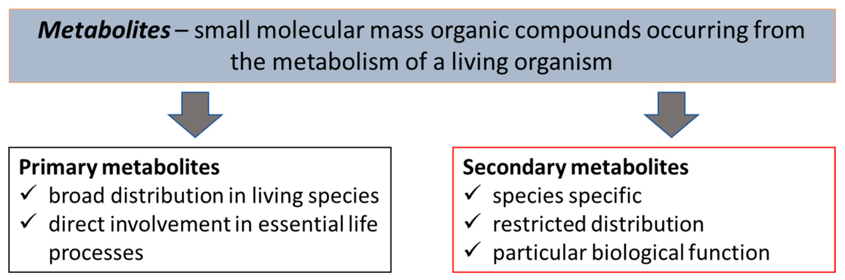
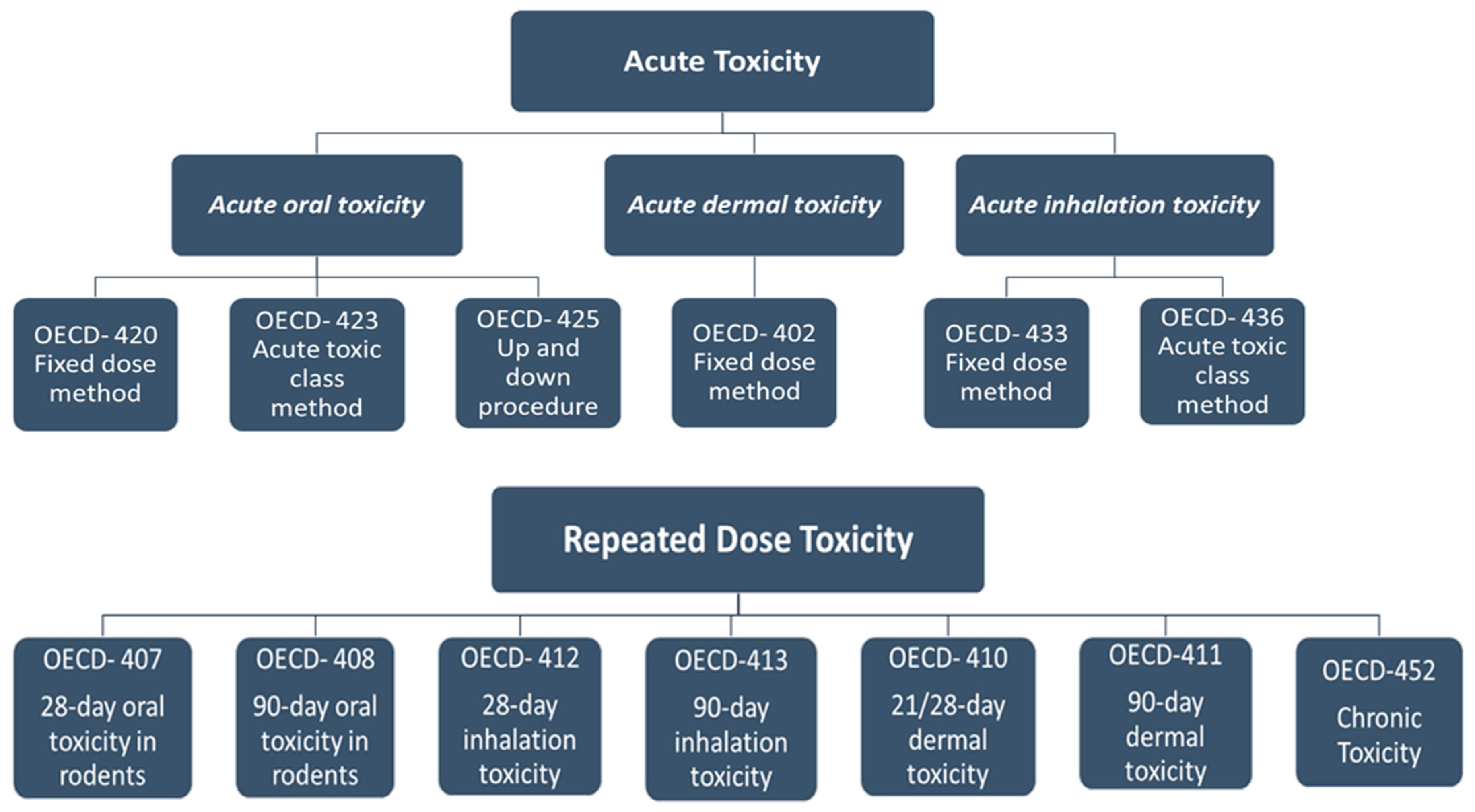
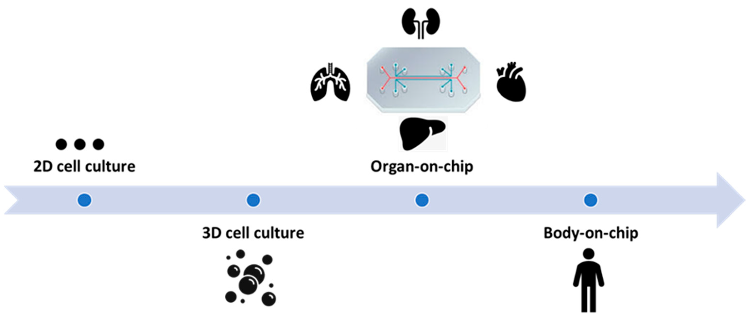
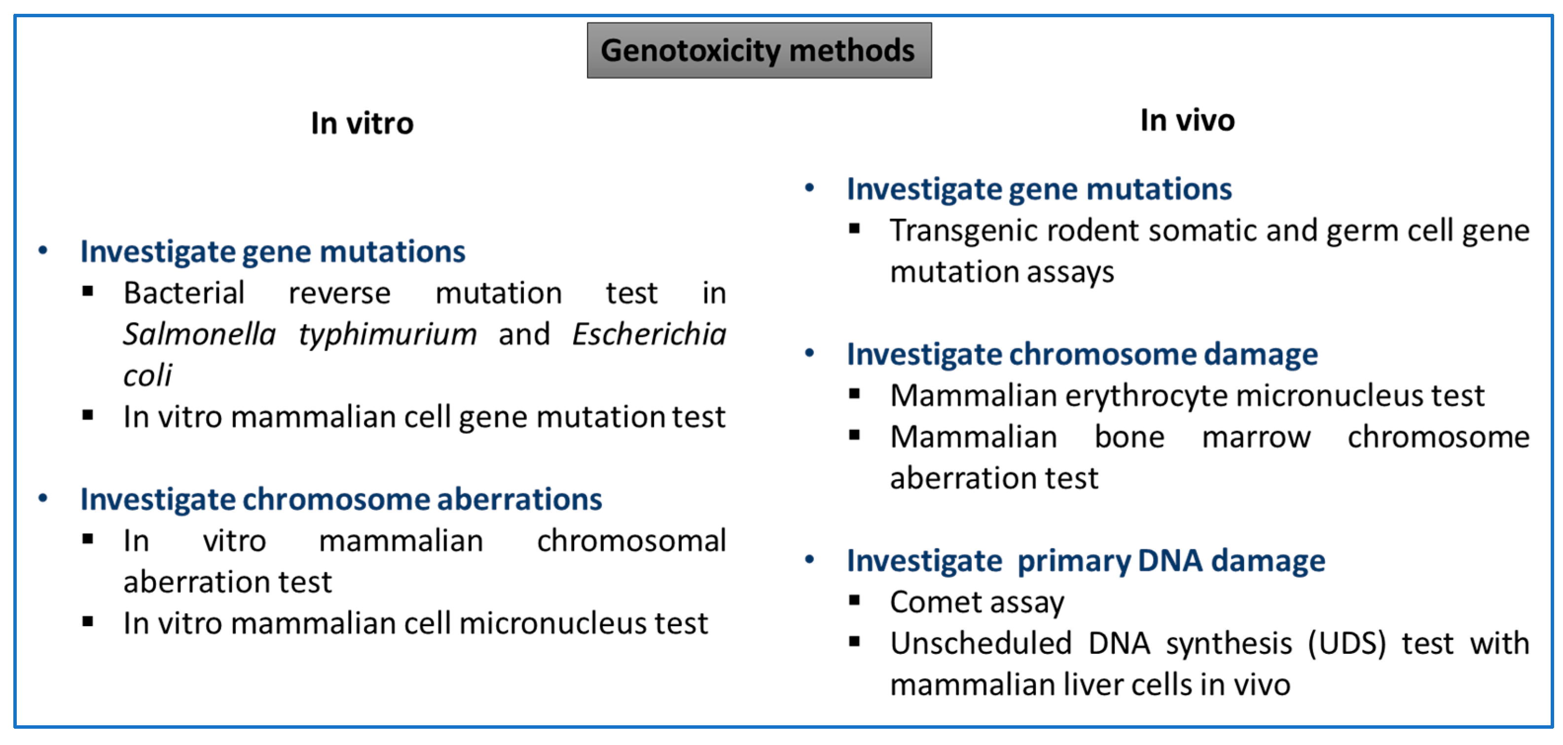
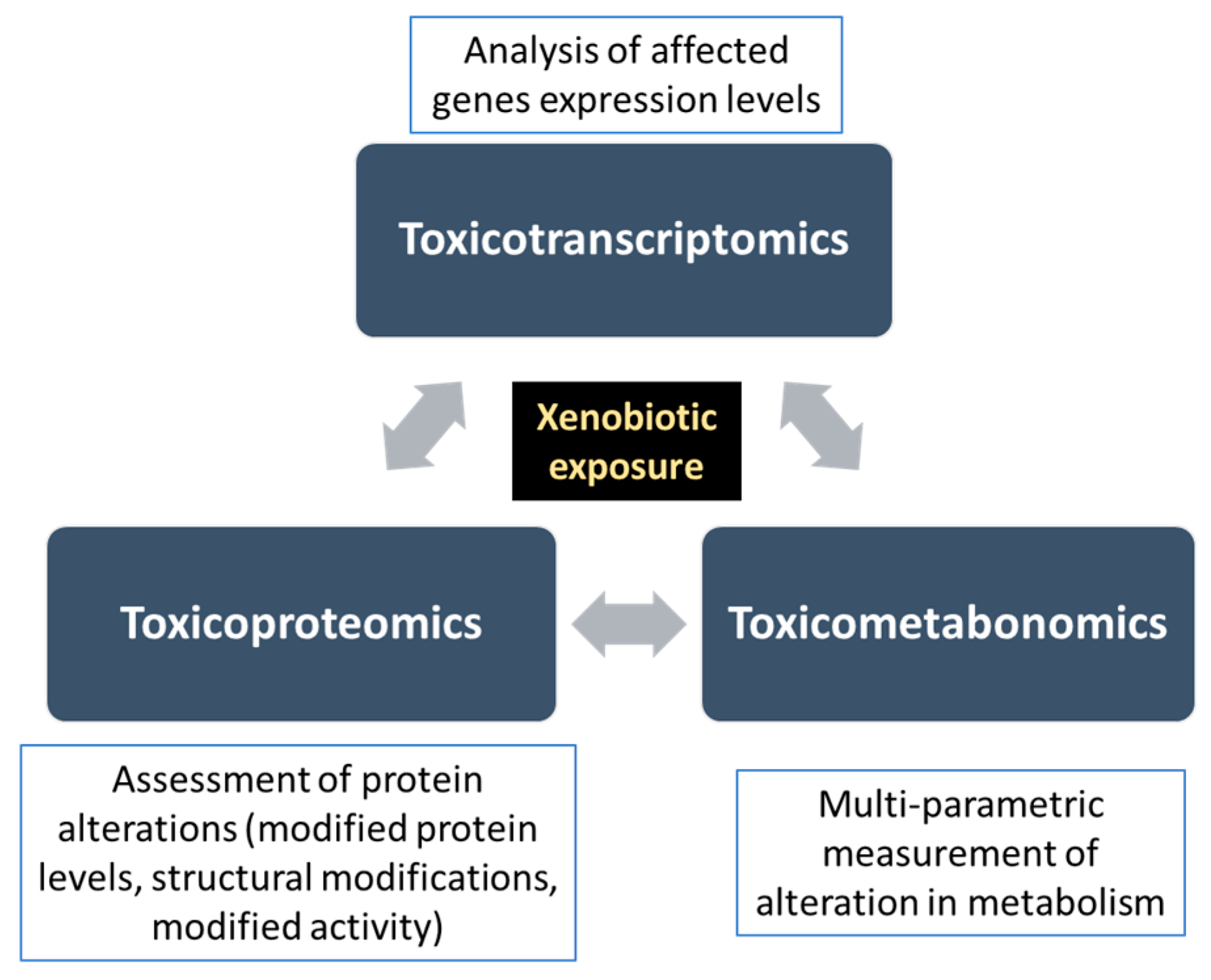
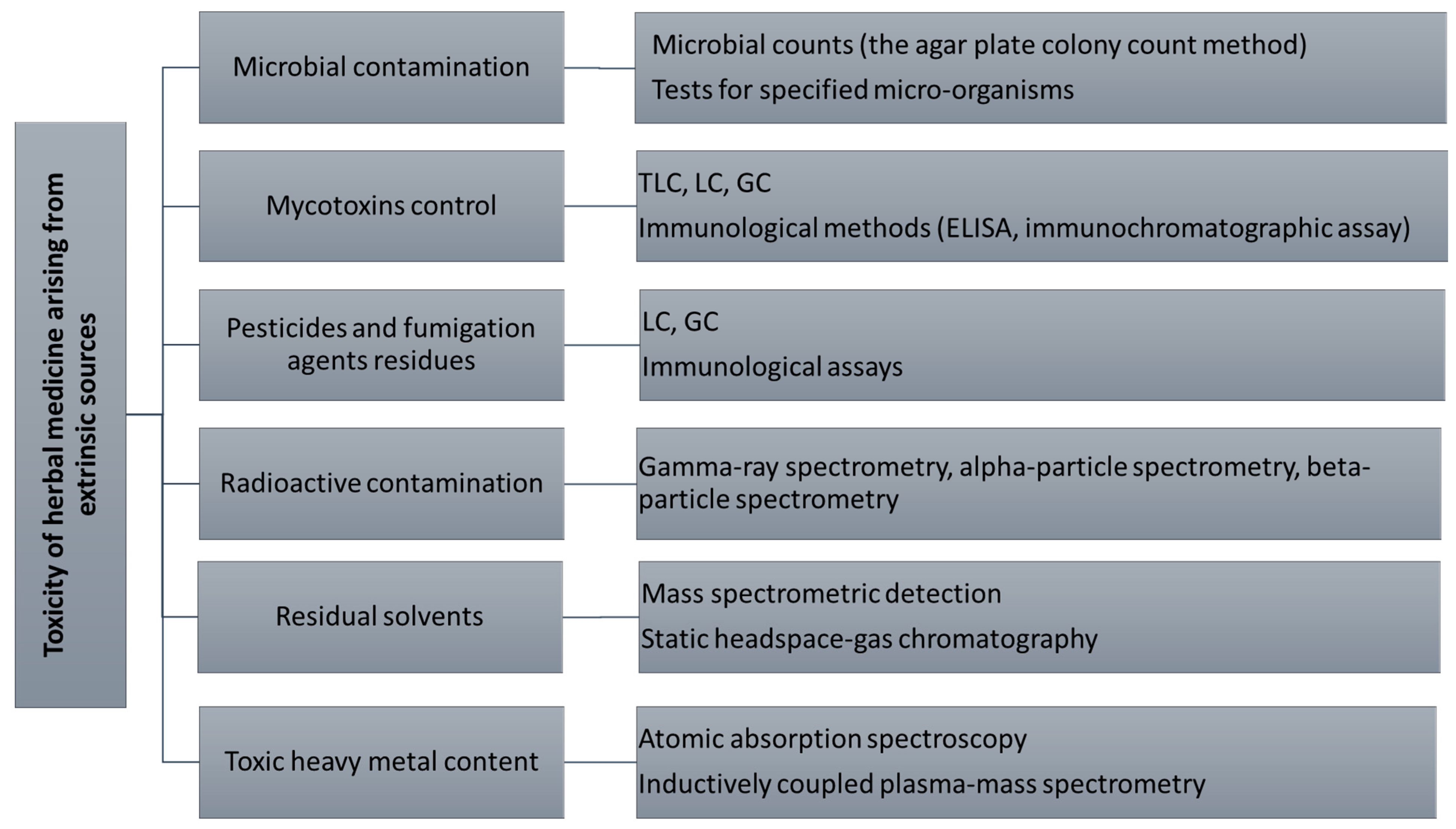
| Analyzed Material | Analytical Technique Used | Reference |
|---|---|---|
| Achillea millefolium | TLC HPLC-UV | [23] |
| Anemone spp. | HPLC-HR-ESI-MS/MS | [24] |
| Allium spp. | NMR HPLC–MS | [25] |
| Alisma plantago-aquatica and A. orientale | UHPLC/Orbitrap-MS | [26] |
| Aphanamixis polystachya | HPTLC | [27] |
| Asparagus officinalis | RP-HPLC-ESI-QTOF/MS/MS | [28] |
| Avicennia marina | HPTLC | [29] |
| Bacopa monnieri | HPLC-PAD HPLC-UV | [30] [31] [32] |
| Calendula officinalis | TLC HPLC-UV | [23] |
| Centella asiatica | HPLC-PAD | [30] |
| Cinnamomum spp. | UHPLC-HRMS | [33] |
| Crocus sativus | 1H-NMR | [34] |
| Curcuma spp. | TLC 1H-NMR | [35,36,37] |
| Dendrobium spp. | 2D NMR | [38] |
| Duboisia spp. | 1H-NMR HPLC-MS | [39] |
| Ephedra spp. | HPTLC GC–MS | [40] |
| 1H-NMR | [41] | |
| Ficus spp. | FTIR, GC-MS NMR | [42] |
| Fritillaria spp. | MALDI-MS | [43] |
| Gastrodia eleta | HPLC-DAD-MS | [44] |
| Ginkgo biloba | LC–MS | [45] |
| Glossostemon bruguier | GC-MS NMR | [46] |
| Glycyrrhiza spp. | HPTLC NMR | [47] |
| GC-MS LC-MS 1H NMR | [48] | |
| HPLC-HRMS | [49] | |
| HPTLC HPLC | [50] | |
| LC-UV LC-MS-MS | [51] | |
| Hedera helix subspp. | HPTLC- image analysis HPTLC-MS | [52] |
| HPLC CE | [53] | |
| Hibiscus sabdariffa | TLC HPLC-UV | [23] |
| Iris domestica, I. tectorum and I. dichotoma | HPLC–DAD-CL ESI-QTOF-MS/MS | [54] |
| Lonicera spp. | RRLC-QTOF-MS | [55] |
| Matricaria recutita | TLC HPLC-UV | [23] |
| Mentha spp. | 1H-NMR | [56] |
| Ocimum sanctum | TLC FTIR 1H-NMR | [57] |
| Panax ginseng | 1H NMR | [58] |
| Phyllanthus spp. | FTIR NMR | [59] |
| Uncaria spp. | UHPLC/QTOF-MS | [21] |
| Zingiber officinale | HPTLC (digitally-enhanced method) | [60] |
| Analyzed Material | Analyzed Genomic Region | Conclusion of the Study | Reference |
|---|---|---|---|
| Angelica spp. | matK, ITS, ITS2, rbcL, psbA-trnH |
| [105] |
| Apiaceae family | rbcL, matK, ITS, ITS2, psbA-trnH |
| [106] |
| Aquilaria spp. | matK, rbcL, rpoB, rpoC1, psbA-trnH, trnL-trnF, ITS, ITS2 |
| [107] |
| Astragalus spp. | ITS |
| [108] |
| Boerhavia diffusa | ITS, ITS1, ITS2, psbA-trnH |
| [109] |
| Canadian Arctic Flora (490 vascular plant species—half of the Canadian Arctic flora and 93% of the flora of the Canadian Arctic Archipelago) | rbcL, matK |
| [110] |
| Caryophyllales | ITS2, rbcL, matK |
| [111] |
| Codiaeum variegatum | rbcL, matK |
| [112] |
| Crocus spp. | rbcL, matK, trnH-psbA, ITS |
| [113] |
| Curcuma spp. | ITS, rbcL, matK |
| [114] |
| Dalbergia spp. | ITS, matK, rbcL |
| [115] |
| Dalbergia odorifera and D. tonkinensis | trnH-psbA |
| [116] |
| Fabaceae family | ITS2 |
| [71] |
| Gentiana spp. | rbcL, matK, ITS, 5S rRNA, trnH-psbA, trnL-F, rpl36-rps8 |
| [117] |
| Glehnia littoralis | ITS2 |
| [118] |
| Hibiscus spp. | matK, rbcL, trnH-psbA, ITS2 |
| [119] |
| Hippophae spp. | ITS2 |
| [102] |
| Hypericum spp. (H. perforatum and H. androsaemum) | ITS1, matK |
| [75] |
| Lonicera spp. | rbcl, matK, psbA-trnH, ITS2, ITS, trnL, trnL-F |
| [120] |
| Matricaria recutita | matK, rbcL, psbA-trnH, ITS, ITS2 |
| [89] |
| Momordica spp. | ITS, matK |
| [121] |
| Nepenthes spp. | ITS, rbcl, matK |
| [122] |
| Panax spp. | ITS2, matK, psbA-trnH |
| [123] |
| atpF-atpH, rbcL, rpoB, and rpoC1, matK, psbK-I, psbM-trnD, rps16 and nad1, psbA-trnH, ITS |
| [124] | |
| Paris polyphylla | ITS2 |
| [97] |
| Phyllanthus spp. | rbcL, trnL |
| [95] |
| psbA-trnH |
| [125] | |
| Piper nigrum | trnL, psbA-trnH |
| [126] |
| psbA-trnH, rbcL, rpoC1 |
| [127] | |
| Rhodiola spp. | rbcL, matK, trnH-psbA, trnL-F, ITS |
| [128] |
| Rhododendron spp. | rbcL, matK, psbA-trnH, ITS2 |
| [129] |
| Ruta graveolens | rpoB, rpoC1, ITS |
| [130] |
| Schisandraceae family | ITS, trnH-psbA, matK, rbcL |
| [131] |
| Senna spp. | trnH-psbA |
| [132] |
| Sida cordifolia | rbcL, matK, psbA-trnH, ITS2 |
| [133] |
| Stephania spp. | ITS, ITS2, psbA-trnH, matK, rbcL, trnL-F |
| [134] |
| Trillium govanianum | ITS, matK, trnH-psbA, rbcL |
| [2] |
| Verbenaceae family | psbA-trnH, rbcL, matK, ITS2, ITS |
| [135] |
| Genotoxic Endpoints | Test | Reference |
|---|---|---|
| Gene mutations | Ames (Bacterial Reverse Mutation Test) | [176,181,182,183,184,185,186,187] |
| Mouse lymphoma assay | [185,187,188] | |
| In Vitro Mammalian Cell Gene Mutation Tests | [189] | |
| DNA damage | Comet assay | [181,188,190,191,192,193,194,195,196] |
| Chromosomal damage (structural and numerical aberrations) | Chromosome aberration test | [184,197,198,199,200] |
| Micronucleus assay | [181,185,187,188,192,197,198,201,202] | |
| Allium cepa test | [182,203,204,205,206] |
| Analyzed Sample | Analytical Methods | Aim of the Study | Reference |
|---|---|---|---|
| Aconitum spp. alkaloids | GC-TOF/MS | Identification of altered metabolites associated with induced metabolic disorders | [257] |
| Aconitum carmichaelii | LC-QTOF-MS | Identification of serum biomarkers of toxicity | [258] |
| Aurantio-obtusin (anthraquinone from Cassia seed) | UPLC-QTOF-MS | Identification of urinary biomarkers associated with hepatotoxicity | [259] |
| Coptidis rhizoma | 1H NMR GC-MS | Investigation of gastro-intestinal toxicity via the alteration of gut microbiota | [260] |
| Dioscorea bulbifera rhizome | 1H NMR GC-MS | Assessment of hepatotoxicity and identification of altered metabolites in blood and tissue samples | [261] |
| GC-MS | Identification of altered metabolites from rat plasma, urine, and feces associated with hepatotoxicity | [262] | |
| Dioscorea bulifera root | 1H NMR | Prediction of hepatotoxicity based on urinary metabolic perturbations | [263] |
| Mesaconitine (diterpenoid alkaloid from Aconitum spp.) | UPLC-Q-Exactive Orbitrap-MS | Investigation of the hepatotoxicity mechanism | [264] |
| Pharbitis nil seeds | UPLC-MS | Investigation of changes of urine metabolite biomarkers associated with nephrotoxicity | [265] |
| Pinellia ternata | UPLC Q-TOF-MS | Investigation of general toxicity (liver, kidney, heart) via profiling serum metabolic alterations | [266] |
| Polygonum multiflorum | UHPLC-MS | Identification of hepatotoxicity biomarkers and pathways | [267] |
| HPLC-MS | [268] | ||
| UPLC-QTOF-MS | [269] | ||
| Realgar (Traditional Chinese medicine) | 1H NMR | Assessment of hepatotoxicity using plasma and urine samples metabolic profiling | [270] |
| 1H NMR | Assessment of hepato- and nephrotoxicity via metabonomic analysis of urine, serum, and liver tissue | [271] | |
| Sophora alopecuroides | 1H NMR | Evaluation of metabonomic profiling alterations associated with hepatic and renal toxicity | [272] |
| Tripterygium wilfordii | GC-MS | Identification of urine biomarkers to elucidate Tripterygium wilfordii poisoning | [273] |
| Xanthii fructus | UPLC-QTOF-MS | Identification of urinary toxicity biomarkers associated with lipid metabolism alteration | [274] |
| Analyzed Sample | Purification and Analysis Methods | Reference |
|---|---|---|
| Brazilian medicinal plants | SFE HRGC–ECD/FPD | [304] |
| Cassia angustifolia | QuEChERS GC–MS | [305] |
| China herbal tea | UPLC-MS/MS coupled with vortex-assisted DLLME | [306] |
| Commercial market medicinal plant samples in India | QuEChERS GC–MS | [307] |
| Codonopsis Radix (in China) | QuEChERS-GC–MS/MS QuEChERS-LC–MS/MS | [308] |
| Fritillaria spp. | QuEChERS LC-MS/MS | [309] |
| Herba epimedii | SPE GC-MS | [310] |
| Herbal Decoctions in Traditional Korean Medicine | GC/ECD GC/MSD | [311] |
| Mentha piperita | QuEChERS GC/ECD/NPD | [312] |
| Polish herbal raw materials | QuEChERS HPLC–MS/MS | [313] |
| Polish products of plant origin | QuEChERS HPLC–MS/MS | [314] |
| A selection of medicine and food herbs | SPLE GC-MS/MS | [315] |
| A selection of medicinal plants—Matricaria chamomilla, Tilia spp., Pulmonaria spp., Melissa spp., Mentha piperita, Thymus vulgaris. | MSPD LSE GC | [303] |
| Traditional Chinese herbal medicines | GC/MS/MS | [302] |
| QuEChERS GC-MS with PTV-LVI-SV | [316] | |
| HPLC-MS/MS GC/MS/MS | [300] | |
| QuEChERS UHPLC–MS/MS | [301] | |
| SPE GC-MS | [317] | |
| GPC GC-MS | [318] |
| Analyzed Material | Analysis Method | Conclusion of the Study | Reference |
|---|---|---|---|
| Emblica officinalis Terminalia chebula Terminalia belerica Withania somnifera | AAS |
| [327] |
| A selection of herbal medicines | ICP-MS |
| [328] |
| A selection of herbal products in Nigeria | AAS |
| [336] |
| A selection of medicinal herbs from Ghana | AAS |
| [331] |
| Petroselinum crispum Ocimum basilicum Salvia officinalis Origanum vulgare Mentha spicata Thymus vulgaris Matricaria chamomilla | AAS |
| [332] |
| A selection of medicinal herbs from India | AAS |
| [333] |
| A selection of health supplement products | ICP-OES |
| [334] |
| A selection of medicinal plants | AAS |
| [335] |
Disclaimer/Publisher’s Note: The statements, opinions and data contained in all publications are solely those of the individual author(s) and contributor(s) and not of MDPI and/or the editor(s). MDPI and/or the editor(s) disclaim responsibility for any injury to people or property resulting from any ideas, methods, instructions or products referred to in the content. |
© 2022 by the authors. Licensee MDPI, Basel, Switzerland. This article is an open access article distributed under the terms and conditions of the Creative Commons Attribution (CC BY) license (https://creativecommons.org/licenses/by/4.0/).
Share and Cite
Jităreanu, A.; Trifan, A.; Vieriu, M.; Caba, I.-C.; Mârțu, I.; Agoroaei, L. Current Trends in Toxicity Assessment of Herbal Medicines: A Narrative Review. Processes 2023, 11, 83. https://doi.org/10.3390/pr11010083
Jităreanu A, Trifan A, Vieriu M, Caba I-C, Mârțu I, Agoroaei L. Current Trends in Toxicity Assessment of Herbal Medicines: A Narrative Review. Processes. 2023; 11(1):83. https://doi.org/10.3390/pr11010083
Chicago/Turabian StyleJităreanu, Alexandra, Adriana Trifan, Mădălina Vieriu, Ioana-Cezara Caba, Ioana Mârțu, and Luminița Agoroaei. 2023. "Current Trends in Toxicity Assessment of Herbal Medicines: A Narrative Review" Processes 11, no. 1: 83. https://doi.org/10.3390/pr11010083