


Microbial Natural Products with Wound-Healing Properties
Abstract
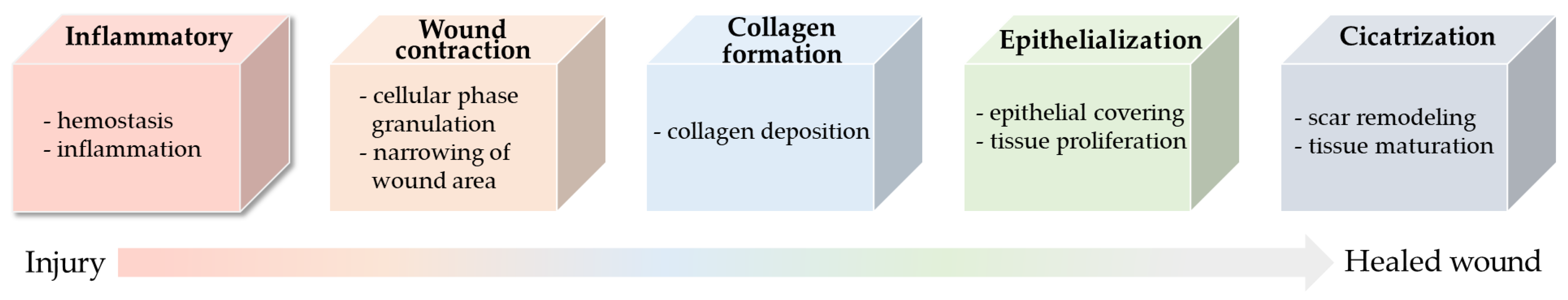
:1. Introduction
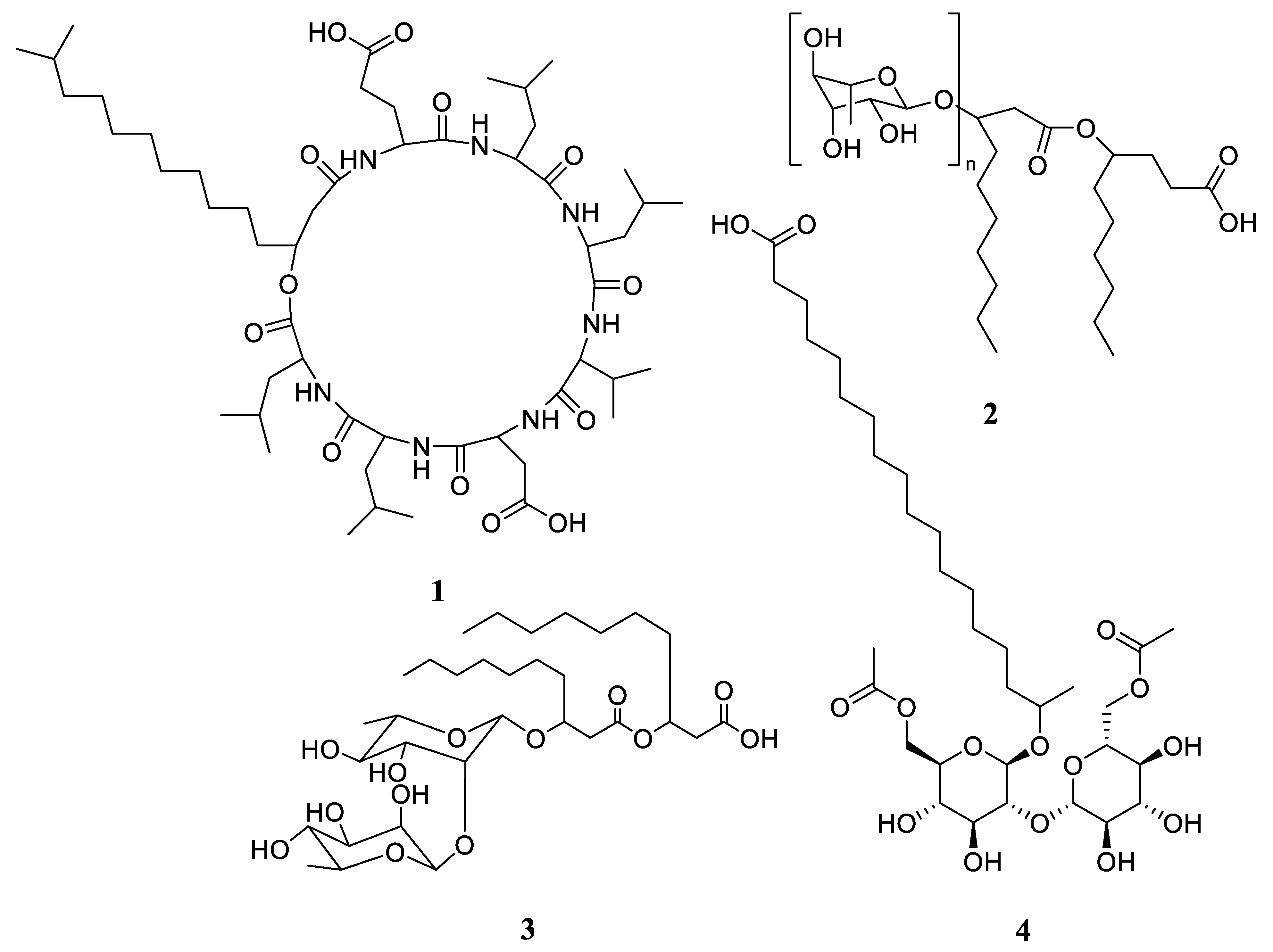
2. Biosurfactant
3. Flavonoids
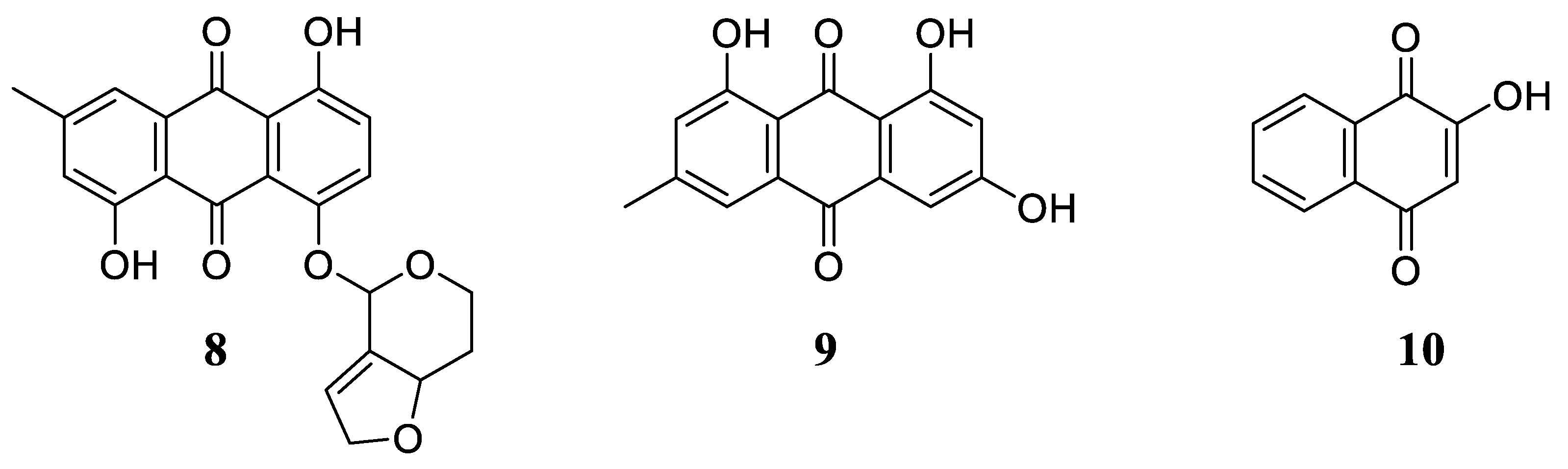
4. Quinones
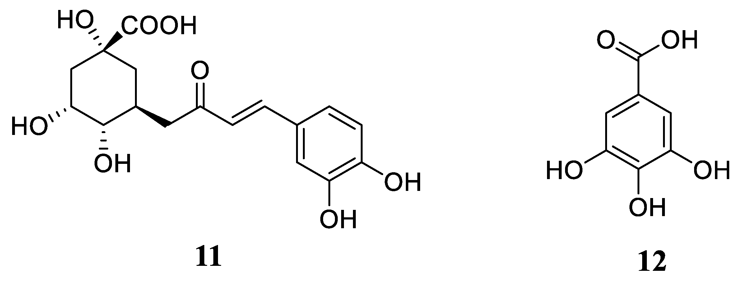
5. Phenolic Acids
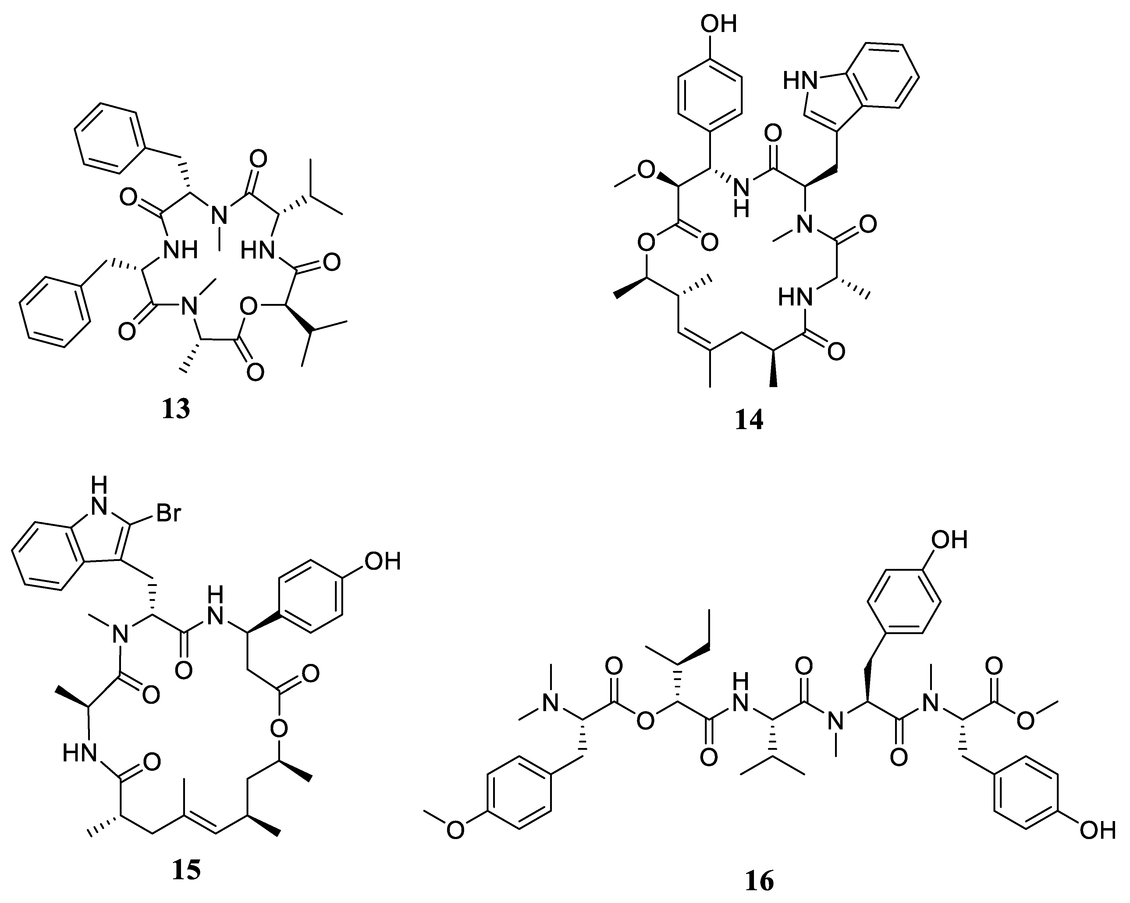
6. Peptides
7. Triterpenoids
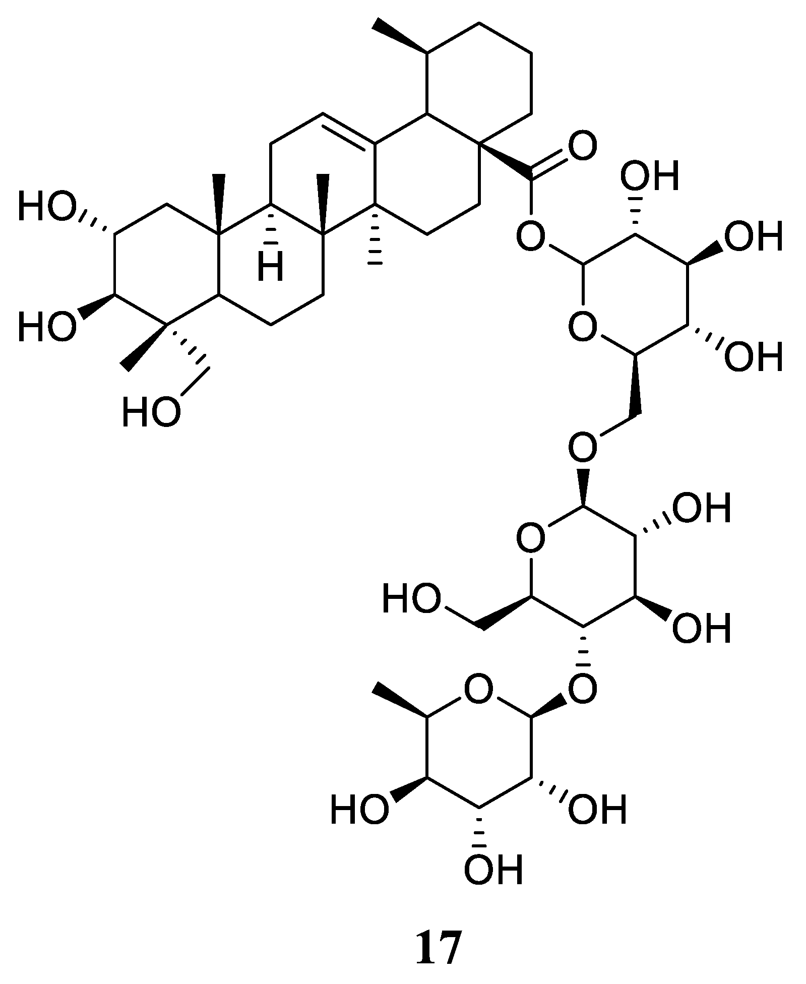
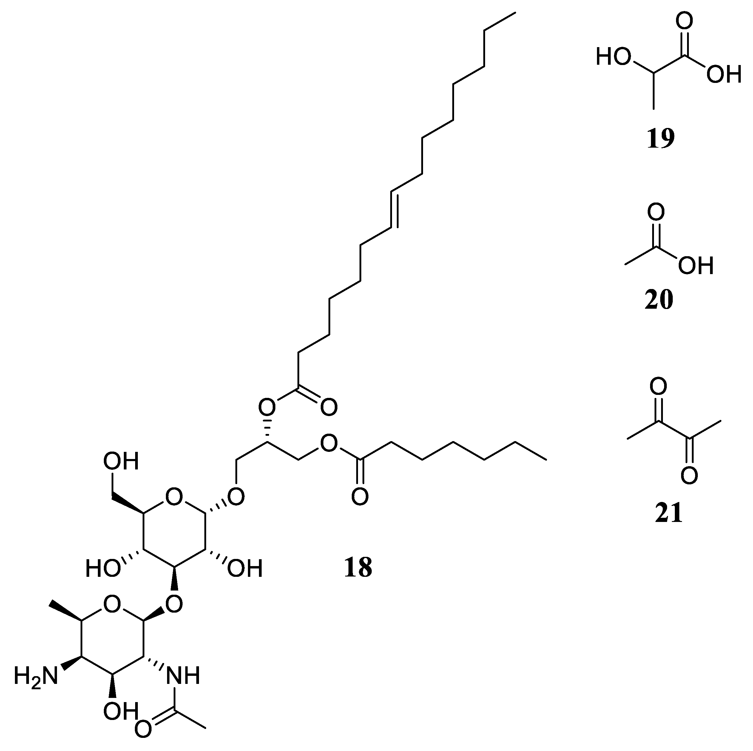
8. Others
9. Conclusions
Author Contributions
Funding
Acknowledgments
Conflicts of Interest
References
- Barreto, R.S.; Albuquerque-Júnior, R.L.; Araújo, A.A.; Almeida, J.R.; Santos, M.R.; Barreto, A.S.; DeSantana, J.M.; Siqueira-Lima, P.S.; Quintans, J.S.; Quintans-Júnior, L.J. A systematic review of the wound-healing effects of monoterpenes and iridoid derivatives. Molecules 2014, 19, 846–862. [Google Scholar] [CrossRef] [PubMed]
- Guo, S.; Dipietro, L.A. Factors affecting wound healing. J. Dent. Res. 2010, 89, 219–229. [Google Scholar] [CrossRef] [PubMed]
- Ibrahim, N.I.; Wong, S.K.; Mohamed, I.N.; Mohamed, N.; Chin, K.-Y.; Ima-Nirwana, S.; Shuid, A.N. Wound Healing Properties of Selected Natural Products. Int J. Environ. Res. Public Health 2018, 15, 2360–2382. [Google Scholar] [CrossRef] [PubMed] [Green Version]
- Kumar, B.; Vijayakumar, M.; Govindarajan, R.; Pushpangadan, P. Ethnopharmacological approaches to wound healing—Exploring medicinal plants of India. J. Ethnopharmacol. 2007, 114, 103–113. [Google Scholar] [CrossRef] [PubMed]
- Trinh, X.-T.; Long, N.-V.; Van Anh, L.T.; Nga, P.T.; Giang, N.N.; Chien, P.N.; Nam, S.-Y.; Heo, C.-Y. A Comprehensive Review of Natural Compounds for Wound Healing: Targeting Bioactivity Perspective. Int. J. Mol. Sci. 2022, 23, 9573. [Google Scholar] [CrossRef] [PubMed]
- Maheswary, T.; Nurul, A.A.; Fauzi, M.B. The Insights of Microbes’ Roles in Wound Healing: A Comprehensive Review. Pharmaceutics 2021, 13, 981–1002. [Google Scholar] [CrossRef] [PubMed]
- Thakur, R.; Jain, N.; Pathak, R.; Sandhu, S.S. Practices in wound healing studies of plants. Evid. Based Complement. Altern. Med. 2011, 2011, 438056. [Google Scholar] [CrossRef] [Green Version]
- Biswas, T.K.; Mukherjee, B. Plant medicines of Indian origin for wound healing activity: A review. Int. J. Low Extrem. Wounds 2003, 2, 25–39. [Google Scholar] [CrossRef]
- Laurano, R.; Boffito, M.; Ciardelli, G.; Chiono, V. Wound dressing products: A translational investigation from the bench to the market. Eng. Regen. 2022, 3, 182–200. [Google Scholar] [CrossRef]
- Fenical, W.; Jensen, P. Developing a new resource for drug discovery: Marine actinomycete bacteria. Nat. Chem. Biol. 2006, 2, 666–673. [Google Scholar] [CrossRef]
- Abdel-Razek, A.S.; El-Naggar, M.E.; Allam, A.; Morsy, O.M.; Othman, S.I. Microbial Natural Products in Drug Discovery. Processes 2020, 8, 470. [Google Scholar] [CrossRef] [Green Version]
- Kong, D.-X.; Jiang, Y.-Y.; Zhang, H.-Y. Marine natural products as sources of novel scaffolds: Achievement and concern. Drug Discov. Today 2010, 15, 884–886. [Google Scholar] [CrossRef] [PubMed]
- Araujo, J.; Monteiro, J.; Silva, D.; Alencar, A.; Silva, K.; Coelho, L.; Pacheco, W.; Silva, D.; Silva, M.; Silva, L.; et al. Surface-Active Compounds Produced by Microorganisms: Promising Molecules for the Development of Antimicrobial, Anti-Inflammatory, and Healing Agents. Antibiotics 2022, 11, 1106–1123. [Google Scholar] [CrossRef] [PubMed]
- Uzoigwe, C.; Burgess, J.G.; Ennis, C.J.; Rahman, P.K.S.M. Bioemulsifiers Are Not Biosurfactants and Require Different Screening Approaches. Front. Microbiol. 2015, 6, 245–250. [Google Scholar] [CrossRef] [Green Version]
- Sharma, D.; Misba, L.; Khan, A.U. Antibiotics versus Biofilm: An Emerging Battleground in Microbial Communities. Antimicrob. Resist. Infect. Control. 2019, 8, 76–86. [Google Scholar] [CrossRef] [PubMed] [Green Version]
- Kumari, A.; Kumari, S.; Prasad, G.S.; Pinnaka, A.K. Production of Sophorolipid Biosurfactant by Insect Derived Novel Yeast Metschnikowia churdharensis f.a., sp. nov., and Its Antifungal Activity against Plant and Human Pathogens. Front. Microbiol. 2021, 12, 678668–678680. [Google Scholar] [CrossRef] [PubMed]
- Ceresa, C.; Tessarolo, F.; Maniglio, D.; Tambone, E.; Carmagnola, I.; Fedeli, E.; Caola, I.; Nollo, G.; Chiono, V.; Allegrone, G.; et al. Medical-Grade Silicone Coated with Rhamnolipid R89 Is Effective against Staphylococcus spp. Biofilms. Molecules 2019, 24, 3843–3860. [Google Scholar] [CrossRef] [Green Version]
- Giugliano, R.; Buonocore, C.; Zannella, C.; Chianese, A.; Palma Esposito, F.; Tedesco, P.; De Filippis, A.; Galdiero, M.; Franci, G.; de Pascale, D. Antiviral Activity of the Rhamnolipids Mixture from the Antarctic Bacterium Pseudomonas gessardii M15 against Herpes simplex Viruses and Coronaviruses. Pharmaceutics 2021, 13, 2121–2138. [Google Scholar] [CrossRef]
- Kim, H.-Y.; Jung, H.; Kim, H.-M.; Jeong, H.-J. Surfactin Exerts an Anti-Cancer Effect through Inducing Allergic Reactions in Melanoma Skin Cancer. Int. Immunopharmacol. 2021, 99, 107934–107942. [Google Scholar] [CrossRef]
- Ceresa, C.; Fracchia, L.; Fedeli, E.; Porta, C.; Banat, I.M. Recent Advances in Biomedical, Therapeutic and Pharmaceutical Applications of Microbial Surfactants. Pharmaceutics 2021, 13, 466–491. [Google Scholar] [CrossRef]
- Rahim, K.; Saleha, S.; Zhu, X.; Huo, L.; Basit, A.; Franco, O.L. Bacterial Contribution in Chronicity of Wounds. Microb. Ecol. 2017, 73, 710–721. [Google Scholar] [CrossRef] [PubMed]
- Pastar, I.; Nusbaum, A.G.; Gil, J.; Patel, S.B.; Chen, J.; Valdes, J.; Stojadinovic, O.; Plano, L.R.; Tomic-Canic, M.; Davis, S.C. Interactions of Methicillin Resistant Staphylococcus aureus USA300 and Pseudomonas aeruginosa in Polymicrobial Wound Infection. PLoS ONE 2013, 8, e56846–e56857. [Google Scholar] [CrossRef] [PubMed]
- Banu, A.; Noorul Hassan, M.M.; Rajkumar, J.; Srinivasa, S. Spectrum of Bacteria Associated with Diabetic Foot Ulcer and Biofilm Formation: A Prospective Study. Australas. Med. J. 2015, 8, 280–285. [Google Scholar] [CrossRef] [PubMed]
- Ohadi, M.; Forootanfar, H.; Rahimi, H.R.; Jafari, E.; Shakibaie, M.; Eslaminejad, T.; Dehghannoudeh, G. Antioxidant Potential and Wound Healing Activity of Biosurfactant Produced by Acinetobacter junii B6. Curr. Pharm. Biotechnol. 2017, 18, 900–908. [Google Scholar] [CrossRef]
- Zouari, R.; Moalla-Rekik, D.; Sahnoun, Z.; Rebai, T.; Ellouze-Chaabouni, S.; Ghribi-Aydi, D. Evaluation of Dermal Wound Healing and in vitro Antioxidant Efficiency of Bacillus Subtilis SPB1 Biosurfactant. Biomed. Pharmacother. 2016, 84, 878–891. [Google Scholar] [CrossRef]
- Yan, L.; Liu, G.; Zhao, B.; Pang, B.; Wu, W.; Ai, C.; Zhao, X.; Wang, X.; Jiang, C.; Shao, D.; et al. Novel Biomedical Functions of Surfactin A from Bacillus subtilis in Wound Healing Promotion and Scar Inhibition. J. Agric. Food Chem. 2020, 68, 6987–6997. [Google Scholar] [CrossRef]
- Gupta, S.; Raghuwanshi, N.; Varshney, R.; Banat, I.M.; Srivastava, A.K.; Pruthi, P.A.; Pruthi, V. Accelerated in Vivo Wound Healing Evaluation of Microbial Glycolipid Containing Ointment as a Transdermal Substitute. Biomed. Pharmacother. 2017, 94, 1186–1196. [Google Scholar] [CrossRef] [PubMed]
- Shen, C.; Jiang, L.; Shao, H.; You, C.; Zhang, G.; Ding, S.; Bian, T.; Han, C.; Meng, Q. Targeted Killing of Myofibroblasts by Biosurfactant di-Rhamnolipid Suggests a Therapy against Scar Formation. Sci. Rep. 2016, 6, 37553–37563. [Google Scholar] [CrossRef] [Green Version]
- Maeng, Y.; Kim, K.T.; Zhou, X.; Jin, L.; Kim, K.S.; Kim, Y.H.; Lee, S.; Park, J.H.; Chen, X.; Kong, M.; et al. A Novel Microbial Technique for Producing High-Quality Sophorolipids from Horse Oil Suitable for Cosmetic Applications. Microb. Biotechnol. 2018, 11, 917–929. [Google Scholar] [CrossRef]
- Kwak, M.-J.; Park, M.-Y.; Kim, J.; Lee, H.; Whang, K.-Y. Curative Effects of Sophorolipid on Physical Wounds: In Vitro and in Vivo Studies. Vet. Med. Sci. 2021, 7, 1400–1408. [Google Scholar] [CrossRef]
- Carvalho, M.T.B.; Araújo-Filho, H.G.; Barreto, A.S.; Quintans-Júnior, L.J.; Quintans, J.S.S.; Barreto, R.S.S. Wound healing properties of flavonoids: A systematic review highlighting the mechanisms of action. Phytomedicine 2021, 90, 153636–153651. [Google Scholar] [CrossRef] [PubMed]
- Menezes, P.D.P.; Frank, L.A.; Lima, B.S.S.; de Carvalho, Y.M.B.G.; Serafini, M.R.; Quintans-Júnior, L.J.; Pohlmann, A.R.; Guterres, S.S.; Araújo, A.A.d.S. Hesperetin-loaded lipid-core nanocapsules in polyamide: A new textile formulation for topical drug delivery. Int. J. Nanomed. 2017, 12, 2069–2079. [Google Scholar] [CrossRef] [PubMed] [Green Version]
- Sak, K. Intake of individual flavonoids and risk of carcinogenesis: Overview of epidemiological evidence. Nutr. Cancer 2017, 69, 1119–1150. [Google Scholar] [CrossRef]
- Pushpavalli, G.; Kalaiarasi, P.; Veeramani, C.; Pugalendi, K.V. Effect of chrysin on hepatoprotective and antioxidant status in D-galactosamine-induced hepatitis in rats. Eur. J. Pharmacol. 2010, 631, 36–41. [Google Scholar] [CrossRef] [PubMed]
- Bagher, Z.; Ehterami, A.; Safdel, M.H.; Khastar, H.; Semiari, H.; Asefnejad, A.; Davachi, S.M.; Mirzaii, M.; Salehi, M. Wound healing with alginate/chitosan hydrogel containing hesperidin in rat model. J. Drug Deliv. Sci. Technol. 2020, 55, 101379. [Google Scholar] [CrossRef]
- Feng, Y.; Sanders, A.J.; Morgan, L.D.; Harding, K.G.; Jiang, W.G. Potential roles of suppressor of cytokine signaling in wound healing. Regen. Med. 2016, 11, 193–209. [Google Scholar] [CrossRef]
- Chen, R.F.; Chang, C.H.; Wang, C.T.; Yang, M.Y.; Wang, C.J.; Kuo, Y.R. Modulation of vascular endothelial growth factor and mitogen-activated protein kinase-related pathway involved in extracorporeal shockwave therapy accelerate diabetic wound healing. Wound Repair Regen. 2019, 27, 69–79. [Google Scholar] [CrossRef] [Green Version]
- Alexandru, V.; Balan, M.; Gaspar, A.; Craciunescu, O.; Moldovan, L. Studies on the antioxidant activity, phenol and flavonoid contents of some selected Romanian medicinal plants used for wound healing. Rom. Biotechnol. Lett. 2007, 12, 3467–3472. [Google Scholar]
- Haddadi, G.; Abbaszadeh, A.; Mosleh-Shirazi, M.A.; Okhovat, M.A.; Salajeghe, A.; Ghorbani, Z. Evaluation of the effect of hesperidin on vascular endothelial growth factor gene expression in rat skin animal models following cobalt-60 gamma irradiation. J. Cancer Res. Ther. 2018, 14, S1098–S1104. [Google Scholar]
- Kandhare, A.D.; Alam, J.; Patil, M.V.K.; Sinha, A.; Bodhankar, S.L. Wound healing potential of naringin ointment formulation via regulating the expression of inflammatory, apoptotic and growth mediators in experimental rats. Pharm. Biol. 2016, 54, 419–432. [Google Scholar] [CrossRef]
- Singh, W.R.; Devil, H.S.; Kumawat, S.; Sadam, A.; Appukuttan, A.; Patel, M.R.; Lingaraju, M.C.; Singh, T.U.; Kumar, D. Angiogenic and MMPs modulatory effects of icariin improved cutaneous wound healing in rats. Eur. J. Pharmacol. 2019, 858, 172466. [Google Scholar] [CrossRef] [PubMed]
- Parasuraman, S.; Perumal, P. Wound Healing Agents from Natural Sources. In Wound Healing Research, 1st ed.; Kumar, P., Kothari, V., Eds.; Springer: Singapore, 2021; pp. 95–148. [Google Scholar]
- Bonesi, M.; Loizzo, M.R.; Menichini, F.; Tundis, R. Flavonoids in treating psoriasis. In Immunity and Inflammation in Health and Disease, 1st ed.; Chatterjee, S., Jungraithmayr, W., Bagchi, D., Eds.; Academic Press: Cambridge, MA, USA, 2018; pp. 281–294. [Google Scholar]
- Manconi, M.; Manca, M.L.; Caddeo, C.; Cencetti, C.; di Meo, C.; Zoratto, N.; Nacher, A.; Fadda, A.M.; Matricardi, P. Preparation of gellan-cholesterol nanohydrogels embedding baicalin and evaluation of their wound healing activity. Eur. J. Pharm. Biopharm. 2018, 127, 244–249. [Google Scholar] [CrossRef] [PubMed]
- Huang, W.Y.; Cai, Y.Z.; Hyde, K.D.; Corke, H.; Sun, M. Biodiversity od endophytic fungi associated with 29 traditional Chinese medicinal plants. Fungal Divers. 2008, 33, 61–75. [Google Scholar]
- Ren, J.; Lu, Y.; Qian, Y.; Chen, B.; Wu, T.; Ji, G. Recent progress regarding kaempferol for the treatment of various diseases. Exp. Med. 2019, 18, 2759–2776. [Google Scholar] [CrossRef] [PubMed] [Green Version]
- Özay, Y.; Güzel, S.; Yumrutaş, Ö.; Pehlivanoğlu, B.; Erdoğdu, İ.H.; Yildirim, Z.; Türk, B.A.; Darcan, S. Wound healing effect of kaempferol in diabetic and nondiabetic rats. J. Surg. Res. 2019, 233, 284–296. [Google Scholar] [CrossRef] [PubMed]
- Ambiga, S.; Narayanan, R.; Gowri, D.; Sukumar, D.; Madhavan, S. Evaluation of wound healing activity of flavonoids from Ipomoea carnea Jacq. Anc. Sci. Life 2007, 26, 45–51. [Google Scholar] [PubMed]
- Calderón-Montaño, J.M.; Burgos-Morón, E.; Pérez-Guerrero, C.; López-Lázaro, M. A review on the dietary flavonoid kaempferol. Mini Rev. Med. Chem. 2011, 11, 298–344. [Google Scholar] [CrossRef]
- Liu, R.H. Health-promoting components of fruits and vegetables in the diet. Adv. Nutr. 2013, 4, 384S–392S. [Google Scholar] [CrossRef] [Green Version]
- Pratibha, C.; Gajbhiye, S.; Roy, S.; Dudhale, R.; Chowdhary, A. Determination of Kaempferol in extracts of Fusarium chlamydosporum, an endophyticfungi of Tylophora indica (Asclepeadaceae) and its anti-microbial activity. Int. J. Pharm. Biol. Sci. 2014, 9, 51–55. [Google Scholar]
- David, A.V.A.; Arulmoli, R.; Parasuraman, S. Overviews of biological importance of quercetin: A bioactive flavonoid. Pharmacogn. Rev. 2016, 10, 84–89. [Google Scholar]
- Gomathi, K.; Gopinath, D.; Ahmed, M.R.; Jayakumar, R. Quercetin incorporated collagen matrices for dermal wound healing processes in rat. Biomaterials 2003, 24, 2767–2772. [Google Scholar] [CrossRef] [PubMed]
- Jangde, R.; Srivastava, S.; Singh, M.R.; Singh, D. In vitro and in vivo characterization of quercetin loaded multiphase hydrogel for wound healing application. Int. J. Biol. Macromol. 2018, 115, 1211–1217. [Google Scholar] [CrossRef] [PubMed]
- Abdel-Raouf, N.; Ibraheem, I.B.M.; Abdel-Tawab, S.; Naser, Y.A.G. Antimicrobial and antihyperlipidemic activities of isolated quercetin from anabaena aequalis(1). J. Phycol. 2011, 47, 955–962. [Google Scholar] [CrossRef] [PubMed]
- Devi, S.S.; Mehendale, H.M. Quinone. In Encyclopedia of Toxicology, 3rd ed.; Wxler, P., Ed.; Elsevier Inc.: Amsterdam, The Netherlands, 2014; pp. 26–28. [Google Scholar]
- Desam, N.R.; Al-Rajab, A.J. Herbal biomolecules: Anticancer agents. In Herbal Biomolecules in Health-Care Application, 1st ed.; Mandal, S.C., Dhara, A.K., Nayak, A.K., Eds.; Elsevier: Amsterdam, The Netherlands, 2022; pp. 435–474. [Google Scholar]
- Chatterjee, T.K.; Chakravorty, A. Wound healing properties of the new antibiotic (MT81) in mice. Indian Drugs 1993, 30, 450–452. [Google Scholar]
- Ismaiel, A.A.; Rabie, G.H.; Abd El-Aal, M.A. Antimicrobial and morphogenic effects of emodin produced by Aspergillus awamori WAIR120. Biologia 2016, 71, 464–474. [Google Scholar] [CrossRef]
- Izhaki, I. Emodin-a secondary metabolite with multiple ecological functions in higher plants. New Phytol. 2002, 155, 205–217. [Google Scholar] [CrossRef]
- Kögl, F.; Postowsky, J.J. Untersuchungen über Pilzfarbstoffe. II. Über die Farbstoffe des blutroten Hautkopfes (Dermocybe sanguinea Wulf.). Justus Liebigs Ann. Chem. 1925, 444, 1–7. [Google Scholar] [CrossRef]
- Agosti, G.; Birkinshaw, J.H.; Chaplen, P. Studies in the biochemistry of micro-organisms. 112. Anthraquinone pigments of strains of Cladosporium fulvum Cooke. Biochem. J. 1962, 85, 528–530. [Google Scholar] [CrossRef] [Green Version]
- Shibata, S.; Shoji, J.; Ohta, A.; Watanable, M. Metabolic products of fungi. XI. Some observations on the occurrence of skyrin and rugulosin in mold metabolites with reference to structural relationships between penicilliopsin and skyrin. Chem. Pharm. Bull. 1957, 5, 380–383. [Google Scholar] [CrossRef] [Green Version]
- Shibata, S.; Udagawa, S. Metabolic products of fungi. XIX. Isolation of rugulosin from Penicillium brunneum Udagawa. Chem. Pharm. Bull. 1963, 11, 402–403. [Google Scholar] [CrossRef] [Green Version]
- Natori, S.; Sato, F.; Udagawa, S. Anthraquinone metabolites of Talaromyces avellanens (Thom et Turreson), C.R. Benjamin and Preussia multispora (Saito et Minoura) Cain. Chem. Pharm. Bull. 1965, 13, 385–389. [Google Scholar] [CrossRef] [PubMed] [Green Version]
- Ghosh, A.C.; Manmade, A.; Demain, A.L. Toxins from Penicillium islandicum Sopp. [in food, feed]. In Mycotoxins in Human and Animal Health; Rodricks, J.V., Hesseltine, C.W., Mehlman, M.A., Eds.; Pathotox: Chicago, IL, USA, 1977; pp. 625–638. [Google Scholar]
- Yamazaki, M.; Maebayashi, Y.; Miyaki, K. The isolation of secalonic acid A from Aspergillus ochraceus cultured on rice. Chem. Pharm. Bull. 1971, 19, 199–201. [Google Scholar] [CrossRef] [PubMed] [Green Version]
- Wells, J.M.; Cole, R.J.; Kirksey, J.W. Emodin, a toxic metabolite of Aspergillus wentii isolated from weevil-damaged chestnuts. Appl. Microbiol. 1975, 30, 26–28. [Google Scholar] [CrossRef]
- Anke, H.; Kolthoum, I.; Laatsch, H. Metabolic products of microorganisms. 192. The anthraquinones of the Aspergillus glaucus group. II. Biological activity. Arch. Microbiol. 1980, 126, 231–236. [Google Scholar] [CrossRef] [PubMed]
- Dong, X.; Fu, J.; Yin, X.; Cao, S.; Li, X.; Lin, L.; Huyiligeqi, N.J. Emodin: A review of its pharmacology, toxicity and pharmacokinetics. Phytother. Res. 2016, 30, 1207–1218. [Google Scholar] [CrossRef] [PubMed]
- Tang, T.; Yin, L.; Yang, J.; Shan, G. Emodin, an anthraquinone derivative from Rheum officinale Baill, enhances cutaneous wound healing in rats. Eur. J. Pharm. 2007, 567, 177–185. [Google Scholar] [CrossRef]
- Sarang, H.; Rajani, P.; Vasanthakumari, M.M.; Kumara, P.M.; Siva, R.; Ravikanth, G.; Shaanker, R.U. An endophytic fungus, Gibberella moniliformis from Lawsonia inermis L. produces lawsone, an orange-red pigment. Antonie Van Leeuwenhoek 2017, 110, 853–862. [Google Scholar] [CrossRef]
- López López, L.I.; Nery Flores, S.D.; Silva Belmares, S.Y.; Sáenz, G.A. Naphthoquinones: Biological properties and synthesis of lawsone and derivatives-a structured review. Vitae 2014, 21, 248–258. [Google Scholar] [CrossRef]
- Mandawgade, S.D.; Patil, K.S. Wound healing potential of some active principles of Lawsonia alba Lam. leaves. Indian J. Pharm. Sci. 2003, 65, 390–394. [Google Scholar]
- Bascha, J.; Murthy, B.R.; Likhitha, P.R.; Ganesh, Y.; Bai, B.G.; Rani, R.J.; Prakash, P.; Shanmugham, V.; Kirthi, A. In vitro and in vivo assessment of lawsone microsphere loaded chitosan scaffolds. Int. J. Phytopharm. 2016, 6, 74–84. [Google Scholar]
- Guimaraes, I.; Baptista-Silva, S.; Pintado, M.; Oliveira, A.L. Poplyphenols: A promising Avenue in Therapeutic Solutions for Wound Care. Appl. Sci. 2021, 11, 1230. [Google Scholar] [CrossRef]
- Działo, M.A.; Mierziak, J.; Korzun, U.; Preisner, M.; Szopa, J.; Kulma, A. The potential of plant phenolics in prevention and therapy of skin disorders. Int. J. Mol. Sci. 2016, 17, 160. [Google Scholar] [CrossRef] [PubMed] [Green Version]
- Ghuman, S.; Ncube, B.; Finnie, J.; McGaw, L.; Njoya, E.M.; Coopoosamy, R.; Van Staden, J. Antioxidant, anti-inflammatory and wound healing properties of medicinal plant extracts used to treat wounds and dermatological disorders. S. Afr. J. Bot. 2019, 126, 232–240. [Google Scholar] [CrossRef]
- Krausz, A.E.; Adler, B.L.; Cabral, V.; Navati, M.; Doerner, J.; Charafeddine, R.A.; Chandra, D.; Liang, H.; Gunther, L.; Clendaniel, A.; et al. Curcumin-encapsulated nanoparticles as innovative antimicrobial and wound healing agent. Nanomed. Nanotechnol. Biol. Med. 2015, 11, 195–206. [Google Scholar] [CrossRef] [Green Version]
- Moghadam, S.E.; Ebrahimi, S.N.; Salehi, P.; Moridi Farimani, M.; Hamburger, M.; Jabbarzadeh, E. Wound healing potential of chlorogenic acid and myricetin-3-O-β-rhamnoside isolated from Parrotia persica. Molecules 2017, 22, 1501. [Google Scholar] [CrossRef] [PubMed] [Green Version]
- Chen, W.C.; Liou, S.S.; Tzeng, T.F.; Lee, S.L.; Liu, I.M. Effect of topical application of chlorogenic acid on excision wound healing in rats. Planta Med. 2013, 79, 616–621. [Google Scholar] [CrossRef] [PubMed]
- Yang, D.J.; Moh, S.H.; Son, D.H.; You, S.; Kinyua, A.W.; Ko, C.M.; Song, M.; Yeo, J.; Choi, Y.H.; Kim, K.W. Gallic acid promotes wound healing in normal and hyperglucidic conditions. Molecules 2016, 21, 899. [Google Scholar] [CrossRef] [Green Version]
- Karatas, O.; Gevrek, F. Gallic acid liposome and powder gels improved wound healing in wistar rats. Ann. Med. Res. 2019, 26, 2720–2727. [Google Scholar] [CrossRef]
- Song, Y.; Wu, C.; Zhang, X.; Bian, W.; Liu, N.; Yin, S.; Yang, M.; Luo, M.; Tang, J.; Yang, X. A short peptide potentially promotes the healing of skin wound. Biosci. Rep. 2019, 39, BSR20181734. [Google Scholar] [CrossRef]
- Larouche, J.; Sheoran, S.; Maruyama, K.; Martino, M.M. Immune regulation of skin wound healing: Mechanisms and novel therapeutic targets. Adv. Wound Care 2018, 7, 209–231. [Google Scholar] [CrossRef]
- Li, X.; Wang, Y.; Zou, Z.; Yang, M.; Wu, C.; Su, Y.; Tang, J.; Yang, X. OM-LV20, a novel peptide from odorous frog skin, accelerates wound healing in vitro and in vivo. Chem. Biol. Drug Des. 2018, 91, 126–136. [Google Scholar] [CrossRef] [PubMed]
- Hardwicke, J.; Schmaljohann, D.; Boyce, D.; Thomas, D. Epidermal growth factor therapy and wound healing–past, present and future perspectives. Surgeon 2008, 6, 172–177. [Google Scholar] [CrossRef] [PubMed]
- Kim, S.; Lee, C.W.; Park, S.-Y.; Asolkar, R.N.; Kim, H.; Kim, G.J.; Oh, S.J.; Kim, Y.; Lee, E.-Y.; Oh, D.-C.; et al. Acremonamide, a Cyclic Pentadepsipeptide with Wound-Healing Properties Isolated from a Marine-Derived Fungus of the Genus Acremonium. J. Nat. Prod. 2021, 84, 2249–2255. [Google Scholar] [CrossRef] [PubMed]
- Wang, X.; Gong, X.; Li, P.; Lai, D.; Zhou, L. Structural Diversity and Biological Activities of Cyclic Depsipeptides from Fungi. Molecules 2018, 23, 169–218. [Google Scholar] [CrossRef] [PubMed] [Green Version]
- Ratnayake, R.; Fremlin, L.J.; Lacey, E.; Gill, J.H.; Capon, R.J. Acremolides A−D, Lipodepsipeptides from an Australian Marine-Derived Fungus, Acremonium sp. J. Nat. Prod. 2008, 71, 403–408. [Google Scholar] [CrossRef]
- Sun, P.; Maloney, K.N.; Nam, S.-J.; Haste, N.M.; Raju, R.; Aalbersberg, W.; Jensen, P.R.; Nizet, V.; Hensler, M.E.; Fenical, W. Fijimycins A–C, three antibacterial etamycin-class depsipeptides from a marine-derived Streptomyces sp. Bioorg. Med. Chem. 2011, 19, 6557–6562. [Google Scholar] [CrossRef] [Green Version]
- Amagata, T.; Morinaka, B.I.; Amagata, A.; Tenney, K.; Valeriote, F.A.; Lobkovsky, E.; Clardy, J.; Crews, P. A Chemical Study of Cyclic Depsipeptides Produced by a Sponge-Derived Fungus. J. Nat. Prod. 2006, 69, 1560–1565. [Google Scholar] [CrossRef] [Green Version]
- Kunze, B.; Jansen, R.; Sasse, F.; Hofle, G.; Reichenbach, H. Chondramides A~D, New Antifungal and Cytostatic Depsipeptides from Chondromyces crocatus (Myxobacteria). J. Antibiot. Res. 1995, 48, 1262–1266. [Google Scholar] [CrossRef]
- Abreu-Blanco, M.T.; Watts, J.J.; Verboon, J.M.; Parkhurst, S.M. Cytoskeleton Response in Wound Repair. Cell Mol. Life Sci. 2012, 69, 2469–2483. [Google Scholar] [CrossRef] [Green Version]
- Ma, C.I.; Diraviyam, K.; Maier, M.E.; Sept, D.; Sibley, L.D. Synthetic chondramide A analogues stabilize filamentous actin and block invasion by Toxoplasma gondii. J. Nat. Prod. 2013, 76, 1565–1572. [Google Scholar] [CrossRef]
- Tannert, R.; Milroy, L.G.; Ellinger, B.; Hu, T.S.; Arndt, H.D.; Waldmann, H. Synthesis and structure-activity correlation of natural-product inspired cyclodepsipeptides stabilizing F-actin. J. Am. Chem. Soc. 2010, 132, 3063–3077. [Google Scholar] [CrossRef]
- Cai, W.; Salvador-Reyes, L.A.; Zhang, W.; Chen, Q.-Y.; Matthew, S.; Ratnayake, R.; Seo, S.J.; Dolles, S.; Gibson, D.J.; Paul, V.J.; et al. Apratyramide, a Marine-Derived Peptidic Stimulator of VEGF-A and Other Growth Factors with Potential Application in Wound Healing. ACS Chem. Biol. 2018, 13, 91–99. [Google Scholar] [CrossRef] [PubMed]
- Ghiulai, R.; Roşca, O.J.; Antal, D.S.; Mioc, M.; Mioc, A.; Racoviceanu, R.; Macaşoi, I.; Olariu, T.; Dehelean, C.; Creţu, O.M.; et al. Tetracyclic and Pentacyclic Triterpenes with High Therapeutic Efficiency in Wound Healing Approaches. Molecules 2020, 25, 5557. [Google Scholar] [CrossRef] [PubMed]
- Isah, M.B.; Tajuddeen, N.; Umar, M.I.; AlHafiz, Z.A.; Mohammed, A.; Ibrahim, M.A. Terpenoids as emerging therapeutic agents: Cellular targets and mechanisms of action against protozoan parasites. In Studies in Natural Products Chemistry, 1st ed.; Atta-ur-Rahman, Ed.; Elsevier: Amsterdam, The Netherlands, 2018; Volume 59, pp. 227–250. [Google Scholar]
- Furtado, N.A.J.C.; Pirson, L.; Edelberg, H.; Miranda, L.M.; Loira-Pastoriza, C.; Préat, V.; Larondelle, Y.; Andre, C.M. Pentacyclic triterpene bioavailability: An overview of in vitro and in vivo studies. Molecules 2017, 22, 400. [Google Scholar] [CrossRef] [Green Version]
- Paduch, R.; Kandefer-Szerszen, M. Antitumor and antiviral activity of pentacyclic triterpenes. Mini-Rev. Org. Chem. 2014, 11, 262–268. [Google Scholar] [CrossRef]
- Chudzik, M.; Korzonek-Szlacheta, I.; Król, W. Triterpenes as potentially cytotoxic compounds. Molecules 2015, 20, 1610–1625. [Google Scholar] [CrossRef] [Green Version]
- Battineni, J.K.; Koneti, P.K.; Bakshi, V.; Boggula, N. Triterpenoids: A review. Int. J. Pharm. Pharm. Sci. 2018, 3, 91–96. [Google Scholar]
- Hill, R.A.; Connolly, J.D. Triterpenoids. Nat. Prod. Rep. 2017, 34, 90–122. [Google Scholar] [CrossRef] [Green Version]
- Agra, L.C.; Ferro, J.N.S.; Barbosa, F.T.; Barreto, E. Triterpenes with healing activity: A systematic review. J. Dermatol. Treat. 2015, 26, 465–470. [Google Scholar] [CrossRef]
- Sh Ahmed, A.; Taher, M.; Mandal, U.K.; Jaffri, J.M.; Susanti, D.; Mahmood, S.; Zakaira, Z.A. Pharmacological properties of Centella asiatica hydrogel in accelerating wound healing in rabbits. BMC Complement. Altern. Med. 2019, 19, 213. [Google Scholar] [CrossRef] [Green Version]
- Gupta, S.; Bhatt, P.; Chaturvedi, P. Determination and quantification of asiaticoside in endophytic fungus from Centella asiatica (L.) Urban. World J. Microbiol. Biotechnol. 2018, 34, 111. [Google Scholar] [CrossRef] [PubMed]
- Ozdemir, O.; Ozkan, K.; Hatipoglu, F.; Uyaroglu, A.; Arican, M. Effect of asiaticoside, collagenase, and alpha-chymotrypsin on wound healing in rabbits. Wounds 2016, 28, 279–286. [Google Scholar] [PubMed]
- Mukherjee, P.K.; Bahadur, S.; Chaudhary, S.K.; Harwansh, R.K.; Nema, N.K. Validation of medicinal herbs for skin aging. In Evidence-Based Validation of Berbal Medicine, 1st ed.; Mukherjee, P.K., Ed.; Elsevier: Amsterdam, The Netherlands, 2015; pp. 119–147. [Google Scholar]
- Lew, L.C.; Liong, M.T. Bioactives from probiotics for dermal health: Functions and benefits. J. Appl. Microbiol. 2013, 114, 1241–1253. [Google Scholar] [CrossRef] [PubMed]
- Arck, P.; Handjiski, B.; Hagen, E.; Pincus, M.; Bruenahl, C.; Bienenstock, J.; Paus, R. Is there a ‘gut-brainskin axis’? Exp. Derm. 2010, 19, 401–405. [Google Scholar] [CrossRef] [PubMed]
- Iordache, F.; Iordache, C.; Chifiriuc, M.C.; Bleotu, C.; Pavel, M.; Smarandache, D.; Sasarman, E.; Laza, V.; Bucu, M.; Dracea, O.; et al. Antimicrobial and immunomodulatory activity of some probiotic fractions with potential clinical application. Arch. Zootc. 2008, 11, 41–51. [Google Scholar]
- Villéger, R.; Saad, N.; Grenier, K.; Falourd, X.; Foucat, L.; Urdaci, M.C.; Bressollier, P.; Ouk, T.S. Characterization of lipoteichoic acid structures from three probiotic Bacillus strains: Involvement of D-alanine in their biological activity. Antonie Van Leeuwenhoek 2014, 106, 693–706. [Google Scholar] [CrossRef] [Green Version]
- Kao, S.J.; Lei, H.C.; Kuo, C.T.; Chang, M.S.; Chen, B.C.; Chang, Y.C.; Chiu, W.T.; Lin, C.H. Lipoteichoic acid induces nuclear factor-κB activation and nitric oxide synthase expression via phosphatidylinositol 3-kinase, Akt, and p38 MAPK in RAW 264.7 macrophages. Immunology 2005, 115, 366–374. [Google Scholar] [CrossRef]
- Lebeer, S.; Claes, I.J.; Vanderleyden, J. Anti-inflammatory potential of probiotics: Lipoteichoic acid makes a difference. Trends Microbiol. 2012, 20, 5–10. [Google Scholar] [CrossRef]
- Sumikawa, Y.; Asada, H.; Hoshino, K.; Azukizawa, H.; Katayama, I.; Akira, S.; Itami, S. Induction of β-defensin 3 in keratinocytes stimulated by bacterial lipopeptides through toll-like receptor 2. Microbes Infect. 2006, 8, 1513–1521. [Google Scholar] [CrossRef]
- Diamond, G.; Beckloff, N.; Weinberg, A.; Kisich, K.O. The roles of antimicrobial peptides in innate host defense. Curr. Pharm. Des. 2009, 15, 2377–2392. [Google Scholar] [CrossRef]
- Wee, Y.J.; Kim, J.N.; Ryu, H.W. Biotechnological production of lactic acid and its recent applications. Food Technol. Biotechnol. 2006, 44, 163–172. [Google Scholar]
- Tang, S.C.; Yang, J.H. Dual effects of alpha-hydroxy acids on the skin. Molecules 2018, 23, 863. [Google Scholar] [CrossRef] [PubMed] [Green Version]
- Lew, L.C.; Gan, C.Y.; Liong, M.T. Dermal bioactives from Lactobacilli and Bifidobacteria. Ann. Microbiol. 2013, 63, 1047–1055. [Google Scholar] [CrossRef]
- Smith, W.P. Epidermal and dermal effects of topical lactic acid. J. Am. Acad. Dermatol. 1996, 35, 388–391. [Google Scholar] [CrossRef]
- Pasricha, A.; Bhalla, P.; Sharma, K.B. Evaluation of lactic acid as an antibacterial agent. Indian J. Dermatol. Venereol. Leprol. 1979, 45, 159–161. [Google Scholar]
- Yamamoto, Y.; Uede, K.; Yonei, N.; Kishioka, A.; Ohtani, T.; Furukawa, F. Effects of alphahydroxy acids on the human skin of Japanese subjects: The rationale for chemical peeling. J. Dermatol. 2006, 33, 16–22. [Google Scholar] [CrossRef]
- Yeo, S.K.; Liong, M.T. Effect of prebiotics on viability and growth characteristics of probiotics in soymilk. J. Sci. Food Agric. 2010, 90, 267–275. [Google Scholar] [CrossRef]
- Nagoba, B.; Wadher, B.; Kulkarni, P.; Kolhe, S. Acetic acid treatment of pseudomonal wound infections. Eur. J. Gen. Med. 2008, 5, 104–106. [Google Scholar] [CrossRef]
- Lanciotti, R.; Patrignani, F.; Bagnolini, F.; Guerzoni, M.E.; Gardini, F. Evaluation of diacetyl antimicrobial activity against Escherichia coli, Listeria monocytogenes and Staphylococcus aureus. Food Microbiol. 2003, 20, 537–543. [Google Scholar] [CrossRef]
- Miller, L.S.; Cho, J.S. Immunity against Staphylococcus aureus cutaneous infections. Nat. Rev. Immunol. 2011, 11, 505–518. [Google Scholar] [CrossRef] [Green Version]
- Doern, G.V.; Jones, R.N.; Pfaller, M.A.; Kugler, K.C.; Beach, M.L. Bacterial pathogens isolated from patients with skin and soft tissue infections: Frequency of occurrence and antimicrobial susceptibility patterns from the SENTRY Antimicrobial Surveillance Program (United States and Canada, 1997). SENTRY Study Group (North America). Diagn. Microbiol. Infect. Dis. 1999, 34, 65–72. [Google Scholar] [PubMed]
- Salama, M.; Elgadir, M.A.; Adam, A. Anti-oxidant, antimicrobial and antiinflammatory effects of selected plants on wound healing. World J. Pharm. Pharm. Sci. 2014, 3, 1–11. [Google Scholar]
- Shafie, N.M.; Shah, R.N.I.R.S.; Krishnan, P.; Haleem, N.A.; Tan, T.Y.C. Scoping Review: Evaluation of Moringa oleifera (Lam.) for Potential Wound Healing in In Vivo Studies. Molecules 2022, 27, 5541. [Google Scholar] [CrossRef] [PubMed]
- Nowak, A.; Zielonka-Brzezicka, J.; Perużyńska, M.; Klimowicz, A. Epilobium angustifolium L. as a Potential Herbal Component of Topical Products for Skin Care and Treatment-A Review. Molecules 2022, 27, 3536. [Google Scholar] [CrossRef] [PubMed]
- Ryall, C.; Duarah, S.; Chen, S.; Yu, H.; Wen, J. Advancements in Skin Delivery of Natural Bioactive Products for Wound Management: A Brief Review of Two Decades. Pharmaceutics 2022, 14, 1072. [Google Scholar] [CrossRef] [PubMed]
- Hang, S.; Chen, H.; Wu, W.; Wang, S.; Fang, Y.; Sheng, R.; Tu, Q.; Guo, R. Progress in Isoindolone Alkaloid Derivatives from Marine Microorganism: Pharmacology, Preparation, and Mechanism. Mar. Drugs. 2022, 20, 405. [Google Scholar] [CrossRef] [PubMed]
- De Sá, J.D.M.; Kumla, D.; Dethoup, T.; Kijjoa, A. Bioactive Compounds from Terrestrial and Marine-Derived Fungi of the Genus Neosartorya. Molecules 2022, 27, 2351. [Google Scholar] [CrossRef]
- Wibowo, J.T.; Ahmadi, P.; Rahmawati, S.I.; Bayu, A.; Putra, M.Y.; Kijjoa, A. Marine-Derived Indole Alkaloids and Their Biological and Pharmacological Activities. Mar. Drugs 2021, 20, 3. [Google Scholar] [CrossRef]
- Zhang, D.; Li, S.; Fan, M.; Zhao, C. The Novel Compounds with Biological Activity Derived from Soil Fungi in the Past Decade. Drug Des. Devel. Ther. 2022, 16, 3493–3555. [Google Scholar] [CrossRef]
- Chukwujekwu, J.C.; Coombes, P.H.; Mulholland, D.A.; van Staden, J. Emodin, an antibacterial anthraquinone from the roots of Cassia occidentalis. S. Afr. J. Bot. 2006, 72, 295–297. [Google Scholar] [CrossRef] [Green Version]
- Xavier, M.R.; Santos, M.M.S.; Queiroz, M.G.; de Lima Silva, M.S.; Goes, A.J.S.; De Morais, M.A., Jr. Lawsone, a 2-hydroxy-1,4-naphthoquinone from Lawsonia inermis (henna), produces mitochondrial dysfunctions and triggers mitophagy in Saccharomyces cerevisiae. Mol. Biol. Rep. 2020, 47, 1173–1185. [Google Scholar] [CrossRef] [PubMed]


Disclaimer/Publisher’s Note: The statements, opinions and data contained in all publications are solely those of the individual author(s) and contributor(s) and not of MDPI and/or the editor(s). MDPI and/or the editor(s) disclaim responsibility for any injury to people or property resulting from any ideas, methods, instructions or products referred to in the content. |
© 2022 by the authors. Licensee MDPI, Basel, Switzerland. This article is an open access article distributed under the terms and conditions of the Creative Commons Attribution (CC BY) license (https://creativecommons.org/licenses/by/4.0/).
Share and Cite
Hillman, P.F.; Lee, C.; Nam, S.-J. Microbial Natural Products with Wound-Healing Properties. Processes 2023, 11, 30. https://doi.org/10.3390/pr11010030
Hillman PF, Lee C, Nam S-J. Microbial Natural Products with Wound-Healing Properties. Processes. 2023; 11(1):30. https://doi.org/10.3390/pr11010030
Chicago/Turabian StyleHillman, Prima F., Chaeyoung Lee, and Sang-Jip Nam. 2023. "Microbial Natural Products with Wound-Healing Properties" Processes 11, no. 1: 30. https://doi.org/10.3390/pr11010030