
Mechanical Behavior of Subcutaneous and Visceral Abdominal Adipose Tissue in Patients with Obesity

 , ,
, , 
Abstract
:1. Introduction
2. Materials and Methods
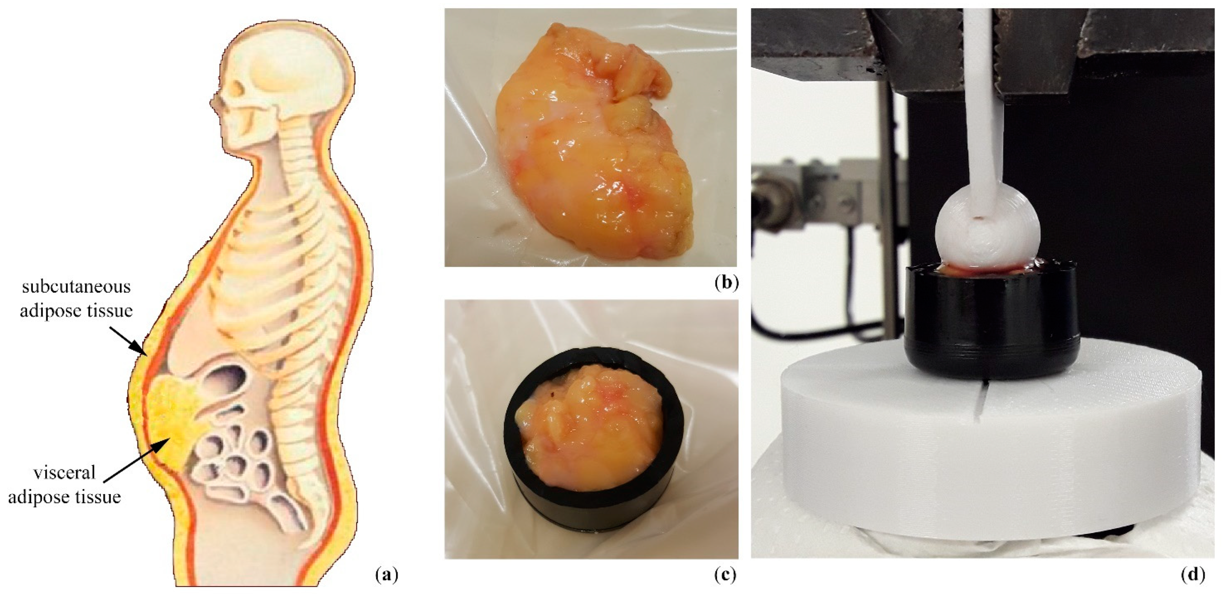
2.1. Specimen Preparation
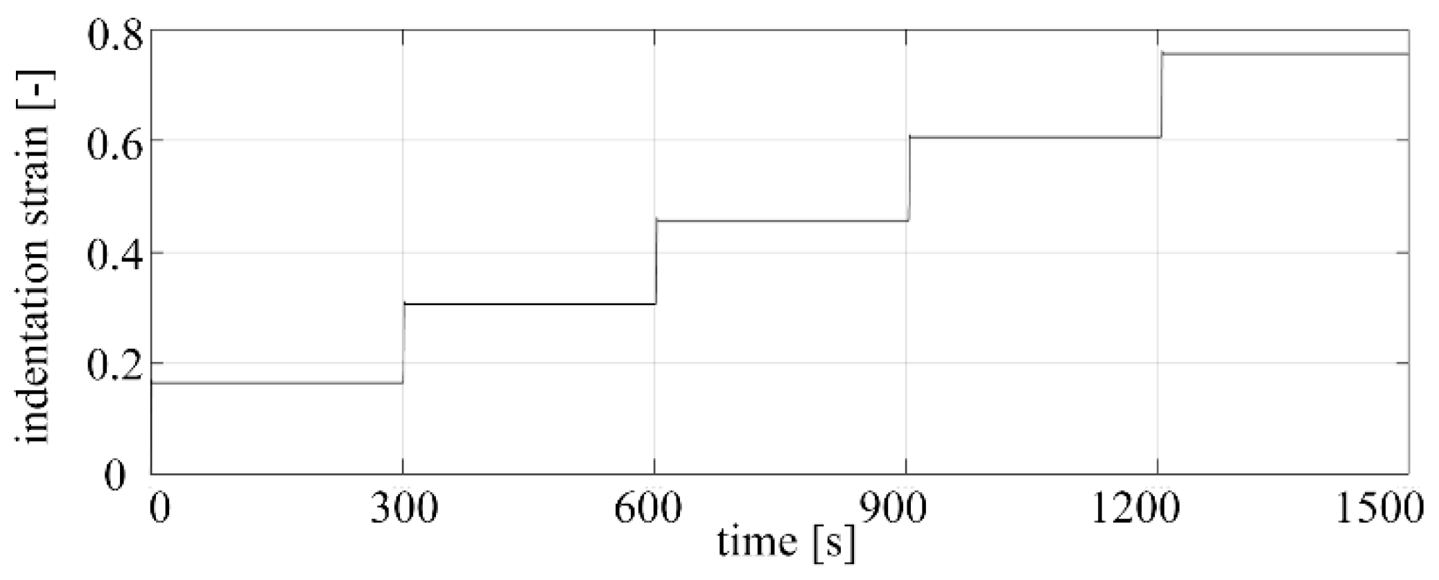
2.2. Mechanical Testing
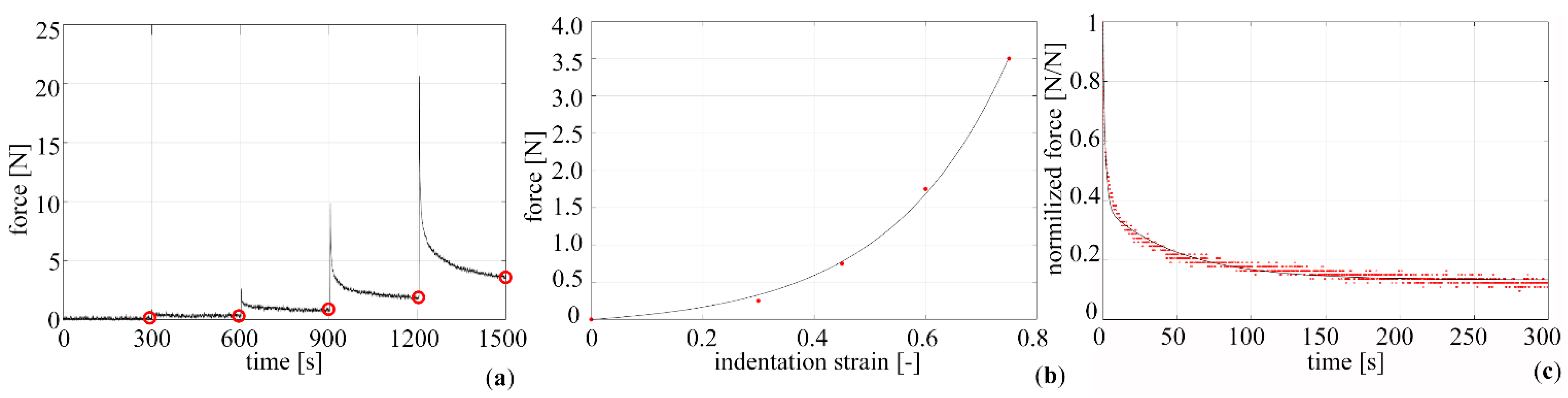
2.3. Data Elaboration
2.4. Constitutive Analysis
2.5. Statistical Analysis
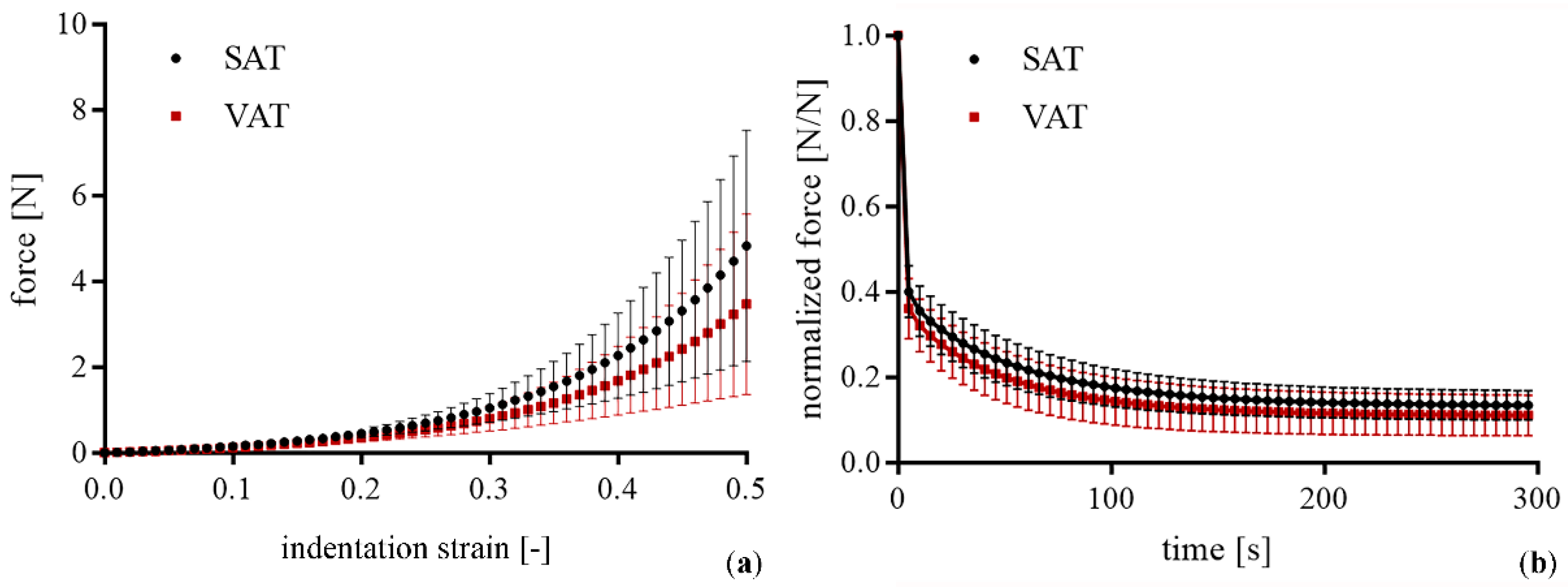
3. Results
4. Discussion
5. Conclusions
Author Contributions
Funding
Institutional Review Board Statement
Informed Consent Statement
Data Availability Statement
Conflicts of Interest
References
- Sommer, G.; Eder, M.; Kovacs, L.; Pathak, H.; Bonitz, L.; Mueller, C.; Regitnig, P.; Holzapfel, G.A. Multiaxial Mechanical Properties and Constitutive Modeling of Human Adipose Tissue: A Basis for Preoperative Simulations in Plastic and Reconstructive Surgery. Acta Biomater. 2013, 9, 9036–9048. [Google Scholar] [CrossRef] [PubMed]
- Carniel, E.L.; Toniolo, I.; Fontanella, C.G. Computational Biomechanics: In-Silico Tools for the Investigation of Surgical Procedures and Devices. Bioengineering 2020, 7, 48. [Google Scholar] [CrossRef]
- Naseri, H.; Iraeus, J.; Johansson, H. The Effect of Adipose Tissue Material Properties on the Lap Belt-Pelvis Interaction: A Global Sensitivity Analysis. J. Mech. Behav. Biomed. Mater. 2020, 107, 103739. [Google Scholar] [CrossRef]
- Gierczycka, D.; Rycman, A.; Cronin, D. Importance of Passive Muscle, Skin, and Adipose Tissue Mechanical Properties on Head and Neck Response in Rear Impacts Assessed with a Finite Element Model. Traffic. Inj. Prev. 2021, 22, 407–412. [Google Scholar] [CrossRef]
- Fox, C.S.; Massaro, J.M.; Hoffmann, U.; Pou, K.M.; Maurovich-Horvat, P.; Liu, C.-Y.; Vasan, R.S.; Murabito, J.M.; Meigs, J.B.; Cupples, L.A.; et al. Abdominal Visceral and Subcutaneous Adipose Tissue Compartments: Association with Metabolic Risk Factors in the Framingham Heart Study. Circulation 2007, 116, 39–48. [Google Scholar] [CrossRef]
- Illouz, F.; Roulier, V.; Rod, A.; Gallois, Y.; Pellé, C.-P.; Aubé, C.; Rohmer, V.; Ritz, P.; Ducluzeau, P.H. Distribution of Adipose Tissue: Quantification and Relationship with Hepatic Steatosis and Vascular Profiles of Type 2 Diabetic Patients with Metabolic Syndrome. Diabetes Metab. 2008, 34, 68–74. [Google Scholar] [CrossRef] [PubMed]
- Kim, Y.J.; Park, J.W.; Kim, J.W.; Park, C.-S.; Gonzalez, J.P.S.; Lee, S.H.; Kim, K.G.; Oh, J.H. Computerized Automated Quantification of Subcutaneous and Visceral Adipose Tissue From Computed Tomography Scans: Development and Validation Study. JMIR Med. Inform. 2016, 4, e2. [Google Scholar] [CrossRef] [PubMed]
- Sun, Z.; Gepner, B.D.; Cottler, P.S.; Lee, S.H.; Kerrigan, J.R. In Vitro Mechanical Characterization and Modeling of Subcutaneous Adipose Tissue: A Comprehensive Review. J. Biomech. Eng. 2021, 143, 070803. [Google Scholar] [CrossRef] [PubMed]
- Macchi, V.; Porzionato, A.; Sarasin, G.; Petrelli, L.; Guidolin, D.; Rossato, M.; Fontanella, C.G.; Natali, A.; De Caro, R. The Infrapatellar Adipose Body: A Histotopographic Study. Cells Tissues Organs 2016, 201, 220–231. [Google Scholar] [CrossRef]
- Natali, A.N.; Fontanella, C.G.; Carniel, E.L. A Numerical Model for Investigating the Mechanics of Calcaneal Fat Pad Region. J. Mech. Behav. Biomed. Mater. 2012, 5, 216–223. [Google Scholar] [CrossRef]
- Frigo, A.; Costantini, M.; Fontanella, C.G.; Salvador, R.; Merigliano, S.; Carniel, E.L. A Procedure for the Automatic Analysis of High-Resolution Manometry Data to Support the Clinical Diagnosis of Esophageal Motility Disorders. IEEE Trans. Biomed. Eng. 2018, 65, 1476–1485. [Google Scholar] [CrossRef] [PubMed]
- Fontanella, C.G.; Macchi, V.; Carniel, E.L.; Frigo, A.; Porzionato, A.; Picardi, E.E.E.; Favero, M.; Ruggieri, P.; de Caro, R.; Natali, A.N. Biomechanical Behavior of Hoffa’s Fat Pad in Healthy and Osteoarthritic Conditions: Histological and Mechanical Investigations. Australas Phys. Eng. Sci. Med. 2018, 41, 657–667. [Google Scholar] [CrossRef] [PubMed]
- Dragoo, J.L.; Shapiro, S.A.; Bradsell, H.; Frank, R.M. The Essential Roles of Human Adipose Tissue: Metabolic, Thermoregulatory, Cellular, and Paracrine Effects. J. Cartil. Jt. Preserv. 2021, 1, 100023. [Google Scholar] [CrossRef]
- Ahrens, W.; Pigeot, I.; Pohlabeln, H.; De Henauw, S.; Lissner, L.; Molnár, D.; Moreno, L.A.; Tornaritis, M.; Veidebaum, T.; Siani, A. Prevalence of Overweight and Obesity in European Children below the Age of 10. Int. J. Obes. 2014, 38 (Suppl. 2), S99–S107. [Google Scholar] [CrossRef]
- Bahia, L.; Coutinho, E.S.F.; Barufaldi, L.A.; Abreu, G.D.A.; Malhão, T.A.; de Souza, C.P.R.; Araujo, D.V. The Costs of Overweight and Obesity-Related Diseases in the Brazilian Public Health System: Cross-Sectional Study. BMC Public Health 2012, 12, 440. [Google Scholar]
- Anderson, M.R.; Shashaty, M.G.S. Impact of Obesity in Critical Illness. Chest 2021, 160, 2135–2145. [Google Scholar] [CrossRef]
- Ibrahim, M.M. Subcutaneous and Visceral Adipose Tissue: Structural and Functional Differences. Obes. Rev. 2010, 11, 11–18. [Google Scholar] [CrossRef] [PubMed]
- Tang, L.; Zhang, F.; Tong, N. The Association of Visceral Adipose Tissue and Subcutaneous Adipose Tissue with Metabolic Risk Factors in a Large Population of Chinese Adults. Clin. Endocrinol. 2016, 85, 46–53. [Google Scholar] [CrossRef]
- Sun, Z.; Lee, S.H.; Gepner, B.D.; Rigby, J.; Hallman, J.J.; Kerrigan, J.R. Comparison of Porcine and Human Adipose Tissue Loading Responses under Dynamic Compression and Shear: A Pilot Study. J. Mech. Behav. Biomed. Mater. 2021, 113, 104112. [Google Scholar] [CrossRef] [PubMed]
- Sun, Z.; Gepner, B.; Lee, S.; Oyen, M.; Rigby, J.; Cottler, P.S.; Hallman, J.; Kerrigan, J. Effect of Temperature and Freezing On Human Adipose Tissue Material Properties Characterized by High-Rate Indentation-Puncture Testing. J. Biomech. Eng. 2021, 144, 034502-1–034502-6. [Google Scholar] [CrossRef] [PubMed]
- Alkhouli, N.; Mansfield, J.; Green, E.; Bel, J.; Knight, B.; Liversedge, N.; Tham, J.C.; Welbourn, R.; Shore, A.C.; Kos, K.; et al. The Mechanical Properties of Human Adipose Tissues and Their Relationships to the Structure and Composition of the Extracellular Matrix. Am. J. Physiol. Endocrinol. Metab. 2013, 305, 1427–1435. [Google Scholar] [CrossRef] [Green Version]
- Comley, K.; Fleck, N.A. A Micromechanical Model for the Young’s Modulus of Adipose Tissue. Int. J. Solids Struct. 2010, 47, 2982–2990. [Google Scholar] [CrossRef]
- Juliar, B.A.; Strieder-barboza, C.; Karmakar, M.; Flesher, C.G.; Baker, N.A.; Varban, O.A.; Lumeng, C.N.; Putnam, A.J.; Robert, W.; Rourke, O. Viscoelastic Characterization of Diabetic and Non-Diabetic Human Adipose Tissue. Biorheology 2021, 57, 15–26. [Google Scholar] [CrossRef]
- Sun, Z.; Gepner, B.D.; Lee, S.H.; Rigby, J.; Cottler, P.S.; Hallman, J.J.; Kerrigan, J.R. Multidirectional Mechanical Properties and Constitutive Modeling of Human Adipose Tissue under Dynamic Loading. Acta Biomater. 2021, 129, 188–198. [Google Scholar] [CrossRef] [PubMed]
- Geerligs, M.; Peters, G.W.M.; Ackermans, P.A.J.; Oomens, C.W.J.; Baaijens, F.P.T. Linear Viscoelastic Behavior of Subcutaneous Adipose Tissue. Biorheology 2008, 45, 677–688. [Google Scholar] [CrossRef] [PubMed]
- Mihai, L.A.; Chin, L.K.; Janmey, P.A.; Goriely, A. A Comparison of Hyperelastic Constitutive Models Applicable to Brain and Fat Tissues. J. R. Soc. Interface 2015, 12, 20150486. [Google Scholar] [CrossRef]
- Unamuno, X.; Gómez-Ambrosi, J.; Becerril, S.; Álvarez-Cienfuegos, F.J.; Ramírez, B.; Rodríguez, A.; Ezquerro, S.; Valentí, V.; Moncada, R.; Mentxaka, A.; et al. Changes in Mechanical Properties of Adipose Tissue after Bariatric Surgery Driven by Extracellular Matrix Remodelling and Neovascularization Are Associated with Metabolic Improvements. Acta Biomater. 2022, 141, 264–279. [Google Scholar] [CrossRef]
- Lanzl, F.; Duddeck, F.; Willuweit, S.; Peldschus, S. Experimental Characterisation of Porcine Subcutaneous Adipose Tissue under Blunt Impact up to Irreversible Deformation. Int. J. Legal Med. 2022, 136, 897–910. [Google Scholar] [CrossRef]
- Busetto, L.; Dicker, D.; Azran, C.; Batterham, R.L.; Farpour-Lambert, N.; Fried, M.; Hjelmesæth, J.; Kinzl, J.; Leitner, D.R.; Makaronidis, J.M.; et al. Obesity Management Task Force of the European Association for the Study of Obesity Released “Practical Recommendations for the Post-Bariatric Surgery Medical Management”. Obes. Surg. 2018, 28, 2117–2121. [Google Scholar] [CrossRef]
- Kueper, M.A.; Kramer, K.M.; Kirschniak, A.; Königsrainer, A.; Pointner, R.; Granderath, F.A. Laparoscopic Sleeve Gastrectomy: Standardized Technique of a Potential Stand-Alone Bariatric Procedure in Morbidly Obese Patients. World J. Surg. 2008, 32, 1462–1465. [Google Scholar] [CrossRef]
- Hayes, K.; Eid, G. Laparoscopic Sleeve Gastrectomy: Surgical Technique and Perioperative Care. Surg. Clin. N. Am. 2016, 96, 763–771. [Google Scholar] [CrossRef]
- Mazurkiewicz, A. The Effect of Trabecular Bone Storage Method on Its Elastic Properties. Acta Bioeng. Biomech. 2018, 20, 21–27. [Google Scholar] [CrossRef]
- Fontanella, C.G.; Belluzzi, E.; Pozzuoli, A.; Favero, M.; Ruggieri, P.; Macchi, V.; Carniel, E.L. Mechanical Behavior of Infrapatellar Fat Pad of Patients Affected by Osteoarthritis. J. Biomech. 2022, 131, 110931. [Google Scholar] [CrossRef]
- Carniel, E.L.; Albanese, A.; Fontanella, C.G.; Pavan, P.G.; Prevedello, L.; Salmaso, C.; Todros, S.; Toniolo, I.; Foletto, M. Biomechanics of Stomach Tissues and Structure in Patients with Obesity. J. Mech. Behav. Biomed. Mater. 2020, 110, 103883. [Google Scholar] [CrossRef] [PubMed]
- Yang, W.; Fung, T.C.; Chian, K.S.; Chong, C.K. Viscoelasticity of Esophageal Tissue and Application of a QLV Model. J. Biomech. Eng. 2006, 128, 909–916. [Google Scholar] [CrossRef]
- Screen, H.R.C.; Toorani, S.; Shelton, J.C. Microstructural Stress Relaxation Mechanics in Functionally Different Tendons. Med. Eng. Phys. 2013, 35, 96–102. [Google Scholar] [CrossRef] [PubMed]
- Natali, A.N.; Carniel, E.L.; Frigo, A.; Fontanella, C.G.; Rubini, A.; Avital, Y.; De Benedictis, G.M. Experimental Investigation of the Structural Behavior of Equine Urethra. Comput. Methods Programs Biomed. 2017, 141, 35–41. [Google Scholar] [CrossRef] [PubMed]
- Fallah, A.; Ahmadian, M.T.; Firozbakhsh, K.; Aghdam, M.M. Micromechanical Modeling of Rate-Dependent Behavior of Connective Tissues. J. Theor. Biol. 2017, 416, 119–128. [Google Scholar] [CrossRef] [PubMed]
- Holzapfel, G.A.; Gasser, T.C.; Ogden, R.W. A New Constitutive Framework for Arterial Wall Mechanics and a Comparative Study of Material Models. J. Elast. 2000, 61, 1–48. [Google Scholar] [CrossRef]
- Natali, A.N.; Fontanella, C.G.; Carniel, E.L. Constitutive Formulation and Numerical Analysis of the Heel Pad Region. Comput Methods Biomech. Biomed. Eng. 2012, 15, 401–409. [Google Scholar] [CrossRef]
- Carniel, E.L.; Fontanella, C.G.; Stefanini, C.; Natali, A.N. A Procedure for the Computational Investigation of Stress-Relaxation Phenomena. Mech. Time Depend. Mater. 2013, 17, 25–38. [Google Scholar] [CrossRef]
- Edwards, L.A.; Bugaresti, J.M.; Buchholz, A.C. Visceral Adipose Tissue and the Ratio of Visceral to Subcutaneous Adipose Tissue Are Greater in Adults with than in Those without Spinal Cord Injury, despite Matching Waist Circumferences. Am. J. Clin. Nutr. 2008, 87, 600–607. [Google Scholar] [CrossRef] [PubMed]
- Pandey, A.; Kumar, P.; Aithal, K.; Sushma, R. Morphometry of Subcutaneous Fat Lobules of the Abdomen and Its Implication in Obesity. Plast. Aesthet. Res. 2015, 2, 286. [Google Scholar]
- Lin, M.; Ge, J.; Wang, X.; Dong, Z.; Xing, M.; Lu, F.; He, Y. Biochemical and Biomechanical Comparisions of Decellularized Scaffolds Derived from Porcine Subcutaneous and Visceral Adipose Tissue. J. Tissue Eng. 2019, 10, 2041731419888168. [Google Scholar] [CrossRef]
- Kim, M.; Lee, C.; Park, J. Extracellular Matrix Remodeling Facilitates Obesity-Associated Cancer Progression. Trends Cell Biol. 2022; 1–10, in press. [Google Scholar] [CrossRef] [PubMed]
- Sturm, R. Increases in Morbid Obesity in the USA: 2000–2005. Public Health 2007, 121, 492–496. [Google Scholar] [CrossRef]
- Calvo-Gallego, J.L.; Domínguez, J.; Gómez Cía, T.; Gómez Ciriza, G.; Martínez-Reina, J. Comparison of Different Constitutive Models to Characterize the Viscoelastic Properties of Human Abdominal Adipose Tissue. A Pilot Study. J. Mech. Behav. Biomed. Mater. 2018, 80, 293–302. [Google Scholar] [CrossRef]
- Naseri, H.; Johansson, H. A Priori Assessment of Adipose Tissue Mechanical Testing by Global Sensitivity Analysis. J. Biomech. Eng. 2018, 140, 051008-1–051008-10. [Google Scholar] [CrossRef]


| VAT | SAT | p-Value | |
|---|---|---|---|
| initial indentation stiffness [N] | 1.29 (±0.30) | 1.65 (±0.29) | 0.003 |
| final indentation stiffness [N] | 21.00 (±16) | 30.30 (±20) | 0.194 |
| VAT | SAT | p-Value | |
|---|---|---|---|
| initial indentation stiffness [N] | |||
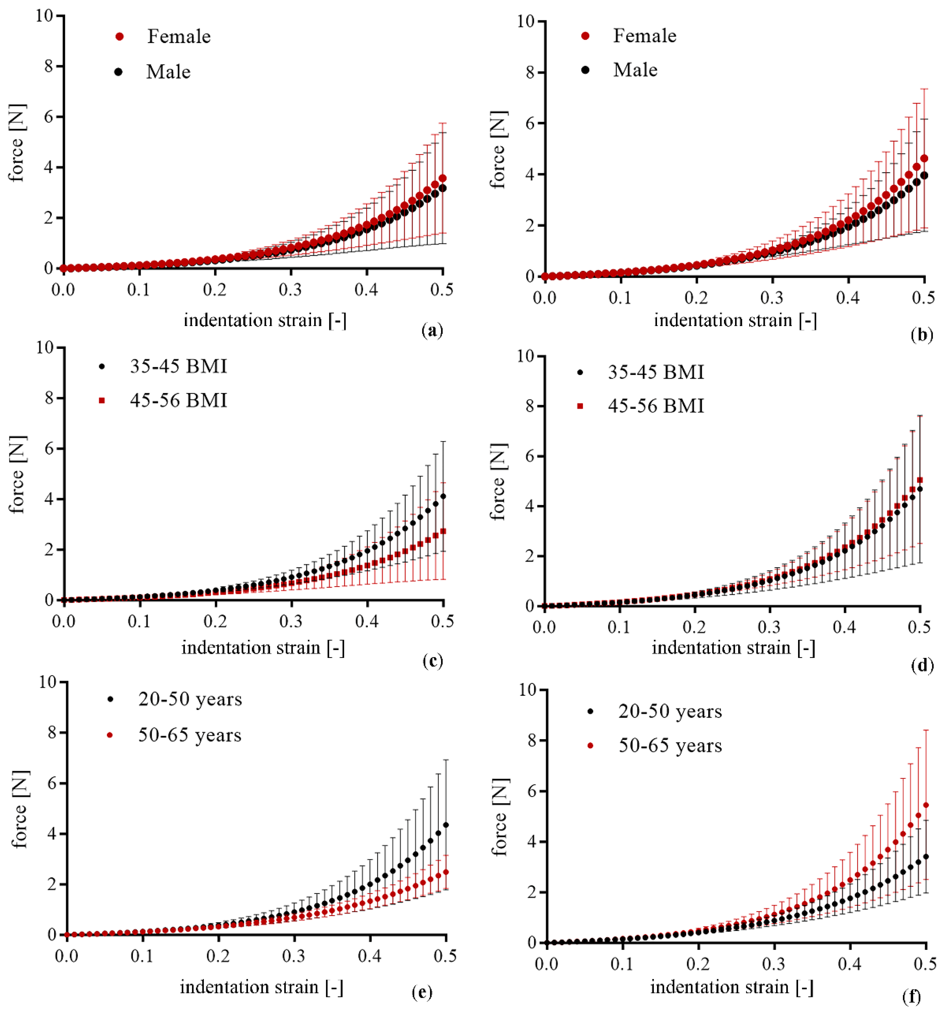
| Male vs. Female | 1.32 (±0.31) vs. 1.18 (±0.27) a | 1.63 (±0.15) vs. 1.65 (±0.32) b | a 0.436 b 0.909 |
| Age <50 vs. >50 years | 1.31 (±0.30) vs. 1.25 (±0.30) a | 1.54 (±0.24) vs. 1.69 (±0.30) b | a 0.690 b 0.384 |
| BMI <45 vs. >45 Kg/m2 | 1.42 (±0.30) vs. 1.14 (±0.24) a | 1.56 (±0.34) vs. 1.75 (±0.17) b | a 0.067 b 0.252 |
| final indentation stiffness [N] | |||
| Male vs. Female | 21.60 (±16.60) vs. 19.10 (±16.10) a | 28.60 (±20) vs. 35.90 (±25) b | a 0.801 b 0.617 |
| Age <50 vs. >50 years | 26.10 (±19.10) vs. 13.21 (±4.50) a | 19.10 (±10.30) vs. 35.32 (±22.91) b | a 0.132 b 0.211 |
| BMI <45 vs. >45 Kg/m2 | 25.40 (±17.11) vs. 15.90 (±14.30) a | 28.51 (±24.32) vs. 32.31 (±18.20) b | a 0.268 b 0.759 |
| (kPa) | α | D (kPa−1) | (s) | (s) | |||
| VAT | 12.55 | 7.23 | 64.66 | 0.63 | 0.27 | 1.21 | 47.48 |
| SAT | 17.50 | 10.11 | 65.66 | 0.60 | 0.26 | 1.41 | 52.63 |
Publisher’s Note: MDPI stays neutral with regard to jurisdictional claims in published maps and institutional affiliations. |
© 2022 by the authors. Licensee MDPI, Basel, Switzerland. This article is an open access article distributed under the terms and conditions of the Creative Commons Attribution (CC BY) license (https://creativecommons.org/licenses/by/4.0/).
Share and Cite
Fontanella, C.G.; Toniolo, I.; Foletto, M.; Prevedello, L.; Carniel, E.L. Mechanical Behavior of Subcutaneous and Visceral Abdominal Adipose Tissue in Patients with Obesity. Processes 2022, 10, 1798. https://doi.org/10.3390/pr10091798
Fontanella CG, Toniolo I, Foletto M, Prevedello L, Carniel EL. Mechanical Behavior of Subcutaneous and Visceral Abdominal Adipose Tissue in Patients with Obesity. Processes. 2022; 10(9):1798. https://doi.org/10.3390/pr10091798
Chicago/Turabian StyleFontanella, Chiara Giulia, Ilaria Toniolo, Mirto Foletto, Luca Prevedello, and Emanuele Luigi Carniel. 2022. "Mechanical Behavior of Subcutaneous and Visceral Abdominal Adipose Tissue in Patients with Obesity" Processes 10, no. 9: 1798. https://doi.org/10.3390/pr10091798