Article

Sterilizer of Knives in the Meat Industry, Working by Activating Aqueous Solutions with Glow Discharge Plasma

by
Sergey V. Belov
1,
Yury K. Danileiko
1,
Alexey B. Egorov
1,
Vladimir I. Lukanin
1,
Anastasia A. Semenova
2,
Andrey B. Lisitsyn
2,
Natalia M. Revutskaya
2,
Victoria V. Nasonova
2,
Yulya K. Yushina
2,
Etery R. Tolordava
2,3,
Nazarbay A. Nasyrov
2,
Alena I. Sinichkina
2,
Eugeny M. Konchekov
1,
Tatyana A. Matveeva
1 and
Sergey V. Gudkov
1,*
1
Prokhorov General Physics Institute of the Russian Academy of Sciences, 38 Vavilova St., Moscow 119991, Russia
2
V.M. Gorbatov Federal Research Center for Food Systems, 26 Talalihina St., Moscow 109316, Russia
3
N.F. Gamaleya Federal Research Centre of Epidemiology and Microbiology, 18 Gamaleya St., Moscow 123098, Russia
*
Author to whom correspondence should be addressed.
Processes 2022, 10(8), 1536; https://doi.org/10.3390/pr10081536
Submission received: 12 July 2022 / Revised: 3 August 2022 / Accepted: 4 August 2022 / Published: 5 August 2022
(This article belongs to the Section Food Process Engineering)

Abstract

:
The development of approaches for the non-thermal sterilization of instruments is an urgent task to ensure the safety of meat industry products, where the use of hot water leads to the formation of condensates and a deterioration in the hygienic condition of the premises. In this study, an installation for sterilizing knives was created, which works by activating aqueous salt solutions with a glow discharge. The power consumption of the installation reactor is only 125–150 Wh. The temperature rise of the sterilizing agent used is about 1.1 ± 0.2 °C/min/L. The effectiveness of the installation for plasma-activation of aqueous solutions of chloride and sodium sulfate by glow discharge (PAW) in relation to the inactivation of microorganisms, including Staphylococcus aureus, Salmonella typhimurium, Pseudomonas gessardii and L. monocytogenes, on steel surfaces was evaluated. Samples of stainless steel (parts of knives) were used in two versions (new and artificially aged). Mono- and polyspecies bacterial biofilms were grown on the surface of the samples. The treatment was carried out by immersing samples of steel plates in plasma-activated aqueous solutions. It was found that the treatment of plates in a knife sterilizer for 1 min had an effective effect on the inhibition of all types of studied bacteria.

1. Introduction

The procedure for disinfecting cutting tools at meat industry enterprises is a necessary measure aimed at preventing cross-contamination of meat raw materials with spoilage microorganisms, as well as pathogenic microorganisms [1]. Of paramount importance is the observance of sanitary and hygienic measures at the stage of slaughter and primary processing of animals. It is known that the gastrointestinal tract and the skin of slaughtered animals are sources of pathogenic microorganisms, such as Escherichia coli and Salmonella typhimurium, which pose a potential threat to human health [2,3,4,5]. Knives that come into contact with animal hide are known to have much higher levels of Salmonella typhimurium when compared to knives used in other operations for butchering beef carcasses [6]. With insufficient sanitary and hygienic treatment of instruments, microorganisms can attach to their surface and form stable biofilms [7], representing a serious problem for the meat industry. Much attention is paid to the study of biofilm formation by such pathogens as L.monocytogenes, Staphylococcus aureus, and Salmonella typhimurium [8,9,10]. Timely preventive measures aimed at cleaning and disinfecting working tools, in particular knives, are a key factor in determining the safety of meat and meat products.
Among the existing methods of sterilization, the method of immersing knives in a container with hot water (sterilizer) is the most widely used in the meat industry [11]. As a rule, in large enterprises, the “two knives” rule is applied, where one knife is used by the operator and the other is in the sterilizer. According to the requirements of EU regulation 853/2004, the water temperature must be at least 82 °C without any indication of exposure time [12]. However, in some studies it was noted that a short-term exposure to a temperature of 82 °C for 1–5 s did not have the proper effect on reducing the number of microbial populations on the surface of knives [13,14,15].
Along with hot water sterilization, alternative procedures with optimized time and temperature parameters can be used if their effectiveness has been proven [11,14]. It is known that the result of sterilization strongly depends on the amount of protein and fat on the surfaces of knives [16]. In this regard, in order to achieve the optimal disinfection of contaminated surfaces, it is necessary to carry out cleaning before sterilization, which consists of the mechanical removal of contaminants using warm running water with a temperature of 20–40 °C [14]. Thus, sterilization of pre-cleaned knives contributed to the complete elimination of microbial contamination on stainless steel plates in 1 s. At the same time, the microbiological indicators of surfaces with a high degree of protein and fat contamination, after immersion in hot water for 10 s, were not satisfactory [16].
The main disadvantages of sterilizing knives with hot water are increased wear of the cutting blades, high energy consumption and condensation of water vapor on the cold surfaces of structures and equipment in work areas. In turn, wetting surfaces due to steam condensation increases microbiological risks in enterprises that seek to reduce the air temperature in industrial premises. It is known that high humidity at relatively low temperatures promotes the survival of microorganisms and the formation of biofilms by them on various surfaces, including metal ones.
In this regard, in the meat industry, there is interest in various non-thermal methods of knife disinfection, for example, using chemical and physical factors of influence [17], as well as their combined use. Several papers have been published to determine the effect of disinfectants on the inhibition of various pathogens. For example, the effectiveness of various concentrations of biguanide and peracetic acid against E. coli on the surface of meat deboning knives was evaluated [18]. Other authors have studied various combinations of time and temperature of disinfection in combination with chemical (lactic acid) and physical (heat, ultrasound) factors [19]. Also, as an alternative non-thermal method of knife disinfection, the use of ultraviolet radiation (UV) in relation to the inhibition of E. coli and S. aureus was proposed, the effectiveness of which depended on the presence of surface contaminants such as blood or fat [20]. The use of electrolysis-activated water (EW) has been proposed, which has shown effective antimicrobial activity against a wide range of microorganisms, including pathogens such as Salmonella typhimurium, E. coli and Yersinia [21,22]. It has been noted that EW contributes to the inhibition of the most common types of viruses, bacteria, fungi, and spores in a short period of time within 5–20 s [23]. EW treatment is considered a simple, effective and safe disinfection method that serves as a good alternative to other more widely used disinfectants [23,24]. The effectiveness of EW has been evaluated on various surfaces, including stainless steel [25]. The authors reported that the disinfection of knives used for slaughtering animals can be achieved by a combination of treatment with alkaline (5 min) and slightly acidic water (10 min) and with sonication. This combination also makes it possible to remove all organic residues without changing the structure of the knife blades [17]. The disadvantage of the EW method is the need to use rather bulky electrolyzers, as well as vessels for storing activated water. At the same time, there are commercially successful solutions on the market [25].
In general, according to our survey of enterprises, the “ideal sterilizer” should meet the following conditions:
be economical, both in terms of the amount of electricity consumed and the amount of water used;
the time for the sterilization cycle (excluding the time of loading the instrument) should be no more than one minute;
the reagents used must be commercially available, of low cost, chemically safe and harmless in the conditions of meat production;
the temperature of the sterilizing agent used should not exceed the ambient temperature in the working room without the use of special cooling devices.
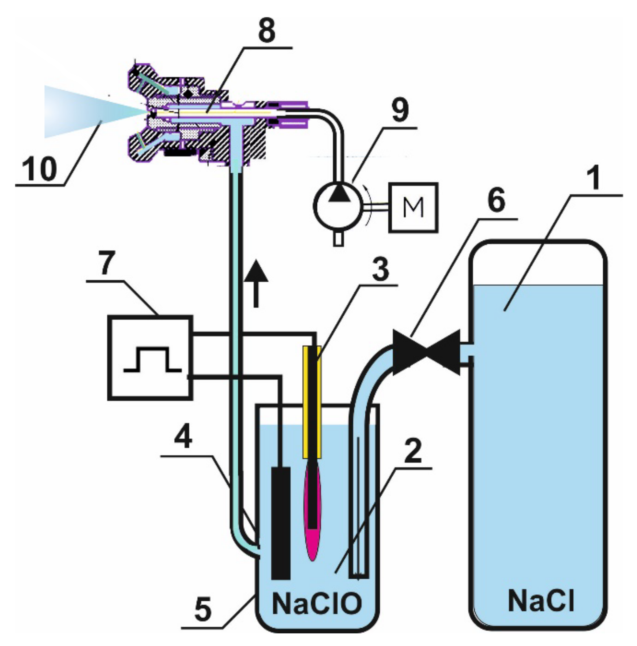
In this regard, we have tried to create a knife sterilizer with characteristics close to the “ideal sterilizer”. We used a compact reactor that treats an aqueous solution with a glow discharge plasma. The processing time is from 1 min. The power consumption of the reactor in stationary mode is up to 150 Wh. Water and any dissociating salt are required as chemical reagents. The temperature rise of the sterilizing agent used is about 1.1 ± 0.2 °C/min/L.

2. Instrumentation

Knife Sterilizer Device

In experiments, an aqueous solution of common salt (NaCl) with a concentration of 1% was used as a medium that is suitable for the ignition and combustion of an underwater glow discharge. In a number of experiments, other salts were used, for example, sodium sulfate (Na2SO4). A schematic diagram of an experimental reactor designed for plasma–chemical activation of weak aqueous solutions of strong electrolytes is shown in Figure 1.
To activate water, a reactor was used, in which two activation methods were implemented—plasma–chemical and electrochemical. The original electrolyte solution is in tank 1 under a slight overpressure of about 0.1 atm. Activation was carried out in a reactor with a volume of 1000 cm3. In the plasma-chemical reactor 2 there is an active electrode 3, on which an underwater glow discharge 5 burns. The active electrode is made of stainless steel and has the shape of a cylinder with a diameter of 0.5 mm and a length of 20 mm. The reactor was powered by a current generator with an operating frequency of 440 kHz. In the plasma discharge ignition mode, the current was about 5 A. In the standard combustion mode, it was about 0.5 A. The main components of the plasma-forming gas are water vapor and its decomposition products [26,27]. Plasma–chemical reactions take place in the volume of the plasma-forming gas. The electrodes for the plasma–chemical part of the reactor are a metal active electrode (cathode) and a plasma–electrolyte interface (electrolyte anode).
The second—the electrochemical part—of the reactor 4 contains a passive (graphite) electrode 5 made of pyrolytic graphite and having an area of about 8 cm2. The passive electrode is the anode of the electrochemical part of the reactor. The cathode of the electrochemical part of the reactor is the plasma–electrolyte interface. The electrodes are connected to a specially designed high-frequency generator 7 operating at a frequency of 440 kHz. The operating voltage on the electrodes was maintained in the range (250 ÷ 350) V. At the same time, the power consumed from the generator could reach 1.2–1.5 kW for a short time (at the time of plasma ignition), which was sufficient for the formation of a vapor–gas bubble on the active electrode and ignition in the vapor phase of a glow discharge. The power consumption of the reactor in stationary mode is up to 150 Wh.
The oscillogram of the current flowing through the reactor is shown in Figure 2. It can be seen from the oscillogram that the current in the negative half-cycle (cathode polarity) greatly exceeds the current in the positive half-cycle (anodic polarity), which is practically close to zero. This leads to the fact that the current flowing through the reactor has a constant component, which causes the occurrence of electrochemical processes on the passive electrode (anode) of the electrolyte part of the reactor. Polyethylene terephthalate was used as the structural material of the reactor.

3. Materials and Methods

3.1. Bacterial Strains and Conditions for Their Cultivation

The material for research was microbial isolates isolated from the production environment of meat industry enterprises. Bacterial cultures were obtained from the laboratory of microbiology of the V.M. Gorbatov Federal Research Center for Food Systems: Staphylococcus aureus, Salmonella typhimurium, Pseudomonas gessardii, and Listeria monocytogenes. Microorganisms were grown (1) on liquid and solid (1.5%) Luria-Bertani (LB) medium, using standard dry nutrient media manufactured by Difco (USA) or (2) in tryptone soy broth (TSB), Panreac, Spain.

3.2. Preparation and Inoculation of Steel Plates

The blades of steel knives used for butchering animal carcasses were cut into identical plates 1 cm × 1 cm in size. Some of the steel plates were electrochemically treated to simulate the aging and wear of steel. It is assumed that the attachment of samples and the formation of biofilms depend on the microrelief of the surface of the plates. New and aged plates were used in experiments to create biofilms on their surface, 5 pieces per experimental point.

3.3. Attachment and Biofilm Formation

To grow bacterial biofilms, an overnight broth culture of bacteria was diluted 100 times in fresh sterile broth, and transferred to a sterile dish (Petri dish or microtiter plate for 12 wells) with sterile plates. After 18–24 h of incubation in a thermostat at 30 °C, a biofilm formed on the surface of the steel plates. The formed biofilms were not washed or fixed.

3.4. Assessment of Cell Viability in Biofilms by Fluorescence Microscopy

For microscopy with a fluorescent lens, the studied biofilm samples were stained with a fluorescent Filmtracer Live/Dead Biofilm Viability Kit. All procedures were performed in accordance with the manufacturer’s recommendations. A Nikon Eclipse Ni direct fluorescence microscope with a Nikon DS-Fi3 camera was used. Figure 3 shows examples of fluorescent images of a completely non-viable biofilm (a), a biofilm with foci of surviving microorganisms (b) and a completely viable biofilm (c).

3.5. Enumeration of Attached and Planktonic Microorganisms

After the experiment, the studied samples of steel plates was placed in test tubes with a physiological raster and shaken vigorously for 10 min. Then the resulting wash was titrated and the number of CFU/mL was determined. Samples of bacterial biofilms not treated with plasma water served as controls.

3.6. Physico-Chemical Tests

Redox potential, pH and electrical conductivity were measured using a high-precision measuring station S470 SevenEx Cellence (Mettler Toledo, Columbus, OH, USA). Sensor electrodes recommended by the manufacturer InLab Expert Pro-ISM and InLab731-ISM (Mettler Toledo) were used. Using a magnetic stirrer, all aqueous solutions were mixed during measurements in a laminar mode (frequency about 3 Hz). The details of experimental measurements were described earlier [28]. The concentration of molecular oxygen dissolved in aqueous solutions was recorded using an AKPM-1-02 polarograph (Bioanalytical Systems and Sensors, Moscow, Russia) [29]. The measurements took into account the atmospheric pressure measured by the PRX-7001t instrument (Casio, Tokyo, Japan) and the temperature of the samples measured with a temperature compensator. The concentration of nitrate anions was determined using a LAQUAtwin B-741 (Horiba, Tokyo, Japan) nitrate meter. The content of free chlorine in solutions was measured using a 2793944 kit (Hach, Loveland, CO, USA). Details of experimental measurements were described earlier [30]. For the quantitative determination of hydrogen peroxide in aqueous solutions, a highly sensitive method of enhanced chemiluminescence in luminol-4-iodophenol-horseradish peroxidase was used [31]. Luminescence intensity was determined on a Biotox-7A chemiluminometer (ANO Ecology, Moscow, Russia). Hydrogen peroxide solutions of known concentration were used for calibration. The details of experimental measurements were described earlier [32].

4. Results and Discussion

The effect of glow discharge plasma on the physicochemical parameters of aqueous solutions is presented in Table 1. A change in the physicochemical parameters of water occurred within 40–50 min of treatment. With further processing, the rate of change in the parameters decreased significantly or, as in the case of hydrogen peroxide, ceased to change, reaching a stationary value of the order of 10−2 M (
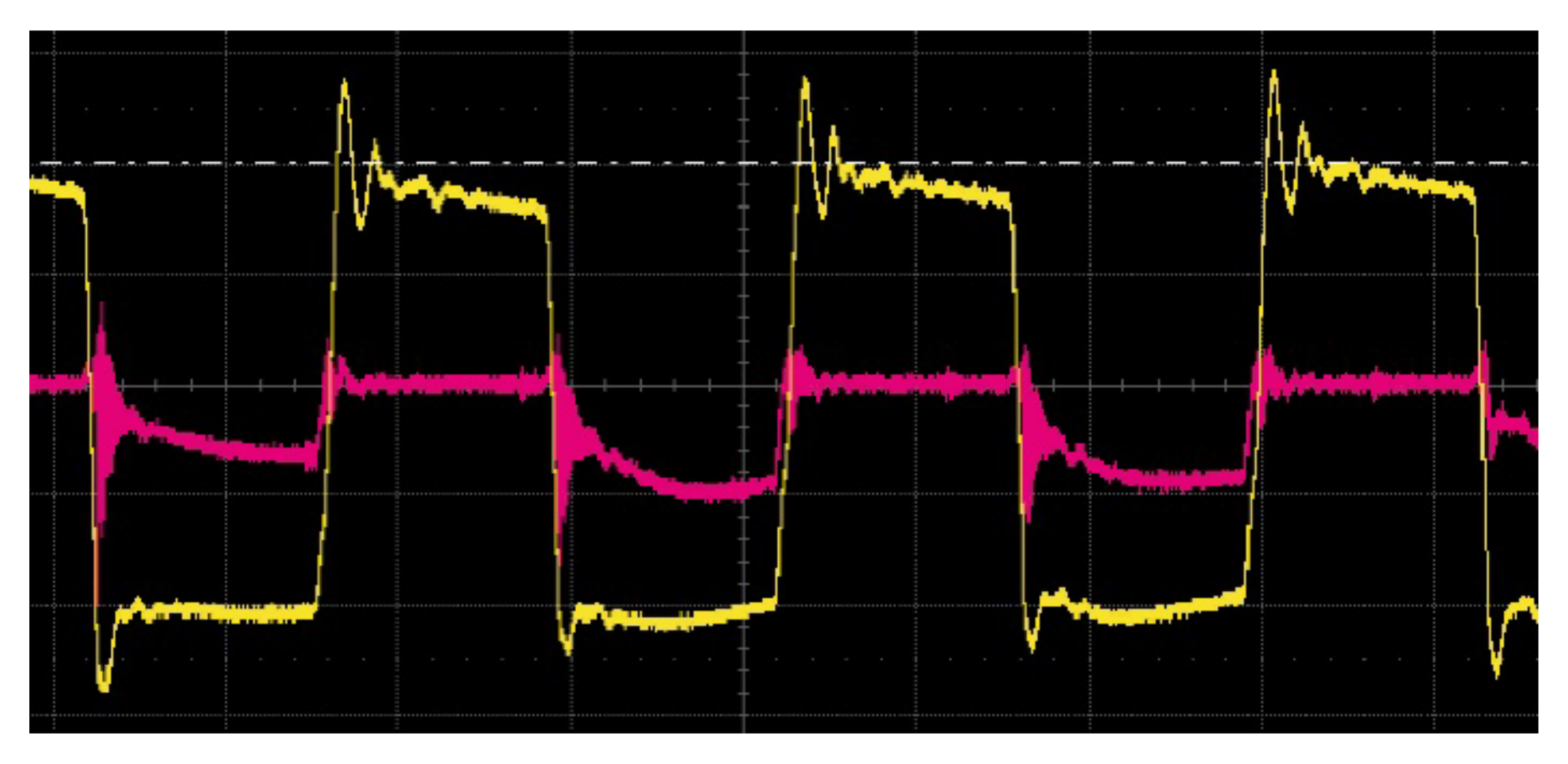
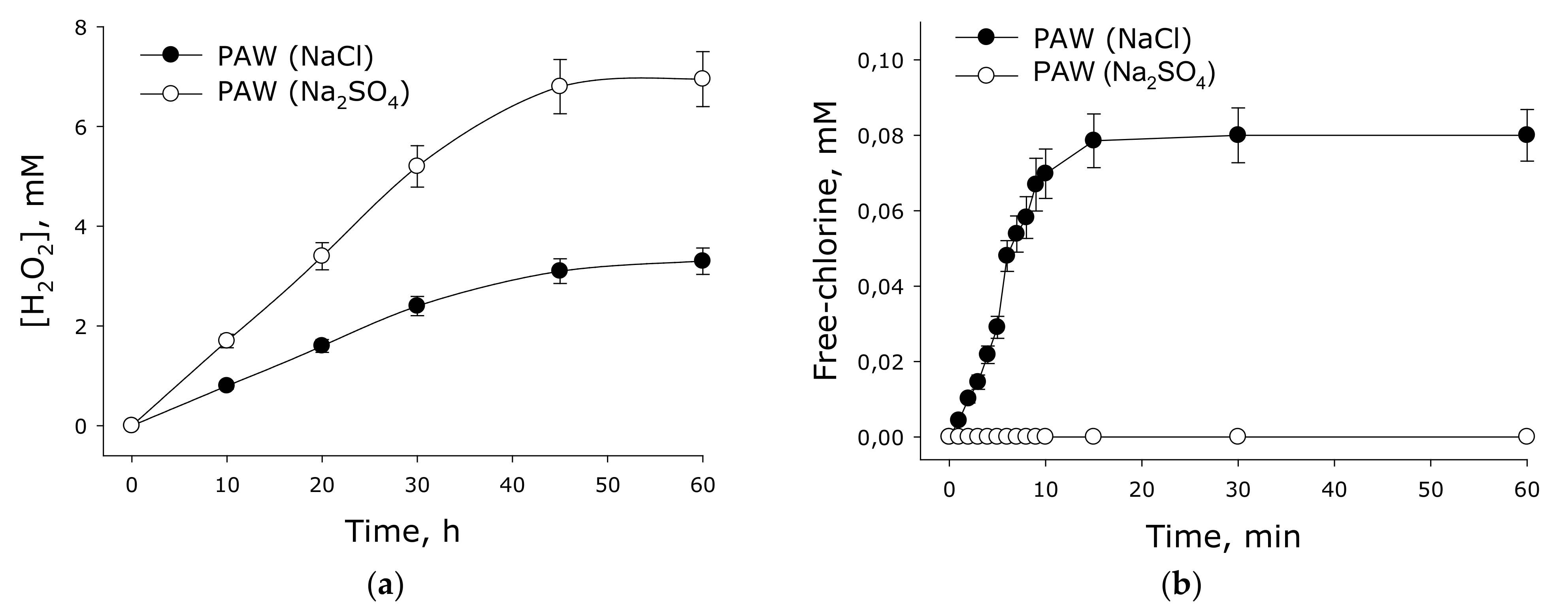
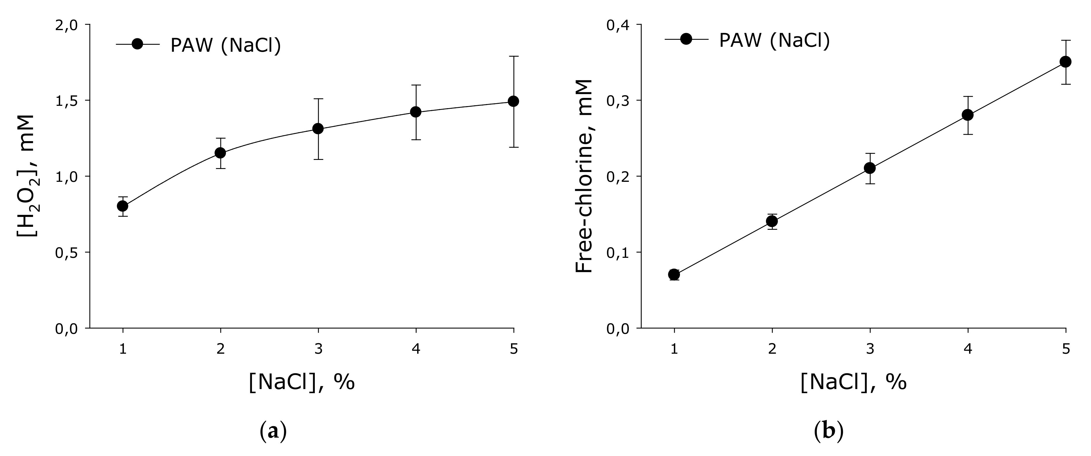
Figure 4). At the same time, free chlorine was intensively farmed only for 10 min. At a long processing times, the concentration of free chlorine became stationary.
During the glow discharge treatment, the electrical conductivity of the solution increased almost linearly. The rate of increase in the specific electrical conductivity of the solution was 400–450 μS/cm/min. At the same time, the concentration of molecular oxygen dissolved in water tended to decrease. In addition, the pH of the solution changed during the activation process. During the first 10 min of treatment, the pH increased by 0.5–0.7 units. For 20 min of treatment, the pH value increased by almost one. With further activation for 40 min, the pH value changed by only 1.5 units. Similar changes were observed in the values of the redox potential. During the first 10 min of treatment, the potential increased by almost 200 mV. In post-processing, it was observed that the increase was much slower. Upon activation, nitrate anions accumulated in aqueous solutions. It is shown that the increase in the concentration of nitrate anions linearly depends on the treatment time. It should be noted that, unlike other studied parameters, the concentration of nitrate anions continued to increase after 50 min of exposure and even after 2 h. No nitrate generation was observed during the experiments in an atmosphere of inert argon gas. Hydrogen peroxide was formed in the medium with NaCl two times less intensively than in the medium with sodium sulfate. In this case, the formation of free chlorine was observed in a medium with sodium chloride.
It should be noted that the concentration of sodium chloride has a significant effect on the generation of hydrogen peroxide and free chlorine (Figure 5). It is shown that with an increase in the concentration of sodium chloride, the generation of hydrogen peroxide increased non-linearly. With an increase in the concentration of sodium chloride, the rate of formation of hydrogen peroxide decreased. Conversely, the free chlorine concentration increased linearly with increasing sodium chloride concentration. Interestingly, the concentration of hydrogen peroxide, in contrast to the concentration of free chlorine, decreased by half in more than one week. On the basis of the obtained results, activated solutions containing the maximum amount of active substances were used in further studies (treatment time 10 min).
The process of decomposition of water vapor in the plasma–chemical reactor 2, operating in the mode of high-frequency glow discharge, apparently occurred as follows. Energy from an electron gas with a temperature of about 1–5 eV is transferred to water molecules through two alternative channels. As a result of both reactions, atomic hydrogen, hydroxyl radical, hydrogen peroxide and other ROS should be formed. Hydrogen peroxide is the only stable ROS molecule. As shown in Figure 4, hydrogen peroxide is capable of accumulating in the reactor.
In the electrolyte part of the reactor 3 at the anode, the process of plasma electrolysis of the NaCl solution with the release of chlorine atoms is likely. Probably, plasma electrolysis of water also occurs with the release of molecular oxygen and molecular hydrogen. This should result in the formation of sodium hypochlorite NaOCl [33]. The latter is due to the fact that, as shown by the measurements, the overvoltage at the anode has a value of more than 1.5 V. In this case, the current density at the anode was chosen such that the chlorine evolution potentials are slightly less than for oxygen. The change in the current density at the anode was carried out by choosing its area, and in our case it was about 1000 A/m2. Sodium hypochlorite in aqueous solutions is largely dissonant with the formation of ClO ions, which are capable of further anodic oxidation with the formation of the chlorate ion ClO3 and further of the formation of the perchlorate ion ClO4. In addition, hypochloride must interact with hydrogen peroxide [34]: H2O2 + 4NaOCl → 2NaCl + 2NaOH + Cl2 + 1O2. As a result of the reaction, molecular chlorine and oxygen in the singlet state (1O2) are formed, and the concentration of hydrogen peroxide in the electrolyte decreases. This explains the different rates of hydrogen peroxide generation in an aqueous solution of sulfate and sodium chloride (Figure 4).
The effect of plasma–activated aqueous solutions (PAW) on planktonic (free-floating) cells of bacteria Staphylococcus aureus, Salmonella typhimurium, Pseudomonas gessardii and L. monocytogenes was studied. To do this, a bacterial culture (107 CFU/mL) in a volume of 100 μL was introduced into freshly prepared and prepared in advance (several days) “plasma water” in a volume of 1 mL. The exposure time was 1 and 10 min. After exposure, the suspension was titrated and sown on a solid nutrient medium to determine CFU/mL. Sterile distilled water served as the control of the experiment. The results are presented in Table 2.
Experiments have shown that freshly prepared PAW exhibits high antibacterial activity. The number of planktonic cells of pathogenic microorganisms decreased by six orders of magnitude, when compared with the control, which indicates effective sterilization. At the same time, PAW prepared a day before the experiment practically did not show its antibacterial properties. It is important that for complete sterilization of the surface it is not necessary to treat the aqueous solution for 10 min. When the treatment time of the aqueous solution is 1 min, complete sterilization was also observed. At the same time, the solution became inactive for less than 24 h, but it remains unclear as to how long PAW activity persists. It should be noted that other methods for controlling the number of microorganisms are described in the literature [35,36].
To study the time during which PAW remains active, bacterial cultures of the genera Pseudomonas gessardii and Staphylococcus aureus were prepared. Microorganisms were grown in liquid LB nutrient medium in a thermostat at 37 °C for 18 h. Further, by the method of tenfold titrations, bacterial broth cultures were prepared in a titer of 107 CFU/mL. Freshly prepared “plasma water” was poured into sterile tubes of 1 mL. Then, at different time intervals from 5 min to 18 h, bacterial cultures were introduced into the tubes with PAW and incubated for 1 h. 1 mL of saline was added to the control tube instead of PAW. Further, the suspensions from the test tubes were sown on solid nutrient media to determine the number of live bacteria per unit volume of the suspension (CFU/mL). The results obtained are presented in Table 3. The results obtained show that the antimicrobial properties of the plasma-treated aqueous solutions were maintained for 2 h.
Thus, the duration of storage of plasma-activated aqueous solutions has been established. This raises the question of whether PAW can be diluted with untreated water. Will such diluted water retain its antibacterial properties? To clarify the answer to this question, freshly prepared PAW was diluted in sterile saline (NaCl 0.9%) by 3, 9 and 27 times. The resulting suspensions were transferred into sterile 1 mL test tubes. Next, bacterial cultures were added to each tube. In control samples, sterile saline was added to the bacterial culture. After 1 h of inoculation, the microorganisms were sown on dense nutrient media to determine the number of living cells per unit volume of the suspension. The results obtained are presented in Table 4. The results obtained show that plasma water has pronounced antimicrobial properties and these properties are fully preserved after a threefold dilution. When diluted nine or more times, the level of antimicrobial properties significantly reduced.
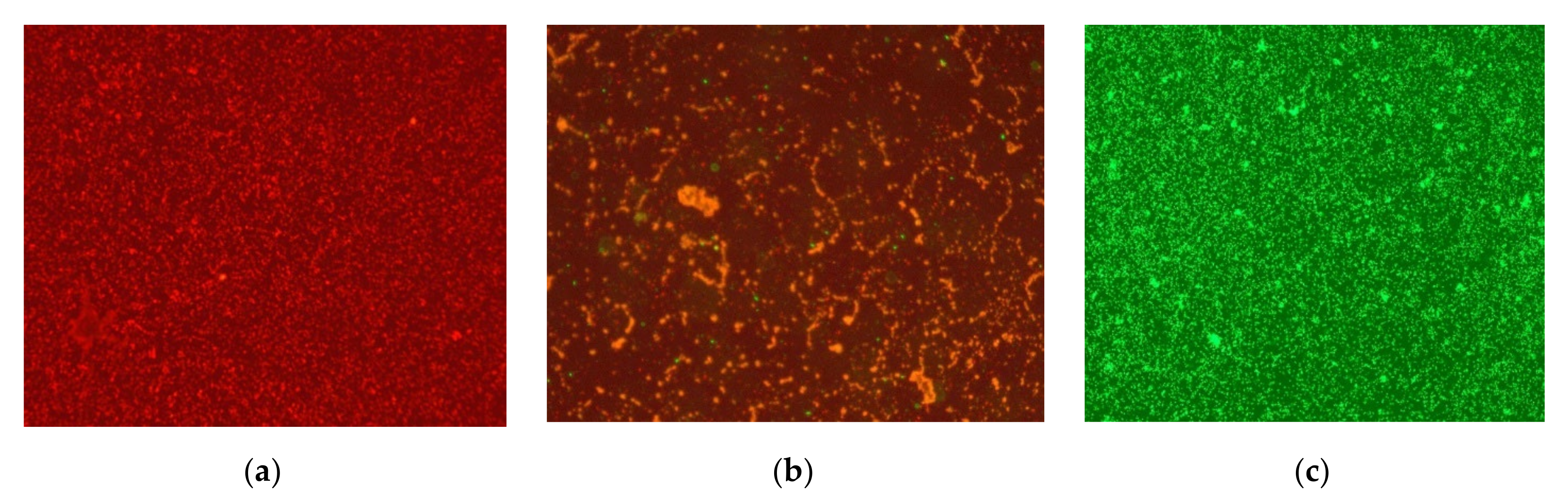
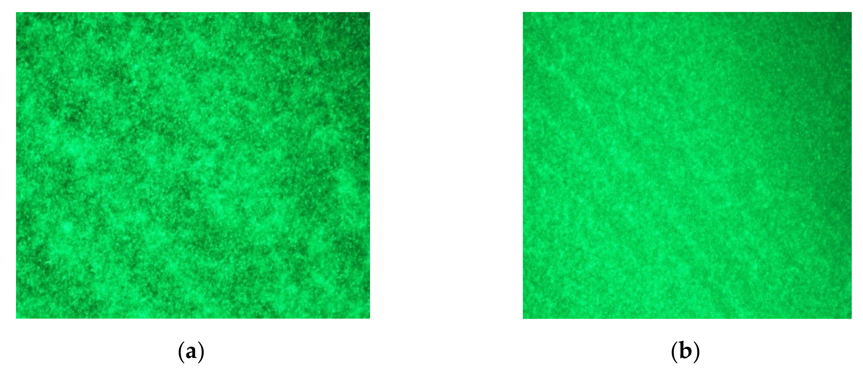
It is known that a large number of microorganisms enter the production environment in the process of slaughter, primary processing of animals and meat processing. As a result, air, floors, equipment and tools, as well as working personnel become sources of pollution. Bacterial biofilms can develop on surfaces. At the same time, microbial contamination of one steel knife can reach up to 107 CFU. It is known that bacterial biofilms do not develop equally successfully on different types of surfaces. We assumed that the development of bacterial films on new and old knives can occur in different ways. To test this assumption, part of the steel plates from the knife steel underwent electrochemical treatment that simulates the aging and wear of steel. Next, bacterial biofilms were grown on “new” and “aged” steel plates for 24 h. Before microscopy, the plates were washed three times with sterile distilled water, excess moisture was removed with lint-free paper, and stained with SYTO 9 fluorescent dye. The degree of biofilm formation was assessed using fluorescent microscopy. The results are presented in Figure 6. It has been shown that microorganisms form a confluent biofilm on the surface of steel plates during the day. At the same time, the formation of biofilms on new and aged steel plates did not differ significantly. In this regard, in order to exclude additional labor costs, only new steel plates were used in further studies.
To study the effect of PAW on mono- and polyspecies biofilms of food pathogens, Staphylococcus aureus, Salmonella typhimurium, Pseudomonas gessardii and L. monocytogenes were grown on steel plates for 48 h. The biofilms obtained on the plates were immersed in freshly prepared PAW for 1 min. Control samples were immersed in sterile water. Next, the plates were placed in test tubes with saline and vigorously shaken for 30 min. The resulting suspension was titrated and sown on a solid nutrient medium to determine CFU. The results of the conducted studies are shown in Table 5.
Research results have shown that freshly prepared PAW has pronounced antimicrobial and antibiofilm properties. At the same time, its antimicrobial effect is the same not only for Gram-positive and Gram-negative bacteria, but also for mono- and multi-species biofilms. PAW prepared in 24 h does not have significant antimicrobial properties against mono- and multi-species biofilms.
To obtain PAW using glow discharge plasma, the designed reactor has an active electrode 3; this electrode can be made of various metals. The dependence of the antibacterial properties of PAW on the electrode material was studied. For these studies, we used stainless steel, platinum (Pt) and silver (Ag) metal electrodes. The design of the experiments was similar to that described above. The antimicrobial effects of PAW have been studied in Listeria monocytogenes biofilms. The research results are shown in Table 6. It has been established that the antibacterial properties of plasma-activated aqueous solutions do not largely depend on the chemical composition of the material used to manufacture the active electrode. It was shown that at the exposure time of 1 min to plasma-activated aqueous solutions on biofilms, the sterilization of samples was observed.

5. Conclusions

Thus, the knife sterilizer for the food industry was created. The sterilizer is based on a reactor that creates plasma in aqueous salt solutions due to a glow discharge, while a plasma-activated aqueous solution (PAW) is formed in the reactor. Studies have shown that PAW has a pronounced antibacterial and antibiofilm activity. The use of the reactor developed by us can become a promising alternative to traditional methods of cleaning and sterilizing knives used at various stages of the technological process of meat processing.

Author Contributions

Conceptualization, A.A.S., Y.K.D. and S.V.G.; methodology, Y.K.D.; software, T.A.M.; formal analysis, T.A.M.; investigation, S.V.B., A.B.E., V.I.L., E.M.K., T.A.M., E.R.T., N.A.N., N.M.R., Y.K.Y., V.V.N. and A.I.S.; writing—original draft preparation, N.M.R., Y.K.Y., V.V.N., T.A.M. and S.V.G.; writing—review and editing, A.B.L.; supervision, A.A.S. and A.B.L.; project administration, A.A.S. and A.B.L.; funding acquisition, A.B.L. All authors have read and agreed to the published version of the manuscript.

Funding

This research was funded by the Ministry of Science and Higher Education of the Russian Federation for large scientific projects in priority areas of scientific and technological development, grant number 075-15-2020-775.

Institutional Review Board Statement

Not applicable.

Informed Consent Statement

Not applicable.

Data Availability Statement

Data available on request due to restrictions.

Acknowledgments

The authors are grateful to the Shared Use Center of the GPI RAS.

Conflicts of Interest

The authors declare no conflict of interest.

References

  1. Eustace, I.; Midgley, J.; Small, A.; Jenson, I.; Sumner, J. Knife sanitizing in abattoirs: The effectiveness of current and alternative practices. Food Prot. Trends 2008, 28, 712–722. [Google Scholar]
  2. De Busser, E.V.; De Zutter, L.; Dewulf, J.; Houf, K.; Maes, D. Salmonella control in live pigs and at slaughter. Vet. J. 2013, 196, 20–27. [Google Scholar] [CrossRef] [PubMed]
  3. Brichta-Harhay, D.M.; Guerini, M.N.; Arthur, T.M.; Bosilevac, J.M.; Kalchayanand, N.; Shackelford, S.D.; Wheeler, T.L.; Koohmaraie, M. Salmonella and Escherichia coli O157: H7 contamination on hides and carcasses of cull cattle presented for slaughter in the United States: An evaluation of prevalence and bacterial loads by immunomagnetic separation and direct plating methods. Appl. Environ. Microbiol. 2008, 74, 6289–6297. [Google Scholar] [CrossRef] [Green Version]
  4. Omisakin, F.; MacRae, M.; Ogden, I.D.; Strachan, N.J.C. Concentration and prevalence of Escherichia coli O157 in cattle feces at slaughter. Appl. Environ. Microbiol. 2003, 69, 2444–2447. [Google Scholar] [CrossRef] [Green Version]
  5. Arthur, T.M.; Nou, X.; Kalchayanand, N.; Bosilevac, J.M.; Wheeler, T.; Koohmaraie, M. Survival of Escherichia coli O157: H7 on cattle hides. Appl. Environ. Microbiol. 2011, 77, 3002–3008. [Google Scholar] [CrossRef] [Green Version]
  6. Peel, B.; Simmons, G.C. Factors in the spread of Salmonellas in meatworks with special reference to contamination of knives. Aust. Vet. J. 1978, 54, 106–110. [Google Scholar] [CrossRef]
  7. Wang, R. Biofilms and meat safety: A mini-review. J. Food Prot. 2019, 82, 120–127. [Google Scholar] [CrossRef] [PubMed]
  8. Fagerlund, A.; Langsrud, S.; Møretrø, T. Microbial diversity and ecology of biofilms in food industry environments associated with Listeria monocytogenes persistence. Curr. Opin. Food Sci. 2021, 37, 171–178. [Google Scholar] [CrossRef]
  9. Rieu, A.; Lemaître, J.P.; Guzzo, J.; Piveteau, P. Interactions in dual species biofilms between Listeria monocytogenes EGD-e and several strains of Staphylococcus aureus. Int. J. Food Microbiol. 2008, 126, 76–82. [Google Scholar] [CrossRef] [PubMed]
  10. Carrasco, E.; Morales-Rueda, A.; García-Gimeno, R.M. Cross-contamination and recontamination by Salmonella in foods: A review. Food Res. Int. 2012, 45, 545–556. [Google Scholar] [CrossRef]
  11. Eustace, I.; Midgley, J.; Giarrusso, C.; Laurent, C.; Jenson, I.; Sumner, J. An alternative process for cleaning knives used on meat slaughter floors. Int. J. Food Microbiol. 2007, 113, 23–27. [Google Scholar] [CrossRef] [PubMed]
  12. Regulation (EC) No. 853/2004 of the European Parliament and of the Councils of 29 April 2004 Laying Down Specific Hygiene Rules for on the Hygiene of Foodstuffs. Available online: http://eur-lex.europa.eu/de/index.htm (accessed on 1 June 2022).
  13. Taormina, P.J.; Dorsa, W.J. Evaluation of hot-water and sanitizer dip treatments of knives contaminated with bacteria and meat residue. J. Food Prot. 2007, 70, 648–654. [Google Scholar] [CrossRef] [PubMed]
  14. Goulter, R.M.; Dykes, G.A.; Small, A. Decontamination of knives used in the meat industry: Effect of different water temperature and treatment time combinations on the reduction of bacterial numbers on knife surfaces. J. Food Prot. 2008, 71, 1338–1342. [Google Scholar] [CrossRef]
  15. Durmuşoğlu, H.; İncili, G.K.; Demir, P.; İlhak, O.İ. Effects of workers’ hand washing and knife disinfection practices on microbiological quality of small animal carcasses in slaughterhouse environment. J. Food Process. Preserv. 2020, 44, e14918. [Google Scholar] [CrossRef]
  16. Snijders, J.M.; Janssen, M.H.; Corstiaensen, G.P.; Gerats, G.E. Cleaning and disinfection of knives in the meat industry. Zentralbl. Bakteriol. Mikrobiol. Hyg. 1985, 181, 121–131. [Google Scholar]
  17. Brasil, C.C.B.; Barin, J.S.; Jacob-Lopes, E.; Menezes, C.R.; Zepka, L.Q.; Wagner, R.; Campagnol, C.B.; Cichoski, A.J. Single step non-thermal cleaning/sanitation of knives used in meat industry with ultrasound. Food Res. Int. 2017, 91, 133–139. [Google Scholar] [CrossRef] [PubMed]
  18. Barbosa, J.; Cuppini, M.; Flach, J.; Steffens, C.; Cansian, R.L.; Toniazzo, G. Removal of Escherichia coli in boning knives with different sanitizers. LWT—Food Sci. Technol. 2016, 71, 309–315. [Google Scholar] [CrossRef]
  19. Leps, J.; Einschütz, K.; Langkabel, N.; Fries, R. Efficacy of knife disinfection techniques in meat processing. Meat Sci. 2013, 95, 185–189. [Google Scholar] [CrossRef]
  20. Raschle, S.; Zweifel, C.; Zurfluh, K.; Stephan, R. Decontamination of knives used in a slaughterhouse by a commercial non-thermal UV-C treatment. Ital. J. Food Saf. 2019, 8, 8107. [Google Scholar] [CrossRef]
  21. Gómez-López, V.M.; Gil, M.I.; Pupunat, L.; Allende, A. Cross-contamination of Escherichia coli O157: H7 is inhibited by electrolyzed water combined with salt under dynamic conditions of increasing organic matter. Food Microbiol. 2015, 46, 471–478. [Google Scholar] [CrossRef]
  22. Han, D.; Hung, Y.C.; Wang, L. Evaluation of the antimicrobial efficacy of neutral electrolyzed water on pork products and the formation of viable but nonculturable (VBNC) pathogens. Food Microbiol. 2018, 73, 227–236. [Google Scholar] [CrossRef] [PubMed]
  23. Rahman, S.M.E.; Khan, I.; Oh, D.H. Electrolyzed water as a novel sanitizer in the food industry: Current trends and future perspectives. Compr. Rev. Food Sci. Food Saf. 2016, 15, 471–490. [Google Scholar] [CrossRef] [PubMed] [Green Version]
  24. Tomasello, F.; Pollesel, M.; Mondo, E.; Savini, F.; Scarpellini, R.; Giacometti, F.; Lorito, L.; Tassinari, M.; Cuomo, S.; Piva, S.; et al. Effectiveness of alkaline electrolyzed water in reducing bacterial load on surfaces intended to come into contact with food. Ital. J. Food Saf. 2021, 10, 9988. [Google Scholar] [CrossRef]
  25. Park, H.; Hung, Y.C.; Kim, C. Effectiveness of electrolyzed water as a sanitizer for treating different surfaces. J. Food Prot. 2002, 65, 1276–1280. [Google Scholar] [CrossRef]
  26. Belov, S.V.; Gudkov, S.V.; Danyleiko, Y.K.; Egorov, A.B.; Lukanin, V.I.; Sidorov, V.A.; Tsvetkov, V.B. A Device for Biological Activation of Aqueous Solutions Using Glow Discharge Plasma in Water Vapor. Biomed. Eng. 2021, 55, 97–102. [Google Scholar] [CrossRef]
  27. Rusanov, V.D.; Fridman, A.A.; Sholin, G.V. The Physics of a Chemically Active Plasma With Nonequilibrium Vibrational Excitationc of Molecules. Sov. Phys. Usp. 1981, 24, 447–474. [Google Scholar] [CrossRef]
  28. Danilejko, Y.K.; Belov, S.V.; Egorov, A.B.; Lukanin, V.I.; Sidorov, V.A.; Apasheva, L.M.; Dushkov, V.Y.; Budnik, M.I.; Belyakov, A.M.; Kulik, K.N.; et al. Increase of Productivity and Neutralization of Pathological Processes in Plants of Grain and Fruit Crops with the Help of Aqueous Solutions Activated by Plasma of High-Frequency Glow Discharge. Plants 2021, 10, 2161. [Google Scholar] [CrossRef]
  29. Baimler, I.V.; Lisitsyn, A.B.; Gudkov, S.V. Influence of Gases Dissolved in Water on the Process of Optical Breakdown of Aqueous Solutions of Cu Nanoparticles. Front. Phys. 2020, 8, 622775. [Google Scholar] [CrossRef]
  30. Belov, S.V.; Danyleiko, Y.K.; Glinushkin, A.P.; Kalinitchenko, V.P.; Egorov, A.V.; Sidorov, V.A.; Konchekov, E.M.; Gudkov, S.V.; Dorokhov, A.S.; Lobachevsky, Y.P.; et al. An Activated Potassium Phosphate Fertilizer Solution for Stimulating the Growth of Agricultural Plants. Front. Phys. 2021, 8, 618320. [Google Scholar] [CrossRef]
  31. Astashev, M.E.; Bolotskova, P.N.; Bunkin, N.F.; Gudkov, S.V.; Kozlov, V.A.; Okuneva, M.A. Swelling of Polymer Membrane in an Aqueous Protein Suspension: Photoluminescence Spectroscopy Experiments. Phys. Wave Phenom. 2021, 29, 123–130. [Google Scholar] [CrossRef]
  32. Baymler, I.V.; Simakin, A.V.; Gudkov, S.V. Investigation of the laser-induced breakdown plasma, acoustic vibrations and dissociation processes of water molecules caused by laser breakdown of colloidal solutions containing Ni nanoparticles. Plasma Sources Sci. Technol. 2021, 30, 125015. [Google Scholar] [CrossRef]
  33. Nikolenko, N.V.; Zakharov, R.I.; Dubenko, A.V.; Moleva, G.V.; Avdienko, T.N. Optimization of plasma treatment of aqueous sodium chloride solutions. High Energy Chem. 2017, 51, 122–127. [Google Scholar] [CrossRef]
  34. Shams El Din, A.M.; Mohammed, R.A. Kinetics of the reaction between hydrogen peroxide and hypochlorite. Desalination 1998, 115, 145–153. [Google Scholar] [CrossRef]
  35. Mohammed, F.S.; Karakas, M.; Akgül, H.; Sevindik, M. Medicinal Properties of Allium calocephalum Collected from Gara Mountain (Iraq). Fresenius Environ. Bull. 2019, 28, 7419–7426. [Google Scholar]
  36. Mohammed, F.S.; Akgül, H.; Sevindik, M.; Khaled, B. Phenolic Content and Biological Activities of Rhus coriaria var. zebaria. Fresenius Environ. Bull. 2018, 27, 5694–5702. [Google Scholar]
Figure 1. Schematic diagram of the reactor for obtaining and spraying the activated solution. Tank with electrolyte solution—1, plasma-chemical reactor—2, active electrode—3, passive electrode—4, glow discharge plasma—5, valves of the electrolyte-filling channel—6, adjustable high-frequency current generator—7, nozzle of the activated solution atomizer—8, air compressor—9, activated solution in water mist—10.
Processes 10 01536 g001
Figure 2. Oscillograms of voltage (yellow curve) at the input of the reactor and current (red curve) flowing through the reactor.
Processes 10 01536 g002
Figure 3. Fluorescent images: (a) A completely non-viable biofilm; (b) A biofilm with foci of surviving microorganisms; (c) A completely viable biofilm.
Processes 10 01536 g003
Figure 4. (a) Influence of time of exposure to glowing plasma on the formation of hydrogen peroxide; (b) Influence of time of exposure to glowing plasma on the formation of free chlorine.
Processes 10 01536 g004
Figure 5. Influence of sodium chloride concentration on generation of hydrogen peroxide (a) and free chlorine (b) by smoldering plasma.
Processes 10 01536 g005
Figure 6. Formation of biofilms by Pseudomonas aeruginosa bacteria on the surface of: (a) new; (b) aged steel plates. Multiplicity of magnification ×10.
Processes 10 01536 g006
Table 1. Changes in the physicochemical parameters of aqueous solutions after exposure to glow discharge plasma for 10 min.
Measured Parameters
EC, mS/cmO2, mg/LpHRedox, mVNO3, mMH2O2, mMFree-Chlorine, mM
Control (NaCl)7.16.56.7300
PAW (NaCl)11.36.47.45205.50.80.08
Control (Na2SO4)6.86.56.7320
PAW (Na2SO4)10.56.37.24805.81.7
EC—Electrical conductivity.
Table 2. Influence of plasma-activated aqueous solution based on sodium chloride on the viability of pathogenic microorganisms.
StrainsTotal Number of Viable Culture Cells, CFU/mL
ControlPAWPAW (24 h)
Exposure Time 1 min
Staphylococcus aureus7 × 106<15 × 106
Salmonella typhimurium2 × 106<12 × 106
Pseudomonas gessardii5 × 106<13 × 106
L. monocytogenes1 × 106<11 × 106
Exposure Time 10 min
Staphylococcus aureus7 × 106<13 × 106
Salmonella typhimurium2 × 106<12 × 106
Pseudomonas gessardii5 × 106<12 × 106
L. monocytogenes1 × 106<11 × 106
<1 CFU/mL—limit of sensitivity; PAW—plasma-activated aqueous solution.
Table 3. Antibacterial activity of a plasma-activated aqueous solution at various time intervals after manufacture.
Total Number of Viable Culture Cells, CFU/mL
StrainsTime between PAW Preparation and Use
Control5 min30 min1 h2 h3 h18 h
Pseudomonas gessardii7 × 106<1<1<1123 × 1053 × 106
Staphylococcus aureus4 × 106<1<1<135 × 1032 × 106
<1 CFU/mL—limit of sensitivity; PAW—plasma-activated aqueous solution.
Table 4. Antibacterial activity of an aqueous solution activated by plasma when diluted with saline solutions by 3, 9 and 27 times.
StrainsTotal Number of Viable Culture Cells, CFU/mL
ControlPAW Dilution Ratio
×1×3×9×27
Pseudomonas gessardii6 × 106<124 × 1053 × 106
Staphylococcus aureus4 × 106<1<15 × 1032 × 106
<1 CFU/mL—limit of sensitivity; PAW—plasma-activated aqueous solution.
Table 5. Effect of plasma-activated aqueous solutions on the survival of foodborne pathogens in bacterial biofilms.
Biofilm-Forming StrainsTotal Number of Viable Culture Cells, CFU/mL
ControlPAWPAW (24 h)
Staphylococcus aureus5 × 107<11 × 107
Salmonella typhimurium3 × 107<17 × 107
Pseudomonas gessardii2 × 108<14 × 108
L.monocytogenes5 × 107<12 × 107
Pseudomonas gessardii + Salmonella typhimurium8 × 108<11 × 108
L.monocytogenes + Salmonella typhimurium6 × 108<16 × 108
<1 CFU/mL—limit of sensitivity; PAW—plasma-activated aqueous solution.
Table 6. Influence of the material of the active electrode of the reactor on the antibacterial properties of plasma-activated aqueous solutions against bacterial biofilms of Listeria monocytogenes.
Active Electrode MaterialTotal Number of Viable Culture Cells, CFU/mL
ControlPAWPAW (24 h)
Stainless steel5 × 107<15 × 107
Platinum5 × 107<12 × 107
Silver5 × 107<18 × 106
<1 CFU/mL—limit of sensitivity; PAW—plasma-activated aqueous solution.
Publisher’s Note: MDPI stays neutral with regard to jurisdictional claims in published maps and institutional affiliations.

