Case Report

Evaluating Dysfunction in Fever-Induced Paroxysmal Weakness and Encephalopathy

Department of Pediatrics, Faculty of Medicine, University of Yamanashi, 1110 Shimokato, Chuo, Yamanashi 409-3898, Japan
*
Author to whom correspondence should be addressed.
Children 2023, 10(4), 703; https://doi.org/10.3390/children10040703
Submission received: 1 February 2023 / Revised: 4 April 2023 / Accepted: 7 April 2023 / Published: 10 April 2023
(This article belongs to the Special Issue Neurological Diseases in Children and Adolescent)

Abstract

:
Heterozygous variants in the ATP1A3 gene are linked to well-known neurological phenotypes. There has been growing evidence for a separate phenotype associated with variants in residue Arg756—fever-induced paroxysmal weakness and encephalopathy (FIPWE) or relapsing encephalopathy with cerebellar ataxia (RECA). With only about 20 cases being reported, the clinical features associated with mutations at Arg756 have not been fully elucidated. We report a case of FIPWE with a p.Arg756Cys change in the ATP1A3 gene and a comparison of the clinical features, including electrophysiological examination, with previous cases. The 3-year-old male patient had normal psychomotor development, presenting with recurrent symptoms of generalized hypotonia with loss of gait, mutism, and dystonic movements only during febrile illnesses since 19 months of age. At 2.7 years of age, a third neurological decompensation episode occurred, during which electroencephalography (EEG) did not reveal high voltage slow waves or epileptiform discharge. Nerve conduction studies (NCS) also did not show latency delay or amplitude reduction. ATP1A3 exon sequencing showed a heterozygous p.Arg756Cys mutation. While the patient experienced repeated encephalopathy-like episodes, including severe hypotonia during febrile illness, EEG and NCS did not reveal any obvious abnormalities. These electrophysiological findings may represent an opportunity to suspect FIPWE and RECA.

1. Introduction

The ATP1A3 gene codes for the α-3 catalytic subunit of the Na+/K+-ATPase pump, which regulates neuronal resting potential. Heterozygous mutations in the ATP1A3 gene cause several well-characterized neurologic syndromes: alternating hemiplegia of childhood, rapid-onset dystonia parkinsonism (RDP), and CAPOS (cerebellar ataxia, areflexia, pes cavus, optic atrophy, and sensorineural hearing loss) syndrome [1]. To date, ATP1A3 mutation-related disorders have been shown to have distinct phenotypes suggesting a unique pathophysiological response to the related ATP1A3 mutations. Similarly, novel phenotypes have been described for the heterozygous missense mutations at Arg756, which usually present as fever-induced paroxysmal weakness and encephalopathy (FIPWE) [2] or relapsing encephalopathy with cerebellar ataxia (RECA) [3]. As only approximately 20 cases have been reported [2,3,4,5,6,7,8,9], the clinical features associated with mutations at Arg756 have not been fully elucidated. To clarify the pathophysiology of weakness and encephalopathy episodes, we report a case of FIPWE caused by Arg756Cys mutation in ATP1A3 and comparison of the clinical features with previous cases, particularly the electrophysiological outcomes.

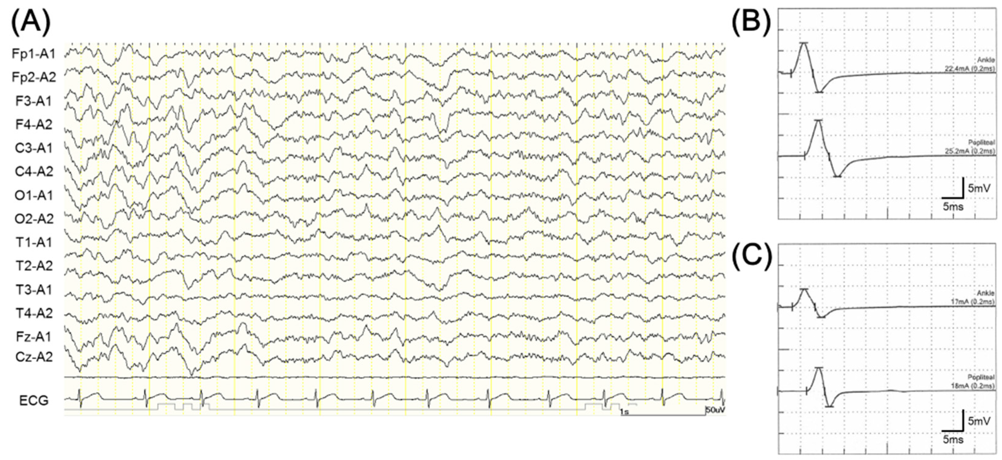
2. Case Presentation

The patient was a 3-year-old boy who was the second among two children from healthy non-consanguineous Japanese parents. He was born following a normal pregnancy without asphyxia or other perinatal events. He had regular psychomotor development until 19 months of age. The patient suffered from a viral infection with fever at 19 and 21 months of age. During those periods of febrile illness, he presented with generalized hypotonia with loss of gait and mutism without altered consciousness. He recovered slowly, and within 9 months was able to sit unassisted. At 2.7 years of age, during a febrile episode of adenovirus enteritis, a third neurological episode occurred, with severe hypotonia, ataxia, dyskinesia of the trunk and upper limbs, swallowing disorder, mutism, and less responsive dystonic movements of the bilateral upper and lower limbs with irritable state. Chorea athetosis was not observed. Routine electroencephalogram (EEG), recorded during awake and natural sleep (stage 1), did not show any high voltage slow wave epileptiform discharge. A photic stimulation showed no significant findings, and hyperventilation was not performed because the patient was unable to follow instructions. Nerve conduction studies (NCS) and auditory evoked potential did not show latency delay or amplitude reduction (Figure 1). Laboratory diagnostic workup, including metabolic investigations and cerebrospinal fluid (CSF) examination, revealed a slight increase in serum and CSF lactate (47.8 and 23.7 mg/dL, respectively) and pyruvate (2.84 and 1.40 mg/dL, respectively) levels. Plasma amino acid analyses revealed elevated plasma alanine levels (497.8 nmol/mL) and increased alanine-to-lysine ratio (Ala/Lys molar ratio = 4.1). Brain and spine magnetic resonance imaging (MRI) and skeletal muscle computed tomography (CT) showed no abnormalities. 99mTc-ethyl cysteinate dimer single photon emission computed tomography (SPECT) showed no significant abnormalities in cerebral blood flow in the cerebrum (right: 57.9 mL/100 g/min and left: 53.4 mL/100 g/min) and basal ganglia (right: 60.1 mL/100 g/min and left: 59.5 mL/100 g/min). However, mild hypoperfusion was observed in the cerebellum (right: 49.5 mL/100 g/min and left: 50.2 mL/100 g/min) (Figure 2). L-carnitine and thiamine were administered, and the patient did not experience any recurrent episodes during the febrile illness. At his most recent follow-up (at 3.5 years of age), he had gradually recovered partially but still had impaired language, generalized mild hypotonia, ataxia, dyskinesias of the trunk and upper limbs, and dystonia of the bilateral upper and lower limbs.
ATP1A3 exon sequencing showed a heterozygous mutation (Arg756Cys). The variant was classified as pathogenic because the same amino acid alternation has previously been identified in cases with similar phenotypic features.

3. Discussion

The α3 subunit of the Na+/K+-ATPase pump, which is the product of the ATP1A3 gene, participates in neuronal resting potential by maintaining the sodium/potassium gradient across the cell membrane. α3 subunit is selectively expressed in neurons and highly enriched in the basal ganglia, thalamus, and cerebellum [10]. Although the mechanisms of pathogenesis are unknown, the anatomical distribution of α3 may correlate with neurological deficits seen in clinical phenotypes, such as ATP1A3-related disorders, including FIPWE and RECA.
Altered consciousness, lack of reaction, bradykinesia, and mutism are sometimes reported in cases of FIPWE and RECA [2,9,10,11,12,13,14,15,16,17]. However, little is known about the pathophysiology of encephalopathy-like episodes, particularly altered consciousness with FIPWE and RECA. The patient, in this case, was also irritable and less responsive to neurological examinations or environmental stimuli. However, EEG recordings showed regular background activity with no encephalopathy-like slow waves in the acute phase of the episode. These EEG findings strongly suggest that cortical dysfunction was unlikely the underlying cause for the lowered responsiveness. Since EEG abnormalities are often lacking in reported cases (Table 1), the transiently altered state of consciousness represented by unresponsiveness and mutism is likely due to thalamic or cerebellar dysfunction rather than cortical dysfunction, considering the distribution of ATP1α3.
There are few reports on cerebral blood flow in ATP1A3-related diseases; a 99mTc- hexamethylpropylene amine oxime scan showed normal CBF in RDP [18]. Although this case was considered to be of FIPWE, there was a slight decrease in cerebellar blood flow, but no significant abnormal findings were observed in the cerebrum, as in previous reports of RDP. The SPECT findings also suggest that the pathophysiology of the lowered responsiveness seen in FIPWE may involve cerebellar dysfunction rather than cortical dysfunction.
Severe hypotonia and flaccid paralysis during febrile illness seems to be a common symptom in FIPWE and RECA caused by mutations at the Arg756 position [2,9,10,11,12,13,14,15,16,17]. However, the pathophysiology of hypotonia with FIPWE and RECA is not well described. In our case, the patient showed severe hypotonia and flaccid paralysis, while NCS, CSF, serum creatine kinase, and skeletal muscle CT did not present abnormalities, in line with previous reports (Table 1). From these findings, muscles and peripheral nerves were unlikely to be involved in the hypotonia. Although significant findings, such as cerebral blood flow abnormalities, cannot be ruled out, basal ganglia, including the internal capsule, may contribute to hypotonia and flaccid paralysis episodes in FIPWE and RECA rather than peripheral nerves.
As reported earlier and in our case, in FIPWE and RECA, it may be difficult to observe characteristic findings by conventional MRI or electrophysiological examination in only the acute phase. In order to understand the pathophysiology of FIPWE and RECA in more detail, it may be necessary to perform these assessments successively through the clinical course.

4. Conclusions

Though the patient in the present study with an ATP1A3-associated Arg756Cys mutation in experienced repeated encephalopathy-like episodes, including severe hypotonia during febrile illness, EEG and NCS did not present any obvious abnormalities. These electrophysiological features may be an opportunity to suspect FIPWE and RECA. Further studies are needed to characterize the features of electrophysiological examination in ATP1A3-related disorders caused by mutations at the Arg756 position, hopefully leading to better diagnostic strategies for this extremely rare disease.

Author Contributions

Conceptualization, F.S., T.F. and T.N.; methodology, T.N.; software, F.S.; validation, F.S., H.Y. and T.N.; formal analysis, T.N.; investigation, T.N.; resources, T.N.; data curation, T.N.; writing—original draft preparation, F.S.; writing—review and editing, Y.K.; visualization, F.S.; supervision, H.Y., H.K. and T.N.; project administration, T.I.; funding acquisition, F.S. All authors have read and agreed to the published version of the manuscript.

Funding

This research was funded by JSPS KAKENHI, grant number JP21K07793.

Institutional Review Board Statement

This work did not need to require ethical committee approval.

Informed Consent Statement

Informed consent was obtained from the patient’s parents. Written informed consent has been obtained from the patient’s parents to publish this paper.

Data Availability Statement

Not applicable.

Conflicts of Interest

The authors declare no conflict of interest.

References

  1. Carecchio, M.; Zorzi, G.; Ragona, F.; Zibordi, F.; Nardocci, N. ATP1A3-Related Disorders: An Update. Eur. J. Paediatr. Neurol. 2018, 22, 257–263. [Google Scholar] [CrossRef] [PubMed]
  2. Yano, S.T.; Silver, K.; Young, R.; DeBrosse, S.D.; Ebel, R.S.; Swoboda, K.J.; Acsadi, G. Fever-Induced Paroxysmal Weakness and Encephalopathy, a New Phenotype of ATP1A3 Mutation. Pediatr. Neurol. 2017, 73, 101–105. [Google Scholar] [CrossRef] [PubMed]
  3. Dard, R.; Mignot, C.; Durr, A.; Lesca, G.; Sanlaville, D.; Roze, E.; Mochel, F. Relapsing Encephalopathy with Cerebellar Ataxia Related to an ATP1A3 Mutation. Dev. Med. Child Neurol. 2015, 57, 1183–1186. [Google Scholar] [CrossRef] [PubMed] [Green Version]
  4. Brashear, A.; Mink, J.W.; Hill, D.F.; Boggs, N.; McCall, W.V.; Stacy, M.A.; Snively, B.; Light, L.S.; Sweadner, K.J.; Ozelius, L.J.; et al. ATP1A3 Mutations in Infants: A New Rapid-Onset Dystonia-Parkinsonism Phenotype Characterized by Motor Delay and Ataxia. Dev. Med. Child Neurol. 2012, 54, 1065–1067. [Google Scholar] [CrossRef] [PubMed] [Green Version]
  5. Fornarino, S.; Stagnaro, M.; Rinelli, M.; Tiziano, D.; Mancardi, M.M.; Traverso, M.; Veneselli, E.; De Grandis, E. Paroxysmal Features Responding to Flunarizine in a Child with Rapid-Onset Dystonia-Parkinsonism. Neurology 2014, 82, 2037–2038. [Google Scholar] [CrossRef] [PubMed]
  6. Kanemasa, H.; Fukai, R.; Sakai, Y.; Torio, M.; Miyake, N.; Lee, S.; Ono, H.; Akamine, S.; Nishiyama, K.; Sanefuji, M.; et al. De Novo p.Arg756Cys Mutation of ATP1A3 Causes an Atypical Form of Alternating Hemiplegia of Childhood with Prolonged Paralysis and Choreoathetosis. BMC Neurol. 2016, 16, 174. [Google Scholar] [CrossRef] [PubMed] [Green Version]
  7. Nicita, F.; Travaglini, L.; Sabatini, S.; Garavaglia, B.; Panteghini, C.; Valeriani, M.; Bertini, E.; Nardocci, N.; Vigevano, F.; Capuano, A. Childhood-Onset ATP1A3-Related Conditions: Report of Two New Cases of Phenotypic Spectrum. Park. Relat. Disord. 2016, 30, 81–82. [Google Scholar] [CrossRef] [PubMed]
  8. Nakamura, Y.; Hattori, A.; Nakashima, M.; Ieda, D.; Hori, I.; Negishi, Y.; Ando, N.; Matsumoto, N.; Saitoh, S. A De Novo p.Arg756Cys Mutation in ATP1A3 Causes a Distinct Phenotype with Prolonged Weakness and Encephalopathy Triggered by Fever. Brain Dev. 2018, 40, 222–225. [Google Scholar] [CrossRef] [PubMed]
  9. Sabouraud, P.; Riquet, A.; Spitz, M.A.; Deiva, K.; Nevsimalova, S.; Mignot, C.; Lesca, G.; Bednarek, N.; Doummar, D.; Pietrement, C.; et al. Relapsing Encephalopathy with Cerebellar Ataxia Are Caused by Variants Involving p.Arg756 in ATP1A3. Eur. J. Paediatr. Neurol. 2019, 23, 448–455. [Google Scholar] [CrossRef] [PubMed]
  10. Dobretsov, M.; Hayar, A.; Kockara, N.T.; Kozhemyakin, M.; Light, K.E.; Patyal, P.; Pierce, D.R.; Wight, P.A. A Transgenic Mouse Model to Selectively Identify α3 Na,K-ATPase Expressing Cells in the Nervous System. Neuroscience 2019, 398, 274–294. [Google Scholar] [CrossRef] [PubMed]
  11. Biela, M.; Rydzanicz, M.; Szymanska, K.; Pieniawska-Smiech, K.; Lewandowicz-Uszynska, A.; Chruszcz, J.; Benben, L.; Kuzior-Plawiak, M.; Szyld, P.; Jakubiak, A.; et al. Variants of ATP1A3 in Residue 756 Cause a Separate Phenotype of Relapsing Encephalopathy with Cerebellar Ataxia (RECA)-Report of Two Cases and Literature Review. Mol. Genet. Genom. Med. 2021, 9, e1772. [Google Scholar] [CrossRef] [PubMed]
  12. Hully, M.; Ropars, J.; Hubert, L.; Boddaert, N.; Rio, N.; Bernadelli, M.; Desguerre, I.; Cornmier, D.; Aire, V.; Munnich, A.; et al. Mosaicism in ATP1A3-related disorders: Not just a theoretical risk. Neurogenetics 2017, 18, 23–28. [Google Scholar] [CrossRef] [PubMed]
  13. Tan, A.H.; Ozelius, L.J.; Brashear, A.; Lang, A.E.; Ahmad-Annuar, A.; Tan, C.T.; Lim, S.Y. Rapid-Onset Dystonia-Parkinsonism in a Chinese Girl with a De Novo ATP1A3 c.2267G>A (p.R756H) Genetic Mutation. Mov. Disord. Clin. Pract. 2015, 2, 74–75. [Google Scholar] [CrossRef] [PubMed] [Green Version]
  14. Sousa, A.L.; Alonso, I.; Magalhães, M. A Portuguese Rapid-Onset Dystonia-Parkinsonism Case with Atypical Features. Neurol. Sci. 2017, 38, 1713–1714. [Google Scholar] [CrossRef] [PubMed]
  15. Jaffer, F.; Fawcett, K.; Sims, D.; Heger, A.; Houlden, H.; Hanna, M.G.; Kingston, H.; Sisodiya, S.M. Familial Childhood-Onset Progressive Cerebellar Syndrome Associated with the ATP1A3 Mutation. Neurol. Genet. 2017, 3, e145. [Google Scholar] [CrossRef] [PubMed] [Green Version]
  16. Schirinzi, T.; Graziola, F.; Nicita, F.; Travaglini, L.; Stregapede, F.; Valeriani, M.; Curatolo, P.; Bertini, E.; Vigevano, F.; Capuano, A. Childhood Rapid-Onset Ataxia: Expanding the Phenotypic Spectrum of ATP1A3 Mutations. Cerebellum 2018, 17, 489–493. [Google Scholar] [CrossRef] [PubMed]
  17. De Vrieze, J.; van de Laar, I.M.B.H.; de Rijk-van Andel, J.F.; Kamsteeg, E.J.; Kotsopoulos, I.A.W.; de Man, S.A. Expanding Phenotype of ATP1A3-Related Disorders: A Case Series. Child Neurol. Open 2021, 8, 2329048X211048068. [Google Scholar] [CrossRef] [PubMed]
  18. Zanotti-Fregonara, P.; Vidailhet, M.; Kas, A.; Ozelius, L.J.; Clot, F.; Hindié, E.; Ravasi, L.; Devaux, J.Y.; Roze, E. [123I]-FP-CIT and [99mTc]-HMPAO Single Photon Emission Computed Tomography in a New Sporadic Case of Rapid-Onset Dystonia-Parkinsonism. J. Neurol. Sci. 2008, 273, 148–151. [Google Scholar] [CrossRef] [PubMed]
Figure 1. Electrophysiological data from the patient on admission at the age of 2.7 years. (A) Electroencephalogram (EEG) on day 2 after admission. Although the patient presented a less responsive status to external stimuli, EEG did not reveal comatose encephalopathy. (B,C) Nerve conduction study of tibial nerve ((B) left; (C) right) did not show latency delay or amplitude reduction.
Children 10 00703 g001
Figure 2. Cerebral blood flow (CBF) evaluated by 99mTc-ethyl cysteinate dimer single photon emission computed tomography (SPECT). (A) No significant CBF abnormality in the cerebrum and basal ganglia is seen. (B) SPECT shows mild decrease in CBF in the cerebellum.
Children 10 00703 g002
Table 1. Clinical characteristics of patients with mutation in ATP1A3 gene at residue 756.
ReferencePresent Case[4][5][3][13][6][7][2][12]
Publication year 20122014201520152016201620172017
Number of patients111111152
MutationR756CR756HR756HR756CR756HR756CR756CR756H 3/5
R756L 2/5
R756C
Age at onset1y7m9m9m1y1m10y1y5m11m13m–3y9m–1y10m
Symptoms
Unresponsive+AnarthriaAnarthria+-++5/51/2
Hypotonia++++-++5/52/2
Mutism++-----0/50/2
Dystonia+++++++3/52/2
Ataxia-+-++-+5/52/2
Electrophysiological examination
EEGNormalNANormalNormalNormalNormalNormalNormal 1/4
Slow wave 3/4
NA
NCSNormalNormalNormalNANANANANormal 3/4
Absent F wave 1/4
NA
ABRNormal NANormalNormalNANA NA NANA
VEPNANANANormalNANANANANA
SEPNANANormalNormalNANANANANA
Reference[14][15][16][8][9][11][17]
Publication year2017201720182018201920212021
Number of patients1321322
MutationR756HR756HR756CR756CR756H 2/3
R756C 1/3
R756HR756C
Age at onset1y2m8m–5y1y6m–1y7m9m1y5m–5y6m1y3m–1y7m
Symptoms
Unresponsive+0/31/2+1/32/20/2
Hypotonia+3/32/2+2/32/22/2
Mutism-0/30/2-3/31/20/2
Dystonia+2/31/2+1/30/21/2
Ataxia+3/32/2+2/32/22/2
Electrophysiological examination
EEGgeneralized
epileptic discharges
NANANormalNormal 3/3NANormal 1/2
NCSNANANormal 1/2NANANANA
ABRNANANANANANANA
VEPNANANANANANANA
SEPNANANormal 1/2NANANANA
EEG: electroencephalography, NCS: nerve conduction study, ABR: auditory brainstem response, VEP: visual evoked potential, SEP: Somatosensory evoked potential, +: Showed symptoms, -: Did not show symptoms.
Disclaimer/Publisher’s Note: The statements, opinions and data contained in all publications are solely those of the individual author(s) and contributor(s) and not of MDPI and/or the editor(s). MDPI and/or the editor(s) disclaim responsibility for any injury to people or property resulting from any ideas, methods, instructions or products referred to in the content.

