Next Issue
Volume 9, August
Previous Issue
Volume 9, June


Biomedicines, Volume 9, Issue 7 (July 2021) – 147 articles

Cover Story (view full-size image): 
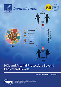
High-density lipoproteins (HDLs) can protect against atherosclerosis through multiple mechanisms, including reverse cholesterol transport, antioxidant, anti-inflammatory, antithrombotic and vasodilatory actions. The function of HDLs does not depend on the levels of a single component, such as cholesterol, but on the total chemical composition. Clinical conditions, such as obesity, metabolic syndrome and type 2 diabetes mellitus (T2DM), can cause an alteration in the chemical composition and consequently in atheroprotective properties of HDLs. View this paper
  • Issues are regarded as officially published after their release is announced to the table of contents alert mailing list.
  • You may sign up for e-mail alerts to receive table of contents of newly released issues.
  • PDF is the official format for papers published in both, html and pdf forms. To view the papers in pdf format, click on the "PDF Full-text" link, and use the free Adobe Reader to open them.
Order results
Result details
Section
Select all
Export citation of selected articles as:
12 pages, 1992 KiB  
Article
Highly Magnetized Encoded Hydrogel Microparticles with Enhanced Rinsing Capabilities for Efficient microRNA Detection
by Wookyoung Jang, Jiwoo Kim, Seok Joon Mun, Sun Min Kim and Ki Wan Bong
Biomedicines 2021, 9(7), 848; https://doi.org/10.3390/biomedicines9070848 - 20 Jul 2021
Cited by 4 | Viewed by 2979
Abstract
Encoded hydrogel microparticles mounting DNA probes are powerful tools for high-performance microRNA (miRNA) detection in terms of sensitivity, specificity, and multiplex detection capability. However, several particle rinsing steps in the assay procedure present challenges for rapid and efficient detection. To overcome this limitation, [...] Read more.
Encoded hydrogel microparticles mounting DNA probes are powerful tools for high-performance microRNA (miRNA) detection in terms of sensitivity, specificity, and multiplex detection capability. However, several particle rinsing steps in the assay procedure present challenges for rapid and efficient detection. To overcome this limitation, we encapsulated dense magnetic nanoparticles to reduce the rinsing steps and duration via magnetic separation. A large number of magnetic nanoparticles were encapsulated into hydrogel microparticles based on a discontinuous dewetting technique combined with degassed micromolding lithography. In addition, we attached DNA probes targeting three types of miRNAs related to preeclampsia to magnetically encoded hydrogel microparticles by post-synthesis conjugation and achieved sensitivity comparable to that of conventional nonmagnetic encoded hydrogel microparticles. To demonstrate the multiplex capability of magnetically encoded hydrogel microparticles while maintaining the advantages of the simplified rinsing process when addressing multiple samples, we conducted a triplex detection of preeclampsia-related miRNAs. In conclusion, the introduction of magnetically encoded hydrogel microparticles not only allowed efficient miRNA detection but also provided comparable sensitivity and multiplexed detectability to conventional nonmagnetic encoded hydrogel microparticles. Full article
Show Figures

Figure 1

9 pages, 692 KiB  
Article
Endothelial Progenitor Cells: Relevant Players in the Vasculopathy and Lung Fibrosis Associated with the Presence of Interstitial Lung Disease in Systemic Sclerosis Patients
by Verónica Pulito-Cueto, Sara Remuzgo-Martínez, Fernanda Genre, Belén Atienza-Mateo, Víctor M. Mora-Cuesta, David Iturbe-Fernández, Leticia Lera-Gómez, Raquel Pérez-Fernández, Diana Prieto-Peña, Virginia Portilla, Ricardo Blanco, Alfonso Corrales, Oreste Gualillo, José M. Cifrián, Raquel López-Mejías and Miguel A. González-Gay
Biomedicines 2021, 9(7), 847; https://doi.org/10.3390/biomedicines9070847 - 20 Jul 2021
Cited by 11 | Viewed by 2495
Abstract
Endothelial progenitor cells (EPC), which are key effectors in the physiologic vascular network, have been described as relevant players in autoimmune diseases. We previously showed that EPC frequency may help to identify the presence of interstitial lung disease (ILD) in rheumatoid arthritis patients. [...] Read more.
Endothelial progenitor cells (EPC), which are key effectors in the physiologic vascular network, have been described as relevant players in autoimmune diseases. We previously showed that EPC frequency may help to identify the presence of interstitial lung disease (ILD) in rheumatoid arthritis patients. Given that ILD constitutes the main cause of mortality in systemic sclerosis (SSc) patients, we aimed to determine the EPC contribution to the pathogenic processes of vasculopathy and lung fibrosis in SSc-ILD+. EPC quantification was performed by flow cytometry on blood from 83 individuals: 21 SSc-ILD+ patients and subjects from comparative groups (20 SSc-ILD and 21 idiopathic pulmonary fibrosis (IPF) patients and 21 healthy controls (HC)). EPC were considered as CD34+, CD45low, CD309+, and CD133+. A significant increase in EPC frequency was found in SSc-ILD+ patients when compared to HC (p < 0.001). SSc-ILD+ patients exhibited a higher EPC frequency than SSc-ILD patients (p = 0.012), whereas it was markedly reduced compared to IPF patients (p < 0.001). EPC frequency was higher in males (p = 0.04) and negatively correlated to SSc duration (p = 0.04) in SSc-ILD+ patients. Our results indicate a role of EPC in the processes of vasculopathy and lung fibrosis in SSc-ILD+. EPC frequency may be considered as a biomarker of ILD in SSc patients. Full article
Show Figures

Figure 1

14 pages, 2511 KiB  
Article
Microbiota Depletion Promotes Human Rotavirus Replication in an Adult Mouse Model
by Roberto Gozalbo-Rovira, Cristina Santiso-Bellón, Javier Buesa, Antonio Rubio-del-Campo, Susana Vila-Vicent, Carlos Muñoz, María J. Yebra, Vicente Monedero and Jesús Rodríguez-Díaz
Biomedicines 2021, 9(7), 846; https://doi.org/10.3390/biomedicines9070846 - 20 Jul 2021
Cited by 5 | Viewed by 2949
Abstract
Intestinal microbiota-virus-host interaction has emerged as a key factor in mediating enteric virus pathogenicity. With the aim of analyzing whether human gut bacteria improve the inefficient replication of human rotavirus in mice, we performed fecal microbiota transplant (FMT) with healthy infants as donors [...] Read more.
Intestinal microbiota-virus-host interaction has emerged as a key factor in mediating enteric virus pathogenicity. With the aim of analyzing whether human gut bacteria improve the inefficient replication of human rotavirus in mice, we performed fecal microbiota transplant (FMT) with healthy infants as donors in antibiotic-treated mice. We showed that a simple antibiotic treatment, irrespective of FMT, resulted in viral shedding for 6 days after challenge with the human rotavirus G1P[8] genotype Wa strain (RVwa). Rotavirus titers in feces were also significantly higher in antibiotic-treated animals with or without FMT but they were decreased in animals subject to self-FMT, where a partial re-establishment of specific bacterial taxons was evidenced. Microbial composition analysis revealed profound changes in the intestinal microbiota of antibiotic-treated animals, whereas some bacterial groups, including members of Lactobacillus, Bilophila, Mucispirillum, and Oscillospira, recovered after self-FMT. In antibiotic-treated and FMT animals where the virus replicated more efficiently, differences were observed in gene expression of immune mediators, such as IL1β and CXCL15, as well as in the fucosyltransferase FUT2, responsible for H-type antigen synthesis in the small intestine. Collectively, our results suggest that antibiotic-induced microbiota depletion eradicates the microbial taxa that restrict human rotavirus infectivity in mice. Full article
(This article belongs to the Special Issue Gut Dysbiosis: Molecular Mechanisms and Therapies)
Show Figures

Figure 1

18 pages, 1911 KiB  
Article
Serum miRNA Profiling for Early PDAC Diagnosis and Prognosis: A Retrospective Study
by Ada Aita, Caterina Millino, Cosimo Sperti, Beniamina Pacchioni, Mario Plebani, Cristiano De Pittà and Daniela Basso
Biomedicines 2021, 9(7), 845; https://doi.org/10.3390/biomedicines9070845 - 20 Jul 2021
Cited by 9 | Viewed by 3290
Abstract
Background: Tumor stage predicts pancreatic cancer (PDAC) prognosis, but prolonged and short survivals have been described in patients with early-stage tumors. Circulating microRNA (miRNA) are an emerging class of suitable biomarkers for PDAC prognosis. Our aim was to identify whether serum miRNA signatures [...] Read more.
Background: Tumor stage predicts pancreatic cancer (PDAC) prognosis, but prolonged and short survivals have been described in patients with early-stage tumors. Circulating microRNA (miRNA) are an emerging class of suitable biomarkers for PDAC prognosis. Our aim was to identify whether serum miRNA signatures predict survival of early-stage PDAC. Methods: Serum RNA from archival 15 stage I-III PDAC patients and 4 controls was used for miRNAs expression profile (Agilent microarrays). PDAC patients with comparable age, gender, diabetes, jaundice and surgery were classified according to survival: less than 14 months (7/15 pts, group A) and more than 22 months (8/15 pts, group B). Bioinformatic data analysis was performed by two-class Significance Analysis of Microarray (SAM) algorithm. Binary logistic regression analyses considering PDAC diagnosis and outcome as dependent variables, and ROC analyses were also performed. Results: 2549 human miRNAs were screened out. At SAM, 76 differentially expressed miRNAs were found among controls and PDAC (FDR = 0.4%), the large majority (50/76, 66%) of them being downregulated in PDAC with respect to controls. Six miRNAs were independently correlated with early PDAC, and among these, hsa-miR-6821-5p was associated with the best ROC curve area in distinguishing controls from early PDAC. Among the 71 miRNAs differentially expressed between groups A and B, the most significant were hsa-miR-3135b expressed in group A only, hsa-miR-6126 and hsa-miR-486-5p expressed in group B only. Eight miRNAs were correlated with the presence of lymph-node metastases; among these, hsa-miR-4669 is of potential interest. hsa-miR-4516, increased in PDAC and found as an independent predictor of survival, has among its putative targets a series of gens involved in key pathways of cancer progression and dissemination, such as Wnt and p53 signalling pathways. Conclusions: A series of serum miRNAs was identified as potentially useful for the early diagnosis of PDAC, and for establishing a prognosis. Full article
(This article belongs to the Special Issue MicroRNA in Solid Tumor and Hematological Diseases)
Show Figures

Figure 1

10 pages, 396 KiB  
Review
Diagnostic Challenges on the Laboratory Detection of Lupus Anticoagulant
by Armando Tripodi
Biomedicines 2021, 9(7), 844; https://doi.org/10.3390/biomedicines9070844 - 20 Jul 2021
Cited by 8 | Viewed by 5792
Abstract
Lupus anticoagulant (LA) is one of the three laboratory parameters (the others being antibodies to either cardiolipin or β2-glycoprotein I) which defines the rare but potentially devastating condition known as antiphospholipid syndrome (APS). Testing for LA is a challenging task for the clinical [...] Read more.
Lupus anticoagulant (LA) is one of the three laboratory parameters (the others being antibodies to either cardiolipin or β2-glycoprotein I) which defines the rare but potentially devastating condition known as antiphospholipid syndrome (APS). Testing for LA is a challenging task for the clinical laboratory because specific tests for its detection are not available. However, proper LA detection is paramount for patients’ management, as its persistent positivity in the presence of (previous or current) thrombotic events, candidate for long term anticoagulation. Guidelines for LA detection have been established and updated over the last two decades. Implementation of these guidelines across laboratories and participation to external quality assessment schemes are required to help standardize the diagnostic procedures and help clinicians for appropriate management of APS. This article aims to review the current state of the art and the challenges that clinical laboratories incur in the detection of LA. Full article
Show Figures

Figure 1

11 pages, 887 KiB  
Article
Clinical Phenotype Classification of Atrial Fibrillation Patients Using Cluster Analysis and Associations with Trial-Adjudicated Outcomes
by Marco Vitolo, Marco Proietti, Alena Shantsila, Giuseppe Boriani and Gregory Y. H. Lip
Biomedicines 2021, 9(7), 843; https://doi.org/10.3390/biomedicines9070843 - 20 Jul 2021
Cited by 17 | Viewed by 3209
Abstract
Background and purpose: Given the great clinical heterogeneity of atrial fibrillation (AF) patients, conventional classification only based on disease subtype or arrhythmia patterns may not adequately characterize this population. We aimed to identify different groups of AF patients who shared common clinical phenotypes [...] Read more.
Background and purpose: Given the great clinical heterogeneity of atrial fibrillation (AF) patients, conventional classification only based on disease subtype or arrhythmia patterns may not adequately characterize this population. We aimed to identify different groups of AF patients who shared common clinical phenotypes using cluster analysis and evaluate the association between identified clusters and clinical outcomes. Methods: We performed a hierarchical cluster analysis in AF patients from AMADEUS and BOREALIS trials. The primary outcome was a composite of stroke/thromboembolism (TE), cardiovascular (CV) death, myocardial infarction, and/or all-cause death. Individual components of the primary outcome and major bleeding were also assessed. Results: We included 3980 AF patients treated with the Vitamin-K Antagonist from the AMADEUS and BOREALIS studies. The analysis identified four clusters in which patients varied significantly among clinical characteristics. Cluster 1 was characterized by patients with low rates of CV risk factors and comorbidities; Cluster 2 was characterized by patients with a high burden of CV risk factors; Cluster 3 consisted of patients with a high burden of CV comorbidities; Cluster 4 was characterized by the highest rates of non-CV comorbidities. After a mean follow-up of 365 (standard deviation 187) days, Cluster 4 had the highest cumulative risk of outcomes. Compared with Cluster 1, Cluster 4 was independently associated with an increased risk for the composite outcome (hazard ratio (HR) 2.43, 95% confidence interval (CI) 1.70–3.46), all-cause death (HR 2.35, 95% CI 1.58–3.49) and major bleeding (HR 2.18, 95% CI 1.19–3.96). Conclusions: Cluster analysis identified four different clinically relevant phenotypes of AF patients that had unique clinical characteristics and different outcomes. Cluster analysis highlights the high degree of heterogeneity in patients with AF, suggesting the need for a phenotype-driven approach to comorbidities, which could provide a more holistic approach to management aimed to improve patients’ outcomes. Full article
Show Figures

Graphical abstract

17 pages, 1742 KiB  
Review
Ovarian Telomerase and Female Fertility
by Simon Toupance, Anne-Julie Fattet, Simon N. Thornton, Athanase Benetos, Jean-Louis Guéant and Isabelle Koscinski
Biomedicines 2021, 9(7), 842; https://doi.org/10.3390/biomedicines9070842 - 20 Jul 2021
Cited by 8 | Viewed by 5204
Abstract
Women’s fertility is characterized both quantitatively and qualitatively mainly by the pool of ovarian follicles. Monthly, gonadotropins cause an intense multiplication of granulosa cells surrounding the oocyte. This step of follicular development requires a high proliferation ability for these cells. Telomere length plays [...] Read more.
Women’s fertility is characterized both quantitatively and qualitatively mainly by the pool of ovarian follicles. Monthly, gonadotropins cause an intense multiplication of granulosa cells surrounding the oocyte. This step of follicular development requires a high proliferation ability for these cells. Telomere length plays a crucial role in the mitotic index of human cells. Hence, disrupting telomere homeostasis could directly affect women’s fertility. Strongly expressed in ovaries, telomerase is the most effective factor to limit telomeric attrition and preserve ovarian reserve. Considering these facts, two situations of infertility could be correlated with the length of telomeres and ovarian telomerase activity: PolyCystic Ovary Syndrome (PCOS), which is associated with a high density of small antral follicles, and Premature Ovarian Failure (POF), which is associated with a premature decrease in ovarian reserve. Several authors have studied this topic, expecting to find long telomeres and strong telomerase activity in PCOS and short telomeres and low telomerase activity in POF patients. Although the results of these studies are contradictory, telomere length and the ovarian telomerase impact in women’s fertility disorders appear obvious. In this context, our research perspectives aimed to explore the stimulation of ovarian telomerase to limit the decrease in the follicular pool while avoiding an increase in cancer risk. Full article
(This article belongs to the Special Issue Telomerase: Role in Health and Aging)
Show Figures

Graphical abstract

11 pages, 797 KiB  
Article
Relationship between Circulating PCSK9 and Markers of Subclinical Atherosclerosis—The IMPROVE Study
by Daniela Coggi, Beatrice Frigerio, Alice Bonomi, Massimiliano Ruscica, Nicola Ferri, Daniela Sansaro, Alessio Ravani, Palma Ferrante, Manuela Damigella, Fabrizio Veglia, Nicolò Capra, Maria Giovanna Lupo, Chiara Macchi, Kai Savonen, Angela Silveira, Sudhir Kurl, Philippe Giral, Matteo Pirro, Rona Juliette Strawbridge, Bruna Gigante, Andries Jan Smit, Elena Tremoli, Mauro Amato, Damiano Baldassarre and on behalf of the IMPROVE Study Groupadd Show full author list remove Hide full author list
Biomedicines 2021, 9(7), 841; https://doi.org/10.3390/biomedicines9070841 - 19 Jul 2021
Cited by 7 | Viewed by 2452
Abstract
(1) Background and purpose: circulating proprotein convertase subtilisin/kexin type 9 (PCSK9) is one of the key regulators of cholesterol metabolism. Despite this, its role as a player in atherosclerosis development is still matter of debate. Here, we investigated the relationships between this protein [...] Read more.
(1) Background and purpose: circulating proprotein convertase subtilisin/kexin type 9 (PCSK9) is one of the key regulators of cholesterol metabolism. Despite this, its role as a player in atherosclerosis development is still matter of debate. Here, we investigated the relationships between this protein and several markers of subclinical atherosclerosis. (2) Methods: the IMPROVE study enrolled 3703 European subjects (54–79 years; 48% men; with ≥3 vascular risk factors), asymptomatic for cardiovascular diseases. PCSK9 levels were measured by ELISA. B-mode ultrasound was used to measure markers of carotid subclinical atherosclerosis. (3) Results: in the crude analysis, PCSK9 levels were associated with several baseline measures of carotid intima-media thickness (cIMT) (all p < 0.0001); with cIMT change over time (Fastest-IMTmax-progr) (p = 0.01); with inter-adventitia common carotid artery diameter (ICCAD) (p < 0.0001); and with the echolucency (Grey Scale Median; GSM) of both carotid plaque and plaque-free common carotid IMT (both p < 0.0001). However, after adjustment for age, sex, latitude, and pharmacological treatment, all the afore-mentioned correlations were no longer statistically significant. The lack of correlation was also observed after stratification for sex, latitude, and pharmacological treatments. (4) Conclusions: in subjects who are asymptomatic for cardiovascular diseases, PCSK9 plasma levels do not correlate with vascular damage and/or subclinical atherosclerosis of extracranial carotid arteries. Full article
Show Figures

Figure 1

10 pages, 15835 KiB  
Article
The Alopecia Areata Phenotype Is Induced by the Water Avoidance Stress Test In cchcr1-Deficient Mice
by Qiaofeng Zhao, Satoshi Koyama, Nagisa Yoshihara, Atsushi Takagi, Etsuko Komiyama, Akino Wada, Akira Oka and Shigaku Ikeda
Biomedicines 2021, 9(7), 840; https://doi.org/10.3390/biomedicines9070840 - 19 Jul 2021
Cited by 5 | Viewed by 2515
Abstract
We recently discovered a nonsynonymous variant in the coiled-coil alpha-helical rod protein 1 (CCHCR1) gene within the alopecia areata (AA) risk haplotype. We also reported that the engineered mice with this risk allele exhibited. To investigate more about the involvement of [...] Read more.
We recently discovered a nonsynonymous variant in the coiled-coil alpha-helical rod protein 1 (CCHCR1) gene within the alopecia areata (AA) risk haplotype. We also reported that the engineered mice with this risk allele exhibited. To investigate more about the involvement of the CCHCR1 gene in AA pathogenesis, we developed an AA model using C57BL/6N cchcr1 gene knockout mice. In this study, mice (6–8 weeks) were divided into two groups: cchcr1/ mice and wild-type (WT) littermates. Both groups were subjected to a water avoidance stress (WAS) test. Eight weeks after the WAS test, 25% of cchcr1/ mice exhibited non-inflammatory foci of alopecia on the dorsal skin. On the other hand, none of wild-type littermates cause hair loss. The foci resembled human AA in terms of gross morphology, trichoscopic findings and histological findings. Additionally, gene expression microarray analysis of cchcr1/ mice revealed abnormalities of hair related genes compared to the control. Our results strongly suggest that CCHCR1 is associated with AA pathogenesis and that cchcr1/ mice are a good model for investigating AA. Full article
(This article belongs to the Special Issue Hair Pathology)
Show Figures

Figure 1

14 pages, 3613 KiB  
Article
TGF-β1 Receptor Inhibitor SB525334 Attenuates the Epithelial to Mesenchymal Transition of Peritoneal Mesothelial Cells via the TGF-β1 Signaling Pathway
by Jung-Yoon Heo, Jun-Young Do, Yunmee Lho, A-Young Kim, Sang-Woon Kim and Seok-Hui Kang
Biomedicines 2021, 9(7), 839; https://doi.org/10.3390/biomedicines9070839 - 19 Jul 2021
Cited by 7 | Viewed by 2507
Abstract
We investigated the effect of SB525334 (TGF-β receptor type 1 (TβRI) inhibitor) on the epithelial to mesenchymal transition (EMT) signaling pathway in human peritoneal mesothelial cells (HPMCs) and a peritoneal fibrosis mouse model. In vitro experiments were performed using HPMCs. HPMCs [...] Read more.
We investigated the effect of SB525334 (TGF-β receptor type 1 (TβRI) inhibitor) on the epithelial to mesenchymal transition (EMT) signaling pathway in human peritoneal mesothelial cells (HPMCs) and a peritoneal fibrosis mouse model. In vitro experiments were performed using HPMCs. HPMCs were treated with TGF-β1 and/or SB525334. In vivo experiments were conducted with male C57/BL6 mice. The 0.1% chlorhexidine gluconate (CG) was intraperitoneally injected with or without SB52534 administration by oral gavage. Mice were euthanized after 28 days. EMT using TGF-β1-treated HPMCs included morphological changes, cell migration and invasion, EMT markers and collagen synthesis. These pathological changes were reversed by co-treatment with SB525334. CG injection was associated with an increase in peritoneal fibrosis and thickness, which functionally resulted in an increase in the glucose absorption via peritoneum. Co-treatment with SB525334 attenuated these changes. The levels of EMT protein markers and immunohistochemical staining for fibrosis showed similar trends. Immunofluorescence staining for EMT markers showed induction of transformed cells with both epithelial and mesenchymal cell markers, which decreased upon co-treatment with SB525334. SB525334 effectively attenuated the TGF-β1-induced EMT in HPMCs. Cotreatment with SB525334 improved peritoneal thickness and fibrosis and recovered peritoneal membrane function in a peritoneal fibrosis mouse model. Full article
Show Figures

Figure 1

14 pages, 533 KiB  
Review
Lipoprotein(a) Where Do We Stand? From the Physiopathology to Innovative Terapy
by Gabriella Iannuzzo, Maria Tripaldella, Vania Mallardo, Mena Morgillo, Nicoletta Vitelli, Arcangelo Iannuzzi, Emilio Aliberti, Francesco Giallauria, Anna Tramontano, Raffaele Carluccio, Ilenia Calcaterra, Matteo Nicola Dario Di Minno and Marco Gentile
Biomedicines 2021, 9(7), 838; https://doi.org/10.3390/biomedicines9070838 - 19 Jul 2021
Cited by 15 | Viewed by 3114
Abstract
A number of epidemiologic studies have demonstrated a strong association between increasing lipoprotein a [Lp(a)] and cardiovascular disease. This correlation was demonstrated independent of other known cardiovascular (CV) risk factors. Screening for Lp(a) in the general population is not recommended, although Lp(a) levels [...] Read more.
A number of epidemiologic studies have demonstrated a strong association between increasing lipoprotein a [Lp(a)] and cardiovascular disease. This correlation was demonstrated independent of other known cardiovascular (CV) risk factors. Screening for Lp(a) in the general population is not recommended, although Lp(a) levels are predominantly genetically determined so a single assessment is needed to identify patients at risk. In 2019 ESC/EAS guidelines recommend Lp(a) measurement at least once a lifetime, fo subjects at very high and high CV risk and those with a family history of premature cardiovascular disease, to reclassify patients with borderline risk. As concerning medications, statins play a key role in lipid lowering therapy, but present poor efficacy on Lp(a) levels. Actually, treatment options for elevated serum levels of Lp(a) are very limited. Apheresis is the most effective and well tolerated treatment in patients with high levels of Lp(a). However, promising new therapies, in particular antisense oligonucleotides have showed to be able to significantly reduce Lp(a) in phase II RCT. This review provides an overview of the biology and epidemiology of Lp(a), with a view to future therapies. Full article
(This article belongs to the Special Issue Lipoproteins and Cardiovascular Diseases)
Show Figures

Figure 1

9 pages, 1217 KiB  
Article
Pitavastatin Ameliorates Lipopolysaccharide-Induced Blood-Brain Barrier Dysfunction
by Takashi Fujimoto, Yoichi Morofuji, Andrej Kovac, Michelle A. Erickson, Mária A. Deli, Masami Niwa and William A. Banks
Biomedicines 2021, 9(7), 837; https://doi.org/10.3390/biomedicines9070837 - 18 Jul 2021
Cited by 7 | Viewed by 3252
Abstract
Statins have neuroprotective effects on neurological diseases, including a pleiotropic effect possibly related to blood–brain barrier (BBB) function. In this study, we investigated the effects of pitavastatin (PTV) on lipopolysaccharide (LPS)-induced BBB dysfunction in an in vitro BBB model comprising cocultured primary mouse [...] Read more.
Statins have neuroprotective effects on neurological diseases, including a pleiotropic effect possibly related to blood–brain barrier (BBB) function. In this study, we investigated the effects of pitavastatin (PTV) on lipopolysaccharide (LPS)-induced BBB dysfunction in an in vitro BBB model comprising cocultured primary mouse brain endothelial cells, pericytes, and astrocytes. LPS (1 ng/mL, 24 h) increased the permeability and lowered the transendothelial electrical resistance of the BBB, and the co-administration of PTV prevented these effects. LPS increased the release of interleukin-6, granulocyte colony-stimulating factor, keratinocyte-derived chemokine, monocyte chemotactic protein-1, and regulated on activation, normal T-cell expressed and secreted from the BBB model. PTV inhibited the LPS-induced release of these cytokines. These results suggest that PTV can ameliorate LPS-induced BBB dysfunction, and these effects might be mediated through the inhibition of LPS-induced cytokine production. Clinically, therapeutic approaches using statins combined with novel strategies need to be designed. Our present finding sheds light on the pharmacological significance of statins in the treatment of central nervous system diseases. Full article
(This article belongs to the Special Issue Cytokines and Cytokine Receptors in Brain Homeostasis)
Show Figures

Figure 1

13 pages, 1092 KiB  
Review
High-Density Lipoprotein Subfractions: Much Ado about Nothing or Clinically Important?
by Knut Tore Lappegård, Christian Abendstein Kjellmo and Anders Hovland
Biomedicines 2021, 9(7), 836; https://doi.org/10.3390/biomedicines9070836 - 18 Jul 2021
Cited by 11 | Viewed by 3147
Abstract
High-density lipoproteins (HDL) are a heterogenous group of plasma molecules with a large variety in composition. There is a wide specter in lipid content and the number of different proteins that has been associated with HDL is approaching 100. Given this heterogeneity and [...] Read more.
High-density lipoproteins (HDL) are a heterogenous group of plasma molecules with a large variety in composition. There is a wide specter in lipid content and the number of different proteins that has been associated with HDL is approaching 100. Given this heterogeneity and the fact that the total amount of HDL is inversely related to the risk of coronary heart disease (CHD), there has been increasing interest in the function of specific HDL subgroups and in what way measuring and quantifying these subgroups could be of clinical importance in determining individual CHD risk. If certain subgroups appear to be more protective than others, it may also in the future be possible to pharmacologically increase beneficial and decrease harmful subgroups in order to reduce CHD risk. In this review we give a short historical perspective, summarize some of the recent clinical findings regarding HDL subclassifications and discuss why such classification may or may not be of clinical relevance. Full article
Show Figures

Figure 1

21 pages, 1579 KiB  
Review
Molecular Mechanisms of Resistance to Immune Checkpoint Inhibitors in Melanoma Treatment: An Update
by Sonja Vukadin, Farah Khaznadar, Tomislav Kizivat, Aleksandar Vcev and Martina Smolic
Biomedicines 2021, 9(7), 835; https://doi.org/10.3390/biomedicines9070835 - 18 Jul 2021
Cited by 22 | Viewed by 5091
Abstract
Over the past decade, immune checkpoint inhibitors (ICI) have revolutionized the treatment of advanced melanoma and ensured significant improvement in overall survival versus chemotherapy. ICI or targeted therapy are now the first line treatment in advanced melanoma, depending on the tumor v-raf murine [...] Read more.
Over the past decade, immune checkpoint inhibitors (ICI) have revolutionized the treatment of advanced melanoma and ensured significant improvement in overall survival versus chemotherapy. ICI or targeted therapy are now the first line treatment in advanced melanoma, depending on the tumor v-raf murine sarcoma viral oncogene homolog B1 (BRAF) mutational status. While these new approaches have changed the outcomes for many patients, a significant proportion of them still experience lack of response, known as primary resistance. Mechanisms of primary drug resistance are not fully elucidated. However, many alterations have been found in ICI-resistant melanomas and possibly contribute to that outcome. Furthermore, some tumors which initially responded to ICI treatment ultimately developed mechanisms of acquired resistance and subsequent tumor progression. In this review, we give an overview of tumor primary and acquired resistance mechanisms to ICI and discuss future perspectives with regards to new molecular targets and combinatorial therapies. Full article
Show Figures

Figure 1

22 pages, 3360 KiB  
Article
Alginate-Agarose Hydrogels Improve the In Vitro Differentiation of Human Dental Pulp Stem Cells in Chondrocytes. A Histological Study
by María Oliver-Ferrándiz, Lara Milián, María Sancho-Tello, José Javier Martín de Llano, Fernando Gisbert Roca, Cristina Martínez-Ramos, Carmen Carda and Manuel Mata
Biomedicines 2021, 9(7), 834; https://doi.org/10.3390/biomedicines9070834 - 17 Jul 2021
Cited by 7 | Viewed by 3034
Abstract
Matrix-assisted autologous chondrocyte implantation (MACI) has shown promising results for cartilage repair, combining cultured chondrocytes and hydrogels, including alginate. The ability of chondrocytes for MACI is limited by different factors including donor site morbidity, dedifferentiation, limited lifespan or poor proliferation in vitro. Mesenchymal [...] Read more.
Matrix-assisted autologous chondrocyte implantation (MACI) has shown promising results for cartilage repair, combining cultured chondrocytes and hydrogels, including alginate. The ability of chondrocytes for MACI is limited by different factors including donor site morbidity, dedifferentiation, limited lifespan or poor proliferation in vitro. Mesenchymal stem cells could represent an alternative for cartilage regeneration. In this study, we propose a MACI scaffold consisting of a mixed alginate-agarose hydrogel in combination with human dental pulp stem cells (hDPSCs), suitable for cartilage regeneration. Scaffolds were characterized according to their rheological properties, and their histomorphometric and molecular biology results. Agarose significantly improved the biomechanical behavior of the alginate scaffolds. Large scaffolds were manufactured, and a homogeneous distribution of cells was observed within them. Although primary chondrocytes showed a greater capacity for chondrogenic differentiation, hDPSCs cultured in the scaffolds formed large aggregates of cells, acquired a rounded morphology and expressed high amounts of type II collagen and aggrecan. Cells cultured in the scaffolds expressed not only chondral matrix-related genes, but also remodeling proteins and chondrocyte differentiation factors. The degree of differentiation of cells was proportional to the number and size of the cell aggregates that were formed in the hydrogels. Full article
(This article belongs to the Section Biomedical Materials and Nanomedicine)
Show Figures

Figure 1

17 pages, 1102 KiB  
Review
Role of PITRM1 in Mitochondrial Dysfunction and Neurodegeneration
by Dario Brunetti, Alessia Catania, Carlo Viscomi, Michela Deleidi, Laurence A. Bindoff, Daniele Ghezzi and Massimo Zeviani
Biomedicines 2021, 9(7), 833; https://doi.org/10.3390/biomedicines9070833 - 17 Jul 2021
Cited by 21 | Viewed by 4742
Abstract
Mounting evidence shows a link between mitochondrial dysfunction and neurodegenerative disorders, including Alzheimer Disease. Increased oxidative stress, defective mitodynamics, and impaired oxidative phosphorylation leading to decreased ATP production, can determine synaptic dysfunction, apoptosis, and neurodegeneration. Furthermore, mitochondrial proteostasis and the protease-mediated quality control [...] Read more.
Mounting evidence shows a link between mitochondrial dysfunction and neurodegenerative disorders, including Alzheimer Disease. Increased oxidative stress, defective mitodynamics, and impaired oxidative phosphorylation leading to decreased ATP production, can determine synaptic dysfunction, apoptosis, and neurodegeneration. Furthermore, mitochondrial proteostasis and the protease-mediated quality control system, carrying out degradation of potentially toxic peptides and misfolded or damaged proteins inside mitochondria, are emerging as potential pathogenetic mechanisms. The enzyme pitrilysin metallopeptidase 1 (PITRM1) is a key player in these processes; it is responsible for degrading mitochondrial targeting sequences that are cleaved off from the imported precursor proteins and for digesting a mitochondrial fraction of amyloid beta (Aβ). In this review, we present current evidence obtained from patients with PITRM1 mutations, as well as the different cellular and animal models of PITRM1 deficiency, which points toward PITRM1 as a possible driving factor of several neurodegenerative conditions. Finally, we point out the prospect of new diagnostic and therapeutic approaches. Full article
(This article belongs to the Special Issue Mitochondria and Brain Disease)
Show Figures

Graphical abstract

10 pages, 1231 KiB  
Article
Glucose-Dependent Insulinotropic Polypeptide Suppresses Foam Cell Formation of Macrophages through Inhibition of the Cyclin-Dependent Kinase 5-CD36 Pathway
by Michishige Terasaki, Hironori Yashima, Yusaku Mori, Tomomi Saito, Yoshie Shiraga, Raichi Kawakami, Makoto Ohara, Tomoyasu Fukui, Tsutomu Hirano, Yuichiro Yamada, Yutaka Seino and Sho-ichi Yamagishi
Biomedicines 2021, 9(7), 832; https://doi.org/10.3390/biomedicines9070832 - 16 Jul 2021
Cited by 7 | Viewed by 2196
Abstract
Glucose-dependent insulinotropic polypeptide (GIP) has been reported to have an atheroprotective property in animal models. However, the effect of GIP on macrophage foam cell formation, a crucial step of atherosclerosis, remains largely unknown. We investigated the effects of GIP on foam cell formation [...] Read more.
Glucose-dependent insulinotropic polypeptide (GIP) has been reported to have an atheroprotective property in animal models. However, the effect of GIP on macrophage foam cell formation, a crucial step of atherosclerosis, remains largely unknown. We investigated the effects of GIP on foam cell formation of, and CD36 expression in, macrophages extracted from GIP receptor-deficient (Gipr−/−) and Gipr+/+ mice and cultured human U937 macrophages by using an agonist for GIP receptor, [D-Ala2]GIP(1–42). Foam cell formation evaluated by esterification of free cholesterol to cholesteryl ester and CD36 gene expression in macrophages isolated from Gipr+/+ mice infused subcutaneously with [D-Ala2]GIP(1–42) were significantly suppressed compared with vehicle-treated mice, while these beneficial effects were not observed in macrophages isolated from Gipr−/− mice infused with [D-Ala2]GIP(1–42). When macrophages were isolated from Gipr+/+ and Gipr−/− mice, and then exposed to [D-Ala2]GIP(1–42), similar results were obtained. [D-Ala2]GIP(1–42) attenuated ox-LDL uptake of, and CD36 gene expression in, human U937 macrophages as well. Gene expression level of cyclin-dependent kinase 5 (Cdk5) was also suppressed by [D-Ala2]GIP(1–42) in U937 cells, which was corelated with that of CD36. A selective inhibitor of Cdk5, (R)-DRF053 mimicked the effects of [D-Ala2]GIP(1–42) in U937 cells. The present study suggests that GIP could inhibit foam cell formation of macrophages by suppressing the Cdk5-CD36 pathway via GIP receptor. Full article
(This article belongs to the Special Issue Macrophages in Health and Non-infectious Disease 2.0)
Show Figures

Figure 1

18 pages, 7219 KiB  
Article
IL-17A Damages the Blood–Retinal Barrier through Activating the Janus Kinase 1 Pathway
by Eimear M. Byrne, María Llorián-Salvador, Miao Tang, Andriana Margariti, Mei Chen and Heping Xu
Biomedicines 2021, 9(7), 831; https://doi.org/10.3390/biomedicines9070831 - 16 Jul 2021
Cited by 19 | Viewed by 2809
Abstract
Blood–retinal barrier (BRB) dysfunction underlies macular oedema in many sight-threatening conditions, including diabetic macular oedema, neovascular age-related macular degeneration and uveoretinitis. Inflammation plays an important role in BRB dysfunction. This study aimed to understand the role of the inflammatory cytokine IL-17A in BRB [...] Read more.
Blood–retinal barrier (BRB) dysfunction underlies macular oedema in many sight-threatening conditions, including diabetic macular oedema, neovascular age-related macular degeneration and uveoretinitis. Inflammation plays an important role in BRB dysfunction. This study aimed to understand the role of the inflammatory cytokine IL-17A in BRB dysfunction and the mechanism involved. Human retinal pigment epithelial (RPE) cell line ARPE19 and murine brain endothelial line bEnd.3 were cultured on transwell membranes to model the outer BRB and inner BRB, respectively. IL-17A treatment (3 days in bEnd.3 cells and 6 days in ARPE19 cells) disrupted the distribution of claudin-5 in bEnd.3 cells and ZO-1 in ARPE19 cells, reduced the transepithelial/transendothelial electrical resistance (TEER) and increased permeability to FITC-tracers in vitro. Intravitreal (20 ng/1 μL/eye) or intravenous (20 ng/g) injection of recombinant IL-17A induced retinal albumin leakage within 48 h in C57BL/6J mice. Mechanistically, IL-17A induced Janus kinase 1 (JAK1) phosphorylation in bEnd.3 but not ARPE19 cells. Blocking JAK1 with Tofacitinib prevented IL-17A-mediated claudin-5 dysmorphia in bEnd.3 cells and reduced albumin leakage in IL-17A-treated mice. Our results suggest that IL-17A can damage the BRB through the activating the JAK1 signaling pathway, and targeting this pathway may be a novel approach to treat inflammation-induced macular oedema. Full article
(This article belongs to the Special Issue Angiogenesis and Inflammation in Biological Barriers 2.0)
Show Figures

Figure 1

18 pages, 3210 KiB  
Article
Pulmonary MicroRNA Changes Alter Angiogenesis in Chronic Obstructive Pulmonary Disease and Lung Cancer
by Clara E. Green, Joseph Clarke, Roy Bicknell and Alice M. Turner
Biomedicines 2021, 9(7), 830; https://doi.org/10.3390/biomedicines9070830 - 16 Jul 2021
Cited by 9 | Viewed by 2351
Abstract
The pulmonary endothelium is dysfunctional in chronic obstructive pulmonary disease (COPD), a known risk factor for lung cancer. The pulmonary endothelium is altered in emphysema, which is disproportionately affected by cancers. Gene and microRNA expression differs between COPD and non-COPD lung. We hypothesised [...] Read more.
The pulmonary endothelium is dysfunctional in chronic obstructive pulmonary disease (COPD), a known risk factor for lung cancer. The pulmonary endothelium is altered in emphysema, which is disproportionately affected by cancers. Gene and microRNA expression differs between COPD and non-COPD lung. We hypothesised that the alteration in microRNA expression in the pulmonary endothelium contributes to its dysfunction. A total of 28 patients undergoing pulmonary resection were recruited and endothelial cells were isolated from healthy lung and tumour. MicroRNA expression was compared between COPD and non-COPD patients. Positive findings were confirmed by quantitative polymerase chain reaction (qPCR). Assays assessing angiogenesis and cellular migration were conducted in Human Umbilical Vein Endothelial Cells (n = 3–4) transfected with microRNA mimics and compared to cells transfected with negative control RNA. Expression of miR-181b-3p, miR-429 and miR-23c (all p < 0.05) was increased in COPD. Over-expression of miR-181b-3p was associated with reduced endothelial sprouting (p < 0.05). miR-429 was overexpressed in lung cancer as well and exhibited a reduction in tubular formation. MicroRNA-driven changes in the pulmonary endothelium thus represent a novel mechanism driving emphysema. These processes warrant further study to determine if they may be therapeutic targets in COPD and lung cancer. Full article
Show Figures

Graphical abstract

16 pages, 5360 KiB  
Article
Biphasic Effects of Blue Light Irradiation on Human Umbilical Vein Endothelial Cells
by Kejia Kan, Yifei Mu, Marielle Bouschbacher, Carsten Sticht, Natalia Kuch, Martin Sigl, Nuh Rahbari, Norbert Gretz, Prama Pallavi and Michael Keese
Biomedicines 2021, 9(7), 829; https://doi.org/10.3390/biomedicines9070829 - 16 Jul 2021
Cited by 4 | Viewed by 2452
Abstract
Blue light regulates biological function in various cells, such as proliferation, oxidative stress, and cell death. We employed blue light illumination on human umbilical vein endothelial cells utilizing a LED device at 453 nm wavelength and revealed a novel biphasic response on human [...] Read more.
Blue light regulates biological function in various cells, such as proliferation, oxidative stress, and cell death. We employed blue light illumination on human umbilical vein endothelial cells utilizing a LED device at 453 nm wavelength and revealed a novel biphasic response on human umbilical vein endothelial cells (HUVECs). The results showed that low fluence blue light irradiation promoted the fundamental cell activities, including cell viability, migration and angiogenesis by activating the angiogenic pathways such as the VEGF signaling pathway. In contrast, high fluence illumination caused the opposite effect on those activities by upregulating pro-apoptotic signaling cascades like ferroptosis, necroptosis and the p53 signaling pathways. Our results provide an underlying insight into photobiomodulation by blue light and may help to implement potential treatment strategies for treating angiogenesis-dependent diseases. Full article
Show Figures

Figure 1

23 pages, 3212 KiB  
Article
Antibody Protection against Long-Term Memory Loss Induced by Monomeric C-Reactive Protein in a Mouse Model of Dementia
by Elisa García-Lara, Samuel Aguirre, Núria Clotet, Xenia Sawkulycz, Clara Bartra, Lidia Almenara-Fuentes, Cristina Suñol, Rubén Corpas, Peter Olah, Florin Tripon, Andrei Crauciuc, Mark Slevin and Coral Sanfeliu
Biomedicines 2021, 9(7), 828; https://doi.org/10.3390/biomedicines9070828 - 16 Jul 2021
Cited by 11 | Viewed by 3182
Abstract
Monomeric C-reactive protein (mCRP), the activated isoform of CRP, induces tissue damage in a range of inflammatory pathologies. Its detection in infarcted human brain tissue and its experimentally proven ability to promote dementia with Alzheimer’s disease (AD) traits at 4 weeks after intrahippocampal [...] Read more.
Monomeric C-reactive protein (mCRP), the activated isoform of CRP, induces tissue damage in a range of inflammatory pathologies. Its detection in infarcted human brain tissue and its experimentally proven ability to promote dementia with Alzheimer’s disease (AD) traits at 4 weeks after intrahippocampal injection in mice have suggested that it may contribute to the development of AD after cerebrovascular injury. Here, we showed that a single hippocampal administration of mCRP in mice induced memory loss, lasting at least 6 months, along with neurodegenerative changes detected by increased levels of hyperphosphorylated tau protein and a decrease of the neuroplasticity marker Egr1. Furthermore, co-treatment with the monoclonal antibody 8C10 specific for mCRP showed that long-term memory loss and tau pathology were entirely avoided by early blockade of mCRP. Notably, 8C10 mitigated Egr1 decrease in the mouse hippocampus. 8C10 also protected against mCRP-induced inflammatory pathways in a microglial cell line, as shown by the prevention of increased generation of nitric oxide. Additional in vivo and in vitro neuroprotective testing with the anti-inflammatory agent TPPU, an inhibitor of the soluble epoxide hydrolase enzyme, confirmed the predominant involvement of neuroinflammatory processes in the dementia induced by mCRP. Therefore, locally deposited mCRP in the infarcted brain may be a novel biomarker for AD prognosis, and its antibody blockade opens up therapeutic opportunities for reducing post-stroke AD risk. Full article
(This article belongs to the Special Issue Biomarkers in Neurodegenerative Diseases 2.0)
Show Figures

Figure 1

14 pages, 881 KiB  
Review
Emerging Evidence of the Functional Impact of the miR379/miR656 Cluster (C14MC) in Breast Cancer
by Elan C. McCarthy and Róisín M. Dwyer
Biomedicines 2021, 9(7), 827; https://doi.org/10.3390/biomedicines9070827 - 16 Jul 2021
Cited by 4 | Viewed by 2870
Abstract
Many microRNAs exist in clusters that share comparable sequence homology and may target genes in a common pathway. The miR-379/miR-656 (C14MC) cluster is imprinted in the DLK1-Dio3 region of 14q32.3 and contains 42 miRNAs. It plays a functional role in numerous biological pathways [...] Read more.
Many microRNAs exist in clusters that share comparable sequence homology and may target genes in a common pathway. The miR-379/miR-656 (C14MC) cluster is imprinted in the DLK1-Dio3 region of 14q32.3 and contains 42 miRNAs. It plays a functional role in numerous biological pathways including vascular remodeling and early development. With many C14MC miRNAs highlighted as potential tumor suppressors in a variety of cancers, the role of this cluster in breast cancer (BC) has garnered increased attention in recent years. This review focuses on C14MC in BC, providing an overview of the constituent miRNAs and addressing each in terms of functional impact, potential target genes/pathways, and, where relevant, biomarker capacity. Studies have revealed the regulation of key factors in disease progression and metastasis including tyrosine kinase pathways and factors critical to epithelial–mesenchymal transition (EMT). This has potentially important clinical implications, with EMT playing a critical role in BC metastasis and tyrosine kinase inhibitors (TKIs) in widespread use for the treatment of BC. While the majority of studies have reported tumor-suppressing roles for these miRNAs, some have highlighted their potential as oncomiRs. Understanding the collective contribution of miRNAs within C14MC to BC may support improved understanding of disease etiology and present novel approaches to targeted therapy. Full article
(This article belongs to the Special Issue MicroRNA in Solid Tumor and Hematological Diseases 2.0)
Show Figures

Figure 1

17 pages, 328 KiB  
Review
Dyslipidaemia in Type 1 Diabetes: Molecular Mechanisms and Therapeutic Opportunities
by Stephen T. O’Brien, Orla M. Neylon and Timothy O’Brien
Biomedicines 2021, 9(7), 826; https://doi.org/10.3390/biomedicines9070826 - 16 Jul 2021
Cited by 6 | Viewed by 2820
Abstract
Cardiovascular disease (CVD) is the leading cause of death in Type 1 Diabetes (T1D). The molecular basis for atherosclerosis in T1D is heavily influenced by hyperglycaemia and its atherogenic effects on LDL. Ongoing research into the distinct pathophysiology of atherosclerosis in T1D offers [...] Read more.
Cardiovascular disease (CVD) is the leading cause of death in Type 1 Diabetes (T1D). The molecular basis for atherosclerosis in T1D is heavily influenced by hyperglycaemia and its atherogenic effects on LDL. Ongoing research into the distinct pathophysiology of atherosclerosis in T1D offers exciting opportunities for novel approaches to calculate CVD risk in patients with T1D and to manage this risk appropriately. Currently, despite the increased risk of CVD in the T1D population, there are few tools available for estimating the risk of CVD in younger patients. This poses significant challenges for clinicians in selecting which patients might benefit from lipid-lowering therapies over the long term. The current best practice guidance for the management of dyslipidaemia in T1D is generally based on evidence from patients with T2D and the opinion of experts in the field. In this review article, we explore the unique pathophysiology of atherosclerosis in T1D, with a specific focus on hyperglycaemia-induced damage and atherogenic LDL modifications. We also discuss the current clinical situation of managing these patients across paediatric and adult populations, focusing on the difficulties posed by a lack of strong evidence and various barriers to treatment. Full article
14 pages, 1447 KiB  
Article
Environmental Enrichment Rescues Endocannabinoid-Dependent Synaptic Plasticity Lost in Young Adult Male Mice after Ethanol Exposure during Adolescence
by Irantzu Rico-Barrio, Sara Peñasco, Leire Lekunberri, Maitane Serrano, Jon Egaña-Huguet, Amaia Mimenza, Edgar Soria-Gomez, Almudena Ramos, Ianire Buceta, Inmaculada Gerrikagoitia, Juan Mendizabal-Zubiaga, Izaskun Elezgarai, Nagore Puente and Pedro Grandes
Biomedicines 2021, 9(7), 825; https://doi.org/10.3390/biomedicines9070825 - 16 Jul 2021
Cited by 5 | Viewed by 3469
Abstract
Binge drinking (BD) is a serious health concern in adolescents as high ethanol (EtOH) consumption can have cognitive sequelae later in life. Remarkably, an enriched environment (EE) in adulthood significantly recovers memory in mice after adolescent BD, and the endocannabinoid, 2-arachydonoyl-glycerol (2-AG), rescues [...] Read more.
Binge drinking (BD) is a serious health concern in adolescents as high ethanol (EtOH) consumption can have cognitive sequelae later in life. Remarkably, an enriched environment (EE) in adulthood significantly recovers memory in mice after adolescent BD, and the endocannabinoid, 2-arachydonoyl-glycerol (2-AG), rescues synaptic plasticity and memory impaired in adult rodents upon adolescent EtOH intake. However, the mechanisms by which EE improves memory are unknown. We investigated this in adolescent male C57BL/6J mice exposed to a drinking in the dark (DID) procedure four days per week for a duration of 4 weeks. After DID, the mice were nurtured under an EE for 2 weeks and were subjected to the Barnes Maze Test performed the last 5 days of withdrawal. The EE rescued memory and restored the EtOH-disrupted endocannabinoid (eCB)-dependent excitatory long-term depression at the dentate medial perforant path synapses (MPP-LTD). This recovery was dependent on both the cannabinoid CB1 receptor and group I metabotropic glutamate receptors (mGluRs) and required 2-AG. Also, the EE had a positive effect on mice exposed to water through the transient receptor potential vanilloid 1 (TRPV1) and anandamide (AEA)-dependent MPP long-term potentiation (MPP-LTP). Taken together, EE positively impacts different forms of excitatory synaptic plasticity in water- and EtOH-exposed brains. Full article
(This article belongs to the Special Issue Biological Aspects of Drug Addiction)
Show Figures

Figure 1

19 pages, 6307 KiB  
Article
Elastomeric Cardiowrap Scaffolds Functionalized with Mesenchymal Stem Cells-Derived Exosomes Induce a Positive Modulation in the Inflammatory and Wound Healing Response of Mesenchymal Stem Cell and Macrophage
by Juan Carlos Chachques, Chiara Gardin, Nermine Lila, Letizia Ferroni, Veronique Migonney, Celine Falentin-Daudre, Federica Zanotti, Martina Trentini, Giulia Brunello, Tiberio Rocca, Vincenzo Gasbarro and Barbara Zavan
Biomedicines 2021, 9(7), 824; https://doi.org/10.3390/biomedicines9070824 - 15 Jul 2021
Cited by 20 | Viewed by 3181
Abstract
A challenge in contractile restoration of myocardial scars is one of the principal aims in cardiovascular surgery. Recently, a new potent biological tool used within healing processes is represented by exosomes derived from mesenchymal stem cells (MSCs). These cells are the well-known extracellular [...] Read more.
A challenge in contractile restoration of myocardial scars is one of the principal aims in cardiovascular surgery. Recently, a new potent biological tool used within healing processes is represented by exosomes derived from mesenchymal stem cells (MSCs). These cells are the well-known extracellular nanovesicles released from cells to facilitate cell function and communication. In this work, a combination of elastomeric membranes and exosomes was obtained and tested as a bioimplant. Mesenchymal stem cells (MSCs) and macrophages were seeded into the scaffold (polycaprolactone) and filled with exosomes derived from MSCs. Cells were tested for proliferation with an MTT test, and for wound healing properties and macrophage polarization by gene expression. Moreover, morphological analyses of their ability to colonize the scaffolds surfaces have been further evaluated. Results confirm that exosomes were easily entrapped onto the surface of the elastomeric scaffolds, increasing the wound healing properties and collagen type I and vitronectin of the MSC, and improving the M2 phenotype of the macrophages, mainly thanks to the increase in miRNA124 and decrease in miRNA 125. We can conclude that the enrichment of elastomeric scaffolds functionalized with exosomes is as an effective strategy to improve myocardial regeneration. Full article
(This article belongs to the Special Issue Tissue Engineering in Cardiology)
Show Figures

Figure 1

18 pages, 6127 KiB  
Article
Glia Not Neurons: Uncovering Brain Dysmaturation in a Rat Model of Alzheimer’s Disease
by Ekaterina A. Rudnitskaya, Tatiana A. Kozlova, Alena O. Burnyasheva, Natalia A. Stefanova and Nataliya G. Kolosova
Biomedicines 2021, 9(7), 823; https://doi.org/10.3390/biomedicines9070823 - 15 Jul 2021
Cited by 8 | Viewed by 2297
Abstract
Sporadic Alzheimer’s disease (AD) is a severe disorder of unknown etiology with no definite time frame of onset. Recent studies suggest that middle age is a critical period for the relevant pathological processes of AD. Nonetheless, sufficient data have accumulated supporting the hypothesis [...] Read more.
Sporadic Alzheimer’s disease (AD) is a severe disorder of unknown etiology with no definite time frame of onset. Recent studies suggest that middle age is a critical period for the relevant pathological processes of AD. Nonetheless, sufficient data have accumulated supporting the hypothesis of “neurodevelopmental origin of neurodegenerative disorders”: prerequisites for neurodegeneration may occur during early brain development. Therefore, we investigated the development of the most AD-affected brain structures (hippocampus and prefrontal cortex) using an immunohistochemical approach in senescence-accelerated OXYS rats, which are considered a suitable model of the most common—sporadic—type of AD. We noticed an additional peak of neurogenesis, which coincides in time with the peak of apoptosis in the hippocampus of OXYS rats on postnatal day three. Besides, we showed signs of delayed migration of neurons to the prefrontal cortex as well as disturbances in astrocytic and microglial support of the hippocampus and prefrontal cortex during the first postnatal week. Altogether, our results point to dysmaturation during early development of the brain—especially insufficient glial support—as a possible “first hit” leading to neurodegenerative processes and AD pathology manifestation later in life. Full article
(This article belongs to the Special Issue Alzheimer's Disease—115 Years after Its Discovery)
Show Figures

Figure 1

17 pages, 3701 KiB  
Article
Stable Gastric Pentadecapeptide BPC 157 Therapy for Monocrotaline-Induced Pulmonary Hypertension in Rats Leads to Prevention and Reversal
by Mario Udovicic, Marko Sever, Lovro Kavur, Kristina Loncaric, Ivan Barisic, Diana Balenovic, Gordana Zivanovic Posilovic, Dean Strinic, Sandra Uzun, Lovorka Batelja Vuletic, Suncana Sikiric, Anita Skrtic, Domagoj Drmic, Alenka Boban Blagaic, Martina Lovric Bencic, Sven Seiwerth and Predrag Sikiric
Biomedicines 2021, 9(7), 822; https://doi.org/10.3390/biomedicines9070822 - 15 Jul 2021
Cited by 14 | Viewed by 4100
Abstract
Background. Monocrotaline selectively injures the lung’s vascular endothelium and induces pulmonary arterial hypertension. The stable gastric pentadecapeptide BPC 157 acts as a prototype cytoprotective agent that maintains endothelium, and its application may be a novel therapy. Besides, BPC 157 prevents and reverses thrombosis [...] Read more.
Background. Monocrotaline selectively injures the lung’s vascular endothelium and induces pulmonary arterial hypertension. The stable gastric pentadecapeptide BPC 157 acts as a prototype cytoprotective agent that maintains endothelium, and its application may be a novel therapy. Besides, BPC 157 prevents and reverses thrombosis formation, maintains platelet function, alleviates peripheral vascular occlusion disturbances, and has anti-arrhythmic and anti-inflammatory effects. Monocrotaline-induced pulmonary arterial hypertension in rats (wall thickness, total vessel area, heart frequency, QRS axis deviation, QT interval prolongation, increase in right ventricle systolic pressure and bodyweight loss) can be counteracted with early or delayed BPC 157 therapy. Methods and Results. After monocrotaline (80 mg/kg subcutaneously), BPC 157 (10 μg/kg or 10 ng/kg, days 1–14 or days 1–30 (early regimens), or days 14–30 (delayed regimen)) was given once daily intraperitoneally (last application 24 h before sacrifice) or continuously in drinking water until sacrifice (day 14 or 30). Without therapy, the outcome was the full monocrotaline syndrome, marked by right-side heart hypertrophy and massive thickening of the precapillary artery’s smooth muscle layer, clinical deterioration, and sometimes death due to pulmonary hypertension and right-heart failure during the 4th week after monocrotaline injection. With all BPC 157 regimens, monocrotaline-induced pulmonary arterial hypertension (including all disturbed parameters) was counteracted, and consistent beneficial effects were documented during the whole course of the disease. Pulmonary hypertension was not even developed (early regimens) as quickly as the advanced pulmonary hypertension was rapidly attenuated and then completely eliminated (delayed regimen). Conclusions. Thus, pentadecapeptide BPC 157 prevents and counteracts monocrotaline-induced pulmonary arterial hypertension and cor pulmonale in rats. Full article
(This article belongs to the Special Issue Frontiers in Pentadecapeptide BPC 157)
Show Figures

Figure 1

16 pages, 1598 KiB  
Review
Dual Roles of the Activin Signaling Pathway in Pancreatic Cancer
by Wanglong Qiu, Chia-Yu Kuo, Yu Tian and Gloria H. Su
Biomedicines 2021, 9(7), 821; https://doi.org/10.3390/biomedicines9070821 - 14 Jul 2021
Cited by 5 | Viewed by 5255
Abstract
Activin, a member of the TGF-β superfamily, is involved in many physiological processes, such as embryonic development and follicle development, as well as in multiple human diseases including cancer. Genetic mutations in the activin signaling pathway have been reported in many cancer types, [...] Read more.
Activin, a member of the TGF-β superfamily, is involved in many physiological processes, such as embryonic development and follicle development, as well as in multiple human diseases including cancer. Genetic mutations in the activin signaling pathway have been reported in many cancer types, indicating that activin signaling plays a critical role in tumorigenesis. Recent evidence reveals that activin signaling may function as a tumor-suppressor in tumor initiation, and a promoter in the later progression and metastasis of tumors. This article reviews many aspects of activin, including the signaling cascade of activin, activin-related proteins, and its role in tumorigenesis, particularly in pancreatic cancer development. The mechanisms regulating its dual roles in tumorigenesis remain to be elucidated. Further understanding of the activin signaling pathway may identify potential therapeutic targets for human cancers and other diseases. Full article
(This article belongs to the Special Issue Feature Papers in Cancer Biology and Therapeutics)
Show Figures

Figure 1

10 pages, 1373 KiB  
Article
Sevoflurane and Desflurane Exposures Following Aneurysmal Subarachnoid Hemorrhage Confer Multifaceted Protection against Delayed Cerebral Ischemia
by Keshav Jayaraman, Meizi Liu, Gregory J. Zipfel and Umeshkumar Athiraman
Biomedicines 2021, 9(7), 820; https://doi.org/10.3390/biomedicines9070820 - 14 Jul 2021
Cited by 8 | Viewed by 1966
Abstract
Numerous studies have demonstrated the ability of isoflurane conditioning to provide multifaceted protection against aneurysmal subarachnoid hemorrhage (SAH)-associated delayed cerebral ischemia (DCI); however, preclinical studies have not yet examined whether other commonly used inhalational anesthetics in neurological patients such as sevoflurane or desflurane [...] Read more.
Numerous studies have demonstrated the ability of isoflurane conditioning to provide multifaceted protection against aneurysmal subarachnoid hemorrhage (SAH)-associated delayed cerebral ischemia (DCI); however, preclinical studies have not yet examined whether other commonly used inhalational anesthetics in neurological patients such as sevoflurane or desflurane are also protective against SAH-induced neurovascular deficits. We therefore sought to identify the potential for sevoflurane and desflurane conditioning to protect against DCI in an endovascular perforation mouse model of SAH. Neurological function was assessed daily via neuroscore. Large artery vasospasm and microvessel thrombosis were assessed three days after SAH or sham surgery. Four groups were examined: Sham, SAH + room air, SAH + 2% Sevoflurane, and SAH + 6% Desflurane. For the SAH groups, one hour after surgery, mice received 2% sevoflurane, 6% desflurane, or room air for one hour. We found that conditioning with sevoflurane or desflurane attenuated large artery vasospasm, reduced microvessel thrombosis, and improved neurologic function. Given their frequent clinical use and strong safety profile in patients (including those with SAH), these data strongly support further studies to validate these findings in preclinical and clinical studies and to elucidate the mechanisms by which these agents might be acting. Full article
(This article belongs to the Section Drug Discovery and Development)
Show Figures

Figure 1

25 pages, 4898 KiB  
Article
Hexapod Assassins’ Potion: Venom Composition and Bioactivity from the Eurasian Assassin Bug Rhynocoris iracundus
by Nicolai Rügen, Timothy P. Jenkins, Natalie Wielsch, Heiko Vogel, Benjamin-Florian Hempel, Roderich D. Süssmuth, Stuart Ainsworth, Alejandro Cabezas-Cruz, Andreas Vilcinskas and Miray Tonk
Biomedicines 2021, 9(7), 819; https://doi.org/10.3390/biomedicines9070819 - 14 Jul 2021
Cited by 6 | Viewed by 4719
Abstract
Assassin bug venoms are potent and exert diverse biological functions, making them potential biomedical goldmines. Besides feeding functions on arthropods, assassin bugs also use their venom for defense purposes causing localized and systemic reactions in vertebrates. However, assassin bug venoms remain poorly characterized. [...] Read more.
Assassin bug venoms are potent and exert diverse biological functions, making them potential biomedical goldmines. Besides feeding functions on arthropods, assassin bugs also use their venom for defense purposes causing localized and systemic reactions in vertebrates. However, assassin bug venoms remain poorly characterized. We collected the venom from the assassin bug Rhynocoris iracundus and investigated its composition and bioactivity in vitro and in vivo. It caused lysis of murine neuroblastoma, hepatoma cells, and healthy murine myoblasts. We demonstrated, for the first time, that assassin bug venom induces neurolysis and suggest that it counteracts paralysis locally via the destruction of neural networks, contributing to tissue digestion. Furthermore, the venom caused paralysis and melanization of Galleria mellonella larvae and pupae, whilst also possessing specific antibacterial activity against Escherichia coli, but not Listeria grayi and Pseudomonas aeruginosa. A combinatorial proteo-transcriptomic approach was performed to identify potential toxins responsible for the observed effects. We identified neurotoxic Ptu1, an inhibitory cystin knot (ICK) toxin homologous to ω-conotoxins from cone snails, cytolytic redulysins homologous to trialysins from hematophagous kissing bugs, and pore-forming hemolysins. Additionally, chitinases and kininogens were found and may be responsible for insecticidal and cytolytic activities. We demonstrate the multifunctionality and complexity of assassin bug venom, which renders its molecular components interesting for potential biomedical applications. Full article
(This article belongs to the Special Issue Recent Advances in the Discovery of Novel Drugs on Natural Molecules)
Show Figures

Graphical abstract

Previous Issue
Next Issue
Back to TopTop