Next Issue
Volume 8, October
Previous Issue
Volume 8, August


Biomedicines, Volume 8, Issue 9 (September 2020) – 80 articles

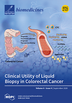
Cover Story (view full-size image): Evaluation of peripheral blood elements, such as circulating tumor cells (CTCs), circulating tumor DNA (ctDNA), microRNAs (miRNAs), and long non-coding RNAs (lncRNAs) could be useful for colorectal cancer screening and diagnosis and for predicting relapse and metastasis. Blood-based liquid biopsy could be helpful in monitoring minimal residual disease and drug resistance in colorectal cancer patients treated with chemotherapy. View this paper
  • Issues are regarded as officially published after their release is announced to the table of contents alert mailing list.
  • You may sign up for e-mail alerts to receive table of contents of newly released issues.
  • PDF is the official format for papers published in both, html and pdf forms. To view the papers in pdf format, click on the "PDF Full-text" link, and use the free Adobe Reader to open them.
Order results
Result details
Section
Select all
Export citation of selected articles as:
24 pages, 771 KiB  
Review
Positron Emission Tomography for Response Evaluation in Microenvironment-Targeted Anti-Cancer Therapy
by Noboru Oriuchi, Shigeyasu Sugawara and Tohru Shiga
Biomedicines 2020, 8(9), 371; https://doi.org/10.3390/biomedicines8090371 - 22 Sep 2020
Cited by 11 | Viewed by 3792
Abstract
Therapeutic response is evaluated using the diameter of tumors and quantitative parameters of 2-[18F] fluoro-2-deoxy-d-glucose positron emission tomography (FDG-PET). Tumor response to molecular-targeted drugs and immune checkpoint inhibitors is different from conventional chemotherapy in terms of temporal metabolic alteration [...] Read more.
Therapeutic response is evaluated using the diameter of tumors and quantitative parameters of 2-[18F] fluoro-2-deoxy-d-glucose positron emission tomography (FDG-PET). Tumor response to molecular-targeted drugs and immune checkpoint inhibitors is different from conventional chemotherapy in terms of temporal metabolic alteration and morphological change after the therapy. Cancer stem cells, immunologically competent cells, and metabolism of cancer are considered targets of novel therapy. Accumulation of FDG reflects the glucose metabolism of cancer cells as well as immune cells in the tumor microenvironment, which differs among patients according to the individual immune function; however, FDG-PET could evaluate the viability of the tumor as a whole. On the other hand, specific imaging and cell tracking of cancer cell or immunological cell subsets does not elucidate tumor response in a complexed interaction in the tumor microenvironment. Considering tumor heterogeneity and individual variation in therapeutic response, a radiomics approach with quantitative features of multimodal images and deep learning algorithm with reference to pathologic and genetic data has the potential to improve response assessment for emerging cancer therapy. Full article
(This article belongs to the Special Issue New Insights in Radiotherapy)
Show Figures

Figure 1

18 pages, 4718 KiB  
Article
Brassicasterol from Edible Aquacultural Hippocampus abdominalis Exerts an Anti-Cancer Effect by Dual-Targeting AKT and AR Signaling in Prostate Cancer
by Yinzhu Xu, Sooin Ryu, You-Kyung Lee and Hyo-Jeong Lee
Biomedicines 2020, 8(9), 370; https://doi.org/10.3390/biomedicines8090370 - 22 Sep 2020
Cited by 12 | Viewed by 3568
Abstract
In the Compendium of Materia Medica, seahorse (Hippocampus) is considered effective for the reinforcement of kidney and men’s health. However, the role of seahorse on human health lacks scientific evidence. Therefore, we evaluated the effect of seahorse on human prostate cancer [...] Read more.
In the Compendium of Materia Medica, seahorse (Hippocampus) is considered effective for the reinforcement of kidney and men’s health. However, the role of seahorse on human health lacks scientific evidence. Therefore, we evaluated the effect of seahorse on human prostate cancer using various in vitro methods and identified bioactive compound. Seahorse lipid extract (SHL) decreased androgen receptor (AR) and prostate-specific antigen (PSA) expression in dihydrotestosterone (DHT)-induced LNCaP cells of prostate cancer. Gas Chromatography (GC)-mass spectrometry data showed that brassicasterol was present in H. abdominalis. Brassicasterol downregulated the expression of AR and PSA in DHT-induced LNCaP cells. Brassicasterol induced apoptosis accompanied by sub-G1 phase arrest and inhibited migration in LNCaP cells. We confirmed that AKT and AR mediated the anti-cancer effect of brassicasterol using siRNA transfection. Brassicasterol exerts an anti-cancer effect in AR-independent cancer as well as in AR-dependent cells by AKT inhibiting. Our findings suggest that SHL has the anticancer potential via inhibition of AR and demonstrated that brassicasterol from H. abdominalis exerted an anti-cancer effect by dual-targeting AKT and AR signaling in prostate cancer. Full article
(This article belongs to the Section Molecular and Translational Medicine)
Show Figures

Figure 1

14 pages, 966 KiB  
Article
Chronic Electrical Stimulation of the Superior Laryngeal Nerve in the Rat: A Potential Therapeutic Approach for Postmenopausal Osteoporosis
by Kaori Iimura, Nobuhiro Watanabe, Philip Milliken, Yee-Hsee Hsieh, Stephen J. Lewis, Arun Sridhar and Harumi Hotta
Biomedicines 2020, 8(9), 369; https://doi.org/10.3390/biomedicines8090369 - 22 Sep 2020
Cited by 4 | Viewed by 2205
Abstract
Electrical stimulation of myelinated afferent fibers of the superior laryngeal nerve (SLN) facilitates calcitonin secretion from the thyroid gland in anesthetized rats. In this study, we aimed to quantify the electrical SLN stimulation-induced systemic calcitonin release in conscious rats and to then clarify [...] Read more.
Electrical stimulation of myelinated afferent fibers of the superior laryngeal nerve (SLN) facilitates calcitonin secretion from the thyroid gland in anesthetized rats. In this study, we aimed to quantify the electrical SLN stimulation-induced systemic calcitonin release in conscious rats and to then clarify effects of chronic SLN stimulation on bone mineral density (BMD) in a rat ovariectomized disease model of osteoporosis. Cuff electrodes were implanted bilaterally on SLNs and after two weeks recovery were stimulated (0.5 ms, 90 microampere) repetitively at 40 Hz for 8 min. Immunoreactive calcitonin release was initially measured and quantified in systemic venous blood plasma samples from conscious healthy rats. For chronic SLN stimulation, stimuli were applied intermittently for 3–4 weeks, starting at five weeks after ovariectomy (OVX). After the end of the stimulation period, BMD of the femur and tibia was measured. SLN stimulation increased plasma immunoreactive calcitonin concentration by 13.3 ± 17.3 pg/mL (mean ± SD). BMD in proximal metaphysis of tibia (p = 0.0324) and in distal metaphysis of femur (p = 0.0510) in chronically SLN-stimulated rats was 4–5% higher than that in sham rats. Our findings demonstrate chronic electrical stimulation of the SLNs produced enhanced calcitonin release from the thyroid gland and partially improved bone loss in OVX rats. Full article
(This article belongs to the Section Molecular and Translational Medicine)
Show Figures

Figure 1

23 pages, 8586 KiB  
Article
Novel 1,3,4-oxadiazole Targets STAT3 Signaling to Induce Antitumor Effect in Lung Cancer
by Vikas H. Malojirao, Swamy S. Girimanchanaika, Muthu K. Shanmugam, Ankith Sherapura, Dukanya, Prashant K. Metri, Vellingiri Vigneshwaran, Arunachalam Chinnathambi, Sulaiman Ali Alharbi, Shobith Rangappa, Chakrabhavi Dhananjaya Mohan, Basappa, Bettadathunga T. Prabhakar and Kanchugarakoppal S. Rangappa
Biomedicines 2020, 8(9), 368; https://doi.org/10.3390/biomedicines8090368 - 21 Sep 2020
Cited by 20 | Viewed by 3223
Abstract
Lung cancer is the leading type of malignancy in terms of occurrence and mortality in the global context. STAT3 is an oncogenic transcription factor that is persistently activated in many types of human malignancies, including lung cancer. In the present report, new oxadiazole [...] Read more.
Lung cancer is the leading type of malignancy in terms of occurrence and mortality in the global context. STAT3 is an oncogenic transcription factor that is persistently activated in many types of human malignancies, including lung cancer. In the present report, new oxadiazole conjugated indazoles were synthesized and examined for their anticancer potential in a panel of cancer cell lines. Among the new compounds, 2-(3-(6-chloro-5-methylpyridin-3-yl)phenyl)-5-(1-methyl-1H-indazol-3-yl)-1,3,4-oxadiazole (CHK9) showed consistently good cytotoxicity towards lung cancer cells with IC50 values ranging between 4.8–5.1 µM. The proapoptotic effect of CHK9 was further demonstrated by Annexin-FITC staining and TUNEL assay. In addition, the effect of CHK9 on the activation of STAT3 in lung cancer cells was examined. CHK9 reduced the phosphorylation of STAT3Y705 in a dose-dependent manner. CHK9 had no effect on the activation and expression of JAK2 and STAT5. It also reduced the STAT3-dependent luciferase reporter gene expression. CHK9 increased the expression of proapoptotic (p53 and Bax) proteins and decreased the expression of the antiapoptotic (Bcl-2, Bcl-xL, BID, and ICAM-1) proteins. CHK9 displayed a significant reduction in the number of tumor nodules in the in vivo lung cancer model with suppression of STAT3 activation in tumor tissues. CHK9 did not show substantial toxicity in the normal murine model. Overall, CHK9 inhibits the growth of lung cancer cells and tumors by interfering with the STAT3 signaling pathway. Full article
Show Figures

Figure 1

13 pages, 2252 KiB  
Article
Tauroursodeoxycholic Acid Protects Retinal Pigment Epithelial Cells from Oxidative Injury and Endoplasmic Reticulum Stress In Vitro
by Reem Hasaballah Alhasani, Mohammad Almarhoun, Xinzhi Zhou, James Reilly, Steven Patterson, Zhihong Zeng and Xinhua Shu
Biomedicines 2020, 8(9), 367; https://doi.org/10.3390/biomedicines8090367 - 21 Sep 2020
Cited by 19 | Viewed by 2585
Abstract
Retinal degeneration is characterized by the dysfunction of retinal cells. Oxidative and endoplasmic reticulum (ER) stress play an important role in the pathogenesis and progression of retinal degeneration. Tauroursodeoxycholic acid (TUDCA) has been demonstrated to have protective effects in in vitro and in [...] Read more.
Retinal degeneration is characterized by the dysfunction of retinal cells. Oxidative and endoplasmic reticulum (ER) stress play an important role in the pathogenesis and progression of retinal degeneration. Tauroursodeoxycholic acid (TUDCA) has been demonstrated to have protective effects in in vitro and in vivo retinal degeneration models. To fully understand the molecular mechanisms of TUDCA’s protection, we first treated human retinal pigment epithelial (RPE) cells, ARPE-19, with H2O2 or H2O2 plus TUDCA for 24 h. RPE cells co-exposed to TUDCA had higher cell viability and lower cell death rate compared to cells exposed to H2O2 alone. TUDCA significantly increased antioxidant capacity in H2O2-treated RPE cells by decreasing the generation of reactive oxygen species (ROS) and Malondialdehyde (MDA), upregulating the expression of antioxidant genes, and increasing the generation of glutathione (GSH). TUDCA also inhibited inflammation in H2O2-challenged RPE cells by decreasing the expression of proinflammatory cytokines. Furthermore, TUDCA suppressed thapsigargin-induced ER stress in RPE cells, as demonstrated by decreased the expression of CCAAT-enhancer-binding protein homologous protein (CHOP) and apoptosis. Our present study suggests that TUDCA can protect RPE cells against oxidative damage, inflammation, and ER stress and may benefit patients with retinal degeneration. Full article
(This article belongs to the Section Molecular and Translational Medicine)
Show Figures

Figure 1

16 pages, 2052 KiB  
Article
Plasma APE1/Ref-1 Correlates with Atherosclerotic Inflammation in ApoE−/− Mice
by Yu Ran Lee, Hee Kyoung Joo, Eun-Ok Lee, Myoung Soo Park, Hyun Sil Cho, Sungmin Kim, Hao Jin, Jin-Ok Jeong, Cuk-Seong Kim and Byeong Hwa Jeon
Biomedicines 2020, 8(9), 366; https://doi.org/10.3390/biomedicines8090366 - 21 Sep 2020
Cited by 15 | Viewed by 3107
Abstract
Apurinic/apyrimidinic endonuclease 1/redox factor-1 (APE1/Ref-1) is involved in DNA base repair and reducing activity. However, the role of APE1/Ref-1 in atherosclerosis is unclear. Herein, we investigated the role of APE1/Ref-1 in atherosclerotic apolipoprotein E (ApoE−/−) mice fed with a Western-type diet. [...] Read more.
Apurinic/apyrimidinic endonuclease 1/redox factor-1 (APE1/Ref-1) is involved in DNA base repair and reducing activity. However, the role of APE1/Ref-1 in atherosclerosis is unclear. Herein, we investigated the role of APE1/Ref-1 in atherosclerotic apolipoprotein E (ApoE−/−) mice fed with a Western-type diet. We found that serologic APE1/Ref-1 was strongly correlated with vascular inflammation in these mice. Neutrophil/lymphocyte ratio (NLR), endothelial cell/macrophage activation, and atherosclerotic plaque formation, reflected by atherosclerotic inflammation, were increased in the ApoE−/− mice fed with a Western-type diet. APE1/Ref-1 expression was upregulated in aortic tissues of these mice, and was co-localized with cells positive for cluster of differentiation 31 (CD31) and galectin-3, suggesting endothelial cell/macrophage expression of APE1/Ref-1. Interestingly, APE1/Ref-1 plasma levels of ApoE−/− mice fed with a Western-type diet were significantly increased compared with those of the mice fed with normal diet (15.76 ± 3.19 ng/mL vs. 3.51 ± 0.50 ng/mL, p < 0.05), and were suppressed by atorvastatin administration. Correlation analysis showed high correlation between plasma APE1/Ref-1 levels and NLR, a marker of systemic inflammation. The cut-off value for APE1/Ref-1 for predicting atherosclerotic inflammation at 4.903 ng/mL showed sensitivity of 100% and specificity of 91%. We conclude that APE1/Ref-1 expression is upregulated in aortic endothelial cells/macrophages of atherosclerotic mice, and that plasma APE1/Ref-1 levels could predict atherosclerotic inflammation. Full article
Show Figures

Graphical abstract

16 pages, 1938 KiB  
Article
Effect of Hypoxia Preconditioned Secretomes on Lymphangiogenic and Angiogenic Sprouting: An in Vitro Analysis
by Philipp Moog, Rahmin Schams, Alexander Schneidinger, Arndt F. Schilling, Hans-Günther Machens, Ektoras Hadjipanayi and Ulf Dornseifer
Biomedicines 2020, 8(9), 365; https://doi.org/10.3390/biomedicines8090365 - 20 Sep 2020
Cited by 11 | Viewed by 2727
Abstract
Hypoxia Preconditioned Plasma (HPP) and Serum (HPS) are two blood-derived autologous growth factor compositions that are being clinically employed as tools for promoting tissue regeneration, and have been extensively examined for their angiogenic activity. As yet, their ability to stimulate/support lymphangiogenesis remains unknown, [...] Read more.
Hypoxia Preconditioned Plasma (HPP) and Serum (HPS) are two blood-derived autologous growth factor compositions that are being clinically employed as tools for promoting tissue regeneration, and have been extensively examined for their angiogenic activity. As yet, their ability to stimulate/support lymphangiogenesis remains unknown, although this is an important but often-neglected process in wound healing and tissue repair. Here we set out to characterize the potential of hypoxia preconditioned secretomes as promoters of angiogenic and lymphangiogenic sprouting in vitro. We first analysed HPP/HPS in terms of pro- (VEGF-C) and anti- (TSP-1, PF-4) angiogenic/lymphangiogenic growth factor concentration, before testing their ability to stimulate microvessel sprouting in the mouse aortic ring assay and lymphatic sprouting in the thoracic duct ring assay. The origin of lymphatic structures was validated with lymph-specific immunohistochemical staining (Anti-LYVE-1) and lymphatic vessel-associated protein (polydom) quantification in culture supernatants. HPP/HPS induced greater angiogenic and lymphatic sprouting compared to non-hypoxia preconditioned samples (normal plasma/serum), a response that was compatible with their higher VEGF-C concentration. These findings demonstrate that hypoxia preconditioned blood-derived secretomes have the ability to not only support sprouting angiogenesis, but also lymphangiogenesis, which underlines their multimodal regenerative potential. Full article
(This article belongs to the Special Issue Hypoxia-Inducible Factors: Regulation and Therapeutic Potential)
Show Figures

Figure 1

21 pages, 3134 KiB  
Article
Capsaicin-Loaded Chitosan Nanocapsules for wtCFTR-mRNA Delivery to a Cystic Fibrosis Cell Line
by A. Katharina Kolonko, Janes Efing, Yadira González-Espinosa, Nadine Bangel-Ruland, Willy van Driessche, Francisco M. Goycoolea and Wolf-Michael Weber
Biomedicines 2020, 8(9), 364; https://doi.org/10.3390/biomedicines8090364 - 20 Sep 2020
Cited by 16 | Viewed by 4349
Abstract
Cystic fibrosis (CF), a lethal hereditary disease caused by mutations in the cystic fibrosis transmembrane conductance regulator (CFTR) gene coding for an epithelial chloride channel, is characterized by an imbalanced homeostasis of ion and water transports in secretory epithelia. As the disease is [...] Read more.
Cystic fibrosis (CF), a lethal hereditary disease caused by mutations in the cystic fibrosis transmembrane conductance regulator (CFTR) gene coding for an epithelial chloride channel, is characterized by an imbalanced homeostasis of ion and water transports in secretory epithelia. As the disease is single-gene based, transcript therapy using therapeutic mRNA is a promising concept of treatment in order to correct many aspects of the fatal pathology on a cellular level. Hence, we developed chitosan nanocapsules surface-loaded with wtCFTR-mRNA to restore CFTR function. Furthermore, we loaded the nanocapsules with capsaicin, aiming to enhance the overall efficiency of transcript therapy by reducing sodium hyperabsorption by the epithelial sodium channel (ENaC). Dynamic light scattering with non-invasive back scattering (DLS-NIBS) revealed nanocapsules with an average hydrodynamic diameter of ~200 nm and a Zeta potential of ~+60 mV. The results of DLS-NIBS measurements were confirmed by asymmetric flow field-flow fractionation (AF4) with multidetection, while transmission electron microscopy (TEM) images confirmed the spherical morphology and size range. After stability measurements showed that the nanocapsules were highly stable in cell culture transfection medium, and cytotoxicity was ruled out, transfection experiments were performed with the CF cell line CFBE41o-. Finally, transepithelial measurements with a new state-of-the-art Ussing chamber confirmed successfully restored CFTR function in transfected cells. This study demonstrates that CS nanocapsules as a natural and non-toxic delivery system for mRNA to target cells could effectively replace risky vectors for gene delivery. The nanocapsules are not only suitable as a transcript therapy for treatment of CF, but open aspiring possibilities for safe gene delivery in general. Full article
(This article belongs to the Section Biomedical Materials and Nanomedicine)
Show Figures

Figure 1

13 pages, 465 KiB  
Article
Impact of Subclinical Congestion on Outcome of Patients Undergoing Mitral Valve Surgery
by Anne-Kristin Schaefer, Thomas Poschner, Martin Andreas, Alfred Kocher, Günther Laufer, Dominik Wiedemann and Markus Mach
Biomedicines 2020, 8(9), 363; https://doi.org/10.3390/biomedicines8090363 - 19 Sep 2020
Cited by 2 | Viewed by 1656
Abstract
Since risk assessment prior to cardiac surgery is based on proven but partly unsatisfactory scores, the need for novel tools in preoperative risk assessment taking into account cardiac decompensation is obvious. Even subclinical chronic heart failure is accompanied by an increase in plasma [...] Read more.
Since risk assessment prior to cardiac surgery is based on proven but partly unsatisfactory scores, the need for novel tools in preoperative risk assessment taking into account cardiac decompensation is obvious. Even subclinical chronic heart failure is accompanied by an increase in plasma volume. This increase is illustrated by means of a plasma volume score (PVS), calculated using weight, gender and hematocrit. A retrospective analysis of 187 consecutive patients with impaired left ventricular function undergoing mitral valve surgery at a single centre between 2013 and 2016 was conducted. Relative preoperative PVS was generated by subtracting the ideal from actual calculated plasma volume. The study population was divided into two cohorts using a relative PVS score > 3.1 as cut-off. Patients with PVS > 3.1 had a significantly higher need for reoperation for bleeding/tamponade (5.5% vs. 16.7%; p = 0.016) and other non-cardiac causes (9.4% vs. 21.7%; p = 0.022). In-hospital as well as 6-month, 1-year and 5-year mortality was significantly increased in PVS > 3.1 (6.3% vs. 18.3%; p = 0.013; 9.4% vs. 23.3%; p = 0.011; 11.5% vs. 23.3%; p = 0.026; 18.1% vs. 33.3%; p = 0.018). Elevated PVS above the defined cut-off used to quantify subclinical congestion was linked to significantly worse outcome after mitral valve surgery and therefore could be a useful addition to current preoperative risk stratification. Full article
(This article belongs to the Section Molecular and Translational Medicine)
Show Figures

Figure 1

30 pages, 3650 KiB  
Review
Protein Dynamics in F-like Bacterial Conjugation
by Nicholas Bragagnolo, Christina Rodriguez, Naveed Samari-Kermani, Alice Fours, Mahboubeh Korouzhdehi, Rachel Lysenko and Gerald F. Audette
Biomedicines 2020, 8(9), 362; https://doi.org/10.3390/biomedicines8090362 - 19 Sep 2020
Cited by 12 | Viewed by 6484
Abstract
Efficient in silico development of novel antibiotics requires high-resolution, dynamic models of drug targets. As conjugation is considered the prominent contributor to the spread of antibiotic resistance genes, targeted drug design to disrupt vital components of conjugative systems has been proposed to lessen [...] Read more.
Efficient in silico development of novel antibiotics requires high-resolution, dynamic models of drug targets. As conjugation is considered the prominent contributor to the spread of antibiotic resistance genes, targeted drug design to disrupt vital components of conjugative systems has been proposed to lessen the proliferation of bacterial antibiotic resistance. Advancements in structural imaging techniques of large macromolecular complexes has accelerated the discovery of novel protein-protein interactions in bacterial type IV secretion systems (T4SS). The known structural information regarding the F-like T4SS components and complexes has been summarized in the following review, revealing a complex network of protein-protein interactions involving domains with varying degrees of disorder. Structural predictions were performed to provide insight on the dynamicity of proteins within the F plasmid conjugative system that lack structural information. Full article
(This article belongs to the Special Issue Protein Structure, Function and Dynamics in Diseases and Therapeutics)
Show Figures

Figure 1

30 pages, 6343 KiB  
Article
Dibenzoylthiamine Has Powerful Antioxidant and Anti-Inflammatory Properties in Cultured Cells and in Mouse Models of Stress and Neurodegeneration
by Margaux Sambon, Anna Gorlova, Alice Demelenne, Judit Alhama-Riba, Bernard Coumans, Bernard Lakaye, Pierre Wins, Marianne Fillet, Daniel C. Anthony, Tatyana Strekalova and Lucien Bettendorff
Biomedicines 2020, 8(9), 361; https://doi.org/10.3390/biomedicines8090361 - 18 Sep 2020
Cited by 19 | Viewed by 3424
Abstract
Thiamine precursors, the most studied being benfotiamine (BFT), have protective effects in mouse models of neurodegenerative diseases. BFT decreased oxidative stress and inflammation, two major characteristics of neurodegenerative diseases, in a neuroblastoma cell line (Neuro2a) and an immortalized brain microglial cell line (BV2). [...] Read more.
Thiamine precursors, the most studied being benfotiamine (BFT), have protective effects in mouse models of neurodegenerative diseases. BFT decreased oxidative stress and inflammation, two major characteristics of neurodegenerative diseases, in a neuroblastoma cell line (Neuro2a) and an immortalized brain microglial cell line (BV2). Here, we tested the potential antioxidant and anti-inflammatory effects of the hitherto unexplored derivative O,S-dibenzoylthiamine (DBT) in these two cell lines. We show that DBT protects Neuro2a cells against paraquat (PQ) toxicity by counteracting oxidative stress at low concentrations and increases the synthesis of reduced glutathione and NADPH in a Nrf2-independent manner. In BV2 cells activated by lipopolysaccharides (LPS), DBT significantly decreased inflammation by suppressing translocation of NF-κB to the nucleus. Our results also demonstrate the superiority of DBT over thiamine and other thiamine precursors, including BFT, in all of the in vitro models. Finally, we show that the chronic administration of DBT arrested motor dysfunction in FUS transgenic mice, a model of amyotrophic lateral sclerosis, and it reduced depressive-like behavior in a mouse model of ultrasound-induced stress in which it normalized oxidative stress marker levels in the brain. Together, our data suggest that DBT may have therapeutic potential for brain pathology associated with oxidative stress and inflammation by novel, coenzyme-independent mechanisms. Full article
(This article belongs to the Section Molecular and Translational Medicine)
Show Figures

Graphical abstract

12 pages, 1005 KiB  
Article
COL2A1 Is a Novel Biomarker of Melanoma Tumor Repopulating Cells
by Bhavana Talluri, Kshitij Amar, Michael Saul, Tasnim Shireen, Vjollca Konjufca, Jian Ma, Taekjip Ha and Farhan Chowdhury
Biomedicines 2020, 8(9), 360; https://doi.org/10.3390/biomedicines8090360 - 18 Sep 2020
Cited by 8 | Viewed by 3980
Abstract
Soft 3D-fibrin-gel selected tumor repopulating cells (TRCs) from the B16F1 melanoma cell line exhibit extraordinary self-renewal and tumor-regeneration capabilities. However, their biomarkers and gene regulatory features remain largely unknown. Here, we utilized the next-generation sequencing-based RNA sequencing (RNA-seq) technique to discover novel biomarkers [...] Read more.
Soft 3D-fibrin-gel selected tumor repopulating cells (TRCs) from the B16F1 melanoma cell line exhibit extraordinary self-renewal and tumor-regeneration capabilities. However, their biomarkers and gene regulatory features remain largely unknown. Here, we utilized the next-generation sequencing-based RNA sequencing (RNA-seq) technique to discover novel biomarkers and active gene regulatory features of TRCs. Systems biology analysis of RNA-seq data identified differentially expressed gene clusters, including the cell adhesion cluster, which subsequently identified highly specific and novel biomarkers, such as Col2a1, Ncam1, F11r, and Negr1. We validated the expression of these genes by real-time qPCR. The expression level of Col2a1 was found to be relatively low in TRCs but twenty-fold higher compared to the parental control cell line, thus making the biomarker very specific for TRCs. We validated the COL2A1 protein by immunofluorescence microscopy, showing a higher expression of COL2A1 in TRCs compared to parental control cells. KEGG pathway analysis showed the JAK/STAT, hypoxia, and Akt signaling pathways to be active in TRCs. Besides, the aerobic glycolysis pathway was found to be very active, indicating a typical Warburg Effect on highly tumorigenic cells. Together, our study revealed highly specific biomarkers and active cell signaling pathways of melanoma TRCs that can potentially target and neutralize TRCs. Full article
(This article belongs to the Special Issue Alternate Therapies and Proteomics/Genomics Studies of Cancer)
Show Figures

Figure 1

16 pages, 1813 KiB  
Article
A Multi-Omics Approach Reveals New Signatures in Obese Allergic Asthmatic Children
by Mª Amelia Gomez-Llorente, Ana Martínez-Cañavate, Natalia Chueca, Mª de la Cruz Rico, Raquel Romero, Augusto Anguita-Ruiz, Concepción Mª Aguilera, Mercedes Gil-Campos, Maria D Mesa, Bekzod Khakimov, Jose Antonio Morillo, Ángel Gil, José Camacho and Carolina Gomez-Llorente
Biomedicines 2020, 8(9), 359; https://doi.org/10.3390/biomedicines8090359 - 18 Sep 2020
Cited by 13 | Viewed by 2954
Abstract
Background: Asthma is a multifactorial condition where patients with identical clinical diagnoses do not have the same clinical history or respond to treatment. This clinical heterogeneity is reflected in the definition of two main endotypes. We aimed to explore the metabolic and microbiota [...] Read more.
Background: Asthma is a multifactorial condition where patients with identical clinical diagnoses do not have the same clinical history or respond to treatment. This clinical heterogeneity is reflected in the definition of two main endotypes. We aimed to explore the metabolic and microbiota signatures that characterize the clinical allergic asthma phenotype in obese children. Methods: We used a multi-omics approach combining clinical data, plasma and fecal inflammatory biomarkers, metagenomics, and metabolomics data in a cohort of allergic asthmatic children. Results: We observed that the obese allergic asthmatic phenotype was markedly associated with higher levels of leptin and lower relative proportions of plasma acetate and a member from the Clostridiales order. Moreover, allergic children with a worse asthma outcome showed higher levels of large unstained cells, fecal D lactate and D/L lactate ratio, and with a higher relative proportion of plasma creatinine and an unclassified family member from the RF39 order belonging to the Mollicutes class. Otherwise, children with persistent asthma presented lower levels of plasma citrate and dimethylsulfone. Conclusion: Our integrative approach shows the molecular heterogeneity of the allergic asthma phenotype while highlighting the use of omics technologies to examine the clinical phenotype at a more holistic level. Full article
(This article belongs to the Section Molecular and Translational Medicine)
Show Figures

Figure 1

17 pages, 2924 KiB  
Article
High-Dimensional Analysis of Immune Cell Composition Predicts Periprosthetic Joint Infections and Dissects Its Pathophysiology
by Maximilian F. Korn, Richard R. Stein, Andreas Dolf, Farhad Shakeri, Andreas Buness, Cäcilia Hilgers, Werner Masson, Sascha Gravius, Hendrik Kohlhof, Christof Burger, Dieter C. Wirtz, Thomas M. Randau and Frank A. Schildberg
Biomedicines 2020, 8(9), 358; https://doi.org/10.3390/biomedicines8090358 - 17 Sep 2020
Cited by 12 | Viewed by 2388
Abstract
Accurate diagnosis of periprosthetic joint infections (PJI) is one of the most widely researched areas in modern orthopedic endoprosthesis. However, our understanding of the immunological basis of this severe complication is still limited. In this study, we developed a flow cytometric approach to [...] Read more.
Accurate diagnosis of periprosthetic joint infections (PJI) is one of the most widely researched areas in modern orthopedic endoprosthesis. However, our understanding of the immunological basis of this severe complication is still limited. In this study, we developed a flow cytometric approach to precisely characterize the immune cell composition in periprosthetic joints. Using high-dimensional multi-parametric data, we defined, for the first time, the local immune cell populations of artificial joints. We identified significant differences in the cellular distribution between infected and non-infected samples, and revealed that myeloid-derived suppressor cells (MDSCs) act as potential regulators of infiltrating immune cells in PJI. Further, we developed an algorithm to predict septic and aseptic samples with high sensitivity and specificity, that may serve as an indispensable addition to the current criteria of the Musculoskeletal Infection Society. This study describes a novel approach to flow cytometrically analyze the immune cell infiltrate of joint fluid that not only improves our understanding of the pathophysiology of PJI, but also enables the development of a novel screening tool to predict infection status. Our data further suggest that pharmacological targeting of MDSCs represents a novel strategy for addressing PJI. Full article
(This article belongs to the Section Molecular and Translational Medicine)
Show Figures

Figure 1

14 pages, 3612 KiB  
Article
Cinnamaldehyde and Hyperthermia Co-Treatment Synergistically Induces ROS-Mediated Apoptosis in ACHN Renal Cell Carcinoma Cells
by Chae Ryeong Ahn, Jinbong Park, Jai-Eun Kim, Kwang Seok Ahn, Young Woo Kim, Minjeong Jeong, Hong Jun Kim, Sun Hyang Park and Seung Ho Baek
Biomedicines 2020, 8(9), 357; https://doi.org/10.3390/biomedicines8090357 - 17 Sep 2020
Cited by 7 | Viewed by 2738
Abstract
Renal cell carcinoma (RCC) represents the most common form of kidney cancer, which accounts for 3–5% newly diagnosed cancer cases. Since limited therapies are available for RCC, a search for new options is required. Therefore, in this study, we evaluated the combination effect [...] Read more.
Renal cell carcinoma (RCC) represents the most common form of kidney cancer, which accounts for 3–5% newly diagnosed cancer cases. Since limited therapies are available for RCC, a search for new options is required. Therefore, in this study, we evaluated the combination effect of cinnamaldehyde (CNM) and hyperthermia treatment. CNM treatment combined with 43 °C hyperthermia synergistically increased cytotoxicity in RCC cell line ACHN cells. Through Western blot assays, we observed increased apoptosis signaling and decreased proliferation/metastasis signaling, along with a repressed heat shock protein 70 level. In flow cytometry analyses, CNM and hyperthermia combination clearly induced apoptosis and mitochondrial potential of ACHN cells, while arresting the cell cycle. Investigation of reactive oxygen species (ROS) suggested a significant increase of ROS generation by CNM and 43 °C hyperthermia co-treatment. We could verify that ROS is crucial in the apoptotic action of combination treatment with CNM and hyperthermia through further experiments regarding an ROS scavenger. Overall, we suggest CNM and hyperthermia combination treatment as an alternative option of anticancer strategies for RCC. Full article
Show Figures

Figure 1

13 pages, 4481 KiB  
Article
Accurate Measurement of Copper Overload in an Experimental Model of Wilson Disease by Laser Ablation Inductively Coupled Plasma Mass Spectrometry
by Philipp Kim, Chengcheng Christine Zhang, Sven Thoröe-Boveleth, Sabine Weiskirchen, Nadine Therese Gaisa, Eva Miriam Buhl, Wolfgang Stremmel, Uta Merle and Ralf Weiskirchen
Biomedicines 2020, 8(9), 356; https://doi.org/10.3390/biomedicines8090356 - 16 Sep 2020
Cited by 8 | Viewed by 2965
Abstract
Wilson disease is a rare inherited autosomal recessive disorder. As a consequence of genetic alterations in the ATP7B gene, copper begins to accumulate in the body, particularly in the liver and brain. Affected persons are prone to develop liver cancer and severe psychiatric [...] Read more.
Wilson disease is a rare inherited autosomal recessive disorder. As a consequence of genetic alterations in the ATP7B gene, copper begins to accumulate in the body, particularly in the liver and brain. Affected persons are prone to develop liver cancer and severe psychiatric and neurological symptoms. Clinically, the development of corneal Kayser-Fleischer rings and low ceruloplasmin concentrations (<20 mg/dL) are indicative of Wilson disease. However, the detection of elevated hepatic copper content (>250 µg/g dry weight) alone is still considered as the best but not exclusive diagnostic test for Wilson disease. Presently, specific copper stains (e.g., rhodanine) or indirect staining for copper-associated proteins (e.g., orcein) are widely used to histochemically visualize hepatic copper deposits. However, these procedures only detect lysosomal copper, while cytosolic copper is not detectable. Similarly, elemental analysis in scanning electron microscope with energy dispersive X-ray analysis (EDX) often leads to false negative results and inconsistencies. Here, we tested the diagnostic potential of laser ablation inductively-coupled mass spectrometry (LA-ICP-MS) that allows quantitative analysis of multiple elements. Comparative studies were performed in wild type and the Atp7b null mouse model. We propose LA-ICP-MS as a versatile and powerful method for the accurate determination of hepatic copper in people with Wilson disease with high spatial resolution. Full article
(This article belongs to the Special Issue Zinc and Copper in Human Health and Disease)
Show Figures

Graphical abstract

21 pages, 2668 KiB  
Article
The Inhibition of Complement System in Formal and Emerging Indications: Results from Parallel One-Stage Pairwise and Network Meta-Analyses of Clinical Trials and Real-Life Data Studies
by Coralina Bernuy-Guevara, Hassib Chehade, Yannick D. Muller, Julien Vionnet, François Cachat, Gabriella Guzzo, Carlos Ochoa-Sangrador, F. Javier Álvarez, Daniel Teta, Débora Martín-García, Marcel Adler, Félix J. de Paz, Frank Lizaraso-Soto, Manuel Pascual and Francisco Herrera-Gómez
Biomedicines 2020, 8(9), 355; https://doi.org/10.3390/biomedicines8090355 - 16 Sep 2020
Cited by 11 | Viewed by 4155
Abstract
This manuscript presents quantitative findings on the actual effectiveness of terminal complement component 5 (C5) inhibitors and complement component 1 (C1) esterase inhibitors through their formal and common “off-label” (compassionate) indications. The results emanated from pairwise and network meta-analyses to present evidence until [...] Read more.
This manuscript presents quantitative findings on the actual effectiveness of terminal complement component 5 (C5) inhibitors and complement component 1 (C1) esterase inhibitors through their formal and common “off-label” (compassionate) indications. The results emanated from pairwise and network meta-analyses to present evidence until September 2019. Clinical trials (CT) and real-life non-randomized studies of the effects of interventions (NRSI) are consistent on the benefits of C5 inhibitors and of the absence of effects of C1 esterase inhibitors (n = 7484): Mathematically, eculizumab (surface under the cumulative ranking area (SUCRA) >0.6) and ravulizumab (SUCRA ≥ 0.7) were similar in terms of their protective effect on hemolysis in paroxysmal nocturnal hemoglobinuria (PNH), thrombotic microangiopathy (TMA) in atypical hemolytic uremic syndrome (aHUS), and acute kidney injury (AKI) in aHUS, in comparison to pre-/off-treatment state and/or placebo (SUCRA < 0.01), and eculizumab was efficacious on thrombotic events in PNH (odds ratio (OR)/95% confidence interval (95% CI) in CT and real-life NRSI, 0.07/0.03 to 0.19, 0.24/0.17 to 0.33) and chronic kidney disease (CKD) occurrence/progression in PNH (0.31/0.10 to 0.97, 0.66/0.44 to 0.98). In addition, meta-analysis on clinical trials shows that eculizumab mitigates a refractory generalized myasthenia gravis (rgMG) crisis (0.29/0.13 to 0.61) and prevents new acute antibody-mediated rejection (AMR) episodes in kidney transplant recipients (0.25/0.13 to 0.49). The update of findings from this meta-analysis will be useful to promote a better use of complement inhibitors, and to achieve personalization of treatments with this class of drugs. Full article
(This article belongs to the Section Molecular and Translational Medicine)
Show Figures

Graphical abstract

14 pages, 758 KiB  
Article
ICP-Mass-Spectrometry Ionic Profile of Whole Saliva in Patients with Untreated and Treated Periodontitis
by Federica Romano, Alexandra Castiblanco, Francesca Spadotto, Federica Di Scipio, Mery Malandrino, Giovanni Nicolao Berta and Mario Aimetti
Biomedicines 2020, 8(9), 354; https://doi.org/10.3390/biomedicines8090354 - 15 Sep 2020
Cited by 18 | Viewed by 2587
Abstract
Over the past decade, there has been growing interest in the association between macro and trace minerals in body fluids and systemic diseases related to chronic inflammation and oxidative stress. Due to the paucity of data in the literature on periodontitis, the aim [...] Read more.
Over the past decade, there has been growing interest in the association between macro and trace minerals in body fluids and systemic diseases related to chronic inflammation and oxidative stress. Due to the paucity of data in the literature on periodontitis, the aim of this cross-sectional study was to assess the relationship between mineral elements in saliva and periodontal status in patients with untreated and treated periodontitis compared to periodontally healthy controls. Salivary samples from 66 nonsmoker healthy patients (20 periodontally healthy, 24 untreated severe periodontitis and 22 treated severe periodontitis) were analyzed by using inductively coupled plasma mass-spectrometry (ICP-MS). Significant increases in copper (Cu), sodium (Na), iron (Fe) and manganese (Mn) concentrations occurred in saliva of severe periodontitis subjects compared to periodontally healthy controls. No differences were detected between healthy controls and treated periodontitis patients apart from levels of zinc (Zn) and lithium (Li) that were found to be increased and reduced, respectively, in periodontitis group. Most subjects were correctly separated by cluster analysis into active periodontitis and periodontally healthy individuals. Treated periodontitis individuals were classified as healthy subjects. Based on these preliminary results, the assessment of salivary concentration of mineral elements might be useful in discriminating periodontal health and disease. Full article
(This article belongs to the Special Issue Models for Oral Biology Research)
Show Figures

Figure 1

14 pages, 1089 KiB  
Article
Polygenic Markers in Patients Diagnosed of Autosomal Dominant Hypercholesterolemia in Catalonia: Distribution of Weighted LDL-c-Raising SNP Scores and Refinement of Variant Selection
by Jesús M. Martín-Campos, Sheila Ruiz-Nogales, Daiana Ibarretxe, Emilio Ortega, Elisabet Sánchez-Pujol, Meritxell Royuela-Juncadella, Àlex Vila, Carolina Guerrero, Alberto Zamora, Cristina Soler i Ferrer, Juan Antonio Arroyo, Gemma Carreras, Susana Martínez-Figueroa, Rosa Roig, Núria Plana, Francisco Blanco-Vaca and Xarxa d’Unitats de Lípids i Arteriosclerosi (XULA)
Biomedicines 2020, 8(9), 353; https://doi.org/10.3390/biomedicines8090353 - 15 Sep 2020
Cited by 6 | Viewed by 2580
Abstract
Familial hypercholesterolemia (FH) is associated with mutations in the low-density lipoprotein (LDL) receptor (LDLR), apolipoprotein B (APOB), and proprotein convertase subtilisin/kexin 9 (PCSK9) genes. A pathological variant has not been identified in 30–70% of clinically diagnosed FH [...] Read more.
Familial hypercholesterolemia (FH) is associated with mutations in the low-density lipoprotein (LDL) receptor (LDLR), apolipoprotein B (APOB), and proprotein convertase subtilisin/kexin 9 (PCSK9) genes. A pathological variant has not been identified in 30–70% of clinically diagnosed FH patients, and a burden of LDL cholesterol (LDL-c)-raising alleles has been hypothesized as a potential cause of hypercholesterolemia in these patients. Our aim was to study the distribution of weighted LDL-c-raising single-nucleotide polymorphism (SNP) scores (weighted gene scores or wGS) in a population recruited in a clinical setting in Catalonia. The study included 670 consecutive patients with a clinical diagnosis of FH and a prior genetic study involving 250 mutation-positive (FH/M+) and 420 mutation-negative (FH/M−) patients. Three wGSs based on LDL-c-raising variants were calculated to evaluate their distribution among FH patients and compared with 503 European samples from the 1000 Genomes Project. The FH/M− patients had significantly higher wGSs than the FH/M+ and control populations, with sensitivities ranging from 42% to 47%. A wGS based only on the SNPs significantly associated with FH (wGS8) showed a higher area under the receiver operating characteristic curve, and higher diagnostic specificity and sensitivity, with 46.4% of the subjects in the top quartile. wGS8 would allow for the assignment of a genetic cause to 66.4% of the patients if those with polygenic FH are added to the 37.3% of patients with monogenic FH. Our data indicate that a score based on 8 SNPs and the75th percentile cutoff point may identify patients with polygenic FH in Catalonia, although with limited diagnostic sensitivity and specificity. Full article
(This article belongs to the Section Molecular and Translational Medicine)
Show Figures

Graphical abstract

16 pages, 4561 KiB  
Article
Honokiol Protects the Kidney from Renal Ischemia and Reperfusion Injury by Upregulating the Glutathione Biosynthetic Enzymes
by Eun Jung Park, Theodomir Dusabimana, Jihyun Je, Kyuho Jeong, Seung Pil Yun, Hye Jung Kim, Hwajin Kim and Sang Won Park
Biomedicines 2020, 8(9), 352; https://doi.org/10.3390/biomedicines8090352 - 15 Sep 2020
Cited by 27 | Viewed by 3760
Abstract
Glutathione (GSH) is an endogenous antioxidant found in plants, animals, fungi, and some microorganisms that protects cells by neutralizing hydrogen peroxide. Honokiol, an active ingredient of Magnolia officinalis, is known for antioxidant, anti-inflammatory, and anti-bacterial properties. We investigated the protective mechanism of [...] Read more.
Glutathione (GSH) is an endogenous antioxidant found in plants, animals, fungi, and some microorganisms that protects cells by neutralizing hydrogen peroxide. Honokiol, an active ingredient of Magnolia officinalis, is known for antioxidant, anti-inflammatory, and anti-bacterial properties. We investigated the protective mechanism of honokiol through regulating cellular GSH in renal proximal tubules against acute kidney injury (AKI). First, we measured cellular GSH levels and correlated them with the expression of GSH biosynthetic enzymes after honokiol treatment in human kidney-2 (HK-2) cells. Second, we used pharmacological inhibitors or siRNA-mediated gene silencing approach to determine the signaling pathway induced by honokiol. Third, the protective effect of honokiol via de novo GSH biosynthesis was investigated in renal ischemia-reperfusion (IR) mice. Honokiol significantly increased cellular GSH levels by upregulating the subunits of glutamate-cysteine ligase (Gcl)—Gclc and Gclm. These increases were mediated by activation of nuclear factor erythroid 2-related factor 2, via PI3K/Akt and protein kinase C signaling. Consistently, honokiol treatment reduced the plasma creatinine, tubular cell death, neutrophil infiltration and lipid peroxidation in IR mice and the effect was correlated with upregulation of Gclc and Gclm. Conclusively, honokiol may benefit to patients with AKI by increasing antioxidant GSH via transcriptional activation of the biosynthetic enzymes. Full article
(This article belongs to the Section Molecular and Translational Medicine)
Show Figures

Figure 1

14 pages, 2161 KiB  
Article
Simvastatin Impairs Glucose Homeostasis in Mice Depending on PGC-1α Skeletal Muscle Expression
by Miljenko Valentin Panajatovic, François Singh, Stephan Krähenbühl and Jamal Bouitbir
Biomedicines 2020, 8(9), 351; https://doi.org/10.3390/biomedicines8090351 - 15 Sep 2020
Cited by 4 | Viewed by 2491
Abstract
Several studies showed an increased risk for diabetes with statin treatment. PGC-1α is an important regulator of muscle energy metabolism and mitochondrial biogenesis. Since statins impair skeletal muscle PGC-1α expression and reduced PGC-1α expression has been observed in diabetic patients, we investigated the [...] Read more.
Several studies showed an increased risk for diabetes with statin treatment. PGC-1α is an important regulator of muscle energy metabolism and mitochondrial biogenesis. Since statins impair skeletal muscle PGC-1α expression and reduced PGC-1α expression has been observed in diabetic patients, we investigated the possibility that skeletal muscle PGC1α expression influences the effect of simvastatin on muscle glucose metabolism. Mice with muscle PGC-1α knockout (KO) or PGC-1α overexpression (OE), and wild-type (WT) mice were investigated. Mice were treated orally for 3 weeks with simvastatin (5 mg/kg/day) and investigated by intraperitoneal glucose tolerance (iGTT), in vivo skeletal muscle glucose uptake, muscle glycogen content, and Glut4 and hexokinase mRNA and protein expression. Simvastatin impaired glucose metabolism in WT mice, as manifested by increased glucose blood concentrations during the iGTT, decreased skeletal muscle glucose uptake and glycogen stores. KO mice showed impaired glucose homeostasis with increased blood glucose concentrations during the iGTT already without simvastatin treatment and simvastatin induced a decrease in skeletal muscle glucose uptake. In OE mice, simvastatin treatment increased blood glucose and insulin concentrations during the iGTT, and increased skeletal muscle glucose uptake, glycogen stores, and Glut4 and hexokinase protein expression. In conclusion, simvastatin impaired skeletal muscle insulin sensitivity in WT mice, while KO mice exhibited impaired skeletal muscle insulin sensitivity already in the absence of simvastatin. In OE mice, simvastatin augmented muscular glucose uptake but impaired whole-body insulin sensitivity. Thus, simvastatin affected glucose homeostasis depending on PGC-1α expression. Full article
(This article belongs to the Section Molecular and Translational Medicine)
Show Figures

Figure 1

29 pages, 6732 KiB  
Article
Mitochondrial Transfer by Human Mesenchymal Stromal Cells Ameliorates Hepatocyte Lipid Load in a Mouse Model of NASH
by Mei-Ju Hsu, Isabel Karkossa, Ingo Schäfer, Madlen Christ, Hagen Kühne, Kristin Schubert, Ulrike E. Rolle-Kampczyk, Stefan Kalkhof, Sandra Nickel, Peter Seibel, Martin von Bergen and Bruno Christ
Biomedicines 2020, 8(9), 350; https://doi.org/10.3390/biomedicines8090350 - 14 Sep 2020
Cited by 20 | Viewed by 4539
Abstract
Mesenchymal stromal cell (MSC) transplantation ameliorated hepatic lipid load; tissue inflammation; and fibrosis in rodent animal models of non-alcoholic steatohepatitis (NASH) by as yet largely unknown mechanism(s). In a mouse model of NASH; we transplanted bone marrow-derived MSCs into the livers; which were [...] Read more.
Mesenchymal stromal cell (MSC) transplantation ameliorated hepatic lipid load; tissue inflammation; and fibrosis in rodent animal models of non-alcoholic steatohepatitis (NASH) by as yet largely unknown mechanism(s). In a mouse model of NASH; we transplanted bone marrow-derived MSCs into the livers; which were analyzed one week thereafter. Combined metabolomic and proteomic data were applied to weighted gene correlation network analysis (WGCNA) and subsequent identification of key drivers. Livers were analyzed histologically and biochemically. The mechanisms of MSC action on hepatocyte lipid accumulation were studied in co-cultures of hepatocytes and MSCs by quantitative image analysis and immunocytochemistry. WGCNA and key driver analysis revealed that NASH caused the impairment of central carbon; amino acid; and lipid metabolism associated with mitochondrial and peroxisomal dysfunction; which was reversed by MSC treatment. MSC improved hepatic lipid metabolism and tissue homeostasis. In co-cultures of hepatocytes and MSCs; the decrease of lipid load was associated with the transfer of mitochondria from the MSCs to the hepatocytes via tunneling nanotubes (TNTs). Hence; MSCs may ameliorate lipid load and tissue perturbance by the donation of mitochondria to the hepatocytes. Thereby; they may provide oxidative capacity for lipid breakdown and thus promote recovery from NASH-induced metabolic impairment and tissue injury. Full article
(This article belongs to the Special Issue NASH and Systemic Complications: From Basic to Clinical Research)
Show Figures

Graphical abstract

17 pages, 3710 KiB  
Article
Lung Microbiome Differentially Impacts Survival of Patients with Non-Small Cell Lung Cancer Depending on Tumor Stroma Phenotype
by Olga Kovaleva, Polina Podlesnaya, Madina Rashidova, Daria Samoilova, Anatoly Petrenko, Irina Zborovskaya, Valeria Mochalnikova, Vladimir Kataev, Yuri Khlopko, Andrey Plotnikov and Alexei Gratchev
Biomedicines 2020, 8(9), 349; https://doi.org/10.3390/biomedicines8090349 - 13 Sep 2020
Cited by 27 | Viewed by 4040
Abstract
The link between a lung tumor and the lung microbiome is a largely unexplored issue. To investigate the relationship between a lung microbiome and the phenotype of an inflammatory stromal infiltrate, we studied a cohort of 89 patients with non-small cell lung cancer. [...] Read more.
The link between a lung tumor and the lung microbiome is a largely unexplored issue. To investigate the relationship between a lung microbiome and the phenotype of an inflammatory stromal infiltrate, we studied a cohort of 89 patients with non-small cell lung cancer. The microbiome was analyzed in tumor and adjacent normal tissue by 16S rRNA amplicon sequencing. Characterization of the tumor stroma was done using immunohistochemistry. We demonstrated that the bacterial load was higher in adjacent normal tissue than in a tumor (p = 0.0325) with similar patterns of taxonomic structure and alpha diversity. Lung adenocarcinomas did not differ in their alpha diversity from squamous cell carcinomas, although the content of Gram-positive bacteria increased significantly in the adenocarcinoma group (p = 0.0419). An analysis of an inflammatory infiltrate of tumor stroma showed a correlation of CD68, iNOS and FOXP3 with a histological type of tumor. For the first time we showed that high bacterial load in the tumor combined with increased iNOS expression is a favorable prognostic factor (HR = 0.1824; p = 0.0123), while high bacterial load combined with the increased number of FOXP3+ cells is a marker of poor prognosis (HR = 4.651; p = 0.0116). Thus, we established that bacterial load of the tumor has an opposite prognostic value depending on the status of local antitumor immunity. Full article
(This article belongs to the Special Issue Macrophages in Health and Non-infectious Disease)
Show Figures

Figure 1

21 pages, 7689 KiB  
Review
Utility of Reactive Species Generation in Plasma Medicine for Neuronal Development
by Sarmistha Mitra, Neha Kaushik, Il Soo Moon, Eun Ha Choi and Nagendra Kumar Kaushik
Biomedicines 2020, 8(9), 348; https://doi.org/10.3390/biomedicines8090348 - 12 Sep 2020
Cited by 8 | Viewed by 3888
Abstract
Reactive oxygen species (ROS) are critical signaling molecules for neuronal physiology that stimulate growth and development and play vital roles in several pathways when in a balanced state, but they cause neurodegeneration when unbalanced. As ROS levels above a certain threshold cause the [...] Read more.
Reactive oxygen species (ROS) are critical signaling molecules for neuronal physiology that stimulate growth and development and play vital roles in several pathways when in a balanced state, but they cause neurodegeneration when unbalanced. As ROS levels above a certain threshold cause the activation of the autophagy system, moderate levels of ROS can be used as treatment strategies. Currently, such treatments are used together with low-level laser or photodynamic therapies, photo-bio modulation, or infrared treatments, in different chronic diseases but not in the treatment of neurodegeneration. Recently, non-thermal plasma has been successfully used in biomedical applications and treatments, and beneficial effects such as differentiation, cell growth, and proliferation, stimulation of ROS based pathways have been observed. Besides the activation of a wide range of biological signaling pathways by generating ROS, plasma application can be an effective treatment in neuronal regeneration, as well as in neuronal diseases. In this review, we summarize the generation and role of ROS in neurons and provide critical insights into their potential benefits on neurons. We also discuss the underlying mechanisms of ROS on neuronal development. Regarding clinical applications, we focus on ROS-based neuronal growth and regeneration strategies and in the usage of non-thermal plasma in neuronal and CNS injury treatments. Full article
Show Figures

Figure 1

22 pages, 300 KiB  
Review
Emerging Nanopharmaceuticals and Nanonutraceuticals in Cancer Management
by Lavinia Salama, Elizabeth R. Pastor, Tyler Stone and Shaker A. Mousa
Biomedicines 2020, 8(9), 347; https://doi.org/10.3390/biomedicines8090347 - 12 Sep 2020
Cited by 45 | Viewed by 4470
Abstract
Nanotechnology is the science of nanoscale, which is the scale of nanometers or one billionth of a meter. Nanotechnology encompasses a broad range of technologies, materials, and manufacturing processes that are used to design and/or enhance many products, including medicinal products. This technology [...] Read more.
Nanotechnology is the science of nanoscale, which is the scale of nanometers or one billionth of a meter. Nanotechnology encompasses a broad range of technologies, materials, and manufacturing processes that are used to design and/or enhance many products, including medicinal products. This technology has achieved considerable progress in the oncology field in recent years. Most chemotherapeutic agents are not specific to the cancer cells they are intended to treat, and they can harm healthy cells, leading to numerous adverse effects. Due to this non-specific targeting, it is not feasible to administer high doses that may harm healthy cells. Moreover, low doses can cause cancer cells to acquire resistance, thus making them hard to kill. A solution that could potentially enhance drug targeting and delivery lies in understanding the complexity of nanotechnology. Engineering pharmaceutical and natural products into nano-products can enhance the diagnosis and treatment of cancer. Novel nano-formulations such as liposomes, polymeric micelles, dendrimers, quantum dots, nano-suspensions, and gold nanoparticles have been shown to enhance the delivery of drugs. Improved delivery of chemotherapeutic agents targets cancer cells rather than healthy cells, thereby preventing undesirable side effects and decreasing chemotherapeutic drug resistance. Nanotechnology has also revolutionized cancer diagnosis by using nanotechnology-based imaging contrast agents that can specifically target and therefore enhance tumor detection. In addition to the delivery of drugs, nanotechnology can be used to deliver nutraceuticals like phytochemicals that have multiple properties, such as antioxidant activity, that protect cells from oxidative damage and reduce the risk of cancer. There have been multiple advancements and implications for the use of nanotechnology to enhance the delivery of both pharmaceutical and nutraceutical products in cancer prevention, diagnosis, and treatment. Full article
(This article belongs to the Section Biomedical Materials and Nanomedicine)
13 pages, 2276 KiB  
Article
Regulation of JAM2 Expression in the Lungs of Streptozotocin-Induced Diabetic Mice and Human Pluripotent Stem Cell-Derived Alveolar Organoids
by Roya Rasaei, Eunbi Kim, Ji-Young Kim, Sunghun Na, Jung-Hyun Kim, Jinbeom Heo, Dong-Myung Shin, Sun Shim Choi and Seok-Ho Hong
Biomedicines 2020, 8(9), 346; https://doi.org/10.3390/biomedicines8090346 - 11 Sep 2020
Cited by 4 | Viewed by 2968
Abstract
Hyperglycemia is a causative factor in the pathogenesis of respiratory diseases, known to induce fibrosis and inflammation in the lung. However, little attention has been paid to genes related to hyperglycemic-induced lung alterations and stem cell applications for therapeutic use. In this study, [...] Read more.
Hyperglycemia is a causative factor in the pathogenesis of respiratory diseases, known to induce fibrosis and inflammation in the lung. However, little attention has been paid to genes related to hyperglycemic-induced lung alterations and stem cell applications for therapeutic use. In this study, our microarray data revealed significantly increased levels of junctional adhesion molecule 2 (JAM2) in the high glucose (HG)-induced transcriptional profile in human perivascular cells (hPVCs). The elevated level of JAM2 in HG-treated hPVCs was transcriptionally and epigenetically reversible when HG treatment was removed. We further investigated the expression of JAM2 using in vivo and in vitro hyperglycemic models. Our results showed significant upregulation of JAM2 in the lungs of streptozotocin (STZ)-induced diabetic mice, which was greatly suppressed by the administration of conditioned medium obtained from human mesenchymal stem cell cultures. Furthermore, JAM2 was found to be significantly upregulated in human pluripotent stem cell-derived multicellular alveolar organoids by exposure to HG. Our results suggest that JAM2 may play an important role in STZ-induced lung alterations and could be a potential indicator for predicting the therapeutic effects of stem cells and drugs in diabetic lung complications. Full article
(This article belongs to the Special Issue Stem Cell Signaling)
Show Figures

Figure 1

18 pages, 2054 KiB  
Article
Identification of Toxicity Parameters Associated with Combustion Produced Soot Surface Chemistry and Particle Structure by in Vitro Assays
by Heba Al Housseiny, Madhu Singh, Shaneeka Emile, Marvin Nicoleau, Randy L. Vander Wal and Patricia Silveyra
Biomedicines 2020, 8(9), 345; https://doi.org/10.3390/biomedicines8090345 - 11 Sep 2020
Cited by 10 | Viewed by 2913
Abstract
Air pollution has become the world’s single biggest environmental health risk of the past decade, causing millions of yearly deaths worldwide. One of the dominant air pollutants is fine particulate matter (PM2.5), which is a product of combustion. Exposure to PM [...] Read more.
Air pollution has become the world’s single biggest environmental health risk of the past decade, causing millions of yearly deaths worldwide. One of the dominant air pollutants is fine particulate matter (PM2.5), which is a product of combustion. Exposure to PM2.5 has been associated with decreased lung function, impaired immunity, and exacerbations of lung disease. Accumulating evidence suggests that many of the adverse health effects of PM2.5 exposure are associated with lung inflammation and oxidative stress. While the physical structure and surface chemistry of PM2.5 are surrogate measures of particle oxidative potential, little is known about their contributions to negative health effects. In this study, we used functionalized carbon black particles as surrogates for atmospherically aged combustion-formed soot to assess the effects of PM2.5 surface chemistry in lung cells. We exposed the BEAS-2B lung epithelial cell line to different soot at a range of concentrations and assessed cell viability, inflammation, and oxidative stress. Our results indicate that exposure to soot with varying particle surface composition results in differential cell viability rates, the expression of pro-inflammatory and oxidative stress genes, and protein carbonylation. We conclude that particle surface chemistry, specifically oxygen content, in soot modulates lung cell inflammatory and oxidative stress responses. Full article
Show Figures

Figure 1

22 pages, 4936 KiB  
Article
Inflammation in Dry Eye Syndrome: Identification and Targeting of Oxylipin-Mediated Mechanisms
by Dmitry V. Chistyakov, Olga S. Gancharova, Viktoriia E. Baksheeva, Veronika V. Tiulina, Sergei V. Goriainov, Nadezhda V. Azbukina, Marina S. Tsarkova, Andrey A. Zamyatnin, Jr., Pavel P. Philippov, Marina G. Sergeeva, Ivan I. Senin and Evgeni Yu. Zernii
Biomedicines 2020, 8(9), 344; https://doi.org/10.3390/biomedicines8090344 - 11 Sep 2020
Cited by 10 | Viewed by 5758
Abstract
Dry eye syndrome (DES) is characterized by decreased tear production and stability, leading to desiccating stress, inflammation and corneal damage. DES treatment may involve targeting the contributing inflammatory pathways mediated by polyunsaturated fatty acids and their derivatives, oxylipins. Here, using an animal model [...] Read more.
Dry eye syndrome (DES) is characterized by decreased tear production and stability, leading to desiccating stress, inflammation and corneal damage. DES treatment may involve targeting the contributing inflammatory pathways mediated by polyunsaturated fatty acids and their derivatives, oxylipins. Here, using an animal model of general anesthesia-induced DES, we addressed these pathways by characterizing inflammatory changes in tear lipidome, in correlation with pathophysiological and biochemical signs of the disease. The decline in tear production was associated with the infiltration of inflammatory cells in the corneal stroma, which manifested one to three days after anesthesia, accompanied by changes in tear antioxidants and cytokines, resulting in persistent damage to the corneal epithelium. The inflammatory response manifested in the tear fluid as a short-term increase in linoleic and alpha-linolenic acid-derived oxylipins, followed by elevation in arachidonic acid and its derivatives, leukotriene B4 (5-lipoxigenase product), 12-hydroxyeicosatetraenoic acid (12-lipoxigeanse product) and prostaglandins, D2, E2 and F2α (cyclooxygenase products) that was observed for up to 7 days. Given these data, DES was treated by a novel ophthalmic formulation containing a dimethyl sulfoxide-based solution of zileuton, an inhibitor of 5-lipoxigenase and arachidonic acid release. The therapy markedly improved the corneal state in DES by attenuating cytokine- and oxylipin-mediated inflammatory responses, without affecting tear production rates. Interestingly, the high efficacy of the proposed therapy resulted from the synergetic action of its components, namely, the general healing activity of dimethyl sulfoxide, suppressing prostaglandins and the more specific effect of zileuton, downregulating leukotriene B4 (inhibition of T-cell recruitment), as well as upregulating docosahexaenoic acid (activation of resolution pathways). Full article
Show Figures

Figure 1

14 pages, 3243 KiB  
Article
Type 1 Diabetes Induces Hearing Loss: Functional and Histological Findings in An Akita Mouse Model
by Yun Yeong Lee, Yeon Ju Kim, Eun Sol Gil, Hantai Kim, Jeong Hun Jang and Yun-Hoon Choung
Biomedicines 2020, 8(9), 343; https://doi.org/10.3390/biomedicines8090343 - 11 Sep 2020
Cited by 11 | Viewed by 3230
Abstract
The relationship between type 1 diabetes and hearing loss is not well known, although based on many pathological studies, type 2 diabetes induced hearing loss is associated with microcirculation problems in the inner ear. The purpose of this study was to investigate the [...] Read more.
The relationship between type 1 diabetes and hearing loss is not well known, although based on many pathological studies, type 2 diabetes induced hearing loss is associated with microcirculation problems in the inner ear. The purpose of this study was to investigate the correlation between type 1 diabetes and hearing loss through hearing function and immunohistochemical analyses using type 1 diabetic Akita or wild-type (WT) mice. The Akita mice had a significant increase in hearing thresholds, blood glucose, and insulin tolerance compared to WT mice. Histological analysis showed that the loss of cells and damage to mitochondria in the spiral ganglion neurons of Akita mice were significantly increased compared to WT. Also, the stria vascularis showed decreased thickness, loss of intermediate cells, and disturbance in blood capillary shape in the Akita mice. Moreover, a reduction in type I, II, and IV fibrocytes and Na+/K+-ATPase α1 expression in spiral ligament was also observed. Cleaved caspase-3 expression was highly expressed in spiral ganglion neurons. In conclusion, hearing loss in type 1 diabetes is caused not only by ion imbalance and blood flow disorders of cochlear endolymph, but through the degenerative nervous system via apoptosis-mediated cell death. Full article
(This article belongs to the Section Molecular and Translational Medicine)
Show Figures

Figure 1

19 pages, 550 KiB  
Review
Algae Polyphenolic Compounds and Modern Antibacterial Strategies: Current Achievements and Immediate Prospects
by Natalya N. Besednova, Boris G. Andryukov, Tatyana S. Zaporozhets, Sergey P. Kryzhanovsky, Tatyana A. Kuznetsova, Ludmila N. Fedyanina, Ilona D. Makarenkova and Tatyana N. Zvyagintseva
Biomedicines 2020, 8(9), 342; https://doi.org/10.3390/biomedicines8090342 - 11 Sep 2020
Cited by 41 | Viewed by 6013
Abstract
The increasing drug resistance of pathogenic microorganisms raises concern worldwide and necessitates the search for new natural compounds with antibacterial properties. Marine algae are considered a natural and attractive biotechnological source of novel antibiotics. The high antimicrobial activity of their polyphenolic compounds is [...] Read more.
The increasing drug resistance of pathogenic microorganisms raises concern worldwide and necessitates the search for new natural compounds with antibacterial properties. Marine algae are considered a natural and attractive biotechnological source of novel antibiotics. The high antimicrobial activity of their polyphenolic compounds is a promising basis for designing innovative pharmaceuticals. They can become both a serious alternative to traditional antimicrobial agents and an effective supplement to antibiotic therapy. The present review summarizes the results of numerous studies on polyphenols from algae and the range of biological activities that determine their biomedical significance. The main focus is put on a group of the polyphenolic metabolites referred to as phlorotannins and, particularly, on their structural diversity and mechanisms of antimicrobial effects. Brown algae are an almost inexhaustible resource with a high biotechnological potential for obtaining these polyfunctional compounds. An opinion is expressed that the effectiveness of the antibacterial activity of phlorotannins depends on the methods of their extraction aimed at preserving the phenolic structure. The use of modern analytical tools opens up a broad range of opportunities for studying the metabolic pathways of phlorotannins and identifying their structural and functional relationships. The high antimicrobial activity of phlorotannins against both Gram-positive and Gram-negative bacteria provides a promising framework for creating novel drugs to be used in the treatment and prevention of infectious diseases. Full article
(This article belongs to the Section Drug Discovery and Development)
Show Figures

Graphical abstract

Previous Issue
Next Issue
Back to TopTop