Next Issue
Volume 8, March
Previous Issue
Volume 8, January


Biomedicines, Volume 8, Issue 2 (February 2020) – 24 articles

Cover Story (view full-size image): 
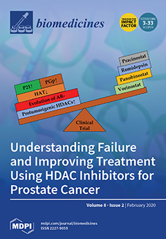
Clinical trials for the HDAC inhibitors vorinostat, panobinostat, romidepsin, and pracinostat have failed in castration-resistant prostate cancers. The emergence of androgen-receptor-negative prostate cancers, protumorigenic HDACs, upregulation of p21 and P-glycoproteins, and a downregulation of histone acetyl transferases may be unexplored pharmacodynamic reasons for this failure. Considerations of these key areas are likely to be vital in upcoming clinical trials. View this paper.
  • Issues are regarded as officially published after their release is announced to the table of contents alert mailing list.
  • You may sign up for e-mail alerts to receive table of contents of newly released issues.
  • PDF is the official format for papers published in both, html and pdf forms. To view the papers in pdf format, click on the "PDF Full-text" link, and use the free Adobe Reader to open them.
Order results
Result details
Section
Select all
Export citation of selected articles as:
12 pages, 2221 KiB  
Article
Improvement Effect of Lotus Leaf Flavonoids on Carbon Tetrachloride-Induced Liver Injury in Mice
by Tongji Liu, Fang Tan, Xingyao Long, Yanni Pan, Jianfei Mu, Xianrong Zhou, Runkun Yi and Xin Zhao
Biomedicines 2020, 8(2), 41; https://doi.org/10.3390/biomedicines8020041 - 24 Feb 2020
Cited by 13 | Viewed by 3381
Abstract
In this study, the effect of lotus leaf flavonoids (LLF) on carbon tetrachloride (CCl4)-induced liver injury in mice was studied. CCl4 was injected intraperitoneally to induce liver injury in Kunming mice. Mice were treated with LLF by gavage, and the [...] Read more.
In this study, the effect of lotus leaf flavonoids (LLF) on carbon tetrachloride (CCl4)-induced liver injury in mice was studied. CCl4 was injected intraperitoneally to induce liver injury in Kunming mice. Mice were treated with LLF by gavage, and the mRNA expression levels in serum and liver were detected. Compared with the model group, LLF significantly reduced the liver index and serum aspartate aminotransferase (AST), alanine aminotransferase (ALT), triglyceride (TG), and total cholesterol (TC) levels in mice with CCl4-induced liver injury. Pathological observation showed that LLF effectively reduced morphological incompleteness and hepatocyte necrosis in CCl4-treated liver tissue. The result of quantitative polymerase chain reaction (qPCR) indicated that LLF significantly up-regulated the mRNA expression levels of copper/zinc superoxide dismutase (Cu/Zn-SOD), manganese superoxide dismutase (Mn-SOD), and catalase (CAT) and down- regulated the expression levels of tumor necrosis factor-alpha (TNF-α), nuclear factor kappa B (NF-κB), and interleukin-1β (IL-1β) (p < 0.05). Thus, LLF is an active ingredient that ameliorates liver injury, and it has good application prospect. Full article
(This article belongs to the Section Drug Discovery and Development)
Show Figures

Figure 1

14 pages, 596 KiB  
Review
Targeting Redox Imbalance as an Approach for Diabetic Kidney Disease
by Keiichiro Matoba, Yusuke Takeda, Yosuke Nagai, Tamotsu Yokota, Kazunori Utsunomiya and Rimei Nishimura
Biomedicines 2020, 8(2), 40; https://doi.org/10.3390/biomedicines8020040 - 22 Feb 2020
Cited by 30 | Viewed by 4135
Abstract
Diabetic kidney disease (DKD) is a worldwide public health problem. It is the leading cause of end-stage renal disease and is associated with increased mortality from cardiovascular complications. The tight interactions between redox imbalance and the development of DKD are becoming increasingly evident. [...] Read more.
Diabetic kidney disease (DKD) is a worldwide public health problem. It is the leading cause of end-stage renal disease and is associated with increased mortality from cardiovascular complications. The tight interactions between redox imbalance and the development of DKD are becoming increasingly evident. Numerous cascades, including the polyol and hexosamine pathways have been implicated in the oxidative stress of diabetes patients. However, the precise molecular mechanism by which oxidative stress affects the progression of DKD remains to be elucidated. Given the limited therapeutic options for DKD, it is essential to understand how oxidants and antioxidants are controlled in diabetes and how oxidative stress impacts the progression of renal damage. This review aims to provide an overview of the current status of knowledge regarding the pathological roles of oxidative stress in DKD. Finally, we summarize recent therapeutic approaches to preventing DKD with a focus on the anti-oxidative effects of newly developed anti-hyperglycemic agents. Full article
Show Figures

Figure 1

14 pages, 2857 KiB  
Review
Correlations between Microbiota Bioactivity and Bioavailability of Functional Compounds: A Mini-Review
by Emanuel Vamanu and Florentina Gatea
Biomedicines 2020, 8(2), 39; https://doi.org/10.3390/biomedicines8020039 - 20 Feb 2020
Cited by 61 | Viewed by 5828
Abstract
Numerous studies have demonstrated the role of the microbiota in supporting the physiological functions, owing to its metabolomic component. The presence of biocomponents generally leads to the correction of the microbial pattern correlated with the reduction of oxidative pressure. This study aims to [...] Read more.
Numerous studies have demonstrated the role of the microbiota in supporting the physiological functions, owing to its metabolomic component. The presence of biocomponents generally leads to the correction of the microbial pattern correlated with the reduction of oxidative pressure. This study aims to present the main processes that correlate the bioavailability and bioactivity of some functional components through the action of the human microbiota. The use of probiotics and prebiotics is an innovative manner involving alternatives that increase the bioavailability of certain natural or metabolic components has been proposed. Probiotic strains (Saccharomyces cerevisiae or Lactobacillus (L.) plantarum) may represent an intermediary for increasing the antioxidant bioactivity, and they may be administered in the form of a biomass enriched with functional compounds, such as phenolic acids. The limiting effect of gastrointestinal transit is, in several cases, the key to the biopharmaceutical value of new products (or supplements). The identification of newer ways of formulating supplements also involves the compatibility of different types of products, the testing of bioaccessibility, and the elimination of biotransformations. Full article
Show Figures

Graphical abstract

11 pages, 1238 KiB  
Article
Long-Term Oral Administration of LLHK, LHK, and HK Alters Gene Expression Profile and Restores Age-Dependent Atrophy and Dysfunction of Rat Salivary Glands
by Yasuko Ishikawa, Tomasz D Pieczonka, Aneta M Bragiel-Pieczonka, Harumichi Seta, Tadahiro Ohkuri, Yumi Sasanuma and Yuji Nonaka
Biomedicines 2020, 8(2), 38; https://doi.org/10.3390/biomedicines8020038 - 20 Feb 2020
Cited by 1 | Viewed by 3043
Abstract
Xerostomia, also known as dry mouth, is caused by a reduction in salivary secretion and by changes in the composition of saliva associated with the malfunction of salivary glands. Xerostomia decreases quality of life. In the present study, we investigated the effects of [...] Read more.
Xerostomia, also known as dry mouth, is caused by a reduction in salivary secretion and by changes in the composition of saliva associated with the malfunction of salivary glands. Xerostomia decreases quality of life. In the present study, we investigated the effects of peptides derived from β-lactoglobulin C on age-dependent atrophy, gene expression profiles, and the dysfunction of salivary glands. Long-term oral administration of Leu57-Leu58-His59-Lys60 (LLHK), Leu58-His59-Lys60 (LHK) and His59-Lys60 (HK) peptides induced salivary secretion and prevented and/or reversed the age-dependent atrophy of salivary glands in older rats. The transcripts of 78 genes were upregulated and those of 81 genes were downregulated by more than 2.0-fold (p ≤ 0.05) after LHK treatment. LHK upregulated major salivary protein genes such as proline-rich proteins (Prpmp5, Prb3, Prp2, Prb1, Prp15), cystatins (Cst5, Cyss, Vegp2), amylases (Amy1a, Amy2a3), and lysozyme (Lyzl1), suggesting that LLHK, LHK, and HK restored normal salivary function. The AP-2 transcription factor gene (Tcfap2b) was also induced significantly by LHK treatment. These results suggest that LLHK, LHK, and HK-administration may prevent and/or reverse the age-dependent atrophy and functional decline of salivary glands by affecting gene expression. Full article
(This article belongs to the Section Molecular and Translational Medicine)
Show Figures

Figure 1

9 pages, 1465 KiB  
Article
Addition of K22 Converts Spider Venom Peptide Pme2a from an Activator to an Inhibitor of NaV1.7
by Kathleen Yin, Jennifer R. Deuis, Zoltan Dekan, Ai-Hua Jin, Paul F. Alewood, Glenn F. King, Volker Herzig and Irina Vetter
Biomedicines 2020, 8(2), 37; https://doi.org/10.3390/biomedicines8020037 - 19 Feb 2020
Cited by 5 | Viewed by 3245
Abstract
Spider venom is a novel source of disulfide-rich peptides with potent and selective activity at voltage-gated sodium channels (NaV). Here, we describe the discovery of μ-theraphotoxin-Pme1a and μ/δ-theraphotoxin-Pme2a, two novel peptides from the venom of the Gooty Ornamental tarantula [...] Read more.
Spider venom is a novel source of disulfide-rich peptides with potent and selective activity at voltage-gated sodium channels (NaV). Here, we describe the discovery of μ-theraphotoxin-Pme1a and μ/δ-theraphotoxin-Pme2a, two novel peptides from the venom of the Gooty Ornamental tarantula Poecilotheria metallica that modulate NaV channels. Pme1a is a 35 residue peptide that inhibits NaV1.7 peak current (IC50 334 ± 114 nM) and shifts the voltage dependence of activation to more depolarised membrane potentials (V1/2 activation: Δ = +11.6 mV). Pme2a is a 33 residue peptide that delays fast inactivation and inhibits NaV1.7 peak current (EC50 > 10 μM). Synthesis of a [+22K]Pme2a analogue increased potency at NaV1.7 (IC50 5.6 ± 1.1 μM) and removed the effect of the native peptide on fast inactivation, indicating that a lysine at position 22 (Pme2a numbering) is important for inhibitory activity. Results from this study may be used to guide the rational design of spider venom-derived peptides with improved potency and selectivity at NaV channels in the future. Full article
(This article belongs to the Special Issue Animal Venoms–Curse or Cure?)
Show Figures

Graphical abstract

11 pages, 1731 KiB  
Article
The Co-Expression of Programmed Death-Ligand 1 (PD-L1) in Untreated EGFR-Mutated Metastatic Lung Adenocarcinoma
by Ping-Chih Hsu, Chih-Wei Wang, Scott Chih-Hsi Kuo, Shu-Min Lin, Yu-Lun Lo, Allen Chung-Cheng Huang, Li-Chung Chiu and Cheng-Ta Yang
Biomedicines 2020, 8(2), 36; https://doi.org/10.3390/biomedicines8020036 - 19 Feb 2020
Cited by 13 | Viewed by 2893
Abstract
Epidermal growth factor receptor (EGFR)-tyrosine kinase inhibitor (TKI) is the standard first-line therapy for metastatic lung adenocarcinoma harboring sensitive EGFR mutations. Tumor surface programmed death-ligand 1 (PD-L1) is expressed in some metastatic EGFR-mutated lung adenocarcinoma, but its impact on the efficacy of EGFR-TKIs [...] Read more.
Epidermal growth factor receptor (EGFR)-tyrosine kinase inhibitor (TKI) is the standard first-line therapy for metastatic lung adenocarcinoma harboring sensitive EGFR mutations. Tumor surface programmed death-ligand 1 (PD-L1) is expressed in some metastatic EGFR-mutated lung adenocarcinoma, but its impact on the efficacy of EGFR-TKIs is unclear. We retrospectively investigated 117 untreated metastatic lung EGFR mutated adenocarcinoma patients with a PD-L1 immunohistochemistry test. The PD-L1 expression level was classified by tumor proportion scores (TPS). Forty-five patients had negative expression (TPS < 1%), 45 had a weak expression (TPS 1–49%), and 27 had a strong expression (≥50%). All patients recruited in this study received EGFR-TKIs as a first-line therapy. No significant differences were observed for objective response rates (68.9% versus 62.2% versus 73.1%, p = 0.807) and median time to treatment failure (TTF) (12.17 versus 13.17 versus 11.0 months, p = 0.443) of first-line EGFR-TKIS among the three groups of patients (negative versus weak versus strong). The median overall survival was 21.27 versus 20.63 versus 19.43 months among the three groups of patients (p = 0.77). Our results demonstrated that PD-L1 did not affect the efficacy of first-line EGFR-TKIs in metastatic EGFR mutated lung adenocarcinoma. Thus, EGFR-TKIs are suggested as the preferred clinical therapy for these patients, despite their PD-L1 levels. Full article
(This article belongs to the Section Cancer Biology and Oncology)
Show Figures

Graphical abstract

17 pages, 2800 KiB  
Article
5-(Carbamoylmethylene)-oxazolidin-2-ones as a Promising Class of Heterocycles Inducing Apoptosis Triggered by Increased ROS Levels and Mitochondrial Dysfunction in Breast and Cervical Cancer
by Biagio Armentano, Rosita Curcio, Matteo Brindisi, Raffaella Mancuso, Vittoria Rago, Ida Ziccarelli, Luca Frattaruolo, Marco Fiorillo, Vincenza Dolce, Bartolo Gabriele and Anna Rita Cappello
Biomedicines 2020, 8(2), 35; https://doi.org/10.3390/biomedicines8020035 - 18 Feb 2020
Cited by 22 | Viewed by 3464
Abstract
Oxazolidinones are antibiotics that inhibit protein synthesis by binding the 50S ribosomal subunit. Recently, numerous worldwide researches focused on their properties and possible involvement in cancer therapy have been conducted. Here, we evaluated in vitro the antiproliferative activity of some 5-(carbamoylmethylene)-oxazolidin-2-ones on MCF-7 [...] Read more.
Oxazolidinones are antibiotics that inhibit protein synthesis by binding the 50S ribosomal subunit. Recently, numerous worldwide researches focused on their properties and possible involvement in cancer therapy have been conducted. Here, we evaluated in vitro the antiproliferative activity of some 5-(carbamoylmethylene)-oxazolidin-2-ones on MCF-7 and HeLa cells. The tested compounds displayed a wide range of cytotoxicity on these cancer cell lines, measured by MTT assay, exhibiting no cytotoxicity on non-tumorigenic MCF-10A cells. Among the nine tested derivatives, four displayed a good anticancer potential. Remarkably, OI compound showed IC50 values of 17.66 and 31.10 µM for MCF-7 and HeLa cancer cells, respectively. Furthermore, we assessed OI effect on the cell cycle by FACS analysis, highlighting a G1 phase arrest after 72 h, supported by a low expression level of Cyclin D1 protein. Moreover, mitochondrial membrane potential was reduced after OI treatment driven by high levels of ROS. These findings demonstrate that OI treatment can inhibit MCF-7 and HeLa cell proliferation and induce apoptosis by caspase-9 activation and cytochrome c release in the cytosol. Hence, 5-(carbamoylmethylene)-oxazolidin-2-ones have a promising anticancer activity, in particular, OI derivative could represent a good candidate for in vivo further studies and potential clinical use. Full article
(This article belongs to the Section Cancer Biology and Oncology)
Show Figures

Graphical abstract

12 pages, 1196 KiB  
Article
Rostral Anterior Cingulate Thickness Predicts the Emotional Psilocybin Experience
by Candace R. Lewis, Katrin H. Preller, B. Blair Braden, Cory Riecken and Franz X. Vollenweider
Biomedicines 2020, 8(2), 34; https://doi.org/10.3390/biomedicines8020034 - 18 Feb 2020
Cited by 12 | Viewed by 10041
Abstract
Psilocybin is the psychoactive compound of mushrooms in the psilocybe species. Psilocybin directly affects a number of serotonin receptors, with highest affinity for the serotonin 2A receptor (5HT-2Ar). Generally, the effects of psilocybin, and its active metabolite psilocin, are well established and include [...] Read more.
Psilocybin is the psychoactive compound of mushrooms in the psilocybe species. Psilocybin directly affects a number of serotonin receptors, with highest affinity for the serotonin 2A receptor (5HT-2Ar). Generally, the effects of psilocybin, and its active metabolite psilocin, are well established and include a range of cognitive, emotional, and perceptual perturbations. Despite the generality of these effects, there is a high degree of inter-individual variability in subjective psilocybin experiences that are not well understood. Others have shown brain morphology metrics derived from magnetic resonance imaging (MRI) can predict individual drug response. Due to high expression of serotonin 2A receptors (5HT-2Ar) in the cingulate cortex, and its prior associations with psilocybin, we investigate if cortical thickness of this structure predicts the psilocybin experience in healthy adults. We hypothesized that greater cingulate thickness would predict higher subjective ratings in sub-scales of the Five-Dimensional Altered State of Consciousness (5D-ASC) with high emotionality in healthy participants (n = 55) who received oral psilocybin (either low dose: 0.160 mg/kg or high dose: 0.215 mg/kg). After controlling for sex, age, and using false discovery rate (FDR) correction, we found the rostral anterior cingulate predicted all four emotional sub-scales, whereas the caudal and posterior cingulate did not. How classic psychedelic compounds induce such large inter-individual variability in subjective states has been a long-standing question in serotonergic research. These results extend the traditional set and setting hypothesis of the psychedelic experience to include brain structure metrics. Full article
(This article belongs to the Section Neurobiology and Clinical Neuroscience)
Show Figures

Graphical abstract

11 pages, 662 KiB  
Article
McArdle Disease: Clinical, Biochemical, Histological and Molecular Genetic Analysis of 60 Patients
by Pushpa Raj Joshi, Marcus Deschauer and Stephan Zierz
Biomedicines 2020, 8(2), 33; https://doi.org/10.3390/biomedicines8020033 - 15 Feb 2020
Cited by 6 | Viewed by 3329
Abstract
A clinical, biochemical, histological and molecular genetic analysis of 60 McArdle patients (33 males and 27 females; mean age at diagnosis: 37 years) was performed. The objective of this study was to identify a possible genotype–phenotype correlation in McArdle disease. All patients complained [...] Read more.
A clinical, biochemical, histological and molecular genetic analysis of 60 McArdle patients (33 males and 27 females; mean age at diagnosis: 37 years) was performed. The objective of this study was to identify a possible genotype–phenotype correlation in McArdle disease. All patients complained of exercise-induced myalgia and fatigue; permanent weakness was present in 47% of the patients. Five percent of patients conveyed of masticatory muscle weakness. Age of onset was <15 years in 92% patients. Serum creatine kinase was elevated 5 to13-fold. Forearm ischemic test showed decreased lactate production but excessively increased ammonia upon exercise (n = 16). Muscle biopsies revealed highly reduced or missing myophosphorylase activity (n = 20) (mean: 0.17 ± 0.35 U/g tissue; normal: 12–61) and histologically, sub-sarcolemmal glycogen accumulation (n = 9). Molecular genetic analysis revealed the common p.Arg50Ter mutation in 68% of the patients. Other rather frequent mutations were p.Arg270Ter (allele frequency: 5%) followed by c.2262delA and p.Met1Val (allele frequencies: 3%). Twenty-four other rare mutations were also identified. No genotype–phenotype correlation was observed. The analysis highlights that testing of the p.Arg50Ter mutation could be performed first in molecular genetic testing of patients with exercise intolerance possibly due to McArdle disease. However, there is enormous mutation heterogeneity in McArdle disease thus sequencing of the myophosphorylase gene is needed in patients highly suspicious of McArdle disease. Full article
(This article belongs to the Section Gene and Cell Therapy)
Show Figures

Figure 1

26 pages, 1790 KiB  
Review
Basic Biology of Hypoxic Responses Mediated by the Transcription Factor HIFs and Its Implication for Medicine
by Kiichi Hirota
Biomedicines 2020, 8(2), 32; https://doi.org/10.3390/biomedicines8020032 - 13 Feb 2020
Cited by 30 | Viewed by 6725
Abstract
Oxygen (O2) is essential for human life. Molecular oxygen is vital for the production of adenosine triphosphate (ATP) in human cells. O2 deficiency leads to a reduction in the energy levels that are required to maintain biological functions. O2 [...] Read more.
Oxygen (O2) is essential for human life. Molecular oxygen is vital for the production of adenosine triphosphate (ATP) in human cells. O2 deficiency leads to a reduction in the energy levels that are required to maintain biological functions. O2 acts as the final acceptor of electrons during oxidative phosphorylation, a series of ATP synthesis reactions that occur in conjunction with the electron transport system in mitochondria. Persistent O2 deficiency may cause death due to malfunctioning biological processes. The above account summarizes the classic view of oxygen. However, this classic view has been reviewed over the last two decades. Although O2 is essential for life, higher organisms such as mammals are unable to biosynthesize molecular O2 in the body. Because the multiple organs of higher organisms are constantly exposed to the risk of “O2 deficiency,” living organisms have evolved elaborate strategies to respond to hypoxia. In this review, I will describe the system that governs oxygen homeostasis in the living body from the point-of-view of the transcription factor hypoxia-inducible factor (HIF). Full article
(This article belongs to the Special Issue Hypoxia-Inducible Factors: Regulation and Therapeutic Potential)
Show Figures

Figure 1

14 pages, 2439 KiB  
Article
The Use of Natural Agents to Counteract Telomere Shortening: Effects of a Multi-Component Extract of Astragalus mongholicus Bunge and Danazol
by Isabelle Guinobert, Claude Blondeau, Bruno Colicchio, Noufissa Oudrhiri, Alain Dieterlen, Eric Jeandidier, Georges Deschenes, Valérie Bardot, César Cotte, Isabelle Ripoche, Patrice Carde, Lucile Berthomier and Radhia M’Kacher
Biomedicines 2020, 8(2), 31; https://doi.org/10.3390/biomedicines8020031 - 12 Feb 2020
Cited by 6 | Viewed by 5687
Abstract
A link between telomere shortening and oxidative stress was found in aging people and patients with cancer or inflammatory diseases. Extracts of Astragalus spp. are known to stimulate telomerase activity, thereby compensating telomere shortening. We characterized a multi-component hydroethanolic root extract (HRE) of [...] Read more.
A link between telomere shortening and oxidative stress was found in aging people and patients with cancer or inflammatory diseases. Extracts of Astragalus spp. are known to stimulate telomerase activity, thereby compensating telomere shortening. We characterized a multi-component hydroethanolic root extract (HRE) of Astragalus mongholicus Bunge and assessed its effects on telomeres compared to those of danazol. Astragalosides I to IV, flavonoids, amino acids and sugars were detected in the HRE. Samples of peripheral blood lymphocytes with short telomeres from 18 healthy donors (mean age 63.5 years; range 32–86 years) were exposed to a single dose of 1 µg/mL HRE or danazol for three days. Telomere length and telomerase expression were then measured. Significant elongation of telomeres associated to a less toxicity was observed in lymphocytes from 13/18 donors following HRE treatment (0.54 kb (0.15–2.06 kb)) and in those from 9/18 donors after danazol treatment (0.95 kb (0.06–2.06 kb)). The rate of cells with short telomeres (<3 kb) decreased in lymphocytes from all donors after exposure to either HRE or danazol, telomere elongation being telomerase-dependent. These findings suggest that the HRE could be used for the management of age-related diseases. Full article
Show Figures

Graphical abstract

13 pages, 1771 KiB  
Article
Impact of Statin Use on Dementia Incidence in Elderly Men and Women with Ischemic Heart Disease
by Mi-Young Kim, Minji Jung, Yoojin Noh, Sooyoung Shin, Chang Hyung Hong, Sukhyang Lee and Yi-Sook Jung
Biomedicines 2020, 8(2), 30; https://doi.org/10.3390/biomedicines8020030 - 09 Feb 2020
Cited by 12 | Viewed by 3296
Abstract
This study aimed to determine the association between statins and the prevention of dementia according to sex differences in elderly patients with ischemic heart disease (IHD). We performed a nationwide retrospective cohort study using the Korean Health Insurance Review and Assessment Service database [...] Read more.
This study aimed to determine the association between statins and the prevention of dementia according to sex differences in elderly patients with ischemic heart disease (IHD). We performed a nationwide retrospective cohort study using the Korean Health Insurance Review and Assessment Service database (2007–2015). Among the 264,036 eligible patients aged ≥65 years with IHD, statin users were compared with non–users by propensity score matching at a 1:1 ratio (71,587 in each group). The primary outcome was dementia risk by estimating hazard ratios (HRs) and 95% confidence intervals (CIs). Differential risks of dementia were assessed by sex in the subgroups of statin types, exposure duration, and patient age, implying that sex is an influential factor for the link between statin use and dementia incidence. Among seven commonly prescribed statins, rosuvastatin was associated with the greatest preventive effect on dementia incidence, with an adjusted HR of 0.82 (95% CI = 0.78–0.87). In a subgroup analysis organized by sex, the differential risk of dementia incidence was assessed in each statin group, implying that sex is an influential factor for the link between statin and dementia. This study suggests that appropriate statin use considering sex differences may have beneficial effects on the development of dementia. Full article
(This article belongs to the Section Molecular and Translational Medicine)
Show Figures

Figure 1

22 pages, 4485 KiB  
Article
Effects of Anchomanes difformis on Inflammation, Apoptosis, and Organ Toxicity in STZ-Induced Diabetic Cardiomyopathy
by Toyin D. Alabi, Novel N. Chegou, Nicole L. Brooks and Oluwafemi O. Oguntibeju
Biomedicines 2020, 8(2), 29; https://doi.org/10.3390/biomedicines8020029 - 08 Feb 2020
Cited by 12 | Viewed by 3100
Abstract
Persistent hyperglycemia is known to cause enhanced generation of reactive oxygen species in diabetes. Several inflammatory cytokines are induced by oxidative stress, and their release also leads to increased oxidative stress; this makes oxidative stress one of the important factors in the development [...] Read more.
Persistent hyperglycemia is known to cause enhanced generation of reactive oxygen species in diabetes. Several inflammatory cytokines are induced by oxidative stress, and their release also leads to increased oxidative stress; this makes oxidative stress one of the important factors in the development of chronic inflammation and other immune responses. These have been implicated in the development of diabetic complications such as nephropathy and cardiomyopathy. Anchomanes difformis has been shown to possess antioxidant and anti-inflammatory potentials. The present study investigated the immunomodulatory potential and the antiapoptotic ability of Anchomanes difformis to ameliorate heart toxicity and injury in type II diabetes. Two weeks of fructose (10%) administration followed by single intraperitoneal injection of streptozotocin (40 mg/kg) were used to induce type II diabetes in male Wistar rats. Leaf extract (aqueous) of Anchomanes difformis (200 and 400 mg/kg) was administered orally for six weeks. Blood glucose concentrations and body weights before and after interventions were determined. Interleukin (IL)-1β, IL-6, IL-10, IL-18, monocyte chemoattractant protein 1 (MCP-1), and tumor necrosis factor alpha (TNFα) were measured in the heart homogenates. Catalase (CAT), superoxide dismutase (SOD), total protein, oxygen radical absorbance capacity (ORAC), ferric reducing antioxidant power (FRAP), thiobarbituric acid reactive substances (TBARS), and heart-type fatty acid-binding protein (H-FABP) levels were determined. Expressions of transcription factors (Nrf 2 and NFkB/p65) and apoptotic markers were also investigated in the heart. Anchomanes difformis administration reduced pro-inflammatory cytokines, increased anti-inflammatory markers, and enhanced antioxidant defense in the heart of diabetic treated animals. Anchomanes difformis is a new, promising therapeutic agent that can be explored for the treatment of pathological conditions associated with immune responses and will be a useful tool in the management of associated diabetic complications. Full article
(This article belongs to the Section Drug Discovery and Development)
Show Figures

Figure 1

23 pages, 918 KiB  
Review
NAFLD Preclinical Models: More than a Handful, Less of a Concern?
by Yvonne Oligschlaeger and Ronit Shiri-Sverdlov
Biomedicines 2020, 8(2), 28; https://doi.org/10.3390/biomedicines8020028 - 08 Feb 2020
Cited by 36 | Viewed by 9101
Abstract
Non-alcoholic fatty liver disease (NAFLD) is a spectrum of liver diseases ranging from simple steatosis to non-alcoholic steatohepatitis, fibrosis, cirrhosis, and/or hepatocellular carcinoma. Due to its increasing prevalence, NAFLD is currently a major public health concern. Although a wide variety of preclinical models [...] Read more.
Non-alcoholic fatty liver disease (NAFLD) is a spectrum of liver diseases ranging from simple steatosis to non-alcoholic steatohepatitis, fibrosis, cirrhosis, and/or hepatocellular carcinoma. Due to its increasing prevalence, NAFLD is currently a major public health concern. Although a wide variety of preclinical models have contributed to better understanding the pathophysiology of NAFLD, it is not always obvious which model is best suitable for addressing a specific research question. This review provides insights into currently existing models, mainly focusing on murine models, which is of great importance to aid in the identification of novel therapeutic options for human NAFLD. Full article
(This article belongs to the Special Issue NASH and Systemic Complications: From Basic to Clinical Research)
Show Figures

Figure 1

21 pages, 4885 KiB  
Review
New Herbal Biomedicines for the Topical Treatment of Dermatological Disorders
by Julia Hoffmann, Fabian Gendrisch, Christoph Mathis Schempp and Ute Wölfle
Biomedicines 2020, 8(2), 27; https://doi.org/10.3390/biomedicines8020027 - 08 Feb 2020
Cited by 45 | Viewed by 19262
Abstract
Herbal extracts and isolated plant compounds play an increasing role in the treatment of skin disorders and wounds. Several new herbal drugs, medicinal products and cosmetic products for the treatment of various skin conditions have been developed in recent years. In this nonsystematic [...] Read more.
Herbal extracts and isolated plant compounds play an increasing role in the treatment of skin disorders and wounds. Several new herbal drugs, medicinal products and cosmetic products for the treatment of various skin conditions have been developed in recent years. In this nonsystematic review, we focus on herbal drugs that were tested in controlled clinical studies or in scientifically sound preclinical studies. The herbal biomedicines are intended to treat atopic dermatitis (St. John’s wort, licorice, tormentil, bitter substances, evening primrose), psoriasis (araroba tree, lace flower, barberry bark, indigo, turmeric, olibanum, St. John’s wort), actinic keratosis (birch bark, petty spurge), herpes simplex (lemon balm, sage and rhubarb), rosacea (green tea, licorice, tormentil) and acne vulgaris (tea tree oil, green tea, hop), or to improve photo protection (green tea, Dyer’s weed, cocoa tree, carotinoids, licorice), aesthetic dermatology (licorice, pine bark, gotu kola) and wound healing (birch bark, onion). Full article
(This article belongs to the Section Drug Discovery and Development)
13 pages, 2647 KiB  
Article
A Versatile Model of Microfluidic Perifusion System for the Evaluation of C-Peptide Secretion Profiles: Comparison Between Human Pancreatic Islets and HLSC-Derived Islet-Like Structures
by Yonathan Gomez, Victor Navarro-Tableros, Ciro Tetta, Giovanni Camussi and Maria Felice Brizzi
Biomedicines 2020, 8(2), 26; https://doi.org/10.3390/biomedicines8020026 - 07 Feb 2020
Cited by 8 | Viewed by 2770
Abstract
A robust and easy-to-use tool for the ex vivo dynamic evaluation of pancreatic islet (PI) function is essential for further development of novel cell-based therapeutic approaches to treating diabetes. Here, we developed four different glucose perifusion protocols (GPPs) in a microfluidic perifusion system [...] Read more.
A robust and easy-to-use tool for the ex vivo dynamic evaluation of pancreatic islet (PI) function is essential for further development of novel cell-based therapeutic approaches to treating diabetes. Here, we developed four different glucose perifusion protocols (GPPs) in a microfluidic perifusion system (MPS), based entirely on commercially available components. After validation, the GPPs were used to evaluate C-peptide secretion profiles of PIs derived from different donors (healthy, obese, and type 2 diabetic) and from human liver stem-cell-derived islet-like structures (HLSC-ILS). Using this device, we demonstrated that PIs derived from healthy donors displayed a physiological C-peptide secretion profile as characterized by the response to (a) different glucose concentrations, (b) consecutive pulses of high-glucose concentrations, (c) a glucose threshold ranging from 5–8 mM, and (d) a constant high-glucose perifusion in a biphasic manner. Moreover, we were able to detect a dysregulated secretion profile in PIs derived from both obese and type 2 diabetes mellitus (T2DM) donors. Finally, we also evaluated the kinetic secretion profiles of HLSC-ILS, demonstrating that, nonetheless, with a lower amplitude of secretion compared to PI derived from healthy donors, they were already glucose-responsive on day seven post-differentiation. In conclusion, we have provided evidence that our MPS is a versatile device and may represent a valuable tool to study insulin-producing cells in vitro. Full article
(This article belongs to the Section Model Systems in Research and Development)
Show Figures

Figure 1

9 pages, 226 KiB  
Review
Almost 40 Years of Tissue Engineering in Russia: Where Are We Now?
by Elena Alpeeva, Yury Sukhanov and Ekaterina Vorotelyak
Biomedicines 2020, 8(2), 25; https://doi.org/10.3390/biomedicines8020025 - 05 Feb 2020
Cited by 1 | Viewed by 2609
Abstract
This review describes achievements of Russian cell-based regenerative medicine in different periods of time depending on the legislation and politics, and future prospects for its commercialization and wide application with an emphasis on products devised for skin regeneration. The world’s experience in tissue [...] Read more.
This review describes achievements of Russian cell-based regenerative medicine in different periods of time depending on the legislation and politics, and future prospects for its commercialization and wide application with an emphasis on products devised for skin regeneration. The world’s experience in tissue engineering began with the development of living skin equivalents, utilizing a biopolymer matrix and cells at the very beginning of the 1980s. During this period, the USSR kept abreast with the times and also conducted studies on skin wound healing, implementing modern cell techniques. However, there soon emerged a gap between scientific advancement and practical application. After the breakup of the USSR, there were no institutions that could implement scientific inventions into full-scale manufacturing for clinical application. At the same time, accumulating scientific and practical experience allowed for the maintenance of biomedical research and its readiness for market entry at present. Recently developed legislation opens up new opportunities in this field in Russia. There are a growing number of studies on the development of novel products for regenerative medicine, bringing hope for its rapid progress. Full article
(This article belongs to the Section Biomedical Materials and Nanomedicine)
11 pages, 1754 KiB  
Article
Physalis angulata Calyces Modulate Macrophage Polarization and Alleviate Chemically Induced Intestinal Inflammation in Mice
by David Rivera, Yanet Ocampo and Luis A. Franco
Biomedicines 2020, 8(2), 24; https://doi.org/10.3390/biomedicines8020024 - 05 Feb 2020
Cited by 4 | Viewed by 2796
Abstract
As part of the search for new bioactive plants from the Colombian Caribbean, the dichloromethane fraction of the calyces of Physalis angulata L. (PADF) was selected for its anti-inflammatory activity. In this work, we investigated the immunomodulatory effect of PADF in activated macrophages [...] Read more.
As part of the search for new bioactive plants from the Colombian Caribbean, the dichloromethane fraction of the calyces of Physalis angulata L. (PADF) was selected for its anti-inflammatory activity. In this work, we investigated the immunomodulatory effect of PADF in activated macrophages and during dextran sulfate sodium (DSS)-induced colitis. PADF displayed a low content of withanolides or phenolic compounds, and a higher content of sucrose esters, representative anti-inflammatory metabolites of the Physalis genus. The PADF fraction at 12.5 μg/mL prevented the induction of interleukin (IL)-1β, tumor necrosis factor (TNF-α), IL-6, IL-12, cyclooxygenase-2 (COX-2), and inducible nitric oxide synthase (iNOS) by lipopolysaccharide (LPS), while increased the levels of arginase (ARG1), IL-10, and mannose receptor C (MRC1). The polarization towards an anti-inflammatory profile was also observed in resting macrophages, without promoting the typical gene profile induced by IL-4, suggesting that PADF promotes a shift to a regulatory status rather than to an alternative one. In vivo, the administration of PADF to mice with chronic DSS-colitis reduced disease signs (i.e., body weight loss and colon shortening), and improved the histology score by diminishing the levels of pro-inflammatory cytokines and increasing the production of IL-10. Overall, results suggest that the regulatory effect on PADF towards macrophages might contribute to the therapeutic activity observed in the murine model of inflammatory bowel disease. Full article
(This article belongs to the Section Drug Discovery and Development)
Show Figures

Figure 1

13 pages, 1999 KiB  
Article
Short-Term Diet Restriction but Not Alternate Day Fasting Prevents Cisplatin-Induced Nephrotoxicity in Mice
by Evrin Gunebakan, Esra Yalcin, Esra Cikler Dulger, Ahmet Yigitbasi, Nilay Ates, Aysun Caglayan, Mustafa C. Beker, Kazim Sahin, Hasan Korkaya and Ertugrul Kilic
Biomedicines 2020, 8(2), 23; https://doi.org/10.3390/biomedicines8020023 - 03 Feb 2020
Cited by 7 | Viewed by 3233
Abstract
Cisplatin (CP) is one of the most preferred platinum-containing antineoplastic drugs. However, even in nontoxic plasma concentrations, it may cause kidney injury. To be able to increase its effective pharmacological dose, its side effects need to be regarded. Diet restriction (DR) has been [...] Read more.
Cisplatin (CP) is one of the most preferred platinum-containing antineoplastic drugs. However, even in nontoxic plasma concentrations, it may cause kidney injury. To be able to increase its effective pharmacological dose, its side effects need to be regarded. Diet restriction (DR) has been demonstrated to improve cellular survival in a number of disorders. In this context, we investigated the role of DR in CP-induced nephrotoxicity (CPN). Besides alternate DR, animals were exposed to DR for 3 days prior or after CP treatment. Here, we observed that both 3 days of DR reverses the nephrotoxic effect of CP, which was associated with improved physiological outcomes, such as serum creatine, blood-urea nitrogen and urea. These treatments significantly increased phosphorylation of survival kinases PI3K/Akt and ERK-1/2 and decreased the level of stress kinase JNK were noted. In addition, the activation level of signal transduction mediator p38 MAPK phosphorylation was higher particularly in both three-day DR groups. Next, animals were fed with carbohydrate-, protein- or fat-enriched diets in the presence of CP. Results indicated that not only fasting but also dietary content itself may play a determinant role in the severity of CPN. Our data suggest that DR is a promising approach to reduce CPN by regulating metabolism and cell signaling pathways. Full article
(This article belongs to the Section Molecular and Translational Medicine)
Show Figures

Figure 1

22 pages, 1509 KiB  
Review
Understanding Failure and Improving Treatment Using HDAC Inhibitors for Prostate Cancer
by Zohaib Rana, Sarah Diermeier, Muhammad Hanif and Rhonda J. Rosengren
Biomedicines 2020, 8(2), 22; https://doi.org/10.3390/biomedicines8020022 - 30 Jan 2020
Cited by 54 | Viewed by 6802
Abstract
Novel treatment regimens are required for castration-resistant prostate cancers (CRPCs) that become unresponsive to standard treatments, such as docetaxel and enzalutamide. Histone deacetylase (HDAC) inhibitors showed promising results in hematological malignancies, but they failed in solid tumors such as prostate cancer, despite the [...] Read more.
Novel treatment regimens are required for castration-resistant prostate cancers (CRPCs) that become unresponsive to standard treatments, such as docetaxel and enzalutamide. Histone deacetylase (HDAC) inhibitors showed promising results in hematological malignancies, but they failed in solid tumors such as prostate cancer, despite the overexpression of HDACs in CRPC. Four HDAC inhibitors, vorinostat, pracinostat, panobinostat and romidepsin, underwent phase II clinical trials for prostate cancers; however, phase III trials were not recommended due to a majority of patients exhibiting either toxicity or disease progression. In this review, the pharmacodynamic reasons for the failure of HDAC inhibitors were assessed and placed in the context of the advancements in the understanding of CRPCs, HDACs and resistance mechanisms. The review focuses on three themes: evolution of androgen receptor-negative prostate cancers, development of resistance mechanisms and differential effects of HDACs. In conclusion, advancements can be made in this field by characterizing HDACs in prostate tumors more extensively, as this will allow more specific drugs catering to the specific HDAC subtypes to be designed. Full article
(This article belongs to the Section Cancer Biology and Oncology)
Show Figures

Graphical abstract

12 pages, 1078 KiB  
Article
Circulating MyomiRs as Potential Biomarkers to Monitor Response to Nusinersen in Pediatric SMA Patients
by Silvia Bonanno, Stefania Marcuzzo, Claudia Malacarne, Eleonora Giagnorio, Riccardo Masson, Riccardo Zanin, Maria Teresa Arnoldi, Francesca Andreetta, Ornella Simoncini, Anna Venerando, Cinzia Gellera, Chiara Pantaleoni, Renato Mantegazza, Pia Bernasconi, Giovanni Baranello and Lorenzo Maggi
Biomedicines 2020, 8(2), 21; https://doi.org/10.3390/biomedicines8020021 - 26 Jan 2020
Cited by 30 | Viewed by 4689
Abstract
Spinal muscular atrophy (SMA) is an autosomal recessive disorder caused by mutations in survival motor neuron (SMN) 1 gene, resulting in a truncated SMN protein responsible for degeneration of brain stem and spinal motor neurons. The paralogous SMN2 gene partially compensates full-length SMN [...] Read more.
Spinal muscular atrophy (SMA) is an autosomal recessive disorder caused by mutations in survival motor neuron (SMN) 1 gene, resulting in a truncated SMN protein responsible for degeneration of brain stem and spinal motor neurons. The paralogous SMN2 gene partially compensates full-length SMN protein production, mitigating the phenotype. Antisense oligonucleotide nusinersen (Spinraza®) enhances SMN2 gene expression. SMN is involved in RNA metabolism and biogenesis of microRNA (miRNA), key gene expression modulators, whose dysregulation contributes to neuromuscular diseases. They are stable in body fluids and may reflect distinct pathophysiological states, thus acting as promising biomarkers. Muscle-specific miRNAs (myomiRs) as biomarkers for clinical use in SMA have not been investigated yet. Here, we analyzed the expression of miR-133a, -133b, -206 and -1, in serum of 21 infantile SMA patients at baseline and after 6 months of nusinersen treatment, and correlated molecular data with response to therapy evaluated by the Hammersmith Functional Motor Scale Expanded (HFMSE). Our results demonstrate that myomiR serological levels decrease over disease course upon nusinersen treatment. Notably, miR-133a reduction predicted patients’ response to therapy. Our findings identify myomiRs as potential biomarkers to monitor disease progression and therapeutic response in SMA patients. Full article
(This article belongs to the Section Molecular and Translational Medicine)
Show Figures

Figure 1

11 pages, 1667 KiB  
Article
2-deoxy-d-glucose Ameliorates Animal Models of Dermatitis
by Soo Young Choi, Min-Jeong Heo, Chanmi Lee, Yeong Min Choi, In-sook An, Seunghee Bae, Sungkwan An and Jin Hyuk Jung
Biomedicines 2020, 8(2), 20; https://doi.org/10.3390/biomedicines8020020 - 24 Jan 2020
Cited by 14 | Viewed by 3945
Abstract
Glucose metabolism is a key metabolic pathway that orchestrates cellular homeostasis by generating ATP, nucleotides, and amino acids. Abnormal glucose signaling has been found in many diseases including cancers and inflammatory diseases. According to recent report, glycolysis contributes to pathogenesis of psoriasis and [...] Read more.
Glucose metabolism is a key metabolic pathway that orchestrates cellular homeostasis by generating ATP, nucleotides, and amino acids. Abnormal glucose signaling has been found in many diseases including cancers and inflammatory diseases. According to recent report, glycolysis contributes to pathogenesis of psoriasis and ablation of Glut1 attenuates animal models of psoriasis. While we were screening a molecular target for atopic dermatitis, we found the levels of glucose transporters including Glut1 (SLC2a1) and Glut3 (SLC2a3) are highly expressed in skin biopsies of dermatitis patients from multiple datasets. We demonstrated that administration of 2-deoxy-d-glucose (2DG) ameliorates animal models of 12-o-tetradecanoylphorbol-13-acetate (TPA) and oxazolone induced dermatitis using morphological and histological analysis. These results suggest that inhibition of glucose metabolism ameliorates dermatitis in animal models. Full article
(This article belongs to the Section Drug Discovery and Development)
Show Figures

Figure 1

23 pages, 4326 KiB  
Article
A Multi-Omics Analysis of Bone Morphogenetic Protein 5 (BMP5) mRNA Expression and Clinical Prognostic Outcomes in Different Cancers Using Bioinformatics Approaches
by Md. Adnan Karim, Abdus Samad, Utpal Kumar Adhikari, Md. Ashraful Kader, Md. Masnoon Kabir, Md. Aminul Islam and Md. Nazmul Hasan
Biomedicines 2020, 8(2), 19; https://doi.org/10.3390/biomedicines8020019 - 21 Jan 2020
Cited by 23 | Viewed by 7036
Abstract
Cumulative studies have provided controversial evidence for the prognostic values of bone morphogenetic protein 5 (BMP5) in different types of cancers such as colon, breast, lung, bladder, and ovarian cancer. To address the inconsistent correlation of BMP5 expression with patient survival [...] Read more.
Cumulative studies have provided controversial evidence for the prognostic values of bone morphogenetic protein 5 (BMP5) in different types of cancers such as colon, breast, lung, bladder, and ovarian cancer. To address the inconsistent correlation of BMP5 expression with patient survival and molecular function of BMP5 in relation to cancer progression, we performed a systematic study to determine whether BMP5 could be used as a prognostic marker in human cancers. BMP5 expression and prognostic values were assessed using different bioinformatics tools such as ONCOMINE, GENT, TCGA, GEPIA, UALCAN, PrognoScan, PROGgene V2 server, and Kaplan–Meier Plotter. In addition, we used cBioPortal database for the identification and analysis of BMP5 mutations, copy number alterations, altered expression, and protein–protein interaction (PPI). We found that BMP5 is frequently down-regulated in our queried cancer types. Use of prognostic analysis showed negative association of BMP5 down-regulation with four types of cancer except for ovarian cancer. The highest mutation was found in the R321*/Q amino acid of BMP5 corresponding to colorectal and breast cancer whereas the alteration frequency was higher in lung squamous carcinoma datasets (>4%). In PPI analysis, we found 31 protein partners of BMP5, among which 11 showed significant co-expression (p-value < 0.001, log odds ratio > 1). Pathway analysis of differentially co-expressed genes with BMP5 in breast, lung, colon, bladder and ovarian cancers revealed the BMP5-correlated pathways. Collectively, this data-driven study demonstrates the correlation of BMP5 expression with patient survival and identifies the involvement of BMP5 pathways that may serve as targets of a novel biomarker for various types of cancers in human. Full article
(This article belongs to the Section Immunology and Immunotherapy)
Show Figures

Figure 1

14 pages, 3219 KiB  
Article
Protective Effects of Astaxanthin Supplementation against Ultraviolet-Induced Photoaging in Hairless Mice
by Xing Li, Tomohiro Matsumoto, Miho Takuwa, Mahmood Saeed Ebrahim Shaiku Ali, Takumi Hirabashi, Hiroyo Kondo and Hidemi Fujino
Biomedicines 2020, 8(2), 18; https://doi.org/10.3390/biomedicines8020018 - 21 Jan 2020
Cited by 27 | Viewed by 6330
Abstract
Ultraviolet (UV) light induces skin photoaging, which is characterized by thickening, wrinkling, pigmentation, and dryness. Astaxanthin (AST), a ketocarotenoid isolated from Haematococcus pluvialis, has been extensively studied owing to its possible effects on skin health as well as UV protection. In addition, [...] Read more.
Ultraviolet (UV) light induces skin photoaging, which is characterized by thickening, wrinkling, pigmentation, and dryness. Astaxanthin (AST), a ketocarotenoid isolated from Haematococcus pluvialis, has been extensively studied owing to its possible effects on skin health as well as UV protection. In addition, AST attenuates the increased generation of reactive oxygen species (ROS) and capillary regression of the skeletal muscle. In this study, we investigated whether AST could protect against UV-induced photoaging and reduce capillary regression in the skin of HR-1 hairless mice. UV light induces wrinkle formation, epidermal thickening, and capillary regression in the dermis of HR-1 hairless mice. The administration of AST reduced the UV-induced wrinkle formation and skin thickening, and increased collagen fibers in the skin. AST supplementation also inhibited the generation of ROS, decreased wrinkle formation, reduced epidermal thickening, and increased the density of capillaries in the skin. We also found an inverse correlation between wrinkle formation and the density of capillaries. An association between photoaging and capillary regression in the skin was also observed. These results suggest that AST can protect against photoaging caused by UV irradiation and the inhibitory effects of AST on photoaging may be associated with the reduction of capillary regression in the skin. Full article
(This article belongs to the Section Drug Discovery and Development)
Show Figures

Graphical abstract

Previous Issue
Next Issue
Back to TopTop