Next Issue
Volume 8, December
Previous Issue
Volume 8, October


Biomedicines, Volume 8, Issue 11 (November 2020) – 86 articles

Cover Story (view full-size image): 
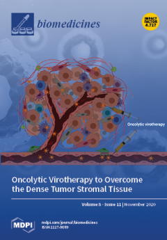
Tumor stroma mainly consists of cancer-associated fibroblasts, extracellular matrix, vasculature, and tumor-associated macrophages. These components all exert key roles in tumor progression, emphasizing their relevance to cancer therapy design. The multifaceted oncolytic viruses (OVs) represent a promising therapeutic platform to overcome the dense stromal tissue. Inherent and genetically modified OVs can directly target both stromal components and tumor cells, facilitating OV penetration, tumor regression, and anti-tumor immune activity. View this paper
  • Issues are regarded as officially published after their release is announced to the table of contents alert mailing list.
  • You may sign up for e-mail alerts to receive table of contents of newly released issues.
  • PDF is the official format for papers published in both, html and pdf forms. To view the papers in pdf format, click on the "PDF Full-text" link, and use the free Adobe Reader to open them.
Order results
Result details
Section
Select all
Export citation of selected articles as:
19 pages, 13247 KiB  
Article
Adenosine A2A and A3 Receptors as Targets for the Treatment of Hypertensive-Diabetic Nephropathy
by Daniela Patinha, Carla Abreu, Carla Carvalho, Olga Mariana Cunha, Mariana Mota, Joana Afonso, Teresa Sousa, António Albino-Teixeira, Carmen Diniz and Manuela Morato
Biomedicines 2020, 8(11), 529; https://doi.org/10.3390/biomedicines8110529 - 23 Nov 2020
Cited by 9 | Viewed by 3892
Abstract
Diabetic nephropathy (DN) and hypertension are prime causes for end-stage renal disease (ESRD) that often coexist in patients, but are seldom studied in combination. Kidney adenosine levels are markedly increased in diabetes, and the expression and function of renal adenosine receptors are altered [...] Read more.
Diabetic nephropathy (DN) and hypertension are prime causes for end-stage renal disease (ESRD) that often coexist in patients, but are seldom studied in combination. Kidney adenosine levels are markedly increased in diabetes, and the expression and function of renal adenosine receptors are altered in experimental diabetes. The aim of this work is to explore the impact of endogenous and exogenous adenosine on the expression/distribution profile of its receptors along the nephron of hypertensive rats with experimentally-induced diabetes. Using spontaneously hypertensive (SHR) rats rendered diabetic with streptozotocin (STZ), we show that treatment of SHR-STZ rats with an agonist of adenosine receptors increases A2A immunoreactivity in superficial glomeruli (SG), proximal tubule (PCT), and distal tubule (DCT). Differently, treatment of SHR-STZ rats with a xanthinic antagonist of adenosine receptors decreases adenosine A3 immunoreactivity in SG, PCT, DCT, and collecting duct. There is no difference in the immunoreactivity against the adenosine A1 and A2B receptors between the experimental groups. The agonist of adenosine receptors ameliorates renal fibrosis, probably via A2A receptors, while the antagonist exacerbates it, most likely due to tonic activation of A3 receptors. The reduction in adenosine A3 immunoreactivity might be due to receptor downregulation in response to prolonged activation. Altogether, these results suggest an opposite regulation exerted by endogenous and exogenous adenosine upon the expression of its A2A and A3 receptors along the nephron of hypertensive diabetic rats, which has a functional impact and should be taken into account when considering novel therapeutic targets for hypertensive-diabetic nephropathy. Full article
Show Figures

Graphical abstract

15 pages, 3692 KiB  
Article
The Potential Effect of Fucoidan on Inhibiting Epithelial-to-Mesenchymal Transition, Proliferation, and Increase in Apoptosis for Endometriosis Treatment: In Vivo and In Vitro Study
by Li-Chun Chang, Yi-Fen Chiang, Hsin-Yuan Chen, Yun-Ju Huang, An-Chieh Liu and Shih-Min Hsia
Biomedicines 2020, 8(11), 528; https://doi.org/10.3390/biomedicines8110528 - 22 Nov 2020
Cited by 10 | Viewed by 3865
Abstract
Endometriosis is common in reproductive-age women and its pathology is to increase proliferation and migration to enhance epithelial-to-mesenchymal transition progression (EMT). However, treatments are currently limited, so it is important to explore new therapeutic drugs. Hence, in this study, we investigate the therapeutic [...] Read more.
Endometriosis is common in reproductive-age women and its pathology is to increase proliferation and migration to enhance epithelial-to-mesenchymal transition progression (EMT). However, treatments are currently limited, so it is important to explore new therapeutic drugs. Hence, in this study, we investigate the therapeutic effect of fucoidan (FC) on the progression and mechanisms of endometriosis. The cell viability of endometrial cell lines End1/E6E7 and Vk2/E6E7 treated with different concentrations of FC were assessed by 3-(4,5-Dimethylthiazol-2-yl)-2,5-diphenyltetrazolium bromide (MTT) assay and cell counting. Cell migration was evaluated using wound-healing assay. In an in vivo experiment, female Balb/c mice received surgically induced endometriosis followed by different concentrations of fucoidan for 6 weeks. High-frequency ultrasound imaging was applied to detect subsequent lesion growth. The results demonstrated that fucoidan inhibited the viability and migration ability of End1/E6E7 and Vk2/E6E7 cells. Additionally, the administration of fucoidan reduced the volume and weight of endometriotic lesions, decreased inflammatory cytokines and vascular endothelial growth factor (VEGF) of serum and lesions, and improved EMT proliferation and apoptosis-related protein expression. For the first time, fucoidan indicated anti-proliferative and anti-inflammatory effects as well as inhibited EMT progression and induced apoptosis, improving endometriosis. Full article
(This article belongs to the Special Issue Advanced Research in Endometriosis)
Show Figures

Graphical abstract

44 pages, 9484 KiB  
Review
Aptamers for Proteins Associated with Rheumatic Diseases: Progress, Challenges, and Prospects of Diagnostic and Therapeutic Applications
by Elizaveta A. Shatunova, Maksim A. Korolev, Vitaly O. Omelchenko, Yuliya D. Kurochkina, Anna S. Davydova, Alya G. Venyaminova and Mariya A. Vorobyeva
Biomedicines 2020, 8(11), 527; https://doi.org/10.3390/biomedicines8110527 - 22 Nov 2020
Cited by 10 | Viewed by 4400
Abstract
Nucleic acid aptamers capable of affine and specific binding to their molecular targets have now established themselves as a very promising alternative to monoclonal antibodies for diagnostic and therapeutic applications. Although the main focus in aptamers’ research and development for biomedicine is made [...] Read more.
Nucleic acid aptamers capable of affine and specific binding to their molecular targets have now established themselves as a very promising alternative to monoclonal antibodies for diagnostic and therapeutic applications. Although the main focus in aptamers’ research and development for biomedicine is made on cardiovascular, infectious, and malignant diseases, the use of aptamers as therapeutic or diagnostic tools in the context of rheumatic diseases is no less important. In this review, we consider the main features of aptamers that make them valuable molecular tools for rheumatologists, and summarize the studies on the selection and application of aptamers for protein biomarkers associated with rheumatic diseases. We discuss the progress in the development of aptamer-based diagnostic assays and targeted therapeutics for rheumatic disorders, future prospects in the field, and issues that have yet to be addressed. Full article
Show Figures

Figure 1

38 pages, 785 KiB  
Review
Mitochondria at Work: New Insights into Regulation and Dysregulation of Cellular Energy Supply and Metabolism
by Volker Schirrmacher
Biomedicines 2020, 8(11), 526; https://doi.org/10.3390/biomedicines8110526 - 22 Nov 2020
Cited by 41 | Viewed by 5671
Abstract
Mitochondria are of great relevance to health, and their dysregulation is associated with major chronic diseases. Research on mitochondria—156 brand new publications from 2019 and 2020—have contributed to this review. Mitochondria have been fundamental for the evolution of complex organisms. As important and [...] Read more.
Mitochondria are of great relevance to health, and their dysregulation is associated with major chronic diseases. Research on mitochondria—156 brand new publications from 2019 and 2020—have contributed to this review. Mitochondria have been fundamental for the evolution of complex organisms. As important and semi-autonomous organelles in cells, they can adapt their function to the needs of the respective organ. They can program their function to energy supply (e.g., to keep heart muscle cells going, life-long) or to metabolism (e.g., to support hepatocytes and liver function). The capacity of mitochondria to re-program between different options is important for all cell types that are capable of changing between a resting state and cell proliferation, such as stem cells and immune cells. Major chronic diseases are characterized by mitochondrial dysregulation. This will be exemplified by cardiovascular diseases, metabolic syndrome, neurodegenerative diseases, immune system disorders, and cancer. New strategies for intervention in chronic diseases will be presented. The tumor microenvironment can be considered a battlefield between cancer and immune defense, competing for energy supply and metabolism. Cancer cachexia is considered as a final stage of cancer progression. Nevertheless, the review will present an example of complete remission of cachexia via immune cell transfer. These findings should encourage studies along the lines of mitochondria, energy supply, and metabolism. Full article
(This article belongs to the Section Immunology and Immunotherapy)
Show Figures

Figure 1

23 pages, 3064 KiB  
Article
Three-Dimensional Biofabrication Models of Endometriosis and the Endometriotic Microenvironment
by Jillian R. H. Wendel, Xiyin Wang, Lester J. Smith and Shannon M. Hawkins
Biomedicines 2020, 8(11), 525; https://doi.org/10.3390/biomedicines8110525 - 21 Nov 2020
Cited by 14 | Viewed by 3825
Abstract
Endometriosis occurs when endometrial-like tissue grows outside the uterine cavity, leading to pelvic pain, infertility, and increased risk of ovarian cancer. The present study describes the optimization and characterization of cellular spheroids as building blocks for Kenzan scaffold-free method biofabrication and proof-of-concept models [...] Read more.
Endometriosis occurs when endometrial-like tissue grows outside the uterine cavity, leading to pelvic pain, infertility, and increased risk of ovarian cancer. The present study describes the optimization and characterization of cellular spheroids as building blocks for Kenzan scaffold-free method biofabrication and proof-of-concept models of endometriosis and the endometriotic microenvironment. The spheroid building blocks must be of a specific diameter (~500 μm), compact, round, and smooth to withstand Kenzan biofabrication. Under optimized spheroid conditions for biofabrication, the endometriotic epithelial-like cell line, 12Z, expressed high levels of estrogen-related genes and secreted high amounts of endometriotic inflammatory factors that were independent of TNFα stimulation. Heterotypic spheroids, composed of 12Z and T-HESC, an immortalized endometrial stromal cell line, self-assembled into a biologically relevant pattern, consisting of epithelial cells on the outside of the spheroids and stromal cells in the core. 12Z spheroids were biofabricated into large three-dimensional constructs alone, with HEYA8 spheroids, or as heterotypic spheroids with T-HESC. These three-dimensional biofabricated constructs containing multiple monotypic or heterotypic spheroids represent the first scaffold-free biofabricated in vitro models of endometriosis and the endometriotic microenvironment. These efficient and innovative models will allow us to study the complex interactions of multiple cell types within a biologically relevant microenvironment. Full article
(This article belongs to the Special Issue Advanced Research in Endometriosis)
Show Figures

Figure 1

13 pages, 1629 KiB  
Article
Cholesterol Efflux Capacity and Cardiovascular Disease: The Ludwigshafen Risk and Cardiovascular Health (LURIC) Study
by Andreas Ritsch, Angela Duerr, Patrick Kahler, Monika Hunjadi, Tatjana Stojakovic, Guenther Silbernagel, Hubert Scharnagl, Marcus E. Kleber and Winfried März
Biomedicines 2020, 8(11), 524; https://doi.org/10.3390/biomedicines8110524 - 21 Nov 2020
Cited by 15 | Viewed by 2512
Abstract
(1) Background and Aims: Efforts to reduce coronary artery disease (CAD) by raising high-density lipoprotein (HDL) cholesterol (HDL-C) have not been uniformly successful. A more important factor than HDL-C may be cellular cholesterol efflux mediated by HDL, which has been shown to be [...] Read more.
(1) Background and Aims: Efforts to reduce coronary artery disease (CAD) by raising high-density lipoprotein (HDL) cholesterol (HDL-C) have not been uniformly successful. A more important factor than HDL-C may be cellular cholesterol efflux mediated by HDL, which has been shown to be associated with CAD. In this report, we analyzed the influence of cardiovascular biomarkers and risk factors on cholesterol efflux in a prospective observational study of patients referred to coronary angiography. (2) Methods: HDL-mediated efflux capacity was determined for 2468 participants of the Ludwigshafen Risk and Cardiovascular Health (LURIC) study who were referred to coronary angiography at baseline between 1997 and 2000. Median follow-up time was 9.9 years. Primary and secondary endpoints were cardiovascular and all-cause mortality, respectively. (3) Results: Cholesterol efflux strongly correlated with HDL-related markers including HDL cholesterol, HDL phospholipids, and apolipoproteins AI and AII, as well as HDL particle concentration, which was not seen for low density lipoprotein (LDL) markers including LDL cholesterol and apoB. Cholesterol efflux was associated negatively with C-reactive protein (CRP), fibrinogen, interleukin-6 (IL-6), and serum amyloid A. Cardiovascular mortality was higher in patients in the lowest cholesterol efflux quartile. This association was weakened, but not fully abolished, after adjustment for HDL cholesterol. (4) Conclusions: We demonstrate that cholesterol efflux was associated with HDL-composition as well as inflammatory burden in patients referred for coronary angiography, and that this inversely predicts cardiovascular mortality independently of HDL cholesterol. Full article
Show Figures

Graphical abstract

11 pages, 687 KiB  
Review
Blood Neurofilament Light Chain: The Neurologist’s Troponin?
by Simon Thebault, Ronald A. Booth and Mark S. Freedman
Biomedicines 2020, 8(11), 523; https://doi.org/10.3390/biomedicines8110523 - 21 Nov 2020
Cited by 52 | Viewed by 8923
Abstract
Blood neurofilament light chain (NfL) is a marker of neuro-axonal injury showing promising associations with outcomes of interest in several neurological conditions. Although initially discovered and investigated in the cerebrospinal fluid (CSF), the recent development of ultrasensitive digital immunoassay technologies has enabled reliable [...] Read more.
Blood neurofilament light chain (NfL) is a marker of neuro-axonal injury showing promising associations with outcomes of interest in several neurological conditions. Although initially discovered and investigated in the cerebrospinal fluid (CSF), the recent development of ultrasensitive digital immunoassay technologies has enabled reliable detection in serum/plasma, obviating the need for invasive lumbar punctures for longitudinal assessment. The most evidence for utility relates to multiple sclerosis (MS) where it serves as an objective measure of both the inflammatory and degenerative pathologies that characterise this disease. In this review, we summarise the physiology and pathophysiology of neurofilaments before focusing on the technological advancements that have enabled reliable quantification of NfL in blood. As the test case for clinical translation, we then highlight important recent developments linking blood NfL levels to outcomes in MS and the next steps to be overcome before this test is adopted on a routine clinical basis. Full article
(This article belongs to the Special Issue Multiple Sclerosis: Diagnosis and Treatment II)
Show Figures

Figure 1

11 pages, 2145 KiB  
Article
Extracellular Vesicles Derived from Kefir Grain Lactobacillus Ameliorate Intestinal Inflammation via Regulation of Proinflammatory Pathway and Tight Junction Integrity
by Eun Ae Kang, Hye-In Choi, Seung Wook Hong, Seokwoo Kang, Hyeon-Young Jegal, Eun Wook Choi, Byung-Soon Park and Joo Sung Kim
Biomedicines 2020, 8(11), 522; https://doi.org/10.3390/biomedicines8110522 - 20 Nov 2020
Cited by 30 | Viewed by 3535
Abstract
The aim of this study was to demonstrate the anti-inflammatory effect of Lactobacillus kefirgranum PRCC-1301-derived extracellular vesicles (PRCC-1301 EVs) on intestinal inflammation and intestinal barrier function. Human intestinal epithelial cells (IECs) Caco-2 were treated with PRCC-1301 EVs and then stimulated with dextran sulfate [...] Read more.
The aim of this study was to demonstrate the anti-inflammatory effect of Lactobacillus kefirgranum PRCC-1301-derived extracellular vesicles (PRCC-1301 EVs) on intestinal inflammation and intestinal barrier function. Human intestinal epithelial cells (IECs) Caco-2 were treated with PRCC-1301 EVs and then stimulated with dextran sulfate sodium (DSS). Real-time RT-PCR revealed that PRCC-1301 EVs inhibited the expression of pro-inflammatory cytokines in Caco-2 cells. PRCC-1301 EVs enhanced intestinal barrier function by maintaining intestinal cell integrity and the tight junction. Loss of Zo-1, claudin-1, and occludin in Caco-2 cells and the colitis tissues was recovered after PRCC-1301 EVs treatment, as evidenced by immunofluorescence analysis. Acute murine colitis was induced using 4% DSS and chronic colitis was generated in piroxicam-treated IL-10-/- mice. PRCC-1301 EVs attenuated body weight loss, colon shortening, and histological damage in acute and chronic colitis models in mice. Immunohistochemistry revealed that phosphorylated NF-κB p65 and IκBα were reduced in the colon tissue sections treated with PRCC-1301 EVs. Our results suggest that PRCC-1301 EVs may have an anti-inflammatory effect on colitis by inhibiting the NF-κB pathway and improving intestinal barrier function. Full article
(This article belongs to the Special Issue Novel Therapeutic Approaches in Inflammatory Bowel Diseases)
Show Figures

Figure 1

13 pages, 3634 KiB  
Article
Photosensitizer-Trapped Gold Nanocluster for Dual Light-Responsive Phototherapy
by Junho Byun, Dongyoon Kim, Jaehyun Choi, Gayong Shim and Yu-Kyoung Oh
Biomedicines 2020, 8(11), 521; https://doi.org/10.3390/biomedicines8110521 - 20 Nov 2020
Cited by 7 | Viewed by 2369
Abstract
Photoresponsive nanomaterials have recently received great attention in the field of cancer therapy. Here, we report a photosensitizer-trapped gold nanocluster that can facilitate dual light-responsive cancer therapy. We utilized methylene blue (MB) as a model photosensitizer, gold nanocluster as a model photothermal agent, [...] Read more.
Photoresponsive nanomaterials have recently received great attention in the field of cancer therapy. Here, we report a photosensitizer-trapped gold nanocluster that can facilitate dual light-responsive cancer therapy. We utilized methylene blue (MB) as a model photosensitizer, gold nanocluster as a model photothermal agent, and a polymerized DNA as the backbone of the nanocluster. We synthesized MB-intercalated gold DNA nanocluster (GMDN) via reduction and clustering of gold ions on a template consisting of MB-intercalated long DNA. Upon GMDN treatment, cancer cells revealed clear cellular uptake of MB and gold clusters; following dual light irradiation (660 nm/808 nm), the cells showed reactive oxygen species generation and increased temperature. Significantly higher cancer cell death was observed in cells treated with GMDN and dual irradiation compared with non-irradiated or single light-irradiated cells. Mice systemically injected with GMDN showed enhanced tumor accumulation compared to that of free MB and exhibited increased temperature upon near infrared irradiation of the tumor site. Tumor growth was almost completely inhibited in GMDN-treated tumor-bearing mice after dual light irradiation, and the survival rate of this group was 100% over more than 60 days. These findings suggest that GMDN could potentially function as an effective phototherapeutic for the treatment of cancer disease. Full article
(This article belongs to the Special Issue Advanced Nanomedicines for Optical Imaging and Phototherapy)
Show Figures

Figure 1

15 pages, 856 KiB  
Article
Predictors of HbA1c among Adipocytokine Biomarkers in African-American Men with Varied Glucose Tolerance
by Elena Barengolts, Arfana Akbar, Brian T. Layden, Yuval Eisenberg, Medha Priyadarshini, Jeffrey A. Borgia, Cristina L. Fhied, Michael Salim and Lara R. Dugas
Biomedicines 2020, 8(11), 520; https://doi.org/10.3390/biomedicines8110520 - 20 Nov 2020
Viewed by 2002
Abstract
This study explored adipocytokine associations with acute and chronic hyperglycemia in African-American men (AAM). Fourteen adipocytokines were measured from men with normal glucose tolerance (NGT) or type 2 diabetes (T2D, drug-naïve MF(−) or using metformin MF(+)). Acute and chronic hyperglycemia were evaluated by [...] Read more.
This study explored adipocytokine associations with acute and chronic hyperglycemia in African-American men (AAM). Fourteen adipocytokines were measured from men with normal glucose tolerance (NGT) or type 2 diabetes (T2D, drug-naïve MF(−) or using metformin MF(+)). Acute and chronic hyperglycemia were evaluated by 120 min oral glucose tolerance test (OGTT) and glycohemoglobin A1c (HbA1c). AAM with T2D (n = 21) compared to NGT (n = 20) were older, had higher BMI and slightly higher glucose and insulin. In the fasted state, TNF-α, IL-6, PAI-1, IL-13, adiponectin, adipsin, and lipocalin were lower in T2D vs. NGT. At 120 min post-glucose load, TNF-α, IL-6, IL-13, IL-8, PAI-1, adiponectin, adipsin, lipocalin, and resistin were lower in T2D vs. NGT. There were no statistical differences for GM-CSF, IL-7, IL-10, IP-10, and MCP-1. Regression analysis showed that fasting IL-8, TNF-α, adiponectin, lipocalin, resistin, adipsin, and PAI-1 were associated with HbA1c. After adjusting for age, BMI, glucose tolerance, and metformin use, only adipsin remained significantly associated with HbA1c (p = 0.021). The model including adipsin, TNF-α, age, BMI, and group designation (i.e., NGT, MF(−), MF(+)) explained 86% of HbA1c variability. The data suggested that adipsin could be associated with HbA1c in AAM with varied glucose tolerance. Full article
(This article belongs to the Section Molecular and Translational Medicine)
Show Figures

Figure 1

16 pages, 5235 KiB  
Article
Modulation of NLRP3 Inflammasome Attenuated Inflammatory Response Associated to Diarrhea-Predominant Irritable Bowel Syndrome
by Sarah Adriana Scuderi, Giovanna Casili, Marika Lanza, Alessia Filippone, Irene Paterniti, Emanuela Esposito and Michela Campolo
Biomedicines 2020, 8(11), 519; https://doi.org/10.3390/biomedicines8110519 - 20 Nov 2020
Cited by 18 | Viewed by 2751
Abstract
Diarrhea-predominant irritable bowel syndrome (IBS-D) is a multifactorial chronic gastrointestinal disorder characterized by inflammation and immune response. In this context, NLRP3 over-activation is associated with a breakdown of enteric-immune balance related to IBS-D. The aim of this study was to evaluate the effect [...] Read more.
Diarrhea-predominant irritable bowel syndrome (IBS-D) is a multifactorial chronic gastrointestinal disorder characterized by inflammation and immune response. In this context, NLRP3 over-activation is associated with a breakdown of enteric-immune balance related to IBS-D. The aim of this study was to evaluate the effect of the inflammasome inhibitor, BAY 11-7082, in a rat model of IBS-D. Syndrome was induced by intracolonic instillation of 1 mL 4% acetic acid at 8 cm proximal to the anus for 30 s and sacrificed 2 weeks after IBS-D induction. BAY 11-7082 (10 and 30 mg/kg) was administered daily by oral gavage. The results obtained showed that the treatment with BAY 11-7082 (30 mg/kg) significantly reduced tissue injury characterized by edema, neutrophil infiltration, and loss of colon structure. We demonstrated that BAY 11-7082 treatment inhibited NLRP3 inflammasome activation and NF-kB translocation, reducing inflammatory mediators. Moreover, treatment with BAY 11-7082 restored tight junction alteration following IBS-D induction and reduced the restraint stress. Taken together, our data demonstrate that IBS-D induced NLRP3 inflammasome pathway activation, accompanied by the production of proinflammatory response. The modulation of the inflammosome pathway with BAY 11-7082 inhibitor significantly reduced pathological signs of IBS-D, therefore, can be considered a valuable strategy to reduce the development of IBS-D. Full article
(This article belongs to the Special Issue Novel Therapeutic Approaches in Inflammatory Bowel Diseases)
Show Figures

Graphical abstract

16 pages, 3901 KiB  
Article
ACTN4 Mediates SEPT14 Mutation-Induced Sperm Head Defects
by Yu-Hua Lin, Chia-Yen Huang, Chih-Chun Ke, Ya-Yun Wang, Tsung-Hsuan Lai, Hsuan-Che Liu, Wei-Chi Ku, Chying-Chyuan Chan and Ying-Hung Lin
Biomedicines 2020, 8(11), 518; https://doi.org/10.3390/biomedicines8110518 - 19 Nov 2020
Cited by 6 | Viewed by 5618
Abstract
Septins (SEPTs) are highly conserved GTP-binding proteins and the fourth component of the cytoskeleton. Polymerized SEPTs participate in the modulation of various cellular processes, such as cytokinesis, cell polarity, and membrane dynamics, through their interactions with microtubules, actin, and other cellular components. The [...] Read more.
Septins (SEPTs) are highly conserved GTP-binding proteins and the fourth component of the cytoskeleton. Polymerized SEPTs participate in the modulation of various cellular processes, such as cytokinesis, cell polarity, and membrane dynamics, through their interactions with microtubules, actin, and other cellular components. The main objective of this study was to dissect the molecular pathological mechanism of SEPT14 mutation-induced sperm head defects. To identify SEPT14 interactors, co-immunoprecipitation (co-IP) and nano-liquid chromatography-mass spectrometry/mass spectrometry were applied. Immunostaining showed that SEPT14 was significantly localized to the manchette structure. The SEPT14 interactors were identified and classified as (1) SEPT-, (2) microtubule-, (3) actin-, and (4) sperm structure-related proteins. One interactor, ACTN4, an actin-holding protein, was selected for further study. Co-IP experiments showed that SEPT14 interacts with ACTN4 in a male germ cell line. SEPT14 also co-localized with ACTN4 in the perinuclear and manchette regions of the sperm head in early elongating spermatids. In the cell model, mutated SEPT14 disturbed the localization pattern of ACTN4. In a clinical aspect, sperm with mutant SEPT14, SEPT14A123T (p.Ala123Thr), and SEPT14I333T (p.Ile333Thr), have mislocalized and fragmented ACTN4 signals. Sperm head defects in donors with SEPT14 mutations are caused by disruption of the functions of ACTN4 and actin during sperm head formation. Full article
(This article belongs to the Section Molecular and Translational Medicine)
Show Figures

Figure 1

27 pages, 2431 KiB  
Review
Molecular Insights into the Multifunctional Role of Natural Compounds: Autophagy Modulation and Cancer Prevention
by Md. Ataur Rahman, MD. Hasanur Rahman, Md. Shahadat Hossain, Partha Biswas, Rokibul Islam, Md Jamal Uddin, Md. Habibur Rahman and Hyewhon Rhim
Biomedicines 2020, 8(11), 517; https://doi.org/10.3390/biomedicines8110517 - 19 Nov 2020
Cited by 31 | Viewed by 4809
Abstract
Autophagy is a vacuolar, lysosomal degradation pathway for injured and damaged protein molecules and organelles in eukaryotic cells, which is controlled by nutrients and stress responses. Dysregulation of cellular autophagy may lead to various diseases such as neurodegenerative disease, obesity, cardiovascular disease, diabetes, [...] Read more.
Autophagy is a vacuolar, lysosomal degradation pathway for injured and damaged protein molecules and organelles in eukaryotic cells, which is controlled by nutrients and stress responses. Dysregulation of cellular autophagy may lead to various diseases such as neurodegenerative disease, obesity, cardiovascular disease, diabetes, and malignancies. Recently, natural compounds have come to attention for being able to modulate the autophagy pathway in cancer prevention, although the prospective role of autophagy in cancer treatment is very complex and not yet clearly elucidated. Numerous synthetic chemicals have been identified that modulate autophagy and are favorable candidates for cancer treatment, but they have adverse side effects. Therefore, different phytochemicals, which include natural compounds and their derivatives, have attracted significant attention for use as autophagy modulators in cancer treatment with minimal side effects. In the current review, we discuss the promising role of natural compounds in modulating the autophagy pathway to control and prevent cancer, and provide possible therapeutic options. Full article
(This article belongs to the Special Issue Natural Medicine in Therapy)
Show Figures

Graphical abstract

17 pages, 773 KiB  
Review
Macrophage Polarization in Chronic Lymphocytic Leukemia: Nurse-Like Cells Are the Caretakers of Leukemic Cells
by Oana Mesaros, Laura Jimbu, Alexandra Neaga, Cristian Popescu, Iulia Berceanu, Ciprian Tomuleasa, Bogdan Fetica and Mihnea Zdrenghea
Biomedicines 2020, 8(11), 516; https://doi.org/10.3390/biomedicines8110516 - 19 Nov 2020
Cited by 13 | Viewed by 3202
Abstract
Macrophages are terminally differentiated innate immune cells. Through their activation, they can be polarized towards the pro-inflammatory M1 type or the wound healing-associated, anti-inflammatory M2 type macrophages. In the tumor microenvironment (TME), M2 is the dominant phenotype and these cells are referred to [...] Read more.
Macrophages are terminally differentiated innate immune cells. Through their activation, they can be polarized towards the pro-inflammatory M1 type or the wound healing-associated, anti-inflammatory M2 type macrophages. In the tumor microenvironment (TME), M2 is the dominant phenotype and these cells are referred to as tumor-associated macrophages (TAMs). TAMs secrete cytokines and chemokines, exerting an antiapoptotic, proliferative and pro-metastatic effect on the tumor cells. TAMs can be found in many cancers, including chronic lymphocytic leukemia (CLL), where they are called nurse-like cells (NLCs). Despite the generally indolent behavior of CLL, the proportion of treatment-refractory patients is significant. As with the majority of cancers, despite significant recent progress, CLL pathogenesis is poorly understood. The emerging role of the TME in nurturing the neoplastic process warrants the investigation of macrophages as a significant pathogenetic element of tumors. In this paper, we review the current knowledge on the role of stromal macrophages in CLL. Full article
(This article belongs to the Special Issue Macrophages in Health and Non-infectious Disease)
Show Figures

Figure 1

15 pages, 1728 KiB  
Article
Gingival Crevicular Fluid Cytokines in Moderate and Deep Sites of Stage III Periodontitis Patients in Different Rates of Clinical Progression
by Federica Romano, Wilma Del Buono, Laura Bianco, Martina Arena, Giulia Maria Mariani, Federica Di Scipio, Giovanni Nicolao Berta and Mario Aimetti
Biomedicines 2020, 8(11), 515; https://doi.org/10.3390/biomedicines8110515 - 18 Nov 2020
Cited by 12 | Viewed by 2294
Abstract
Clinical criteria are inappropriate to measure the degree of susceptibility to progression of periodontal damage. Thus, the aim of this study was to assess whether gingival crevicular fluid (GCF) levels of cytokines could discriminate patients suffering from stage III periodontitis with moderate (Grade [...] Read more.
Clinical criteria are inappropriate to measure the degree of susceptibility to progression of periodontal damage. Thus, the aim of this study was to assess whether gingival crevicular fluid (GCF) levels of cytokines could discriminate patients suffering from stage III periodontitis with moderate (Grade B) and rapid rates of progression (Grade C) prior to and 6 months after non-surgical periodontal treatment. GCF samples were obtained from moderate and deep sites of 20 patients diagnosed as Grade B and 20 patients as grade C stage III periodontitis and analyzed for interleukin (IL)-1β, IL-9, tumor necrosis factor (TNF)-α, and vascular endothelial growth factor (VEGF) using a high-sensitivity Bio-Plex Suspension Array System. At baseline, higher IL-1β but lower IL-9 GCF levels were observed in moderate sites of the grade C compared to the grade B group. In spite of comparable clinical improvement, this difference maintained after treatment, suggesting a residual pro-inflammatory state. In deep sites, no differences were observed between periodontitis groups except for VEGF levels that decreased more in Grade B periodontitis at 6 months post-therapy. A mathematical model was constructed to identify Grade C periodontitis patients based on the subjects’ GCF levels of IL-1β and IL-9, which achieved an area under the receiver-operating characteristic (ROC) curve of 0.94. This study can contribute to the early assessment of risk of future breakdown in periodontitis patients. Full article
(This article belongs to the Special Issue Models for Oral Biology Research)
Show Figures

Figure 1

10 pages, 2486 KiB  
Article
SGLT2 Inhibitor-Induced Sympathoexcitation in White Adipose Tissue: A Novel Mechanism for Beiging
by Jennifer R. Matthews, Lakshini Y. Herat, Aaron L. Magno, Shelley Gorman, Markus P. Schlaich and Vance B. Matthews
Biomedicines 2020, 8(11), 514; https://doi.org/10.3390/biomedicines8110514 - 18 Nov 2020
Cited by 11 | Viewed by 2475
Abstract
Recent preclinical data show that sodium glucose cotransporter 2 (SGLT2) inhibitors are able to reduce weight gain and induce beiging in white adipose tissue (WAT). We have previously shown that in neurogenic hypertensive Schlager (BPH/2J) mice, treatment with the SGLT2 inhibitor, Dapagliflozin, reduced [...] Read more.
Recent preclinical data show that sodium glucose cotransporter 2 (SGLT2) inhibitors are able to reduce weight gain and induce beiging in white adipose tissue (WAT). We have previously shown that in neurogenic hypertensive Schlager (BPH/2J) mice, treatment with the SGLT2 inhibitor, Dapagliflozin, reduced blood pressure and prevented weight gain. Here we show that chemical sympathetic denervation achieved by systemic administration of 6-hydroxy-dopamine (6-OHDA) reduces body weight and the heightened sympathetic nervous system (SNS) innervation in WAT. Furthermore, we demonstrate that 2 weeks of Dapagliflozin treatment increases SNS innervation in WAT of hypertensive mice. This increase is accompanied by a non-significant elevation in mRNA levels of the Ucp1 and Pgc-1α genes, which are markers of beiging. No significant difference in the mRNA levels of the inflammatory mediators Il-6 and Tnf-α were detected in WAT of Dapagliflozin treated mice. These findings suggest that SGLT-2 inhibitor-associated prevention of weight gain may be mediated, at least in part, by inducing the beiging of WAT. Full article
Show Figures

Figure 1

21 pages, 4696 KiB  
Article
Anti-MAP Triple Therapy Supports Immunomodulatory Therapeutic Response in Crohn’s Disease through Downregulation of NF-κB Activation in the Absence of MAP Detection
by Ahmad Qasem, Erij Elkamel and Saleh A. Naser
Biomedicines 2020, 8(11), 513; https://doi.org/10.3390/biomedicines8110513 - 18 Nov 2020
Cited by 15 | Viewed by 6045
Abstract
We previously reported that the triple antibiotic formulation, known as anti-MAP therapy, exhibits unique synergistic antimicrobial activity and should be effective for treatment of Crohn’s disease (CD) associated with Mycobacterium avium subspecies paratuberculosis (MAP). The absence of MAP detection in some CD cases [...] Read more.
We previously reported that the triple antibiotic formulation, known as anti-MAP therapy, exhibits unique synergistic antimicrobial activity and should be effective for treatment of Crohn’s disease (CD) associated with Mycobacterium avium subspecies paratuberculosis (MAP). The absence of MAP detection in some CD cases may be linked to poor diagnostics or lack of association with the disease. To understand the therapeutic response of some CD patients to anti-MAP therapy in absence of MAP detection, we investigated the immunomodulatory potency of anti-MAP therapy and its major ingredients, clarithromycin (CLA) and rifabutin (RIF), in THP-1, Caco-2, and Jurkat T-cells. Anti-MAP formulation at 2.0 μg/mL decreased MAP viability in macrophages by 18-fold over 72 h. Additionally, M1/M2 macrophage polarization ratio was reduced by 6.7-fold, and expression and protein levels of TNF-α and IL-6 were reduced by 2.9-fold, whereas IL-10 increased by 5.0-fold in these cells. Mechanistically, the effect of anti-MAP formulation on NF-κB p65 activation was dose-dependent and decreased to 13.4% at 2.0 μg/mL. Most importantly, anti-MAP therapy also reversed pro-inflammatory response in lipopolysaccharide (LPS)-induced macrophages, which shows that the anti-inflammatory effect of the treatment is not just due to a decrease in MAP viability. To study the anti-cytotoxic effects of anti-MAP therapy in Caco-2 monolayers infected with MAP or treated with dextran sodium sulfate (DSS), we showed a 45% decrease in lactate dehydrogenase (LDH) activity and an 84% increase in glutathione (GSH) activity, which supports anti-apoptotic activity of the drug. In Jurkat T-cells, anti-MAP therapy decreased T-cell proliferation by 4.8-fold following treatment with phytohemagglutinin (PHA) and by 2.9-fold with MAP purified protein derivative (PPD). Overall, the data demonstrate that anti-MAP therapy plays a significant role in modulating and eliciting a protective immune response in macrophages, endothelial cells, and T lymphocytes, even in absence of infection. This may explain the therapeutic response of some CD patients to treatment, even in absence of MAP detection, infection, or total eradication. The study supports anti-MAP therapy as an alternate treatment option in CD patients, especially in absence of reliable MAP diagnostics. Full article
(This article belongs to the Special Issue Novel Therapeutic Approaches in Inflammatory Bowel Diseases)
Show Figures

Figure 1

15 pages, 1858 KiB  
Article
Effect of Ezetimibe on Glucose Metabolism and Inflammatory Markers in Adipose Tissue
by Yongin Cho, Ryeong-Hyeon Kim, Hyunki Park, Hye Jin Wang, Hyangkyu Lee and Eun Seok Kang
Biomedicines 2020, 8(11), 512; https://doi.org/10.3390/biomedicines8110512 - 18 Nov 2020
Cited by 12 | Viewed by 4122
Abstract
Despite numerous studies, the effects of ezetimibe on glucose metabolism are poorly understood. Here, we aimed to investigate the effects of ezetimibe on glucose metabolism and the expression of inflammatory markers. Thirteen rats were randomly assigned to an ezetimibe (n = 6) [...] Read more.
Despite numerous studies, the effects of ezetimibe on glucose metabolism are poorly understood. Here, we aimed to investigate the effects of ezetimibe on glucose metabolism and the expression of inflammatory markers. Thirteen rats were randomly assigned to an ezetimibe (n = 6) or control group (n = 7). The control group received a high fat diet (HFD; 60 Kcal%), whereas the ezetimibe group received an HFD (60 Kcal%) containing 160 mg/kg of ezetimibe. After 14 weeks, adipose and liver tissues, along with plasma, were collected and comparatively analyzed. The effects of combination therapy with ezetimibe and statins on glucose metabolism were investigated over a 1-year period using data from patients with hyperlipidemia. Several indices of glucose metabolism partially improved in the ezetimibe group. The sizes of adipocytes and the accumulation of pro-inflammatory cytokines were reduced in the ezetimibe group. Ezetimibe treatment induced anti-inflammatory cytokines and fatty acid oxidation in adipocytes and reduced serum levels of free fatty acids. Clinical data analysis revealed that statin monotherapy significantly increased insulin resistance. However, combination therapy with ezetimibe and statins did not increase insulin resistance. In conclusion, ezetimibe was found to reduce the sizes of adipocytes in visceral fat and serum levels of free fatty acids, to induce fatty acid oxidation, to improve adipocytic inflammation, and to partially improve glycemic index values. Full article
Show Figures

Figure 1

11 pages, 1322 KiB  
Article
Detection Rate of 68Ga-PSMA Ligand PET/CT in Patients with Recurrent Prostate Cancer and Androgen Deprivation Therapy
by Joachim Brumberg, Melanie Beckl, Alexander Dierks, Andreas Schirbel, Markus Krebs, Andreas Buck, Hubert Kübler, Constantin Lapa and Anna Katharina Seitz
Biomedicines 2020, 8(11), 511; https://doi.org/10.3390/biomedicines8110511 - 18 Nov 2020
Cited by 9 | Viewed by 1979
Abstract
Prostate-specific membrane antigen (PSMA) ligand PET/CT enables the localization of tumor lesions in patients with recurrent prostate cancer, but it is unclear whether androgen deprivation therapy (ADT) influences diagnostic accuracy. The aim of this study was to evaluate the effect of ADT on [...] Read more.
Prostate-specific membrane antigen (PSMA) ligand PET/CT enables the localization of tumor lesions in patients with recurrent prostate cancer, but it is unclear whether androgen deprivation therapy (ADT) influences diagnostic accuracy. The aim of this study was to evaluate the effect of ADT on the detection rate of 68Ga-PSMA ligand PET/CT. Thus, 399 patients with initial radical prostatectomy and 68Ga-PSMA ligand PET/CT during PSA relapse were retrospectively evaluated. Propensity score matching was used to create two balanced groups of 62 subjects who either did or did not receive ADT within six months before imaging. All 68Ga-PSMA ligand PET/CT were evaluated visually and with semiquantitative measures. The detection rate of tumor recurrence was significantly higher in the group with ADT (88.7% vs. 72.6%, p = 0.02) and improved with increasing PSA-levels in both groups. In subjects with pathological PET/CT and ADT, whole-body total lesion PSMA (p < 0.01) and PSMA-derived tumor volume (p < 0.01) were significantly higher than in those without ADT. More PSMA-positive lesions and higher PSMA-derived volumetric parameters in patients with ADT suggest that a better detection rate is related to a (biologically) more advanced disease stage. Due to high detection rates in patients with PSA-levels < 2 ng/mL, the withdrawal of ADT before PSMA ligand PET/CT cannot be recommended. Full article
(This article belongs to the Special Issue Biomarkers and Therapy Targets in Urologic Cancer)
Show Figures

Figure 1

14 pages, 6912 KiB  
Article
Leptin, Leptin Receptor, KHDRBS1 (KH RNA Binding Domain Containing, Signal Transduction Associated 1), and Adiponectin in Bone Metastasis from Breast Carcinoma: An Immunohistochemical Study
by Paola Maroni, Alessandro Luzzati, Giuseppe Perrucchini, Luca Cannavò and Paola Bendinelli
Biomedicines 2020, 8(11), 510; https://doi.org/10.3390/biomedicines8110510 - 17 Nov 2020
Cited by 4 | Viewed by 2248
Abstract
Breast cancer patients are at a high risk of complications from bone metastasis. Molecular characterization of bone metastases is essential for the discovery of new therapeutic targets. Here, we investigated the expression and the intracellular distribution of KH RNA binding domain containing, signal [...] Read more.
Breast cancer patients are at a high risk of complications from bone metastasis. Molecular characterization of bone metastases is essential for the discovery of new therapeutic targets. Here, we investigated the expression and the intracellular distribution of KH RNA binding domain containing, signal transduction associated 1 (KHDRBS1), leptin, leptin receptor (LEPR), and adiponectin in bone metastasis from breast carcinoma and looked for correlations between the data. The expression of these proteins is known in breast carcinoma, but it has not been investigated in bone metastatic tissue to date. Immunohistochemical analysis was carried out on bone metastasis specimens, then semiquantitative evaluation of the results and the Pearson test were performed to determine eventual correlations. KHDRBS1 expression was significantly higher in the nuclei than in the cytosol of metastatic cells; LEPR was prevalently observed in the cytosol and the nuclei; leptin and adiponectin were found in metastatic cells and stromal cells; the strongest positive correlation was between nuclear KHDRBS1 and nuclear LEPR expression. Taken together, our findings support the importance of the leptin/LEPR/KHDRBS1 axis and of adiponectin in the progression of bone metastasis and suggest their potential application in pharmacological interventions. Full article
Show Figures

Figure 1

16 pages, 1517 KiB  
Article
Early Depression Independently of Other Neuropsychiatric Conditions, Influences Disability and Mortality after Stroke (Research Study—Part of PROPOLIS Study)
by Katarzyna Kowalska, Łukasz Krzywoszański, Jakub Droś, Paulina Pasińska, Aleksander Wilk and Aleksandra Klimkowicz-Mrowiec
Biomedicines 2020, 8(11), 509; https://doi.org/10.3390/biomedicines8110509 - 17 Nov 2020
Cited by 15 | Viewed by 3612
Abstract
Post-stroke depression (PSD) is the most frequent neuropsychiatric consequence of stroke. The nature of the relationship between PSD and mortality still remains unknown. One hypothesis is that PSD could be more frequent in those patients who are more vulnerable to physical disability, a [...] Read more.
Post-stroke depression (PSD) is the most frequent neuropsychiatric consequence of stroke. The nature of the relationship between PSD and mortality still remains unknown. One hypothesis is that PSD could be more frequent in those patients who are more vulnerable to physical disability, a mediator variable for higher level of physical damage related to higher risk of mortality. Therefore, the authors’ objective was to explore the assumption that PSD increases disability after stroke, and secondly, that mortality is higher among patients with PSD regardless of stroke severity and other neuropsychiatric conditions. We included 524 consecutive patients with acute stroke or transient ischemic attack, who were screened for depression between 7–10 days after stroke onset. Physical impairment and death were the outcomes measures at evaluation check points three and 12 months post-stroke. PSD independently increased the level of disability three (OR = 1.94, 95% CI 1.31–2.87, p = 0.001), and 12 months post-stroke (OR = 1.61, 95% CI 1.14–2.48, p = 0.009). PSD was also an independent risk factor for death three (OR = 5.68, 95% CI 1.58–20.37, p = 0.008) and 12 months after stroke (OR = 4.53, 95% CI 2.06–9.94, p = 0.001). Our study shows the negative impact of early PSD on the level of disability and survival rates during first year after stroke and supports the assumption that depression may act as an independent mediator for disability leading to death in patients who are more vulnerable for brain injury. Full article
Show Figures

Figure 1

17 pages, 378 KiB  
Review
Management of Antiphospholipid Syndrome
by Amine Ghembaza and David Saadoun
Biomedicines 2020, 8(11), 508; https://doi.org/10.3390/biomedicines8110508 - 17 Nov 2020
Cited by 17 | Viewed by 6053
Abstract
Antiphospholipid syndrome (APS), is an acquired autoimmune disorder characterised by thrombosis, pregnancy morbidity, and the presence of antiphospholipid antibodies (aPL). Although venous thromboembolism is the most common manifestation, thrombotic events in APS may also occur in virtually any vascular bed, with cerebral circulation [...] Read more.
Antiphospholipid syndrome (APS), is an acquired autoimmune disorder characterised by thrombosis, pregnancy morbidity, and the presence of antiphospholipid antibodies (aPL). Although venous thromboembolism is the most common manifestation, thrombotic events in APS may also occur in virtually any vascular bed, with cerebral circulation being the arterial territory most commonly affected. As APS is a heterogeneous condition, its management should be tailored with a patient-centred approach based on individual risk assessment, which includes the aPL profile, concomitant auto-immune diseases, and traditional cardiovascular risk factors. Although literature data are conflicting regarding primary prophylaxis, there is some evidence indicating that antiplatelet agents may reduce the risk of a first thrombotic event in individuals with a high-risk profile. In patients with thrombotic APS, current evidence-based guidelines recommend lifelong vitamin K antagonists (VKAs), preferably warfarin. The optimal intensity of anticoagulation following arterial thrombosis remains controversial. Arterial thrombosis should be treated either with high-intensity warfarin at a target INR > 3.0, or low-dose aspirin (LDA) combined with moderate-intensity warfarin (INR 2.0–3.0). It is recommended to avoid direct oral anticoagulants (DOACs) in patients with high-risk APS, mainly those with triple-positive PL and previous arterial events. They would only be used exceptionally in selected patients with low-risk venous thromboembolism (VTE). In low-risk VTE patients currently treated with a DOAC due to warfarin intolerance or a previous unstable International Normalized Ratio on warfarin, the decision of continuing DOACs would be taken in carefully selected patients. In women with obstetric APS, the combination therapy with LDA plus heparin remains the conventional strategy. Full article
Show Figures

Figure 1

13 pages, 1795 KiB  
Article
Increased Expressions of Matrix Metalloproteinases (MMPs) in Prostate Cancer Tissues of Men with Type 2 Diabetes
by Andras Franko, Lucia Berti, Jörg Hennenlotter, Steffen Rausch, Marcus O. Scharpf, Martin Hrabĕ de Angelis, Arnulf Stenzl, Andreas Peter, Andreas L. Birkenfeld, Stefan Z. Lutz, Hans-Ulrich Häring and Martin Heni
Biomedicines 2020, 8(11), 507; https://doi.org/10.3390/biomedicines8110507 - 16 Nov 2020
Cited by 6 | Viewed by 2435
Abstract
Type 2 diabetes (T2D) is associated with worse prognosis of prostate cancer (PCa). The molecular mechanisms behind this association are still not fully understood. The aim of this study was to identify key factors, which contribute to the more aggressive PCa phenotype in [...] Read more.
Type 2 diabetes (T2D) is associated with worse prognosis of prostate cancer (PCa). The molecular mechanisms behind this association are still not fully understood. The aim of this study was to identify key factors, which contribute to the more aggressive PCa phenotype in patients with concurrent T2D. Therefore, we investigated benign and PCa tissue of PCa patients with and without diabetes using real time qPCR. Compared to patients without diabetes, patients with T2D showed a decreased E-cadherin/N-cadherin (CDH1/CDH2) ratio in prostate tissue, indicating a switch of epithelial-mesenchymal transition (EMT), which is a pivotal process in carcinogenesis. In addition, the gene expression levels of matrix metalloproteinases (MMPs) and CC chemokine ligands (CCLs) were higher in prostate samples of T2D patients. Next, prostate adenocarcinoma PC3 cells were treated with increasing glucose concentrations to replicate hyperglycemia in vitro. In these cells, high glucose induced expressions of MMPs and CCLs, which showed significant positive associations with the proliferation marker proliferating cell nuclear antigen (PCNA). These results indicate that in prostate tissue of men with T2D, hyperglycemia may induce EMT, increase MMP and CCL gene expressions, which in turn activate invasion and inflammatory processes accelerating the progression of PCa. Full article
(This article belongs to the Special Issue Biomarkers and Therapy Targets in Urologic Cancer)
Show Figures

Figure 1

23 pages, 4919 KiB  
Article
8-Hydroxydaidzein, an Isoflavone from Fermented Soybean, Induces Autophagy, Apoptosis, Differentiation, and Degradation of Oncoprotein BCR-ABL in K562 Cells
by Pei-Shan Wu, Jui-Hung Yen, Chih-Yang Wang, Pei-Yi Chen, Jui-Hsiang Hung and Ming-Jiuan Wu
Biomedicines 2020, 8(11), 506; https://doi.org/10.3390/biomedicines8110506 - 16 Nov 2020
Cited by 17 | Viewed by 3028
Abstract
8-Hydroxydaidzein (8-OHD, 7,8,4′-trihydoxyisoflavone) is a hydroxylated derivative of daidzein isolated from fermented soybean products. The aim of this study is to investigate the anti-proliferative effects and the underlying mechanisms of 8-OHD in K562 human chronic myeloid leukemia (CML) cells. We found that 8-OHD [...] Read more.
8-Hydroxydaidzein (8-OHD, 7,8,4′-trihydoxyisoflavone) is a hydroxylated derivative of daidzein isolated from fermented soybean products. The aim of this study is to investigate the anti-proliferative effects and the underlying mechanisms of 8-OHD in K562 human chronic myeloid leukemia (CML) cells. We found that 8-OHD induced reactive oxygen species (ROS) overproduction and cell cycle arrest at the S phase by upregulating p21Cip1 and downregulating cyclin D2 (CCND2) and cyclin-dependent kinase 6 (CDK6) expression. 8-OHD also induced autophagy, caspase-7-dependent apoptosis, and the degradation of BCR-ABL oncoprotein. 8-OHD promoted Early Growth Response 1 (EGR1)-mediated megakaryocytic differentiation as an increased expression of marker genes, CD61 and CD42b, and the formation of multi-lobulated nuclei in enlarged K562 cells. A microarray-based transcriptome analysis revealed a total of 3174 differentially expressed genes (DEGs) after 8-OHD (100 μM) treatment for 48 h. Bioinformatics analysis of DEGs showed that hemopoiesis, cell cycle regulation, nuclear factor-κB (NF-κB), and mitogen-activated protein kinase (MAPK) and Janus kinase/signal transducers and activators of transcription (JAK-STAT)-mediated apoptosis/anti-apoptosis networks were significantly regulated by 8-OHD. Western blot analysis confirmed that 8-OHD significantly induced the activation of MAPK and NF-κB signaling pathways, both of which may be responsible, at least in part, for the stimulation of apoptosis, autophagy, and differentiation in K562 cells. This is the first report on the anti-CML effects of 8-OHD and the combination of experimental and in silico analyses could provide a better understanding for the development of 8-OHD on CML therapy. Full article
Show Figures

Figure 1

17 pages, 4642 KiB  
Article
β-Hexachlorocyclohexane: A Small Molecule with a Big Impact on Human Cellular Biochemistry
by Elisabetta Rubini, Giuliano Paglia, David Cannella, Alberto Macone, Antonella Di Sotto, Marco Gullì, Fabio Altieri and Margherita Eufemi
Biomedicines 2020, 8(11), 505; https://doi.org/10.3390/biomedicines8110505 - 16 Nov 2020
Cited by 17 | Viewed by 2680
Abstract
Organochlorine pesticides (OCPs) belong to a heterogeneous class of organic compounds blacklisted by the Stockholm Convention in 2009 due to their harmful impact on human health. Among OCPs, β-hexachlorocyclohexane (β-HCH) is one of the most widespread and, at the same time, poorly studied [...] Read more.
Organochlorine pesticides (OCPs) belong to a heterogeneous class of organic compounds blacklisted by the Stockholm Convention in 2009 due to their harmful impact on human health. Among OCPs, β-hexachlorocyclohexane (β-HCH) is one of the most widespread and, at the same time, poorly studied environmental contaminant. Due to its physicochemical properties, β-HCH is the most hazardous of all HCH isomers; therefore, clarifying the mechanisms underlying its molecular action could provide further elements to draw the biochemical profile of this OCP. For this purpose, LNCaP and HepG2 cell lines were used as models and were subjected to immunoblot, immunofluorescence, and RT-qPCR analysis to follow the expression and mRNA levels, together with the distribution, of key biomolecules involved in the intracellular responses to β-HCH. In parallel, variations in redox homeostasis and cellular bioenergetic profile were monitored to have a complete overview of β-HCH effects. Obtained results strongly support the hypothesis that β-HCH could be an endocrine disrupting chemical as well as an activator of AhR signaling, promoting the establishment of an oxidative stress condition and a cellular metabolic shift toward aerobic glycolysis. In this altered context, β-HCH can also induce DNA damage through H2AX phosphorylation, demonstrating its multifaceted mechanisms of action. Full article
(This article belongs to the Section Molecular and Translational Medicine)
Show Figures

Graphical abstract

15 pages, 572 KiB  
Review
Quercetin-Based Nanocomposites as a Tool to Improve Dental Disease Management
by Giuseppe Angellotti, Denise Murgia, Giuseppina Campisi and Viviana De Caro
Biomedicines 2020, 8(11), 504; https://doi.org/10.3390/biomedicines8110504 - 16 Nov 2020
Cited by 11 | Viewed by 3381
Abstract
The restoration and prosthetic rehabilitation of missing teeth are commonly performed using dental implants, which are extremely effective and long-lasting techniques due to their osteointegration ability with the preimplant tissues. Quercetin is a phytoestrogen-like flavonoid well known for its several positive effects on [...] Read more.
The restoration and prosthetic rehabilitation of missing teeth are commonly performed using dental implants, which are extremely effective and long-lasting techniques due to their osteointegration ability with the preimplant tissues. Quercetin is a phytoestrogen-like flavonoid well known for its several positive effects on human health, mostly linked to the anti-inflammatory, antioxidant, and antibacterial activities against both Gram-positive and Gram-negative bacteria. Moreover, many studies in dentistry and the maxillofacial fields have highlighted the positive effects of quercetin on osteogenesis, acting on osteoblast activity and angiogenetic process, and promoting soft and hard tissue regeneration. This review focuses on the role of quercetin on the healing and restoration of bony defects, considering the experimental findings of its application both in vitro and in vivo as a mere compound or in association with scaffolds and dental implants having functionalized surfaces. Full article
(This article belongs to the Special Issue Soft and Hard Tissue Regeneration)
Show Figures

Graphical abstract

15 pages, 1395 KiB  
Review
Potential Use of Anti-Inflammatory Synthetic Heparan Sulfate to Attenuate Liver Damage
by Katelyn Arnold, Yi-En Liao and Jian Liu
Biomedicines 2020, 8(11), 503; https://doi.org/10.3390/biomedicines8110503 - 16 Nov 2020
Cited by 6 | Viewed by 3577
Abstract
Heparan sulfate is a highly sulfated polysaccharide abundant on the surface of hepatocytes and surrounding extracellular matrix. Emerging evidence demonstrates that heparan sulfate plays an important role in neutralizing the activities of proinflammatory damage associate molecular patterns (DAMPs) that are released from hepatocytes [...] Read more.
Heparan sulfate is a highly sulfated polysaccharide abundant on the surface of hepatocytes and surrounding extracellular matrix. Emerging evidence demonstrates that heparan sulfate plays an important role in neutralizing the activities of proinflammatory damage associate molecular patterns (DAMPs) that are released from hepatocytes under pathological conditions. Unlike proteins and nucleic acids, isolation of homogenous heparan sulfate polysaccharides from biological sources is not possible, adding difficulty to study the functional role of heparan sulfate. Recent advancement in the development of a chemoenzymatic approach allows production of a large number of structurally defined oligosaccharides. These oligosaccharides are used to probe the physiological functions of heparan sulfate in liver damage under different pathological conditions. The findings provide a potential new therapeutic agent to treat liver diseases that are associated with excessive inflammation. Full article
(This article belongs to the Special Issue Experimental and Translational Research on Liver Disease)
Show Figures

Figure 1

15 pages, 761 KiB  
Review
Effects of Antibiotics upon the Gut Microbiome: A Review of the Literature
by Theocharis Konstantinidis, Christina Tsigalou, Alexandros Karvelas, Elisavet Stavropoulou, Chrissoula Voidarou and Eugenia Bezirtzoglou
Biomedicines 2020, 8(11), 502; https://doi.org/10.3390/biomedicines8110502 - 16 Nov 2020
Cited by 65 | Viewed by 9879
Abstract
The human gastrointestinal tract carries a large number of microorganisms associated with complex metabolic processes and interactions. Although antibiotic treatment is crucial for combating infections, its negative effects on the intestinal microbiota and host immunity have been shown to be of the utmost [...] Read more.
The human gastrointestinal tract carries a large number of microorganisms associated with complex metabolic processes and interactions. Although antibiotic treatment is crucial for combating infections, its negative effects on the intestinal microbiota and host immunity have been shown to be of the utmost importance. Multiple studies have recognized the adverse consequences of antibiotic use upon the gut microbiome in adults and neonates, causing dysbiosis of the microbiota. Repeated antibiotic treatments in clinical care or low-dosage intake from food could be contributing factors in this issue. Researchers in both human and animal studies have strived to explain this multifaceted relationship. The present review intends to elucidate the axis of the gastrointestinal microbiota and antibiotics resistance and to highlight the main aspects of the issue. Full article
(This article belongs to the Special Issue Microbial Ecology in Health and Disease)
Show Figures

Figure 1

23 pages, 1614 KiB  
Review
The Gut Microbiota: How Does It Influence the Development and Progression of Liver Diseases
by Paulraj Kanmani, Kanmani Suganya and Hojun Kim
Biomedicines 2020, 8(11), 501; https://doi.org/10.3390/biomedicines8110501 - 16 Nov 2020
Cited by 26 | Viewed by 4015
Abstract
The gut–liver axis plays important roles in both the maintenance of a healthy liver and the pathogenesis of liver diseases, where the gut microbiota acts as a major determinant of this relationship. Gut bacteria-derived metabolites and cellular components are key molecules that affect [...] Read more.
The gut–liver axis plays important roles in both the maintenance of a healthy liver and the pathogenesis of liver diseases, where the gut microbiota acts as a major determinant of this relationship. Gut bacteria-derived metabolites and cellular components are key molecules that affect the function of the liver and modulate the pathology of liver diseases. Accumulating evidence showed that gut microbiota produces a myriad of molecules, including lipopolysaccharide, lipoteichoic acid, peptidoglycan, and DNA, as well as short-chain fatty acids, bile acids, trimethylamine, and indole derivatives. The translocation of these components to the liver exerts beneficial or pathogenic effects by interacting with liver immune cells. This is a bidirectional relationship. Therefore, the existence of crosstalk between the gut and liver and its implications on host health and diseases are essential for the etiology and treatment of diseases. Several mechanisms have been proposed for the pathogenesis of liver diseases, but still, the mechanisms behind the pathogenic role of gut-derived components on liver pathogenesis remain elusive and not understandable. This review discusses the current progress on the gut microbiota and its components in terms of the progression of liver diseases, and in turn, how liver diseases indirectly affect the intestinal function and induce intestinal inflammation. Moreover, this paper highlights the current therapeutic and preventive strategies used to restore the gut microbiota composition and improve host health. Full article
(This article belongs to the Section Molecular and Translational Medicine)
Show Figures

Figure 1

12 pages, 1464 KiB  
Article
SEZ6L2 Is an Important Regulator of Drug-Resistant Cells and Tumor Spheroid Cells in Lung Adenocarcinoma
by Jang-Seok Lee, Hee Yeon Kim, Bomyi Won, Sang Won Kang, Yong-Nyun Kim and Hyonchol Jang
Biomedicines 2020, 8(11), 500; https://doi.org/10.3390/biomedicines8110500 - 13 Nov 2020
Cited by 7 | Viewed by 3176
Abstract
Many lung cancer deaths result from relapses in distant organs, such as the brain or bones, after standard chemotherapy. For cancer cells to spread to other organs, they must survive as circulating tumor cells (CTCs) in blood vessels. Thus, reducing distant recurrence after [...] Read more.
Many lung cancer deaths result from relapses in distant organs, such as the brain or bones, after standard chemotherapy. For cancer cells to spread to other organs, they must survive as circulating tumor cells (CTCs) in blood vessels. Thus, reducing distant recurrence after chemotherapy requires simultaneously inhibiting drug resistance and CTC survival. Here, we investigated the molecular pathways and genes that are commonly altered in drug-resistant lung cancer cells and lung tumor spheroid (TS) cells. First, RNA sequencing was performed in drug-resistant cells and TS cells originating from H460 and A549 lung cancer cells. Bioinformatic pathway analysis showed that cell cycle-related pathways were downregulated in drug-resistant cells, and cholesterol biosynthesis-related pathways were upregulated in TS cells. Seizure-related 6 homolog-like 2 (SEZ6L2) was selected as a gene that was commonly upregulated in both drug-resistant cells and TS cells, and that showed elevated expression in samples from lung adenocarcinoma patients. Second, the protein expression of SEZ6L2 was analyzed by flow cytometry. The proportions of SEZ6L2 positive cells among both drug-resistant cells and TS cells was increased. Finally, as SEZ6L2 is a transmembrane protein with an extracellular region, the function of SEZ6L2 was disrupted by treatment with an anti-SEZ6L2 antibody. Treatment with the anti-SEZ6L2 antibody reduced drug resistance and TS formation. Overall, our data showed that SEZ6L2 plays an important role in drug resistance and TS formation and may be a therapeutic target for reducing distant recurrence of lung adenocarcinoma. Full article
(This article belongs to the Special Issue Innate Immunity Orchestration in Lung Health and Diseases)
Show Figures

Figure 1

Previous Issue
Next Issue
Back to TopTop