Next Issue
Volume 8, February
Previous Issue
Volume 7, December


Biomedicines, Volume 8, Issue 1 (January 2020) – 17 articles

Cover Story (view full-size image): 
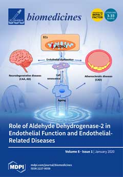
ALDH2 down-regulation (down arrow) or inhibition (dashed red lines) into mitochondria of endothelial cells (ECs) leads to inefficient aldehyde catabolism, affecting ECs functions and accelerating senescence. Senescence itself might further aggravate endothelial dysfunction. Age-related cardiovascular diseases (atherosclerosis and coronary artery disease, CAD) and cerebrovascular unit dysfunction contributing to neurodegenerative disorders (Cerebral Amyloid Angiopathy, CAA, and Alzheimer’s disease, AD) are linked to ALDH2 impairment.View this paper.
  • Issues are regarded as officially published after their release is announced to the table of contents alert mailing list.
  • You may sign up for e-mail alerts to receive table of contents of newly released issues.
  • PDF is the official format for papers published in both, html and pdf forms. To view the papers in pdf format, click on the "PDF Full-text" link, and use the free Adobe Reader to open them.
Order results
Result details
Section
Select all
Export citation of selected articles as:
3 pages, 285 KiB  
Editorial
Acknowledgement to Reviewers of Biomedicines in 2019
by Biomedicines Editorial Office
Biomedicines 2020, 8(1), 17; https://doi.org/10.3390/biomedicines8010017 - 19 Jan 2020
Cited by 1 | Viewed by 1634
Abstract
The editorial team greatly appreciates the reviewers who have dedicated their considerable time and expertise to the journal’s rigorous editorial process over the past 12 months, regardless of whether the papers are finally published or not [...] Full article
23 pages, 7472 KiB  
Article
Comparative Evaluation of the Angiogenic Potential of Hypoxia Preconditioned Blood-Derived Secretomes and Platelet-Rich Plasma: An In Vitro Analysis
by Philipp Moog, Katharina Kirchhoff, Sanjar Bekeran, Anna-Theresa Bauer, Sarah von Isenburg, Ulf Dornseifer, Hans-Günther Machens, Arndt F. Schilling and Ektoras Hadjipanayi
Biomedicines 2020, 8(1), 16; https://doi.org/10.3390/biomedicines8010016 - 16 Jan 2020
Cited by 12 | Viewed by 4674
Abstract
Blood-derived factor preparations are being clinically employed as tools for promoting tissue repair and regeneration. Here we set out to characterize the in vitro angiogenic potential of two types of frequently used autologous blood-derived secretomes: platelet-rich plasma (PRP) and hypoxia preconditioned plasma (HPP)/serum [...] Read more.
Blood-derived factor preparations are being clinically employed as tools for promoting tissue repair and regeneration. Here we set out to characterize the in vitro angiogenic potential of two types of frequently used autologous blood-derived secretomes: platelet-rich plasma (PRP) and hypoxia preconditioned plasma (HPP)/serum (HPS). The concentration of key pro-angiogenic (VEGF) and anti-angiogenic (TSP-1, PF-4) protein factors in these secretomes was analyzed via ELISA, while their ability to induce microvessel formation and sprouting was examined in endothelial cell and aortic ring cultures, respectively. We found higher concentrations of VEGF in PRP and HPP/HPS compared to normal plasma and serum. This correlated with improved induction of microvessel formation by PRP and HPP/HPS. HPP had a significantly lower TSP-1 and PF-4 concentration than PRP and HPS. PRP and HPP/HPS appeared to induce similar levels of microvessel sprouting; however, the length of these sprouts was greater in HPP/HPS than in PRP cultures. A bell-shaped angiogenic response profile was observed with increasing HPP/HPS dilutions, with peak values significantly exceeding the PRP response. Our findings demonstrate that optimization of peripheral blood cell-derived angiogenic factor signalling through hypoxic preconditioning offers an improved alternative to simple platelet concentration and release of growth factors pre-stored in platelets. Full article
(This article belongs to the Special Issue Hypoxia-Inducible Factors: Regulation and Therapeutic Potential)
Show Figures

Figure 1

16 pages, 1071 KiB  
Article
Antioxidant, Antifungal Activities of Ethnobotanical Ficus hirta Vahl. and Analysis of Main Constituents by HPLC-MS
by Chuying Chen, Xuan Peng, Jinyin Chen and Chunpeng Wan
Biomedicines 2020, 8(1), 15; https://doi.org/10.3390/biomedicines8010015 - 15 Jan 2020
Cited by 14 | Viewed by 3310
Abstract
The medicinal and edible plant, Ficus hirta Vahl. (also called hairy fig), is used for the treatment of constipation, inflammation, postpartum hypogalactia, tumors, and cancer. There is an urgent need for scientific evaluation to verify the pharmacological properties of F. hirta. Therefore, [...] Read more.
The medicinal and edible plant, Ficus hirta Vahl. (also called hairy fig), is used for the treatment of constipation, inflammation, postpartum hypogalactia, tumors, and cancer. There is an urgent need for scientific evaluation to verify the pharmacological properties of F. hirta. Therefore, in vitro assays evaluated the antioxidant and antifungal activities of various solvent extracts of hairy fig fruits (HFF). HFF extracts had abundant antioxidant components for a significant amount of total phenolic (TPC) and flavonoid contents (TFC) (TPC from 17.75 ± 0.52 to 85.25 ± 1.72 mg gallic acid/g dw and TFC from 15.80 ± 0.59 to 144.22 ± 8.46 mg rutin/g dw, respectively). The ethyl acetate extract (EAE) and acetone extract (AE) of HFF demonstrated potent antioxidant activities against 1,1-diphenyl-2-picrylhydrazyl (DPPH) (IC50 values of 2.52 and 2.02 mg/mL, respectively) and ABTS radicals (IC50 values of 3.06 and 9.26 mg/mL, respectively). Moreover, the AE with a high TFC showed a prominent in vitro and in vivo antifungal activity against Penicillium italicum, causing citrus blue mold. Eighteen metabolites were identified or putatively identified from six HFF extracts. Current findings indicated that HFF extracts had significant antioxidant and antifungal activities and could potentially be used as an alternative agent for the preservation of agricultural products. Full article
(This article belongs to the Section Drug Discovery and Development)
Show Figures

Figure 1

13 pages, 3459 KiB  
Article
Aptamer-Conjugated Tb(III)-Doped Silica Nanoparticles for Luminescent Detection of Leukemia Cells
by Yaroslav A. Grechkin, Svetlana L. Grechkina, Emil A. Zaripov, Svetlana V. Fedorenko, Asiya R. Mustafina and Maxim V. Berezovski
Biomedicines 2020, 8(1), 14; https://doi.org/10.3390/biomedicines8010014 - 13 Jan 2020
Cited by 14 | Viewed by 3732
Abstract
DNA aptamers have many benefits for cell imaging, such as high affinity and specificity, easiness of chemical functionalization, and low cost of production. Among known aptamers, Sgc8-aptamer was selected against acute lymphoblastic leukemia cells with a dissociation constant in a nanomolar range. The [...] Read more.
DNA aptamers have many benefits for cell imaging, such as high affinity and specificity, easiness of chemical functionalization, and low cost of production. Among known aptamers, Sgc8-aptamer was selected against acute lymphoblastic leukemia cells with a dissociation constant in a nanomolar range. The aptamer was previously used for the covalent coupling with fluorescent and magnetic nanoparticles, as well as for the fabrication of aptamer-based biosensors. Among commonly used fluorescent tags, lanthanide nanoparticles offer stable luminescence with narrow, well-resolved emission peaks and the absence of photoblinking. In other words, lanthanide nanoparticles could serve as luminescence reporters and be used in biosensing. In our study, we conjugated amino- and carboxyl-modified silica-coated terbium (III) thiacalix[4]arenesulfonate luminescent nanoparticles with Sgc8-aptamer and showed the ability of the aptamer-conjugated nanoparticles to detect leukemia cells using fluorescence microscopy. In addition, we conducted a cell viability assay and confirmed that the nanoparticles do not induce spontaneous cell apoptosis or necrosis and could be potentially used for bioimaging applications. Full article
(This article belongs to the Special Issue Engineering Aptamers for Biomedical Applications II)
Show Figures

Figure 1

29 pages, 2315 KiB  
Review
Multifunctional Polymeric Nanoplatforms for Brain Diseases Diagnosis, Therapy and Theranostics
by Shahryar Shakeri, Milad Ashrafizadeh, Ali Zarrabi, Rasoul Roghanian, Elham Ghasemipour Afshar, Abbas Pardakhty, Reza Mohammadinejad, Anuj Kumar and Vijay Kumar Thakur
Biomedicines 2020, 8(1), 13; https://doi.org/10.3390/biomedicines8010013 - 13 Jan 2020
Cited by 87 | Viewed by 6281
Abstract
The blood–brain barrier (BBB) acts as a barrier to prevent the central nervous system (CNS) from damage by substances that originate from the blood circulation. The BBB limits drug penetration into the brain and is one of the major clinical obstacles to the [...] Read more.
The blood–brain barrier (BBB) acts as a barrier to prevent the central nervous system (CNS) from damage by substances that originate from the blood circulation. The BBB limits drug penetration into the brain and is one of the major clinical obstacles to the treatment of CNS diseases. Nanotechnology-based delivery systems have been tested for overcoming this barrier and releasing related drugs into the brain matrix. In this review, nanoparticles (NPs) from simple to developed delivery systems are discussed for the delivery of a drug to the brain. This review particularly focuses on polymeric nanomaterials that have been used for CNS treatment. Polymeric NPs such as polylactide (PLA), poly (D, L-lactide-co-glycolide) (PLGA), poly (ε-caprolactone) (PCL), poly (alkyl cyanoacrylate) (PACA), human serum albumin (HSA), gelatin, and chitosan are discussed in detail. Full article
Show Figures

Figure 1

11 pages, 388 KiB  
Article
Combined Effects of NMES and Mendelsohn Maneuver on the Swallowing Function and Swallowing–Quality of Life of Patients with Stroke-Induced Sub-Acute Swallowing Disorders
by Haewon Byeon
Biomedicines 2020, 8(1), 12; https://doi.org/10.3390/biomedicines8010012 - 12 Jan 2020
Cited by 14 | Viewed by 5202
Abstract
It is necessary to identify how to improve the swallowing-related quality of life, as well as the swallowing function, in order to evaluate the effect of treatments on swallowing disorders. This study aimed to prove the effects of a compound swallowing intervention (Mendelsohn [...] Read more.
It is necessary to identify how to improve the swallowing-related quality of life, as well as the swallowing function, in order to evaluate the effect of treatments on swallowing disorders. This study aimed to prove the effects of a compound swallowing intervention (Mendelsohn maneuver + neuromuscular electrical stimulation (NMES)) on the swallowing function and the quality of life by applying the compound swallowing intervention to patients with sub-acute swallowing disorders due to cerebral infarction for eight weeks. This study analyzed 43 subjects who were diagnosed with swallowing disorders due to cerebral infarction. The experiment consisted of the Mendelsohn maneuver treatment group (n = 15), the NMES treatment group (n = 13), the compound intervention group (Mendelsohn maneuver + NMES; n = 15). The results of ANCOVA showed that the changes in Functional Dysphagia Scale (FDS) scores and Swallowing–Quality of Life (SWAL–QOL) score were different among groups. The compound intervention group had the highest FDS scores and SWAL–QOL score followed by Mendelsohn, and the NMES group had the lowest. The result of this study suggests that NMES can be more effective when it is combined with a traditional swallowing rehabilitation therapy rather than a single intervention method. Full article
(This article belongs to the Special Issue Recent Advances in Brain Vascular Diseases Management and Therapy)
Show Figures

Figure 1

9 pages, 1438 KiB  
Article
Anti-Inflammatory Effects of Diospyrin on Lipopolysaccharide-Induced Inflammation Using RAW 264.7 Mouse Macrophages
by Adnan Shahidullah, Ji-Young Lee, Young-Jin Kim, Syed Muhammad Ashhad Halimi, Abdur Rauf, Hyun-Ju Kim, Bong-Youn Kim and Wansu Park
Biomedicines 2020, 8(1), 11; https://doi.org/10.3390/biomedicines8010011 - 11 Jan 2020
Cited by 6 | Viewed by 4779
Abstract
Diospyrin is a bisnaphthoquinonoid medicinal compound derived from Diospyros lotus, with known anti-cancer, anti-tubercular, and anti-leishmanial activities against Leishmania donovani. However, the effects of diospyrin on lipopolysaccharide (LPS)-induced macrophage activation and inflammation are not fully reported. In this study, the anti-inflammatory [...] Read more.
Diospyrin is a bisnaphthoquinonoid medicinal compound derived from Diospyros lotus, with known anti-cancer, anti-tubercular, and anti-leishmanial activities against Leishmania donovani. However, the effects of diospyrin on lipopolysaccharide (LPS)-induced macrophage activation and inflammation are not fully reported. In this study, the anti-inflammatory effects of diospyrin on LPS-induced macrophages were examined. Diospyrin showed no toxicity in RAW 264.7 at concentrations of up to 10 μM. Diospyrin moderated the production of nitric oxide (NO), monocyte chemotactic protein-1, macrophage inflammatory protein-1β, interleukin (IL)-6, IL-10, granulocyte colony-stimulating factor, granulocyte macrophage colony-stimulating factor, vascular endothelial growth factor, leukemia inhibitory factor, and RANTES/CCL5, as well as calcium release in LPS-induced RAW 264.7, at concentrations of up to 10 μM significantly (p < 0.05). Diospyrin also significantly inhibited the phosphorylation of p38 mitogen-activated protein kinase (MAPK) and mRNA expression of C/EBP homologous protein (CHOP), as well as tumor necrosis factor receptor superfamily member 6 (Fas), in LPS-induced RAW 264.7 cells at concentrations of up to 10 μM (p < 0.05). Diospyrin exhibits anti-inflammatory properties mediated via inhibition of NO, and cytokines in LPS-induced mouse macrophages via the ER-stressed calcium-p38 MAPK/CHOP/Fas pathway. Full article
Show Figures

Graphical abstract

23 pages, 1766 KiB  
Review
Emerging Concepts and Challenges in Rheumatoid Arthritis Gene Therapy
by Andrei A. Deviatkin, Yulia A. Vakulenko, Ludmila V. Akhmadishina, Vadim V. Tarasov, Marina I. Beloukhova, Andrey A. Zamyatnin Jr. and Alexander N. Lukashev
Biomedicines 2020, 8(1), 9; https://doi.org/10.3390/biomedicines8010009 - 09 Jan 2020
Cited by 28 | Viewed by 6912
Abstract
Rheumatoid arthritis (RA) is a systemic inflammatory joint disease affecting about 1% of the population worldwide. Current treatment approaches do not ensure a cure for every patient. Moreover, classical regimens are based on nontargeted systemic immune suppression and have significant side effects. Biological [...] Read more.
Rheumatoid arthritis (RA) is a systemic inflammatory joint disease affecting about 1% of the population worldwide. Current treatment approaches do not ensure a cure for every patient. Moreover, classical regimens are based on nontargeted systemic immune suppression and have significant side effects. Biological treatment has advanced considerably but efficacy and specificity issues remain. Gene therapy is one of the potential future directions for RA therapy, which is rapidly developing. Several gene therapy trials done so far have been of moderate success, but experimental and genetics studies have yielded novel targets. As a result, the arsenal of gene therapy tools keeps growing. Currently, both viral and nonviral delivery systems are used for RA therapy. Herein, we review recent approaches for RA gene therapy. Full article
(This article belongs to the Special Issue Gene Therapy Coming of Age)
Show Figures

Figure 1

11 pages, 2843 KiB  
Article
Meta-Analysis of Hypoxic Transcriptomes from Public Databases
by Hidemasa Bono and Kiichi Hirota
Biomedicines 2020, 8(1), 10; https://doi.org/10.3390/biomedicines8010010 - 09 Jan 2020
Cited by 23 | Viewed by 7143
Abstract
Hypoxia is the insufficiency of oxygen in the cell, and hypoxia-inducible factors (HIFs) are central regulators of oxygen homeostasis. In order to obtain functional insights into the hypoxic response in a data-driven way, we attempted a meta-analysis of the RNA-seq data from the [...] Read more.
Hypoxia is the insufficiency of oxygen in the cell, and hypoxia-inducible factors (HIFs) are central regulators of oxygen homeostasis. In order to obtain functional insights into the hypoxic response in a data-driven way, we attempted a meta-analysis of the RNA-seq data from the hypoxic transcriptomes archived in public databases. In view of methodological variability of archived data in the databases, we first manually curated RNA-seq data from appropriate pairs of transcriptomes before and after hypoxic stress. These included 128 human and 52 murine transcriptome pairs. We classified the results of experiments for each gene into three categories: upregulated, downregulated, and unchanged. Hypoxic transcriptomes were then compared between humans and mice to identify common hypoxia-responsive genes. In addition, meta-analyzed hypoxic transcriptome data were integrated with public ChIP-seq data on the known human HIFs, HIF-1 and HIF-2, to provide insights into hypoxia-responsive pathways involving direct transcription factor binding. This study provides a useful resource for hypoxia research. It also demonstrates the potential of a meta-analysis approach to public gene expression databases for selecting candidate genes from gene expression profiles generated under various experimental conditions. Full article
(This article belongs to the Special Issue Hypoxia-Inducible Factors: Regulation and Therapeutic Potential)
Show Figures

Figure 1

14 pages, 1970 KiB  
Review
The Gut Microbiome and Type 2 Diabetes Mellitus: Discussing A Complex Relationship
by Angelos K. Sikalidis and Adeline Maykish
Biomedicines 2020, 8(1), 8; https://doi.org/10.3390/biomedicines8010008 - 07 Jan 2020
Cited by 109 | Viewed by 18305
Abstract
Type 2 diabetes mellitus (T2DM) is a disease that affects over 9% of the United States population and is closely linked to obesity. While obesity was once thought to stem from a sedentary lifestyle and diets high in fat, recent evidence supports the [...] Read more.
Type 2 diabetes mellitus (T2DM) is a disease that affects over 9% of the United States population and is closely linked to obesity. While obesity was once thought to stem from a sedentary lifestyle and diets high in fat, recent evidence supports the idea that there is more complexity pertinent to the issue. The human gut microbiome has recently been the focus in terms of influencing disease onset. Evidence has shown that the microbiome may be more closely related to T2DM than what was originally thought. High fat diets typically result in poor microbiome heath, which then shifts the gut into a state of dysbiosis. Dysbiosis can then lead to metabolic deregulation, including increased insulin resistance and inflammation, two key factors in the development of T2DM. The purpose of this review is to discuss how microbiome relates to T2DM onset, especially considering obesity, insulin resistance, and inflammation. Full article
Show Figures

Figure 1

10 pages, 646 KiB  
Review
Gastric Cancer Stem Cells: Current Insights into the Immune Microenvironment and Therapeutic Targets
by Lingfeng Fu, Luke Bu, Tadahito Yasuda, Mayu Koiwa, Takahiko Akiyama, Tomoyuki Uchihara, Hideo Baba and Takatsugu Ishimoto
Biomedicines 2020, 8(1), 7; https://doi.org/10.3390/biomedicines8010007 - 06 Jan 2020
Cited by 29 | Viewed by 5495
Abstract
Gastric cancer (GC) is a leading cause of cancer-related death worldwide. Cancer stem cells (CSCs) are known to be involved in chemotherapy resistance and the development of metastases. Although CSCs harbor self-renewal and tumorigenic abilities, the immune microenvironment surrounding CSCs provides various factors [...] Read more.
Gastric cancer (GC) is a leading cause of cancer-related death worldwide. Cancer stem cells (CSCs) are known to be involved in chemotherapy resistance and the development of metastases. Although CSCs harbor self-renewal and tumorigenic abilities, the immune microenvironment surrounding CSCs provides various factors and supports the maintenance of CSC properties. The current review summarizes the accumulating findings regarding the relationship between the immune microenvironment and gastric CSCs (GCSCs), which will support the possibility of developing novel therapeutic strategies for targeting GCSCs. Full article
(This article belongs to the Special Issue Gastric Cancer Research: From Basic Science to the Clinic)
Show Figures

Graphical abstract

16 pages, 1425 KiB  
Article
Familial Combined Hyperlipidemia (FCH) Patients with High Triglyceride Levels Present with Worse Lipoprotein Function Than FCH Patients with Isolated Hypercholesterolemia
by Núria Puig, Inka Miñambres, Sonia Benítez, Pedro Gil, Margarida Grau-Agramunt, Andrea Rivas-Urbina, Antonio Pérez and José Luis Sánchez-Quesada
Biomedicines 2020, 8(1), 6; https://doi.org/10.3390/biomedicines8010006 - 06 Jan 2020
Cited by 6 | Viewed by 3629
Abstract
Lipoprotein characteristics were analyzed in familial combined hyperlipidemia (FCH) patients before and after statin treatment. Twenty-six FCH patients were classified according to the presence (HTG group, n = 13) or absence (normotriglyceridemic (NTG) group, n = 13) of hypertriglyceridemia. Fifteen healthy subjects comprised [...] Read more.
Lipoprotein characteristics were analyzed in familial combined hyperlipidemia (FCH) patients before and after statin treatment. Twenty-six FCH patients were classified according to the presence (HTG group, n = 13) or absence (normotriglyceridemic (NTG) group, n = 13) of hypertriglyceridemia. Fifteen healthy subjects comprised the control group. Lipid profile, inflammation markers, and qualitative characteristics of lipoproteins were assessed. Both groups of FCH subjects showed high levels of plasma C-reactive protein (CRP), lipoprotein-associated phospholipase A2 (Lp-PLA2) activity and apolipoprotein J. Statins reverted the increased levels of Lp-PLA2 and CRP. Lipoprotein composition alterations detected in FCH subjects were much more frequent in the HTG group, leading to dysfunctional low-density lipoproteins (LDL) and high-density lipoproteins (HDL). In the HTG group, LDL was smaller, more susceptible to oxidation, and contained more electronegative LDL (LDL(-)) compared to the NTG and control groups. Regarding HDL, the HTG group had less Lp-PLA2 activity than the NTG and control groups. HDL from both FCH groups was less anti-inflammatory than HDL from the control group. Statins increased LDL size, decreased LDL(-), and lowered Lp-PLA2 in HDL from HTG. In summary, pro-atherogenic alterations were more frequent and severe in the HTG group. Statins improved some alterations, but many remained unchanged in HTG. Full article
Show Figures

Figure 1

15 pages, 2435 KiB  
Article
Early Exposure to THC Alters M-Cell Development in Zebrafish Embryos
by Md Ruhul Amin, Kazi T. Ahmed and Declan W. Ali
Biomedicines 2020, 8(1), 5; https://doi.org/10.3390/biomedicines8010005 - 04 Jan 2020
Cited by 13 | Viewed by 4686
Abstract
Cannabis is one of the most commonly used illicit recreational drugs that is often taken for medicinal purposes. The psychoactive ingredient in cannabis is Δ9-Tetrahydrocannabinol (Δ9-THC, hereafter referred to as THC), which is an agonist at the endocannabinoid receptors [...] Read more.
Cannabis is one of the most commonly used illicit recreational drugs that is often taken for medicinal purposes. The psychoactive ingredient in cannabis is Δ9-Tetrahydrocannabinol (Δ9-THC, hereafter referred to as THC), which is an agonist at the endocannabinoid receptors CB1R and CB2R. Here, we exposed zebrafish embryos to THC during the gastrulation phase to determine the long-term effects during development. We specifically focused on reticulospinal neurons known as the Mauthner cells (M-cell) that are involved in escape response movements. The M- cells are born during gastrulation, thus allowing us to examine neuronal morphology of neurons born during the time of exposure. After the exposure, embryos were allowed to develop normally and were examined at two days post-fertilization for M-cell morphology and escape responses. THC treated embryos exhibited subtle alterations in M-cell axon diameter and small changes in escape response dynamics to touch. Because escape responses were altered, we also examined muscle fiber development. The fluorescent labelling of red and white muscle fibers showed that while muscles were largely intact, the fibers were slightly disorganized with subtle but significant changes in the pattern of expression of nicotinic acetylcholine receptors. However, there were no overt changes in the expression of nicotinic receptor subunit mRNA ascertained by qPCR. Embryos were allowed to further develop until 5 dpf, when they were examined for overall levels of movement. Animals exposed to THC during gastrulation exhibited reduced activity compared with vehicle controls. Together, these findings indicate that zebrafish exposed to THC during the gastrula phase exhibit small changes in neuronal and muscle morphology that may impact behavior and locomotion. Full article
(This article belongs to the Special Issue Zebrafish Models for Development and Disease 2.0)
Show Figures

Figure 1

12 pages, 578 KiB  
Review
Endothelial Aldehyde Dehydrogenase 2 as a Target to Maintain Vascular Wellness and Function in Ageing
by Ginevra Nannelli, Marina Ziche, Sandra Donnini and Lucia Morbidelli
Biomedicines 2020, 8(1), 4; https://doi.org/10.3390/biomedicines8010004 - 03 Jan 2020
Cited by 14 | Viewed by 3880
Abstract
Endothelial cells are the main determinants of vascular function, since their dysfunction in response to a series of cardiovascular risk factors is responsible for disease progression and further consequences. Endothelial dysfunction, if not resolved, further aggravates the oxidative status and vessel wall inflammation, [...] Read more.
Endothelial cells are the main determinants of vascular function, since their dysfunction in response to a series of cardiovascular risk factors is responsible for disease progression and further consequences. Endothelial dysfunction, if not resolved, further aggravates the oxidative status and vessel wall inflammation, thus igniting a vicious cycle. We have furthermore to consider the physiological manifestation of vascular dysfunction and chronic low-grade inflammation during ageing, also known as inflammageing. Based on these considerations, knowledge of the molecular mechanism(s) responsible for endothelial loss-of-function can be pivotal to identify novel targets of intervention with the aim of maintaining endothelial wellness and vessel trophism and function. In this review we have examined the role of the detoxifying enzyme aldehyde dehydrogenase 2 (ALDH2) in the maintenance of endothelial function. Its impairment indeed is associated with oxidative stress and ageing, and in the development of atherosclerosis and neurodegenerative diseases. Strategies to improve its expression and activity may be beneficial in these largely diffused disorders. Full article
Show Figures

Figure 1

20 pages, 1987 KiB  
Review
Beneficial Biological Activities of Cinnamomum osmophloeum and Its Potential Use in the Alleviation of Oral Mucositis: A Systematic Review
by Abu Bakar, Pin-Chuan Yao, Valendriyani Ningrum, Cheng-Tzu Liu and Shih-Chieh Lee
Biomedicines 2020, 8(1), 3; https://doi.org/10.3390/biomedicines8010003 - 01 Jan 2020
Cited by 9 | Viewed by 4214
Abstract
The aim of this review was to provide an updated overview of studies on the medical-biological activities of Cinnamomum osmophloeum (C. osmophloeum) in vitro and in vivo and the potential therapeutic use of natural agents prepared from this plant for the [...] Read more.
The aim of this review was to provide an updated overview of studies on the medical-biological activities of Cinnamomum osmophloeum (C. osmophloeum) in vitro and in vivo and the potential therapeutic use of natural agents prepared from this plant for the alleviation of oral mucositis (OM). Reported articles were collected using web search engine tools. The systematic review was organized according to the preferred reporting items for reviews and meta-analyses (PRISMA) statement. Additional sources were identified through cross-referencing to identify the potential use of C. osmophloeum in the alleviation of OM. The results disclosed that C. osmophloeum is comprised of bioactive ingredients that could act diversely as a reagent in anti-inflammation, antibacterial, antioxidant, anti-hyperglycemic, antidyslipidemia, anti-cancer, renal disease therapy and anti-hyperuricemia capacities. Recent studies revealed that the overall effects on anti-inflammation, wound repair, and the antibacterial and antioxidant activities of its constituents would act as a potential remedy for oral mucositis. Up-to-date in vitro and in vivo studies on the medical-biological activities of C. osmophloeum suggested that C. osmophloeum and its constituents could be promising remedies as adjuvants in OM therapy and warrant further investigation. Full article
(This article belongs to the Section Drug Discovery and Development)
Show Figures

Graphical abstract

14 pages, 1875 KiB  
Article
Cytotoxic Effects of the Dual ErbB Tyrosine Kinase Inhibitor, Lapatinib, on Walker 256 Rat Breast Tumour and IEC-6 Rat Normal Small Intestinal Cell Lines
by Wan Nor I′zzah Wan Mohamad Zain, Joanne Bowen, Emma Bateman and Dorothy Keefe
Biomedicines 2020, 8(1), 2; https://doi.org/10.3390/biomedicines8010002 - 30 Dec 2019
Cited by 4 | Viewed by 3104
Abstract
Lapatinib is an orally administered, dual ErbB1/ErbB2 tyrosine kinase inhibitor (TKI). It is effective in ErbB2 + ve breast cancer treatment. However, lapatinib is associated with diarrhoea with an incidence of 47–75%. The mechanism of ErbB1 TKI-induced diarrhoea remains unclear. ErbB1 or epidermal [...] Read more.
Lapatinib is an orally administered, dual ErbB1/ErbB2 tyrosine kinase inhibitor (TKI). It is effective in ErbB2 + ve breast cancer treatment. However, lapatinib is associated with diarrhoea with an incidence of 47–75%. The mechanism of ErbB1 TKI-induced diarrhoea remains unclear. ErbB1 or epidermal growth factor receptor (EGFR) is expressed in gastrointestinal mucosa whereby the primary site for drug absorption is intestine. Thus, administration of ErbB1 oral TKI may disrupt gut homeostasis, leading to diarrhoea. Nevertheless, further investigations are required. We observed that lapatinib inhibited 50% Walker 256 breast tumour cells and IEC-6 small intestinal cell growth. Higher percentage of necrosis was observed in lapatinib-treated Walker 256. Lapatinib-treated IEC-6 showed higher percentage of late apoptosis. Only ErbB2 mRNA was detected in Walker 256 but both ErbB1 and ErbB2 mRNAs were detected in IEC-6, yet both protein staining were detected in both cells. Lapatinib exhibited cytotoxic properties on ErbB1/ErbB2 expressing cell lines, with intestinal cells being more sensitive to lapatinib compared to tumour cells. Lapatinib induced necrosis in tumour cells, while inducing late apoptosis in intestinal cells may explain lapatinib-induced diarrhoea in patients administered with the drug which could be due to apoptosis of intestinal epithelial cells leading to barrier disruption and consequently diarrhoea. Full article
Show Figures

Figure 1

16 pages, 2045 KiB  
Article
Comprehensive Analysis of Neurotoxin-Induced Ablation of Dopaminergic Neurons in Zebrafish Larvae
by Michael Kalyn, Khang Hua, Suzita Mohd Noor, Chee Ern David Wong and Marc Ekker
Biomedicines 2020, 8(1), 1; https://doi.org/10.3390/biomedicines8010001 - 28 Dec 2019
Cited by 26 | Viewed by 5233
Abstract
Neurotoxin exposure of zebrafish larvae has been used to mimic a Parkinson’s disease (PD) phenotype and to facilitate high-throughput drug screening. However, the vulnerability of zebrafish to various neurotoxins was shown to be variable. Here, we provide a direct comparison of ablative effectiveness [...] Read more.
Neurotoxin exposure of zebrafish larvae has been used to mimic a Parkinson’s disease (PD) phenotype and to facilitate high-throughput drug screening. However, the vulnerability of zebrafish to various neurotoxins was shown to be variable. Here, we provide a direct comparison of ablative effectiveness in order to identify the optimal neurotoxin-mediated dopaminergic (DAnergic) neuronal death in larval zebrafish. Transgenic zebrafish, Tg(dat:eGFP), were exposed to different concentrations of the neurotoxins MPTP, MPP+, paraquat, 6-OHDA, and rotenone for four days, starting at three days post-fertilization. The LC50 of each respective neurotoxin concentration was determined. Confocal live imaging on Tg(dat:eGFP) showed that MPTP, MPP+, and rotenone caused comparable DAnergic cell loss in the ventral diencephalon (vDC) region while, paraquat and 6-OHDA caused fewer losses of DAnergic cells. These results were further supported by respective gene expression analyses of dat, th, and p53. Importantly, the loss of DAnergic cells from exposure to MPTP, MPP+, and rotenone impacted larval locomotor function. MPTP induced the largest motor deficit, but this was accompanied by the most severe morphological impairment. We conclude that, of the tested neurotoxins, MPP+ recapitulates a substantial degree of DAnergic ablation and slight locomotor perturbations without systemic defects indicative of a Parkinsonian phenotype. Full article
(This article belongs to the Collection Neurotoxicity: Mechanisms and Potential Therapeutic Strategies)
Show Figures

Figure 1

Previous Issue
Next Issue
Back to TopTop