
Isolated Progression of Multiple Myeloma into the Extramedullary Plasmacytoma of Dura Mater: A Case Report and Review of the Literature


Abstract
:1. Introduction
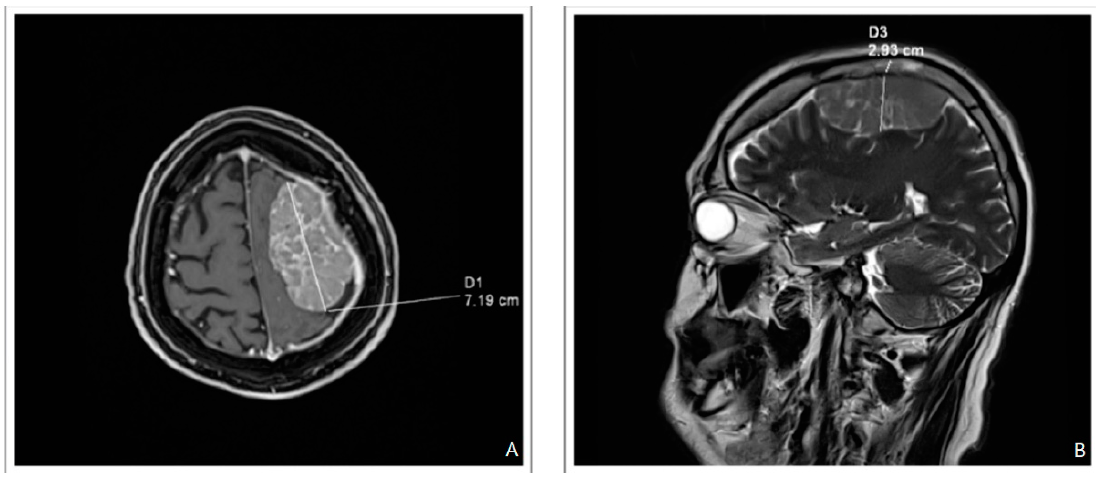
2. Case Report
3. Discussion

| Author | Country | No. of Patient | Year | OS (Months) |
|---|---|---|---|---|
| Jurczyszyn et al., 2016 [6] | International | 172 | 2000–2015 | 6.7 |
| Dias et al., 2017 [23] | Brazil | 20 | 2008–2016 | 7.4 |
| Badros et al., 2016 [34] | USA | 2 | 2008–2016 | na |
| Katodritou et al., 2015 [28] | Greece | 31 | 2000–2013 | 3 |
| Paludo et al., 2016 [24] | USA, Mayo | 29 | 1998–2014 | 5 |
| Varga et al., 2018 [22] | Hungary | 13 | 2007–2017 | 4 |
| Chen et al., 2013 [18] | Canada | 37 | 1999–2010 | 5 |
| Gozzetti et al., 2012 [46] | Italy | 50 | 2000–2010 | 6 |
| Abdallah et al., 2014 [4] | USA | 35 | 1996–2012 | 4 |
| Median-OS by CNS-MM by Treatment [Months] | CNS-MM Treatment | |||
|---|---|---|---|---|
| IMID Immunomodulatory Drugs | PIs Proteasome Inhibitors | SCT Stem Cell Transplant | XRT Radiotherapy | |
| 5.1 | X | X | ||
| 4.7 | X | X | ||
| 7.3 | X | X | X | |
| 5.3 | X | |||
| 9.0 | X | X | ||
| 6.0 | X | X | ||
Author Contributions
Funding
Institutional Review Board Statement
Informed Consent Statement
Data Availability Statement
Conflicts of Interest
References
- Egan, P.A.; Elder, P.T.; Deighan, W.I.; O’Connor, S.J.; Alexander, H.D. Multiple myeloma with central nervous system relapse. Haematologica 2020, 105, 1780–1790. [Google Scholar] [CrossRef]
- Fassas, A.B.T.; Ward, S.; Muwalla, F.; Van Hemert, R.; Schluterman, K.; Harik, S.; Tricot, G. Myeloma of the central nervous system: Strong association with unfavorable chromosomal abnormalities and other high-risk disease features. Leuk. Lymphoma 2004, 45, 291–300. [Google Scholar] [CrossRef]
- Varettoni, M.; Corso, A.; Pica, G.; Mangiacavalli, S.; Pascutto, C.; Lazzarino, M. Incidence, presenting features and outcome of extramedullary disease in multiple myeloma: A longitudinal study on 1003 consecutive patients. Ann. Oncol. 2010, 21, 325–330. [Google Scholar] [CrossRef]
- Abdallah, A.-O.; Atrash, S.; Shahid, Z.; Jameel, M.; Grazziutti, M.; Apewokin, S.; Kumar, N.S.; Restrepo, A.; Waheed, S.; Van Rhee, F.; et al. Patterns of Central Nervous System Involvement in Relapsed and Refractory Multiple Myeloma. Clin. Lymphoma Myeloma Leuk. 2014, 14, 211–214. [Google Scholar] [CrossRef] [PubMed]
- Lee, D.; Kalff, A.; Low, M.; Gangatharan, S.; Ho, P.; Bajel, A.; Ritchie, D.; Grigg, A.; Spencer, A. Central nervous system multiple myeloma–potential roles for intrathecal therapy and measurement of cerebrospinal fluid light chains. Br. J. Haematol. 2013, 162, 371–375. [Google Scholar] [CrossRef] [PubMed]
- Jurczyszyn, A.; Grzasko, N.; Gozzetti, A.; Czepiel, J.; Cerase, A.; Hungria, V.; Crusoe, E.; Dias, A.L.M.S.; Vij, R.; Fiala, M.A.; et al. Central nervous system involvement by multiple myeloma: A multi-institutional retrospective study of 172 patients in daily clinical practice. Am. J. Hematol. 2016, 91, 575–580. [Google Scholar] [CrossRef] [PubMed]
- Lasocki, A.; Gangatharan, S.; Gaillard, F.; Harrison, S. Intracranial involvement by multiple myeloma. Clin. Radiol. 2015, 70, 890–897. [Google Scholar] [CrossRef] [PubMed]
- Okamoto, K.; Ito, J.; Furusawa, T.; Sakai, K.; Tokiguchi, S.; Sato, M.; Tanaka, R.; Nemoto, K.; Oyanagi, K. Solitary plasmacytomas of the occipital bone: A report of two cases. Eur. Radiol. 1997, 7, 503–506. [Google Scholar] [CrossRef] [PubMed]
- Pardridge, W.M. Drug Transport across the Blood–Brain Barrier. J. Cereb. Blood Flow Metab. 2012, 32, 1959–1972. [Google Scholar] [CrossRef]
- Chen, A.I.; Negrin, R.S.; McMillan, A.; Shizuru, J.A.; Johnston, L.J.; Lowsky, R.; Miklos, D.B.; Arai, S.; Weng, W.-K.; Laport, G.G.; et al. Tandem chemo-mobilization followed by high-dose melphalan and carmustine with single autologous hematopoietic cell transplantation for multiple myeloma. Bone Marrow Transplant. 2012, 47, 516–521. [Google Scholar] [CrossRef]
- Sammartano, V.; Cerase, A.; Venanzi, V.; Mazzei, M.A.; Vangone, B.E.; Gentili, F.; Chiarotti, I.; Bocchia, M.; Gozzetti, A. Central Nervous System Myeloma and Unusual Extramedullary Localizations: Real Life Practical Guidance. Front. Oncol. 2022, 12, 3276. [Google Scholar] [CrossRef]
- Marini, A.; Carulli, G.; Lari, T.; Buda, G.; Lambelet, P.; Ciancia, E.M.; Benedetti, E.; Caracciolo, F.; Ferreri, M.I.; Pesaresi, I.; et al. Myelomatous Meningitis Evaluated by Multiparameter Flow Cytometry: Report of a Case and Review of the Literature. J. Clin. Exp. Hematop. 2014, 54, 129–136. [Google Scholar] [CrossRef] [PubMed]
- Tsang, C.-S.; Ho, L.; Tan, T.-C. Intracranial multiple myeloma involving the dura. J. Clin. Neurosci. 2006, 13, 122–123. [Google Scholar] [CrossRef]
- Yellu, M.R.; Engel, J.M.; Ghose, A.; Onitilo, A.A. Overview of recent trends in diagnosis and management of leptomeningeal multiple myeloma. Hematol. Oncol. 2016, 34, 2–8, Retraction in: Hematol Oncol. 2017, 35, 142. [Google Scholar] [CrossRef] [PubMed]
- Cerase, A.; Tarantino, A.; Gozzetti, A.; Muccio, C.F.; Gennari, P.; Monti, L.; Di Blasi, A.; Venturi, C. Intracranial involvement in plasmacytomas and multiple myeloma: A pictorial essay. Neuroradiology 2008, 50, 665–674. [Google Scholar] [CrossRef] [PubMed]
- Jurczyszyn, A.; Malkowski, B.; Czepiel, J.; Skotnicki, A.B. The importance of imaging techniques in the modern treatment of multiple myeloma. Przeglad Lek. 2014, 71, 221–230. [Google Scholar]
- Nieuwenhuizen, L.; Biesma, D.H. Central nervous system myelomatosis: Review of the literature. Eur. J. Haematol. 2008, 80, 1–9. [Google Scholar] [CrossRef]
- Chen, C.I.; Masih-Khan, E.; Jiang, H.; Rabea, A.; Cserti-Gazdewich, C.; Jimenez-Zepeda, V.H.; Chu, C.-M.; Kukreti, V.; Trudel, S.; Tiedemann, R.; et al. Central nervous system involvement with multiple myeloma: Long term survival can be achieved with radiation, intrathecal chemotherapy, and immunomodulatory agents. Br. J. Haematol. 2013, 162, 483–488. [Google Scholar] [CrossRef]
- Méndez, C.E.; Hwang, B.J.; Destian, S.; Mazumder, A.; Jagannath, S.; Vesole, D.H. Intracranial Multifocal Dural Involvement in Multiple Myeloma: Case Report and Review of the Literature. Clin. Lymphoma Myeloma Leuk. 2010, 10, 220–223. [Google Scholar] [CrossRef]
- Haegelen, C.; Riffaud, L.; Bernard, M.; Carsin-Nicol, B.; Morandi, X. Dural plasmacytoma revealing multiple myeloma: Case report. J. Neurosurg. 2006, 104, 608–610. [Google Scholar] [CrossRef]
- Wavre, A.; Baur, A.S.; Betz, M.; Mühlematter, D.; Jotterand, M.; Zaman, K.; Ketterer, N. Case study of intracerebral plasmacytoma as an initial presentation of multiple myeloma. Neuro-Oncology 2007, 9, 370–372. [Google Scholar] [CrossRef] [PubMed]
- Varga, G.; Mikala, G.; Gopcsa, L.; Csukly, Z.; Kollai, S.; Balázs, G.; Botond, T.; Wohner, N.; Horváth, L.; Szombath, G.; et al. Multiple Myeloma of the Central Nervous System: 13 Cases and Review of the Literature. J. Oncol. 2018, 2018, 3970169. [Google Scholar] [CrossRef] [PubMed]
- Dias, A.L.M.S.; Higashi, F.; Peres, A.L.M.; Cury, P.; Crusoé, E.D.Q.; Hungria, V.T.D.M. Multiple myeloma and central nervous system involvement: Experience of a Brazilian center. Hematol. Transfus. Cell Ther. 2018, 40, 30–36. [Google Scholar] [CrossRef] [PubMed]
- Paludo, J.; Painuly, U.; Kumar, S.; Gonsalves, W.I.; Rajkumar, V.; Buadi, F.; Lacy, M.Q.; Dispenzieri, A.; Kyle, R.A.; Mauermann, M.L.; et al. Myelomatous Involvement of the Central Nervous System. Clin. Lymphoma Myeloma Leuk. 2016, 16, 644–654. [Google Scholar] [CrossRef] [PubMed]
- Pontikoglou, C.; Fragos, C.; Kolyvaki, E.; Samonis, G.; Eliopoulos, G.D.; Papadaki, H.A. Multiple myeloma involving the central nervous system: A report of two cases with unusual manifestations. Leuk. Lymphoma 2005, 46, 737–741. [Google Scholar] [CrossRef]
- Chen, Y.; Qiu, Y.; Fu, H.; Li, J.; Chen, L.; Liao, S.; Liu, T. IgD multiple myeloma with central nervous system involvement: A case report and literature review. Mol. Clin. Oncol. 2021, 15, 1–6. [Google Scholar] [CrossRef]
- Jurczyszyn, A.; Olszewska-Szopa, M.; Fornagiel, S.; Skotnicki, A. Zajęcie ośrodkowego układu nerwowego w przebiegu szpiczaka plazmocytowego–Opis przypadku i przegląd literatury. Acta Haematol. Pol. 2015, 46, 242–247. [Google Scholar] [CrossRef]
- Katodritou, E.; Terpos, E.; Kastritis, E.; Delimpasis, S.; Symeonidis, A.S.; Repousis, P.; Kyrtsonis, M.-C.; Vadikolia, C.; Michalis, E.; Polychronidou, G.; et al. Lack of survival improvement with novel anti-myeloma agents for patients with multiple myeloma and central nervous system involvement: The Greek Myeloma Study Group experience. Ann. Hematol. 2015, 94, 2033–2042. [Google Scholar] [CrossRef]
- Renfrow, J.J.; DeTroye, A.; Chan, M.; Tatter, S.; Ellis, T.; McMullen, K.; Johnson, A.; Mott, R.; Lesser, G.J. Initial experience with bendamustine in patients with recurrent primary central nervous system lymphoma: A case report. J. Neurooncol. 2012, 107, 659–663. [Google Scholar] [CrossRef]
- Annibali, O.; Nobile, C.; Greco, R.; Cellini, F.; Quattrocchi, C.C.; Tirindelli, M.C.; Petrucci, M.T.; Avvisati, G. The combination topotecan, temozolomide and dexamethasone associated with radiotherapy as treatment of central nervous system myeloma relapse. Int. J. Hematol. 2009, 89, 513–516. [Google Scholar] [CrossRef]
- Richardson, P.G.; Spencer, A.; Cannell, P.; Harrison, S.J.; Catley, L.; Underhill, C.; Zimmerman, T.M.; Hofmeister, C.C.; Jakubowiak, A.J.; Laubach, J.P.; et al. Phase 1 Clinical Evaluation of Twice-Weekly Marizomib (NPI-0052), a Novel Proteasome Inhibitor, in Patients with Relapsed/Refractory Multiple Myeloma (MM). Blood 2011, 118, 302. [Google Scholar] [CrossRef]
- Williamson, M.J.; Blank, J.L.; Bruzzese, F.J.; Cao, Y.; Daniels, J.S.; Dick, L.R.; Labutti, J.; Mazzola, A.M.; Patil, A.D.; Reimer, C.L.; et al. Comparison of biochemical and biological effects of ML858 (salinosporamide A) and bortezomib. Mol. Cancer Ther. 2006, 5, 3052–3061. [Google Scholar] [CrossRef] [PubMed]
- Mele, G.; Pinna, S.; Alloro, E.; Brocca, M.C.; Coppi, M.R.; Quarta, G. Inefficacy of bortezomib therapy for CNS involvement of refractory multiple myeloma. Leuk. Res. 2007, 31, 721–723. [Google Scholar] [CrossRef]
- Badros, A.; Singh, Z.; Dhakal, B.; Kwok, Y.; MacLaren, A.; Richardson, P.; Trikha, M.; Hari, P. Marizomib for central nervous system-multiple myeloma. Br. J. Haematol. 2017, 177, 221–225. [Google Scholar] [CrossRef]
- Mussetti, A.; Dalto, S.; Montefusco, V. Effective treatment of pomalidomide in central nervous system myelomatosis. Leuk. Lymphoma 2013, 54, 864–866. [Google Scholar] [CrossRef]
- Yutaka, H.; Mariko, Y.; Shinichiro, O.; Kunihiko, M.; Yusuke, T.; Yasuo, I. Thalidomide for the treatment of leptomeningeal multiple myeloma. Eur. J. Haematol. 2006, 76, 358–359. [Google Scholar] [CrossRef] [PubMed]
- Li, Z.; Qiu, Y.; Personett, D.; Huang, P.; Edenfield, B.; Katz, J.; Babusis, D.; Tang, Y.; Shirely, M.A.; Moghaddam, M.F.; et al. Pomalidomide Shows Significant Therapeutic Activity against CNS Lymphoma with a Major Impact on the Tumor Microenvironment in Murine Models. PLoS ONE 2013, 8, e71754. [Google Scholar] [CrossRef] [PubMed]
- Zajec, M.; Frerichs, K.A.; Van Duijn, M.M.; Nijhof, I.S.; Stege, C.A.; Avet-Loiseau, H.; Luider, T.M.; De Rijke, Y.B.; Jacobs, J.F.; Van De Donk, N.W. Cerebrospinal Fluid Penetrance of Daratumumab in Leptomeningeal Multiple Myeloma. Hemasphere 2020, 4, e413. [Google Scholar] [CrossRef]
- Gozzetti, A.; Cerase, A. Send Orders for Reprints to reprints@benthamscience.net Central Nervous System Agents in Medicinal Chemistry Novel Agents in CNS Myeloma Treatment. Curr. Top. Med. Chem. 2014, 14, 1923–1938. [Google Scholar]
- Wang, T.; He, T.; Ma, L.; Yang, Y.; Feng, R.; Ding, Y.; Shan, Y.; Bu, B.; Qi, F.; Wu, F.; et al. Clinical Outcomes of BCMA CAR-T Cells in a Multiple Myeloma Patient With Central Nervous System Invasion. Front. Oncol. 2022, 12, 854448. [Google Scholar] [CrossRef]
- Raje, N.; Berdeja, J.; Lin, Y.; Siegel, D.; Jagannath, S.; Madduri, D.; Liedtke, M.; Rosenblatt, J.; Maus, M.V.; Turka, A.; et al. Anti-BCMA CAR T-Cell Therapy bb2121 in Relapsed or Refractory Multiple Myeloma. N. Engl. J. Med. 2019, 380, 1726–1737. [Google Scholar] [CrossRef] [PubMed]
- Berdeja, J.G.; Madduri, D.; Usmani, S.Z.; Jakubowiak, A.; Agha, M.; Cohen, A.D.; Stewart, A.K.; Hari, P.; Htut, M.; Lesokhin, A.; et al. Ciltacabtagene autoleucel, a B-cell maturation antigen-directed chimeric antigen receptor T-cell therapy in patients with relapsed or refractory multiple myeloma (CARTITUDE-1): A phase 1b/2 open-label study. Lancet 2021, 398, 314–324. [Google Scholar] [CrossRef] [PubMed]
- Brudno, J.N.; Maric, I.; Hartman, S.D.; Rose, J.J.; Wang, M.; Lam, N.; Stetler-Stevenson, M.; Salem, D.; Yuan, C.; Pavletic, S.; et al. T Cells Genetically Modified to Express an Anti–B-Cell Maturation Antigen Chimeric Antigen Receptor Cause Remissions of Poor-Prognosis Relapsed Multiple Myeloma. J. Clin. Oncol. 2018, 36, 2267–2280. [Google Scholar] [CrossRef] [PubMed]
- Wang, Y.; Zu, C.; Teng, X.; Yang, L.; Zhang, M.; Hong, R.; Zhao, H.; Cui, J.; Xu, H.; Hongsheng, A.C.; et al. BCMA CAR-T Therapy Is Safe and Effective for Refractory/Relapsed Multiple Myeloma With Central Nervous System Involvement. J. Immunother. 2022, 45, 25–34. [Google Scholar] [CrossRef] [PubMed]
- Cohen, A.D.; Parekh, S.; Santomasso, B.D.; Pérez-Larraya, J.G.; van de Donk, N.W.C.J.; Arnulf, B.; Mateos, M.-V.; Lendvai, N.; Jackson, C.C.; De Braganca, K.C.; et al. Incidence and management of CAR-T neurotoxicity in patients with multiple myeloma treated with ciltacabtagene autoleucel in CARTITUDE studies. Blood Cancer J. 2022, 12, 32. [Google Scholar] [CrossRef]
- Gozzetti, A.; Cerase, A.; Lotti, F.; Rossi, D.; Palumbo, A.; Petrucci, M.T.; Patriarca, F.; Nozzoli, C.; Cavo, M.; Offidani, M.; et al. Extramedullary intracranial localization of multiple myeloma and treatment with novel agents: A retrospective survey of 50 patients. Cancer 2012, 118, 1574–1584. [Google Scholar] [CrossRef] [PubMed]

| Drug | Crosses the BBB (In Vivo Tests, Laboratory Tests on Animal Patterns, etc.) | Prospective Clinical Trials |
|---|---|---|
| Thalidomide | Yes | No |
| Bortezomib | No | No |
| Lenalidomide | Yes | No |
| Pomalidomide | Yes | No |
| Carfilzomib | No | No |
| Marizomib | Yes | No |
| Daratumumab | Yes | No |
| Bendamustine | Yes | No |
| Topotecan | Yes | No |
| Melphalan (high dose) | Yes | No |
Disclaimer/Publisher’s Note: The statements, opinions and data contained in all publications are solely those of the individual author(s) and contributor(s) and not of MDPI and/or the editor(s). MDPI and/or the editor(s) disclaim responsibility for any injury to people or property resulting from any ideas, methods, instructions or products referred to in the content. |
© 2023 by the authors. Licensee MDPI, Basel, Switzerland. This article is an open access article distributed under the terms and conditions of the Creative Commons Attribution (CC BY) license (https://creativecommons.org/licenses/by/4.0/).
Share and Cite
Tyczyńska, A.; Turski, M.; Zarzycka, E.; Zaucha, J.M. Isolated Progression of Multiple Myeloma into the Extramedullary Plasmacytoma of Dura Mater: A Case Report and Review of the Literature. Biomedicines 2023, 11, 1225. https://doi.org/10.3390/biomedicines11041225
Tyczyńska A, Turski M, Zarzycka E, Zaucha JM. Isolated Progression of Multiple Myeloma into the Extramedullary Plasmacytoma of Dura Mater: A Case Report and Review of the Literature. Biomedicines. 2023; 11(4):1225. https://doi.org/10.3390/biomedicines11041225
Chicago/Turabian StyleTyczyńska, Agata, Mikołaj Turski, Ewa Zarzycka, and Jan Maciej Zaucha. 2023. "Isolated Progression of Multiple Myeloma into the Extramedullary Plasmacytoma of Dura Mater: A Case Report and Review of the Literature" Biomedicines 11, no. 4: 1225. https://doi.org/10.3390/biomedicines11041225