
Multi-Omics and Management of Follicular Carcinoma of the Thyroid
 ,
,  ,
,
Abstract
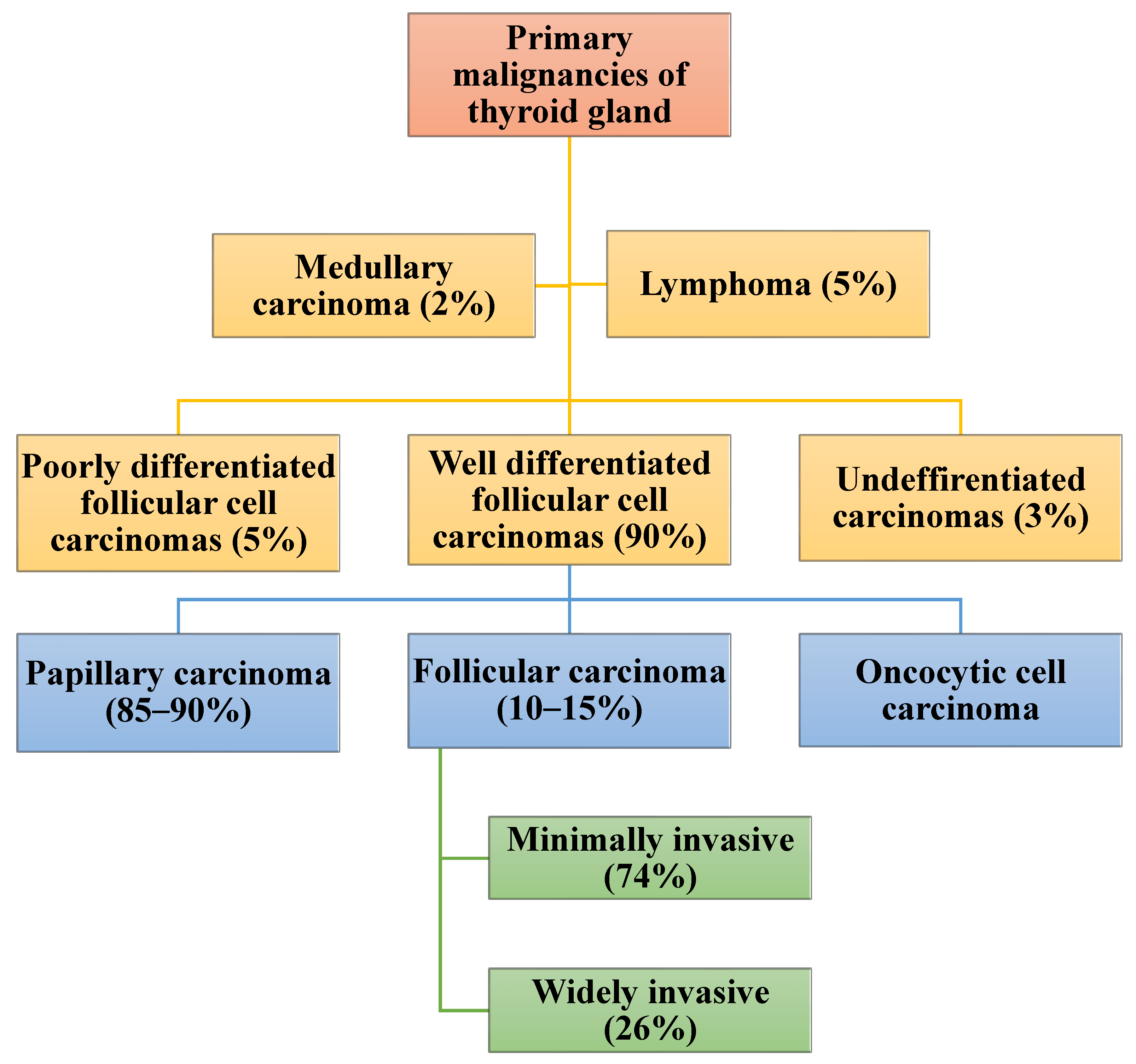
:1. Introduction
2. Diagnosis and Management of Follicular Carcinoma
2.1. Diagnostic Work-Up of Suspected Follicular Carcinoma
2.2. Staging of Follicular Carcinoma
2.3. Risk Stratification and Associated Challenges in Follicular Carcinoma
2.4. Management of Follicular Carcinoma and Related Challenges
2.4.1. Lobectomy versus Total Thyroidectomy in FTC
2.4.2. Radioactive Iodine Therapy and Dosimetry in the Management of FTC
2.4.3. Thyroid Stimulating Hormone Suppression during Management of FTC
2.4.4. Management of FTC with Extensive Extra-Thyroidal Extension
2.5. Management of Metastatic Follicular Carcinoma and Related Challenges
2.5.1. Pulmonary Metastases
2.5.2. Bone Metastases
2.5.3. Brain Metastasis
2.6. Management of De-Differentiated FTC
2.7. Follow-Up of Patients Who Have Follicular Carcinoma and Associated Challenges
2.8. Outcome of Treatment of Follicular Carcinoma
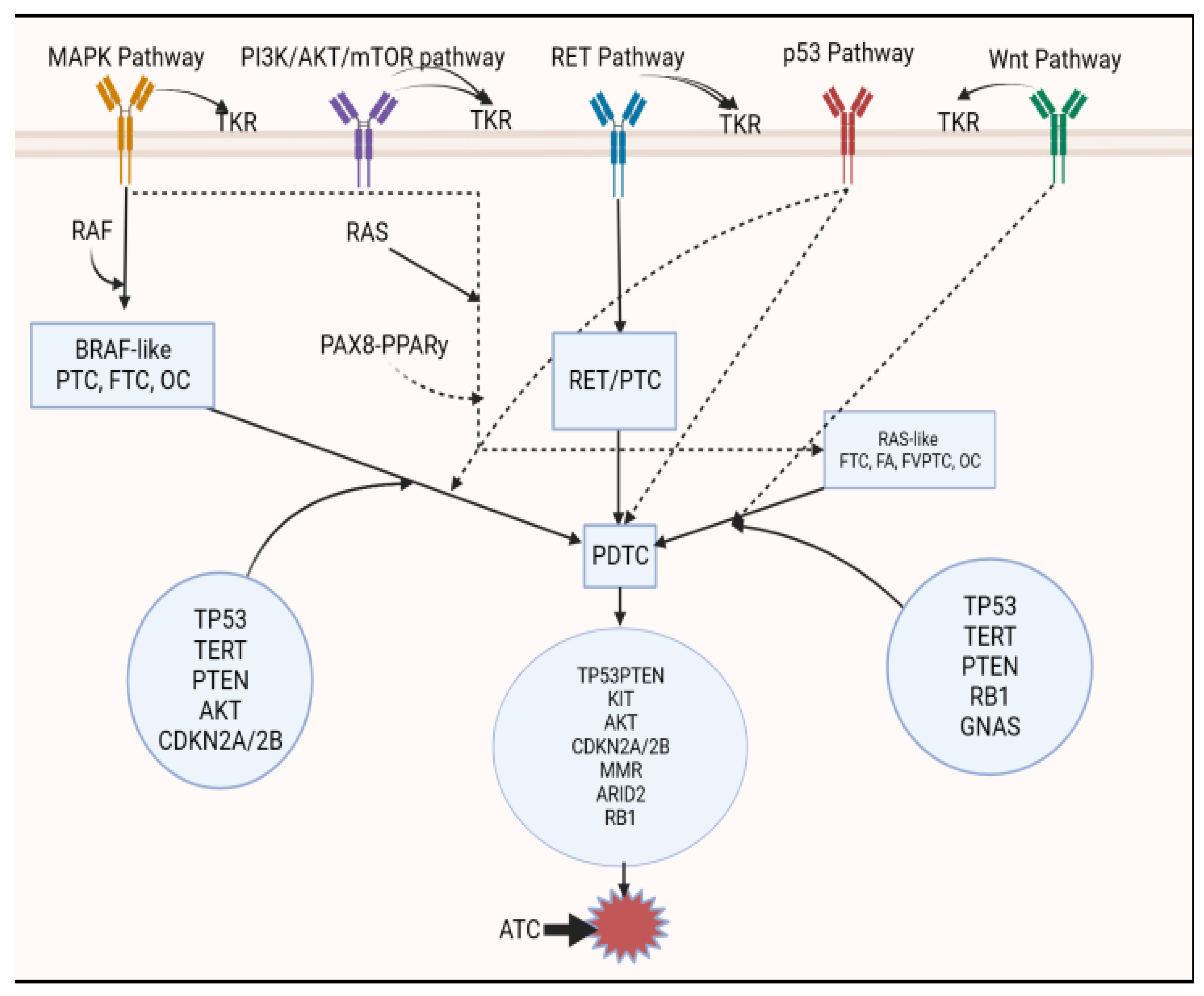
3. Recent Development in the Genomics of Follicular Carcinoma and Other Thyroid Tumors
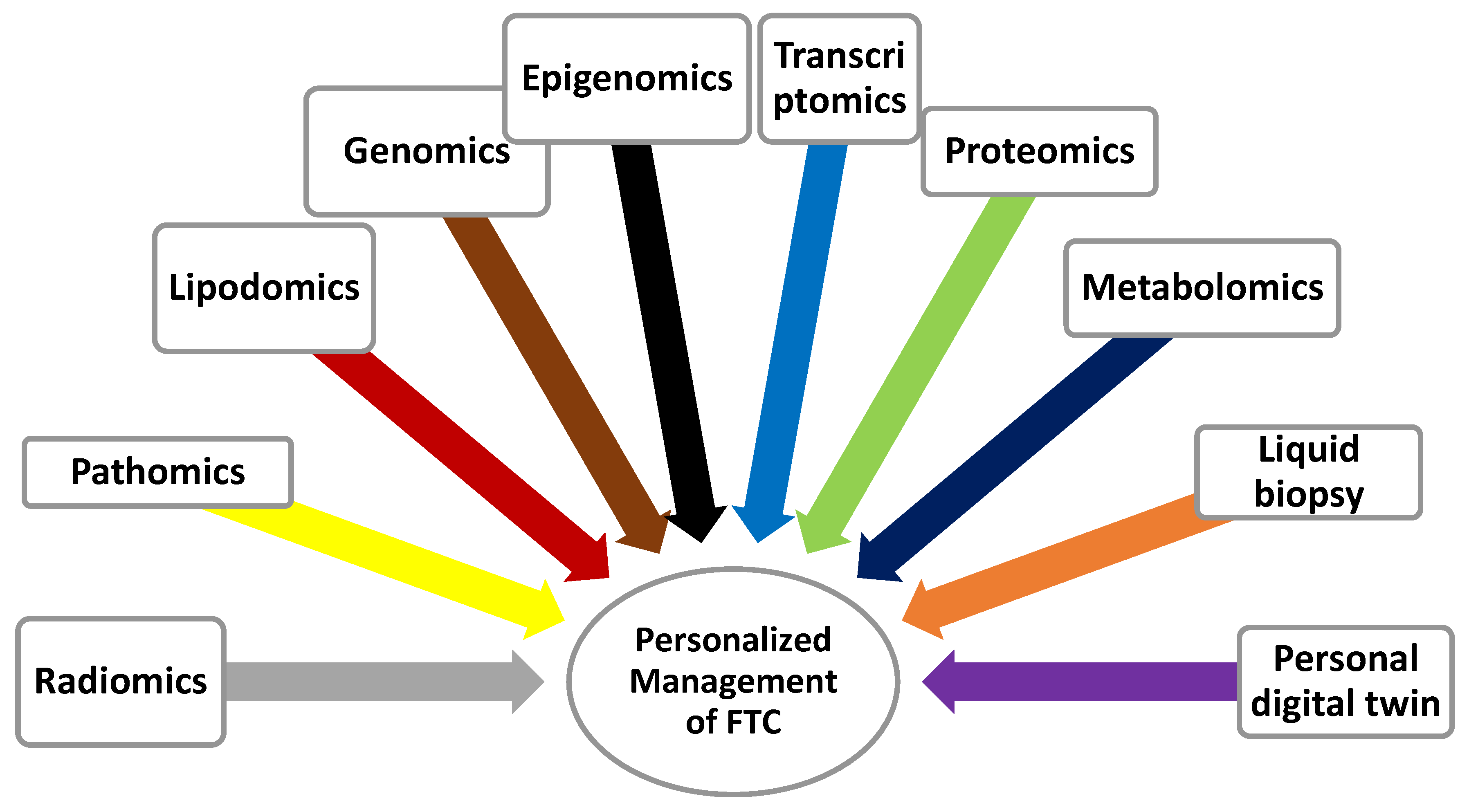
4. Multi-Omics of Follicular Carcinoma and Other Thyroid Tumors
4.1. Pathomics in the Diagnostic Work-Up of Follicular Carcinoma
4.2. Genomics of Follicular Carcinoma and Other Thyroid Malignancies
4.3. Epigenomics of Follicular Carcinoma and Other Thyroid Tumors
4.3.1. Histone Modification
4.3.2. Non-Coding RNAs
4.4. Proteomics of Follicular Carcinoma and Other Thyroid Malignancies
4.5. Metabolomics of Follicular Carcinoma and Other Thyroid Tumors
4.6. Liquid Biopsy in Follicular Carcinoma and Other Thyroid Tumors
4.7. Radiomics and FTC
5. Artificial Intelligence and Management of FTC
6. Conclusions
Author Contributions
Funding
Institutional Review Board Statement
Informed Consent Statement
Acknowledgments
Conflicts of Interest
References
- Haugen, B.R.; Alexander, E.K.; Bible, K.C.; Doherty, G.M.; Mandel, S.J.; Nikiforov, Y.E.; Pacini, F.; Randolph, G.W.; Sawka, A.M.; Schlumberger, M. 2015 American Thyroid Association management guidelines for adult patients with thyroid nodules and differentiated thyroid cancer: The American Thyroid Association guidelines task force on thyroid nodules and differentiated thyroid cancer. Thyroid 2016, 26, 1–133. [Google Scholar] [CrossRef] [PubMed]
- Lamartina, L.; Grani, G.; Durante, C.; Filetti, S. Recent advances in managing differentiated thyroid cancer. F1000Research 2018, 7. [Google Scholar] [CrossRef] [PubMed]
- Bolin, J. Thyroid follicular epithelial cell–derived cancer: New approaches and treatment strategies. J. Nucl. Med. Technol. 2021, 49, 199–208. [Google Scholar] [CrossRef] [PubMed]
- Katoh, H.; Yamashita, K.; Enomoto, T.; Watanabe, M. Classification and general considerations of thyroid cancer. Ann. Clin. Pathol. 2015, 3, 1045. [Google Scholar]
- Pulcrano, M.; Boukheris, H.; Talbot, M.; Caillou, B.; Dupuy, C.; Virion, A.; De Vathaire, F.; Schlumberger, M. Poorly differentiated follicular thyroid carcinoma: Prognostic factors and relevance of histological classification. Thyroid 2007, 17, 639–646. [Google Scholar] [CrossRef]
- Romano, C.; Martorana, F.; Pennisi, M.S.; Stella, S.; Massimino, M.; Tirrò, E.; Vitale, S.R.; Di Gregorio, S.; Puma, A.; Tomarchio, C. Opportunities and challenges of liquid biopsy in thyroid cancer. Int. J. Mol. Sci. 2021, 22, 7707. [Google Scholar] [CrossRef]
- Pozdeyev, N.; Gay, L.M.; Sokol, E.S.; Hartmaier, R.; Deaver, K.E.; Davis, S.; French, J.D.; Borre, P.V.; LaBarbera, D.V.; Tan, A.-C. Genetic analysis of 779 advanced differentiated and anaplastic thyroid cancers. Clin. Cancer Res. 2018, 24, 3059–3068. [Google Scholar] [CrossRef]
- Ferrari, S.M.; Fallahi, P.; Politti, U.; Materazzi, G.; Baldini, E.; Ulisse, S.; Miccoli, P.; Antonelli, A. Molecular targeted therapies of aggressive thyroid cancer. Front. Endocrinol. 2015, 6, 176. [Google Scholar] [CrossRef]
- Singh, A.; Ham, J.; Po, J.W.; Niles, N.; Roberts, T.; Lee, C.S. The genomic landscape of thyroid cancer tumourigenesis and implications for immunotherapy. Cells 2021, 10, 1082. [Google Scholar] [CrossRef]
- Kakudo, K.; Bai, Y.; Liu, Z.; Ozaki, T. Encapsulated papillary thyroid carcinoma, follicular variant: A misnomer. Pathol. Int. 2012, 62, 155–160. [Google Scholar] [CrossRef]
- Podda, M.; Saba, A.; Porru, F.; Reccia, I.; Pisanu, A. Follicular thyroid carcinoma: Differences in clinical relevance between minimally invasive and widely invasive tumors. World J. Surg. Oncol. 2015, 13, 1–7. [Google Scholar] [CrossRef] [PubMed]
- Tallini, G.; Tuttle, R.M.; Ghossein, R.A. The History of the Follicular Variant of Papillary Thyroid Carcinoma. J. Clin. Endocrinol. Metab. 2017, 102, 15–22. [Google Scholar] [CrossRef] [PubMed]
- Araque, K.A.; Gubbi, S.; Klubo-Gwiezdzinska, J. Updates on the management of thyroid cancer. Horm. Metab. Res. 2020, 52, 562–577. [Google Scholar] [CrossRef] [PubMed]
- Huszno, B.; Szybiński, Z.; Przybylik-Mazurek, E.; Stachura, J.; Trofimiuk, M.; Buziak-Bereza, M.; Gołkowski, F.; Pantoflinski, J. Influence of iodine deficiency and iodine prophylaxis on thyroid cancer histotypes and incidence in endemic goiter area. J. Endocrinol. Investig. 2003, 26, 71–76. [Google Scholar]
- Nicol, F.; McLaren, K.M.; Toft, A.D. Multifocal follicular carcinoma of thyroid following radiotherapy for Hodgkin’s disease. Postgrad. Med. J. 1982, 58, 180–181. [Google Scholar] [CrossRef] [PubMed]
- Ogbera, A.O.; Kuku, S.F. Epidemiology of thyroid diseases in Africa. Indian J. Endocrinol. Metab. 2011, 15, S82. [Google Scholar] [CrossRef] [PubMed]
- Sáez, J.M.G. Hashimoto’s Thyroiditis and thyroid cancer. J. Hum. Endocrinol. 2016, 1. [Google Scholar]
- Deivanathan, N.; Rathinam, S.; Gopal, K.A.; Anandan, H. Incidence of Types of Thyroid Carcinoma in an Iodine-rich Area: Thoothukudi Southern Coastal City. Int. J. Sci. Study 2016, 4, 137–139. [Google Scholar]
- Zhang, W.; Ruan, X.; Li, Y.; Zhi, J.; Hu, L.; Hou, X.; Shi, X.; Wang, X.; Wang, J.; Ma, W. KDM1A promotes thyroid cancer progression and maintains stemness through the Wnt/β-catenin signaling pathway. Theranostics 2022, 12, 1500. [Google Scholar] [CrossRef]
- Ivanova, L.B.; Vukov, M.I.; Vassileva-Valerianova, Z.G. Thyroid Cancer Incidence in Bulgaria before and after the Introduction of Universal Salt Iodization: An Analysis of the National Cancer Registry Data. Balk. Med. J. 2020, 37, 330. [Google Scholar] [CrossRef]
- Mulaudzi, T.V.; Ramdial, P.K.; Madiba, T.E.; Callaghan, R.A. Thyroid carcinoma at King Edward VIII Hospital, Durban, South Africa. East Afr. Med. J. 2001, 78, 242–245. [Google Scholar] [CrossRef]
- Mitro, S.D.; Rozek, L.S.; Vatanasapt, P.; Suwanrungruang, K.; Chitapanarux, I.; Srisukho, S.; Sriplung, H.; Meza, R. Iodine deficiency and thyroid cancer trends in three regions of Thailand, 1990–2009. Cancer Epidemiol. 2016, 43, 92–99. [Google Scholar] [CrossRef] [PubMed]
- Shin, D.Y.; Jo, Y.S. Clinical implications of follicular and Hurthle cell carcinoma in an iodine-sufficient area. Korean J. Intern. Med. 2014, 29, 305–306. [Google Scholar] [CrossRef] [PubMed]
- Zimmermann, M.B.; Galetti, V. Iodine intake as a risk factor for thyroid cancer: A comprehensive review of animal and human studies. Thyroid Res. 2015, 8, 1–21. [Google Scholar] [CrossRef] [PubMed]
- Widder, S.; Guggisberg, K.; Khalil, M.; Pasieka, J.L. A pathologic re-review of follicular thyroid neoplasms: The impact of changing the threshold for the diagnosis of the follicular variant of papillary thyroid carcinoma. Surgery 2008, 144, 80–85. [Google Scholar] [CrossRef] [PubMed]
- Grigsby, P.W.; Gal-or, A.; Michalski, J.M.; Doherty, G.M. Childhood and adolescent thyroid carcinoma. Cancer Interdiscip. Int. J. Am. Cancer Soc. 2002, 95, 724–729. [Google Scholar] [CrossRef]
- Oka, K.; Shien, T.; Otsuka, F. Thyroid follicular carcinoma in a teenager: A case report. J. Gen. Fam. Med. 2018, 19, 170–172. [Google Scholar] [CrossRef]
- Lin, J.-D.; Lin, S.-F.; Chen, S.-T.; Hsueh, C.; Li, C.-L.; Chao, T.-C. Long-term follow-up of papillary and follicular thyroid carcinomas with bone metastasis. PLoS ONE 2017, 12, e0173354. [Google Scholar] [CrossRef]
- Parameswaran, R.; Shulin Hu, J.; Min En, N.; Tan, W.B.; Yuan, N.K. Patterns of metastasis in follicular thyroid carcinoma and the difference between early and delayed presentation. Ann. R. Coll. Surg. Engl. 2017, 99, 151–154. [Google Scholar] [CrossRef]
- Bombil, I.; Bentley, A.; Kruger, D.; Luvhengo, T.E. Incidental cancer in multinodular goitre post thyroidectomy. South Afr. J. Surg. 2014, 52, 5–9. [Google Scholar]
- Karimifar, M. A case of functional metastatic follicular thyroid carcinoma that presented with hip fracture and hypercalcemia. Adv. Biomed. Res. 2018, 7. [Google Scholar] [CrossRef] [PubMed]
- Liu, J.; Wang, Y.; Da, D.; Zheng, M. Hyperfunctioning thyroid carcinoma: A systematic review. Mol. Clin. Oncol. 2019, 11, 535–550. [Google Scholar] [CrossRef] [PubMed]
- Cibas, E.S.; Ali, S.Z. The 2017 Bethesda system for reporting thyroid cytopathology. Thyroid 2017, 27, 1341–1346. [Google Scholar] [CrossRef] [PubMed]
- Ustun, B.; Chhieng, D.; Prasad, M.L.; Holt, E.; Hammers, L.; Carling, T.; Udelsman, R.; Adeniran, A.J. Follicular variant of papillary thyroid carcinoma: Accuracy of FNA diagnosis and implications for patient management. Endocr. Pathol. 2014, 25, 257–264. [Google Scholar] [CrossRef]
- Baloch, Z.W.; Asa, S.L.; Barletta, J.A.; Ghossein, R.A.; Juhlin, C.C.; Jung, C.K.; LiVolsi, V.A.; Papotti, M.G.; Sobrinho-Simoes, M.; Tallini, G. Overview of the 2022 WHO classification of thyroid neoplasms. Endocr. Pathol. 2022, 33, 27–63. [Google Scholar] [CrossRef] [PubMed]
- Armanious, H.; Adam, B.; Meunier, D.; Formenti, K.; Izevbaye, I. Digital gene expression analysis might aid in the diagnosis of thyroid cancer. Curr. Oncol. 2020, 27, 93–99. [Google Scholar] [CrossRef]
- Endo, M.; Sipos, J.A.; Ringel, M.D.; Porter, K.; Nagaraja, H.N.; Phay, J.E.; Shirley, L.A.; Long, C.; Wright, C.L.; Roll, K. Prevalence of cancer and the benign call rate of afirma gene classifier in 18F-Fluorodeoxyglucose positron emission tomography positive cytologically indeterminate thyroid nodules. Cancer Med. 2021, 10, 1084–1090. [Google Scholar] [CrossRef]
- Kim, K.; Jung, C.K.; Lim, D.-J.; Bae, J.S.; Kim, J.S. Clinical and pathologic features for predicting malignancy in thyroid follicular neoplasms. Gland Surg. 2021, 10, 50. [Google Scholar] [CrossRef]
- Zhao, L.; Zhu, X.-Y.; Jiang, R.; Xu, M.; Wang, N.; Chen, G.G.; Liu, Z.-M. Role of GPER1, EGFR and CXCR1 in differentiating between malignant follicular thyroid carcinoma and benign follicular thyroid adenoma. Int. J. Clin. Exp. Pathol. 2015, 8, 11236. [Google Scholar]
- Zaydfudim, V.; Feurer, I.D.; Griffin, M.R.; Phay, J.E. The impact of lymph node involvement on survival in patients with papillary and follicular thyroid carcinoma. Surgery 2008, 144, 1070–1078. [Google Scholar] [CrossRef]
- Matsuura, D.; Yuan, A.; Harris, V.; Shaha, A.R.; Tuttle, R.M.; Patel, S.G.; Shah, J.P.; Ganly, I. Surgical management of low-/intermediate-risk node negative thyroid cancer: A single-institution study using propensity matching analysis to compare thyroid lobectomy and total thyroidectomy. Thyroid 2022, 32, 28–36. [Google Scholar] [CrossRef] [PubMed]
- Schneider, D.F.; Elfenbein, D.; Lloyd, R.V.; Chen, H.; Sippel, R.S. Lymph node metastases do not impact survival in follicular variant papillary thyroid cancer. Ann. Surg. Oncol. 2015, 22, 158–163. [Google Scholar] [CrossRef] [PubMed]
- Xu, B.; Farhat, N.; Barletta, J.A.; Hung, Y.P.; Biase, D.d.; Casadei, G.P.; Onenerk, A.M.; Tuttle, R.M.; Roman, B.R.; Katabi, N. Should subcentimeter non-invasive encapsulated, follicular variant of papillary thyroid carcinoma be included in the noninvasive follicular thyroid neoplasm with papillary-like nuclear features category? Endocrine 2018, 59, 143–150. [Google Scholar] [CrossRef]
- Brecelj, E.; Grazio, S.F.; Auersperg, M.; Bračko, M. Prognostic value of E-cadherin expression in thyroid follicular carcinoma. Eur. J. Surg. Oncol. 2005, 31, 544–548. [Google Scholar] [CrossRef]
- Dai, Y.; Miao, Y.; Zhu, Q.; Gao, M.; Hao, F. Expression of long non-coding RNA H19 predicts distant metastasis in minimally invasive follicular thyroid carcinoma. Bioengineered 2019, 10, 383–389. [Google Scholar] [CrossRef] [PubMed]
- Gupta, A.; Jain, S.; Khurana, N.; Kakar, A.K. Expression of p63 and Bcl-2 in malignant thyroid tumors and their correlation with other diagnostic immunocytochemical markers. J. Clin. Diagn. Res. 2016, 10, EC04. [Google Scholar] [CrossRef]
- Heriyanto, D.S.; Laiman, V.; Limantara, N.V.; Anantawikrama, W.P.; Yuliani, F.S.; Cempaka, R.; Anwar, S.L. High frequency of KRAS and EGFR mutation profiles in BRAF-negative thyroid carcinomas in Indonesia. BMC Res. Notes 2022, 15, 369. [Google Scholar] [CrossRef]
- Indrasena, B.S.H. Use of thyroglobulin as a tumour marker. World J. Biol. Chem. 2017, 8, 81. [Google Scholar] [CrossRef]
- McCaffrey, J.C. Aerodigestive tract invasion by well-differentiated thyroid carcinoma: Diagnosis, management, prognosis, and biology. Laryngoscope 2006, 116, 1–11. [Google Scholar] [CrossRef]
- Mahmoudian-Sani, M.-R.; Jalali, A.; Jamshidi, M.; Moridi, H.; Alghasi, A.; Shojaeian, A.; Mobini, G.-R. Long non-coding RNAs in thyroid cancer: Implications for pathogenesis, diagnosis, and therapy. Oncol. Res. Treat. 2019, 42, 136–142. [Google Scholar] [CrossRef]
- Wang, X.; Zheng, X.; Zhu, J.; Li, Z.; Wei, T. Impact of Extent of Surgery on Long-Term Prognosis of Follicular Thyroid Carcinoma Without Extrathyroidal Extension and Distant Metastasis. World J. Surg. 2022, 46, 104–111. [Google Scholar] [CrossRef] [PubMed]
- Bains, A.; Mur, T.; Wallace, N.; Noordzij, J.P. The role of vitamin D as a prognostic marker in papillary thyroid cancer. Cancers 2021, 13, 3516. [Google Scholar] [CrossRef] [PubMed]
- Riguetto, C.M.; Barreto, I.S.; Maia, F.F.R.; Assumpção, L.V.M.d.; Zantut-Wittmann, D.E. Usefulness of pre-thyroidectomy neutrophil–lymphocyte, platelet–lymphocyte, and monocyte–lymphocyte ratios for discriminating lymph node and distant metastases in differentiated thyroid cancer. Clinics 2021, 76. [Google Scholar] [CrossRef]
- Fakhar, Y.; Khooei, A.; Aghaee, A.; Mohammadzadeh Kosari, H.; Wartofsky, L.; Zakavi, S.R. Bone metastasis from noninvasive follicular thyroid neoplasm with papillary-like nuclear features (NIFTP); a case report. BMC Endocr. Disord. 2021, 21, 1–5. [Google Scholar] [CrossRef] [PubMed]
- Nwaeze, O.; Obidike, S.; Mullen, D.; Aftab, F. Follicular variant papillary thyroid carcinoma with a twist. Int. J. Surg. Case Rep. 2015, 8, 107–110. [Google Scholar] [CrossRef]
- Kammori, M.; Fukumori, T.; Sugishita, Y.; Hoshi, M.; Yamada, T. Therapeutic strategy for low-risk thyroid cancer in Kanaji Thyroid Hospital. Endocr. J. 2014, 61, 1–12. [Google Scholar] [CrossRef] [PubMed]
- Omar, B.; Yassir, H.; Youssef, O.; Sami, R.; Larbi, A.R.; Mohamed, R.; Mohamed, M. A rare case of follicular thyroid carcinoma metastasis to the sacral region: A case report with literature review. Int. J. Surg. Case Rep. 2022, 94, 107001. [Google Scholar] [CrossRef]
- Swierniak, M.; Pfeifer, A.; Stokowy, T.; Rusinek, D.; Chekan, M.; Lange, D.; Krajewska, J.; Oczko-Wojciechowska, M.; Czarniecka, A.; Jarzab, M. Somatic mutation profiling of follicular thyroid cancer by next generation sequencing. Mol. Cell. Endocrinol. 2016, 433, 130–137. [Google Scholar] [CrossRef]
- Vaisman, F.; Momesso, D.; Bulzico, D.A.; Pessoa, C.H.C.N.; Cruz, M.D.G.d.; Dias, F.; Corbo, R.; Vaisman, M.; Tuttle, R.M. Thyroid lobectomy is associated with excellent clinical outcomes in properly selected differentiated thyroid cancer patients with primary tumors greater than 1 cm. J. Thyroid Res. 2013, 2013. [Google Scholar] [CrossRef]
- Pignatti, E.; Vighi, E.; Magnani, E.; Kara, E.; Roncati, L.; Maiorana, A.; Santi, D.; Madeo, B.; Cioni, K.; Carani, C. Expression and clinicopathological role of miR146a in thyroid follicular carcinoma. Endocrine 2019, 64, 575–583. [Google Scholar] [CrossRef]
- Capdevila, J.; Awada, A.; Führer-Sakel, D.; Leboulleux, S.; Pauwels, P. Molecular diagnosis and targeted treatment of advanced follicular cell-derived thyroid cancer in the precision medicine era. Cancer Treat. Rev. 2022, 102380. [Google Scholar] [CrossRef] [PubMed]
- Fallahi, P.; Ferrari, S.M.; Galdiero, M.R.; Varricchi, G.; Elia, G.; Ragusa, F.; Paparo, S.R.; Benvenga, S.; Antonelli, A. Molecular targets of tyrosine kinase inhibitors in thyroid cancer. Semin. Cancer Biol. 2022, 79, 180–196. [Google Scholar] [CrossRef] [PubMed]
- Aboelnaga, E.M.; Ahmed, R.A. Difference between papillary and follicular thyroid carcinoma outcomes: An experince from Egyptian institution. Cancer Biol. Med. 2015, 12, 53–59. [Google Scholar]
- Vuong, H.G.; Kondo, T.; Oishi, N.; Nakazawa, T.; Mochizuki, K.; Inoue, T.; Tahara, I.; Kasai, K.; Hirokawa, M.; Tran, T.M. Genetic alterations of differentiated thyroid carcinoma in iodine-rich and iodine-deficient countries. Cancer Med. 2016, 5, 1883–1889. [Google Scholar] [CrossRef] [PubMed]
- Battoo, A.J.; Rasool, Z.; Sheikh, Z.A.; Haji, A.G. Follicular thyroid carcinoma presenting as solitary liver metastasis: A case report. J. Med. Case Rep. 2016, 10, 1–7. [Google Scholar] [CrossRef]
- Kato, S.; Demura, S.; Shinmura, K.; Yokogawa, N.; Shimizu, T.; Tsuchiya, H. Current management of bone metastases from differentiated thyroid cancer. Cancers 2021, 13, 4429. [Google Scholar] [CrossRef] [PubMed]
- Sharma, P.; Kumar, N.; Gupta, R.; Jain, S. Follicular Carcinoma of the Thyroid with Hyperthyroidism. Acta Cytol. 2004, 48, 219–222. [Google Scholar] [CrossRef]
- Oyer, S.L.; Fritsch, V.A.; Lentsch, E.J. Comparison of survival rates between papillary and follicular thyroid carcinomas among 36,725 patients. Ann. Otol. Rhinol. Laryngol. 2014, 123, 94–100. [Google Scholar] [CrossRef]
- Chow, S.M.; Law, S.C.K.; Au, S.K.; Leung, T.W.; Chan, P.T.M.; Mendenhall, W.M.; Lau, W.H. Differentiated thyroid carcinoma: Comparison between papillary and follicular carcinoma in a single institute. J. Sci. Spec. Head Neck 2002, 24, 670–677. [Google Scholar] [CrossRef]
- Nicolson, N.G.; Paulsson, J.O.; Juhlin, C.C.; Carling, T.; Korah, R. Transcription factor profiling identifies spatially heterogenous mediators of follicular thyroid cancer invasion. Endocr. Pathol. 2020, 31, 367–376. [Google Scholar] [CrossRef]
- Schmidbauer, B.; Menhart, K.; Hellwig, D.; Grosse, J. Differentiated thyroid cancer—Treatment: State of the art. Int. J. Mol. Sci. 2017, 18, 1292. [Google Scholar] [CrossRef] [PubMed]
- Puliafito, I.; Esposito, F.; Prestifilippo, A.; Marchisotta, S.; Sciacca, D.; Vitale, M.P.; Giuffrida, D. Target therapy in thyroid cancer: Current challenge in clinical use of tyrosine kinase inhibitors and management of side effects. Front. Endocrinol. 2022, 13. [Google Scholar] [CrossRef] [PubMed]
- Choudhury, P.S.; Gupta, M. Differentiated thyroid cancer theranostics: Radioiodine and beyond. Br. J. Radiol. 2018, 91, 20180136. [Google Scholar] [CrossRef] [PubMed]
- Girolami, I.; Marletta, S.; Pantanowitz, L.; Torresani, E.; Ghimenton, C.; Barbareschi, M.; Scarpa, A.; Brunelli, M.; Barresi, V.; Trimboli, P. Impact of image analysis and artificial intelligence in thyroid pathology, with particular reference to cytological aspects. Cytopathology 2020, 31, 432–444. [Google Scholar] [CrossRef]
- Madabhushi, A.; Lee, G. Image analysis and machine learning in digital pathology: Challenges and opportunities. Med. Image Anal. 2016, 33, 170–175. [Google Scholar] [CrossRef]
- Rudzińska, M.; Czarnocka, B. The impact of transcription factor prospero homeobox 1 on the regulation of thyroid cancer malignancy. Int. J. Mol. Sci. 2020, 21, 3220. [Google Scholar] [CrossRef]
- Liang, P.; Wang, S.; Chen, K.B.; Li, M.; Liu, Y.; Li, S.; Pan, Y.W.; Zhang, Y.X.; Jiang, Y. The diagnosis and treatment of primary thyroid lymphoma. Chin. J. Otorhinolaryngol. Head Neck Surg. 2016, 51, 313–316. [Google Scholar]
- Badulescu, C.I.; Piciu, D.; Apostu, D.; Badan, M.; Piciu, A. Follicular thyroid carcinoma-clinical and diagnostic findings in a 20-year follow up study. Acta Endocrinol. 2020, 16, 170. [Google Scholar] [CrossRef]
- Miasaki, F.Y.; Saito, K.C.; Yamamoto, G.L.; Boguszewski, C.L.; de Carvalho, G.A.; Kimura, E.T.; Kopp, P.A. Thyroid and Breast Cancer in 2 Sisters With Monoallelic Mutations in the Ataxia Telangiectasia Mutated (ATM) Gene. J. Endocr. Soc. 2022, 6, bvac026. [Google Scholar] [CrossRef]
- Nosé, V. Thyroid cancer of follicular cell origin in inherited tumor syndromes. Adv. Anat. Pathol. 2010, 17, 428–436. [Google Scholar] [CrossRef]
- Howell, G.M.; Hodak, S.P.; Yip, L. RAS mutations in thyroid cancer. Oncology 2013, 18, 926–932. [Google Scholar] [CrossRef] [PubMed]
- Motoi, N.; Sakamoto, A.; Yamochi, T.; Horiuchi, H.; Motoi, T.; Machinami, R. Role of ras mutation in the progression of thyroid carcinoma of follicular epithelial origin. Pathol. Res. Pract. 2000, 196, 1–7. [Google Scholar] [CrossRef] [PubMed]
- Bhuiyan, M.; Machowski, A. Nodular thyroid disease and thyroid malignancy: Experience at Polokwane Mankweng Hospital Complex, Limpopo Province, South Africa. South Afr. Med. J. 2015, 105, 570–572. [Google Scholar] [CrossRef] [PubMed]
- Trovato, M.; Campennì, A.; Giovinazzo, S.; Siracusa, M.; Ruggeri, R.M. Hepatocyte growth factor/C-met axis in thyroid cancer: From diagnostic biomarker to therapeutic target. Biomark. Insights 2017, 12, 1177271917701126. [Google Scholar] [CrossRef]
- Livolsi, V.A.; Asa, S.L. The demise of follicular carcinoma of the thyroid gland. Thyroid 1994, 4, 233–236. [Google Scholar] [CrossRef]
- Cipriani, N.A.; Nagar, S.; Kaplan, S.P.; White, M.G.; Antic, T.; Sadow, P.M.; Aschebrook-Kilfoy, B.; Angelos, P.; Kaplan, E.L.; Grogan, R.H. Follicular thyroid carcinoma: How have histologic diagnoses changed in the last half-century and what are the prognostic implications? Thyroid 2015, 25, 1209–1216. [Google Scholar] [CrossRef]
- De Crea, C.; Raffaelli, M.; Sessa, L.; Ronti, S.; Fadda, G.; Bellantone, C.; Lombardi, C.P. Actual incidence and clinical behaviour of follicular thyroid carcinoma: An institutional experience. Sci. World J. 2014, 2014. [Google Scholar] [CrossRef]
- DeMay, R.M. Follicular Lesions of the Thyroid: W(h)ither Follicular Carcinoma? Am. J. Clin. Pathol. 2000, 114, 681–683. [Google Scholar] [CrossRef]
- Lee, S.R.; Jung, C.K.; Kim, T.E.; Bae, J.S.; Jung, S.L.; Choi, Y.J.; Kang, C.S. Molecular genotyping of follicular variant of papillary thyroid carcinoma correlates with diagnostic category of fine-needle aspiration cytology: Values of RAS mutation testing. Thyroid 2013, 23, 1416–1422. [Google Scholar] [CrossRef]
- Kapur, U.; Wojcik, E.M. Follicular neoplasm of the thyroid—Vanishing cytologic diagnosis? Diagn. Cytopathol. 2007, 35, 525–528. [Google Scholar] [CrossRef]
- Ahmadi, S.; Stang, M.; Jiang, X.S.; Sosa, J.A. Hürthle cell carcinoma: Current perspectives. OncoTargets Ther. 2016, 6873–6884. [Google Scholar] [CrossRef] [PubMed]
- Lau, L.W.; Ghaznavi, S.; Frolkis, A.D.; Stephenson, A.; Robertson, H.L.; Rabi, D.M.; Paschke, R. Malignancy risk of hyperfunctioning thyroid nodules compared with non-toxic nodules: Systematic review and a meta-analysis. Thyroid Res. 2021, 14, 1–16. [Google Scholar] [CrossRef] [PubMed]
- Saito, F.; Uruno, T.; Shibuya, H.; Kitagawa, W.; Nagahama, M.; Sugino, K.; Ito, K. Prognosis After Brain Metastasis from Differentiated Thyroid Carcinoma. World J. Surg. 2016, 40, 574–581. [Google Scholar] [CrossRef] [PubMed]
- Sugino, K.; Kameyama, K.; Nagahama, M.; Kitagawa, W.; Shibuya, H.; Ohkuwa, K.; Uruno, T.; Akaishi, J.; Suzuki, A.; Masaki, C. Follicular thyroid carcinoma with distant metastasis: Outcome and prognostic factor. Endocr. J. 2014, 61, 273–279. [Google Scholar] [CrossRef]
- Tang, J.; Kong, D.; Cui, Q.; Wang, K.; Zhang, D.; Liao, X.; Gong, Y.; Wu, G. Racial disparities of differentiated thyroid carcinoma: Clinical behavior, treatments, and long-term outcomes. World J. Surg. Oncol. 2018, 16, 1–9. [Google Scholar] [CrossRef]
- Abe, I.; Lam, A.K. Anaplastic thyroid carcinoma: Updates on WHO classification, clinicopathological features and staging. Histol Histopathol 2021, 36, 239–248. [Google Scholar]
- Bible, K.C.; Kebebew, E.; Brierley, J.; Brito, J.P.; Cabanillas, M.E.; Clark Jr, T.J.; Di Cristofano, A.; Foote, R.; Giordano, T.; Kasperbauer, J. 2021 American thyroid association guidelines for management of patients with anaplastic thyroid cancer: American thyroid association anaplastic thyroid cancer guidelines task force. Thyroid 2021, 31, 337–386. [Google Scholar] [CrossRef]
- Feldkamp, J. The role of genetic alterations in thyroid carcinoma. Front. Endocrinol. 2022, 13. [Google Scholar] [CrossRef]
- Landa, I.; Ibrahimpasic, T.; Boucai, L.; Sinha, R.; Knauf, J.A.; Shah, R.H.; Dogan, S.; Ricarte-Filho, J.C.; Krishnamoorthy, G.P.; Xu, B. Genomic and transcriptomic hallmarks of poorly differentiated and anaplastic thyroid cancers. J. Clin. Investig. 2016, 126, 1052–1066. [Google Scholar] [CrossRef]
- Penna, G.C.; Vaisman, F.; Vaisman, M.; Sobrinho-Simões, M.; Soares, P. Molecular markers involved in tumorigenesis of thyroid carcinoma: Focus on aggressive histotypes. Cytogenet. Genome Res. 2016, 150, 194–207. [Google Scholar] [CrossRef]
- Kostoglou-Athanassiou, I.; Athanassiou, P.; Vecchini, G.; Gogou, L.; Kaldrymides, P. Mixed medullary-follicular thyroid carcinoma. Horm. Res. Paediatr. 2004, 61, 300–304. [Google Scholar] [CrossRef]
- Tohidi, M.; Pourbehi, G.; Bahmanyar, M.; Eghbali, S.S.; Kalantar Hormozi, M.; Nabipour, I. Mixed medullary-follicular carcinoma of the thyroid. Case Rep. Endocrinol. 2013, 2013. [Google Scholar] [CrossRef] [PubMed]
- Grani, G.; Lamartina, L.; Durante, C.; Filetti, S.; Cooper, D.S. Follicular thyroid cancer and Hürthle cell carcinoma: Challenges in diagnosis, treatment, and clinical management. Lancet Diabetes Endocrinol. 2018, 6, 500–514. [Google Scholar] [CrossRef] [PubMed]
- Nabhan, F.; Ringel, M.D. Thyroid nodules and cancer management guidelines: Comparisons and controversies. Endocr. Relat. Cancer 2017, 24, R13. [Google Scholar] [CrossRef] [PubMed]
- Ashamallah, G.A.; El-Adalany, M.A. Risk for malignancy of thyroid nodules: Comparative study between TIRADS and US based classification system. Egypt. J. Radiol. Nucl. Med. 2016, 47, 1373–1384. [Google Scholar] [CrossRef]
- Kwak, J.Y.; Han, K.H.; Yoon, J.H.; Moon, H.J.; Son, E.J.; Park, S.H.; Jung, H.K.; Choi, J.S.; Kim, B.M.; Kim, E.-K. Thyroid imaging reporting and data system for US features of nodules: A step in establishing better stratification of cancer risk. Radiology 2011, 260, 892–899. [Google Scholar] [CrossRef]
- Kuo, T.-C.; Wu, M.-H.; Chen, K.-Y.; Hsieh, M.-S.; Chen, A.; Chen, C.-N. Ultrasonographic features for differentiating follicular thyroid carcinoma and follicular adenoma. Asian J. Surg. 2020, 43, 339–346. [Google Scholar] [CrossRef]
- Fernández Sánchez, J. TI-RADS classification of thyroid nodules based on a score modified regarding the ultrasound criteria for malignancy. Rev. Argent. De Radiol. 2014, 78, 138–148. [Google Scholar] [CrossRef]
- Li, M.; Wei, L.; Li, F.; Kan, Y.; Liang, X.; Zhang, H.; Liu, J. High Risk Thyroid Nodule Discrimination and Management by Modified TI-RADS. Cancer Manag. Res. 2021, 225–234. [Google Scholar] [CrossRef]
- Mistry, R.; Hillyar, C.R.; Nibber, A.; Sooriyamoorthy, T.; Kumar, N.; Hillyar, C. Ultrasound classification of thyroid nodules: A systematic review. Cureus J. Med. Sci. 2020, 12. [Google Scholar] [CrossRef]
- Qiao, J.; Li, C.; Zhang, Y.; Wang, S.; Gao, S. HBME-1 expression in differentiated thyroid carcinoma and its correlation with the ultrasonic manifestation of thyroid. Oncol. Lett. 2017, 14, 6505–6510. [Google Scholar] [CrossRef] [PubMed]
- Spinelli, C.; Rallo, L.; Morganti, R.; Mazzotti, V.; Inserra, A.; Cecchetto, G.; Massimino, M.; Collini, P.; Strambi, S. Surgical management of follicular thyroid carcinoma in children and adolescents: A study of 30 cases. J. Pediatr. Surg. 2019, 54, 521–526. [Google Scholar] [CrossRef] [PubMed]
- Cibas, E.S.; Ali, S.Z. The Bethesda system for reporting thyroid cytopathology. Thyroid 2009, 19, 1159–1165. [Google Scholar] [CrossRef]
- Misiakos, E.P.; Margari, N.; Meristoudis, C.; Machairas, N.; Schizas, D.; Petropoulos, K.; Spathis, A.; Karakitsos, P.; Machairas, A. Cytopathologic diagnosis of fine needle aspiration biopsies of thyroid nodules. World J. Clin. Cases 2016, 4, 38. [Google Scholar] [CrossRef] [PubMed]
- Elsherbini, N.; Kim, D.H.; Payne, R.J.; Hudson, T.; Forest, V.-I.; Hier, M.P.; Payne, A.E.; Pusztaszeri, M.P. EIF1AX mutation in thyroid tumors: A retrospective analysis of cytology, histopathology and co-mutation profiles. J. Otolaryngol. Head Neck Surg. 2022, 51, 1–9. [Google Scholar] [CrossRef] [PubMed]
- Dom, G.; Frank, S.; Floor, S.; Kehagias, P.; Libert, F.; Hoang, C.; Andry, G.; Spinette, A.; Craciun, L.; de Saint Aubin, N. Thyroid follicular adenomas and carcinomas: Molecular profiling provides evidence for a continuous evolution. Oncotarget 2018, 9, 10343. [Google Scholar] [CrossRef]
- Sobrinho-Simoes, M.; Eloy, C.; Magalhaes, J.; Lobo, C.; Amaro, T. Follicular thyroid carcinoma. Mod. Pathol. 2011, 24, S10–S18. [Google Scholar] [CrossRef]
- Filetti, S.; Durante, C.; Hartl, D.; Leboulleux, S.; Locati, L.D.; Newbold, K.; Papotti, M.G.; Berruti, A. Thyroid cancer: ESMO Clinical Practice Guidelines for diagnosis, treatment and follow-up. Ann. Oncol. 2019, 30, 1856–1883. [Google Scholar] [CrossRef]
- McLeod, D.S.; Jonklaas, J.; Brierley, J.D.; Ain, K.B.; Cooper, D.S.; Fein, H.G.; Haugen, B.R.; Ladenson, P.W.; Magner, J.; Ross, D.S.; et al. Reassessing the NTCTCS Staging Systems for Differentiated Thyroid Cancer, Including Age at Diagnosis. Thyroid 2015, 25, 1097–1105. [Google Scholar] [CrossRef]
- Glikson, E.; Alon, E.; Bedrin, L.; Talmi, Y.P. Prognostic Factors in Differentiated Thyroid Cancer Revisited. Isr. Med. Assoc. J. 2017, 19, 114–118. [Google Scholar]
- Jung, C.K.; Bychkov, A.; Kakudo, K. Update from the 2022 world health organization classification of thyroid tumors: A standardized diagnostic approach. Endocrinol. Metab. 2022, 37, 703–718. [Google Scholar] [CrossRef] [PubMed]
- Papaleontiou, M.; Haymart, M.R. New insights in risk stratification of differentiated thyroid cancer. Curr. Opin. Oncol. 2014, 26, 1. [Google Scholar] [CrossRef] [PubMed]
- Shaha, A.R.; Tuttle, R.M. Thyroid cancer staging and genomics. Ann. Transl. Med. 2019, 7. [Google Scholar] [CrossRef] [PubMed]
- Vaisman, F.; Corbo, R.; Vaisman, M. Thyroid carcinoma in children and adolescents—Systematic review of the literature. J. Thyroid Res. 2011, 2011. [Google Scholar] [CrossRef]
- Naoum, G.E.; Morkos, M.; Kim, B.; Arafat, W. Novel targeted therapies and immunotherapy for advanced thyroid cancers. Mol. Cancer 2018, 17, 1–15. [Google Scholar] [CrossRef]
- Teo, K.W.; Yuan, N.K.; Tan, W.B.; Parameswaran, R. Comparison of prognostic scoring systems in follicular thyroid cancer. Ann. R. Coll. Surg. Engl. 2017, 99, 479–484. [Google Scholar] [CrossRef]
- Nabhan, F.; Dedhia, P.H.; Ringel, M.D. Thyroid cancer, recent advances in diagnosis and therapy. Int. J. Cancer 2021, 149, 984–992. [Google Scholar] [CrossRef]
- Lee, S.L. Complications of radioactive iodine treatment of thyroid carcinoma. J. Natl. Compr. Cancer Netw. 2010, 8, 1277–1287. [Google Scholar] [CrossRef]
- van Velsen, E.F.S.; Stegenga, M.T.; van Kemenade, F.J.; Kam, B.L.R.; van Ginhoven, T.M.; Visser, W.E.; Peeters, R.P. Comparing the prognostic value of the eighth edition of the American Joint Committee on cancer/tumor node metastasis staging system between papillary and follicular thyroid cancer. Thyroid 2018, 28, 976–981. [Google Scholar] [CrossRef]
- Megwalu, U.C.; Green, R.W. Total Thyroidectomy Versus Lobectomy for the Treatment of Follicular Thyroid Microcarcinoma. Anticancer Res 2016, 36, 2899–2902. [Google Scholar]
- Coelho, S.M.; Vaisman, F.; Buescu, A.; Mello, R.C.R.; Carvalho, D.P.; Vaisman, M. Follow-up of patients treated with retinoic acid for the control of radioiodine non-responsive advanced thyroid carcinoma. Braz. J. Med. Biol. Res. 2011, 44, 73–77. [Google Scholar] [CrossRef] [PubMed]
- Lorusso, L.; Cappagli, V.; Valerio, L.; Giani, C.; Viola, D.; Puleo, L.; Gambale, C.; Minaldi, E.; Campopiano, M.C.; Matrone, A. Thyroid cancers: From surgery to current and future systemic therapies through their molecular identities. Int. J. Mol. Sci. 2021, 22, 3117. [Google Scholar] [CrossRef] [PubMed]
- Poma, A.M.; Giannini, R.; Piaggi, P.; Ugolini, C.; Materazzi, G.; Miccoli, P.; Vitti, P.; Basolo, F. A six-gene panel to label follicular adenoma, low-and high-risk follicular thyroid carcinoma. Endocr. Connect. 2018, 7, 124. [Google Scholar] [CrossRef] [PubMed]
- Tong, G.-X.; Mody, K.; Wang, Z.; Hamele-Bena, D.; Nikiforova, M.N.; Nikiforov, Y.E. Mutations of TSHR and TP53 genes in an aggressive clear cell follicular carcinoma of the thyroid. Endocr. Pathol. 2015, 26, 315–319. [Google Scholar] [CrossRef]
- Iizuka, Y.; Katagiri, T.; Ogura, K.; Inoue, M.; Nakamura, K.; Mizowaki, T. Comparison of thyroid hormone withdrawal and recombinant human thyroid-stimulating hormone administration for adjuvant therapy in patients with intermediate-to high-risk differentiated thyroid cancer. Ann. Nucl. Med. 2020, 34, 736–741. [Google Scholar] [CrossRef]
- Ciarallo, A.; Rivera, J. Radioactive iodine therapy in differentiated thyroid cancer: 2020 update. Am. J. Roentgenol. 2020, 215, 285–291. [Google Scholar] [CrossRef]
- Silaghi, H.; Lozovanu, V.; Georgescu, C.E.; Pop, C.; Nasui, B.A.; Cătoi, A.F.; Silaghi, C.A. State of the art in the current management and future directions of targeted therapy for differentiated thyroid cancer. Int. J. Mol. Sci. 2022, 23, 3470. [Google Scholar] [CrossRef]
- Corrêa, N.L.; de Sá, L.V.; de Mello, R.C.R. Estimation of second primary cancer risk after treatment with radioactive iodine for differentiated thyroid carcinoma. Thyroid 2017, 27, 261–270. [Google Scholar] [CrossRef]
- Fard-Esfahani, A.; Emami-Ardekani, A.; Fallahi, B.; Fard-Esfahani, P.; Beiki, D.; Hassanzadeh-Rad, A.; Eftekhari, M. Adverse effects of radioactive iodine-131 treatment for differentiated thyroid carcinoma. Nucl. Med. Commun. 2014, 35, 808–817. [Google Scholar] [CrossRef]
- Chmielik, E.; Rusinek, D.; Oczko-Wojciechowska, M.; Jarzab, M.; Krajewska, J.; Czarniecka, A.; Jarzab, B. Heterogeneity of thyroid cancer. Pathobiology 2018, 85, 117–129. [Google Scholar] [CrossRef]
- Masui, T.; Uemura, H.; Ota, I.; Kimura, T.; Nishikawa, D.; Yamanaka, T.; Yane, K.; Kitahara, T. A study of 17 cases for the identification of prognostic factors for anaplastic thyroid carcinoma. Mol. Clin. Oncol. 2021, 14, 1. [Google Scholar] [CrossRef] [PubMed]
- Saji, M.; Ringel, M.D. The PI3K-Akt-mTOR pathway in initiation and progression of thyroid tumors. Mol. Cell. Endocrinol. 2010, 321, 20–28. [Google Scholar] [CrossRef]
- Zampella, E.; Klain, M.; Pace, L.; Cuocolo, A. PET/CT in the management of differentiated thyroid cancer. Diagn. Interv. Imaging 2021, 102, 515–523. [Google Scholar] [CrossRef] [PubMed]
- Batrinos, M.L. The problem of exogenous subclinical hyperthyroidism. Horm. Athens 2006, 5, 119. [Google Scholar] [CrossRef] [PubMed]
- Yavuz, D.G.; Yazan, C.D.; Hekimsoy, Z.; Aydin, K.; Gokkaya, N.; Ersoy, C.; Akalın, A.; Topaloglu, O.; Aydogan, B.I.; Dilekci, E.N.A. Assesment of attainment of recommended TSH levels and levothyroxine compliance in differentiated thyroid cancer patients. Clin. Endocrinol. 2022, 97, 833–840. [Google Scholar] [CrossRef] [PubMed]
- Bartalena, L.; Pinchera, A. Effects of thyroxine excess on peripheral organs. Acta Med. Austriaca 1994, 21, 60–65. [Google Scholar]
- Yang, X.; Guo, N.; Gao, X.; Liang, J.; Fan, X.; Zhao, Y. Meta-analysis of TSH suppression therapy and the risk of cardiovascular events after thyroid cancer surgery. Front. Endocrinol. 2022, 13. [Google Scholar] [CrossRef]
- Miccoli, P.; Materazzi, G.; Rossi, L. Levothyroxine therapy in thyrodectomized patients. Front. Endocrinol. 2021, 11, 626268. [Google Scholar] [CrossRef]
- Rajan, N.; Khanal, T.; Ringel, M.D. Progression and dormancy in metastatic thyroid cancer: Concepts and clinical implications. Endocrine 2020, 70, 24–35. [Google Scholar] [CrossRef]
- Brauckhoff, M. Classification of aerodigestive tract invasion from thyroid cancer. Langenbeck’s Arch. Surg. 2014, 399, 209–216. [Google Scholar] [CrossRef]
- Wu, H.-S.; Young, M.T.; Ituarte, P.H.G.; D’Avanzo, A.; Duh, Q.-Y.; Greenspan, F.S.; Loh, K.C.; Clark, O.H. Death from thyroid cancer of follicular cell origin. J. Am. Coll. Surg. 2000, 191, 600–606. [Google Scholar] [CrossRef] [PubMed]
- Allen, M.; Spillinger, A.; Arianpour, K.; Johnson, J.; Johnson, A.P.; Folbe, A.J.; Hotaling, J.; Svider, P.F. Tracheal Resection in the Management of Thyroid Cancer: An Evidence-Based Approach. Laryngoscope 2021, 131, 932–946. [Google Scholar] [CrossRef] [PubMed]
- Chen, W.-C.; Chou, C.-K.; Chang, Y.-H.; Chiang, P.-L.; Lim, L.-S.; Chi, S.-Y.; Luo, S.-D.; Lin, W.-C. Efficacy of radiofrequency ablation for metastatic papillary thyroid cancer with and without initial biochemical complete status. Front. Endocrinol. 2022, 13. [Google Scholar] [CrossRef] [PubMed]
- Ohkuwa, K.; Sugino, K.; Nagahama, M.; Kitagawa, W.; Matsuzu, K.; Suzuki, A.; Tomoda, C.; Hames, K.; Akaishi, J.; Masaki, C. Risk stratification in differentiated thyroid cancer with RAI-avid lung metastases. Endocr. Connect. 2021, 10, 825. [Google Scholar] [CrossRef] [PubMed]
- Papanikolaou, V.; Kyrodimos, E.; Mastronikolis, N.; Asimakopoulos, A.D.; Papanastasiou, G.; Tsiambas, E.; Spyropoulou, D.; Katsinis, S.; Manoli, A.; Papouliakos, S. Anti-EGFR/BRAF-Tyrosine Kinase Inhibitors in Thyroid Carcinoma. Cancer Diagn. Progn. 2023, 3, 151. [Google Scholar] [CrossRef] [PubMed]
- Broecker-Preuss, M.; Müller, S.; Britten, M.; Worm, K.; Schmid, K.W.; Mann, K.; Fuhrer, D. Sorafenib inhibits intracellular signaling pathways and induces cell cycle arrest and cell death in thyroid carcinoma cells irrespective of histological origin or BRAF mutational status. BMC Cancer 2015, 15, 1–13. [Google Scholar] [CrossRef]
- Kiyota, N.; Robinson, B.; Shah, M.; Hoff, A.O.; Taylor, M.H.; Li, D.; Dutcus, C.E.; Lee, E.K.; Kim, S.-B.; Tahara, M. Defining radioiodine-refractory differentiated thyroid cancer: Efficacy and safety of lenvatinib by radioiodine-refractory criteria in the SELECT trial. Thyroid 2017, 27, 1135–1141. [Google Scholar] [CrossRef]
- Hong, Y.-W.; Lin, J.-D.; Yu, M.-C.; Hsu, C.-C.; Lin, Y.-S. Outcomes and prognostic factors in thyroid cancer patients with cranial metastases: A retrospective cohort study of 4,683 patients. Int. J. Surg. 2018, 55, 182–187. [Google Scholar] [CrossRef]
- Slutzky-Shraga, I.; Gorshtein, A.; Popovitzer, A.; Robenshtok, E.; Tsvetov, G.; Akirov, A.; Hirsch, D.; Benbassat, C. Clinical characteristics and disease outcome of patients with non-medullary thyroid cancer and brain metastases. Oncol. Lett. 2018, 15, 672–676. [Google Scholar] [CrossRef]
- Choi, J.; Kim, J.W.; Keum, Y.S.; Lee, I.J. The largest known survival analysis of patients with brain metastasis from thyroid cancer based on prognostic groups. PLoS ONE 2016, 11, e0154739. [Google Scholar]
- Wong, K.; Di Cristofano, F.; Ranieri, M.; De Martino, D.; Di Cristofano, A. PI3K/mTOR inhibition potentiates and extends palbociclib activity in anaplastic thyroid cancer. Endocr. Relat. Cancer 2019, 26, 425. [Google Scholar] [CrossRef] [PubMed]
- Thakur, S.; Daley, B.; Millo, C.; Cochran, C.; Jacobson, O.; Lu, H.; Wang, Z.; Kiesewetter, D.; Chen, X.; Vasko, V. 177Lu-DOTA-EB-TATE, a Radiolabeled Analogue of Somatostatin Receptor Type 2, for the Imaging and Treatment of Thyroid CancerRadiolabeled Somatostatin Analogues and Thyroid Cancer. Clin. Cancer Res. 2021, 27, 1399–1409. [Google Scholar] [CrossRef]
- Panda, S.K.; Patro, B.; Samantaroy, M.R.; Mishra, J.; Mohapatra, K.C.; Meher, R.K. Unusual presentation of follicular carcinoma thyroid with special emphasis on their management. Int. J. Surg. Case Rep. 2014, 5, 408–411. [Google Scholar] [CrossRef] [PubMed]
- Prete, A.; Borges de Souza, P.; Censi, S.; Muzza, N.N.; Sponziello, M. Update on fundamental mechanisms of thyroid cancer. Front. Endocrinol. 2020, 11, 102. [Google Scholar] [CrossRef] [PubMed]
- Segkos, K.; Porter, K.; Senter, L.; Ringel, M.D.; Nabhan, F.A. Neck ultrasound in patients with follicular thyroid carcinoma. Horm. Cancer 2018, 9, 433–439. [Google Scholar] [CrossRef]
- Wynford-Thomas, D. Origin and progression of thyroid epithelial tumours: Cellular and molecular mechanisms. Horm. Res. Paediatr. 1997, 47, 145–157. [Google Scholar] [CrossRef]
- Zhang, J.; Li, Y.; Lyu, N.; Ying, J.M. The analysis of genetic and clinicopathologic characteristics in patients with follicular thyroid neoplasm. Chin. J. Oncol. 2019, 41, 594–598. [Google Scholar]
- Ludvíková, M.; Kalfeřt, D.; Kholová, I. Pathobiology of microRNAs and their emerging role in thyroid fine-needle aspiration. Acta Cytol. 2015, 59, 435–444. [Google Scholar] [CrossRef]
- Yoo, S.-K.; Song, Y.S.; Park, Y.J.; Seo, J.-S. Recent improvements in genomic and transcriptomic understanding of anaplastic and poorly differentiated thyroid cancers. Endocrinol. Metab. 2020, 35, 44–54. [Google Scholar] [CrossRef]
- Lee, K.-S.; Park, H. Machine learning on thyroid disease: A review. Front. Biosci.-Landmark 2022, 27, 101. [Google Scholar] [CrossRef]
- Agrawal, R.; Prabakaran, S. Big data in digital healthcare: Lessons learnt and recommendations for general practice. Heredity 2020, 124, 525–534. [Google Scholar] [CrossRef] [PubMed]
- Kamel Boulos, M.N.; Zhang, P. Digital twins: From personalised medicine to precision public health. J. Pers. Med. 2021, 11, 745. [Google Scholar] [CrossRef] [PubMed]
- Dlamini, Z.; Francies, F.Z.; Hull, R.; Marima, R. Artificial intelligence (AI) and big data in cancer and precision oncology. Comput. Struct. Biotechnol. J. 2020, 18, 2300–2311. [Google Scholar] [CrossRef] [PubMed]
- Kim, H.-S.; Kim, D.-J.; Yoon, K.-H. Medical big data is not yet available: Why we need realism rather than exaggeration. Endocrinol. Metab. 2019, 34, 349–354. [Google Scholar] [CrossRef] [PubMed]
- Sahal, R.; Alsamhi, S.H.; Brown, K.N. Personal digital twin: A close look into the present and a step towards the future of personalised healthcare industry. Sensors 2022, 22, 5918. [Google Scholar] [CrossRef] [PubMed]
- Shimizu, H.; Nakayama, K.I. Artificial intelligence in oncology. Cancer Sci. 2020, 111, 1452–1460. [Google Scholar] [CrossRef]
- Campennì, A.; Siracusa, M.; Ruggeri, R.M.; Laudicella, R.; Pignata, S.A.; Baldari, S.; Giovanella, L. Differentiating malignant from benign thyroid nodules with indeterminate cytology by 99mTc-MIBI scan: A new quantitative method for improving diagnostic accuracy. Sci. Rep. 2017, 7, 6147. [Google Scholar] [CrossRef]
- Cao, Y.; Zhong, X.; Diao, W.; Mu, J.; Cheng, Y.; Jia, Z. Radiomics in differentiated thyroid cancer and nodules: Explorations, application, and limitations. Cancers 2021, 13, 2436. [Google Scholar] [CrossRef]
- Kezlarian, B.; Lin, O. Artificial intelligence in thyroid fine needle aspiration biopsies. Acta Cytol. 2021, 65, 324–329. [Google Scholar] [CrossRef]
- Wang, S.; Yang, D.M.; Rong, R.; Zhan, X.; Xiao, G. Pathology image analysis using segmentation deep learning algorithms. Am. J. Pathol. 2019, 189, 1686–1698. [Google Scholar] [CrossRef]
- Wang, X.-Y.; Qu, H.-Z.; Fang, X.-D. Omics big data and medical artificial intelligence. Yi Chuan Hered. 2021, 43, 930–937. [Google Scholar]
- Abdel-Rahman, H.M.; Abowarda, M.H.; Abdel-Aal, S.M. Diffusion-weighted MRI and apparent diffusion coefficient in differentiation of benign from malignant solitary thyroid nodule. Egypt. J. Radiol. Nucl. Med. 2016, 47, 1385–1390. [Google Scholar] [CrossRef]
- Elliott Range, D.D.; Dov, D.; Kovalsky, S.Z.; Henao, R.; Carin, L.; Cohen, J. Application of a machine learning algorithm to predict malignancy in thyroid cytopathology. Cancer Cytopathol. 2020, 128, 287–295. [Google Scholar] [CrossRef] [PubMed]
- Savala, R.; Dey, P.; Gupta, N. Artificial neural network model to distinguish follicular adenoma from follicular carcinoma on fine needle aspiration of thyroid. Diagn. Cytopathol. 2018, 46, 244–249. [Google Scholar] [CrossRef]
- Saji, M.; Kim, C.S.; Wang, C.; Zhang, X.; Khanal, T.; Coombes, K.; La Perle, K.; Cheng, S.-Y.; Tsichlis, P.N.; Ringel, M.D. Akt isoform-specific effects on thyroid cancer development and progression in a murine thyroid cancer model. Sci. Rep. 2020, 10, 18316. [Google Scholar] [CrossRef]
- Li, J.; Zhang, S.; Zheng, S.; Zhang, D.; Qiu, X. The BRAF V600E mutation predicts poor survival outcome in patients with papillary thyroid carcinoma: A meta analysis. Int. J. Clin. Exp. Med. 2015, 8, 22246. [Google Scholar]
- Su, X.; Li, P.; Han, B.; Jia, H.; Liang, Q.; Wang, H.; Gu, M.; Cai, J.; Li, S.; Zhou, Y. Vitamin C sensitizes BRAFV600E thyroid cancer to PLX4032 via inhibiting the feedback activation of MAPK/ERK signal by PLX4032. J. Exp. Clin. Cancer Res. 2021, 40, 1–12. [Google Scholar] [CrossRef]
- Lee, R.C.; Feinbaum, R.L.; Ambros, V. elegans heterochronic gene lin-4 encodes small RNAs with antisense complementarity to lin-14. Cell 1993, 75, 843–854. [Google Scholar] [CrossRef]
- Vuong, H.G.; Altibi, A.M.A.; Duong, U.N.P.; Hassell, L. Prognostic implication of BRAF and TERT promoter mutation combination in papillary thyroid carcinoma—A meta-analysis. Clin. Endocrinol. 2017, 87, 411–417. [Google Scholar] [CrossRef]
- Ibrahimpasic, T.; Xu, B.; Landa, I.; Dogan, S.; Middha, S.; Seshan, V.; Deraje, S.; Carlson, D.L.; Migliacci, J.; Knauf, J.A. Genomic Alterations in Fatal Forms of Non-Anaplastic Thyroid Cancer: Identification of MED12 and RBM10 as Novel Thyroid Cancer Genes Associated with Tumor VirulenceGenomics of Fatal Forms of Non-Anaplastic Thyroid Cancer. Clin. Cancer Res. 2017, 23, 5970–5980. [Google Scholar] [CrossRef]
- Rodriguez-Rodero, S.; Delgado-Alvarez, E.; Diaz-Naya, L.; Nieto, A.M.; Torre, E.M. Epigenetic modulators of thyroid cancer. Endocrinol. Diabetes Y Nutr. 2017, 64, 44–56. [Google Scholar] [CrossRef] [PubMed]
- Zhu, P.; Liao, L.-Y.; Zhao, T.-T.; Mo, X.-M.; Chen, G.G.; Liu, Z.-M. GPER/ERK&AKT/NF-κB pathway is involved in cadmium-induced proliferation, invasion and migration of GPER-positive thyroid cancer cells. Mol. Cell. Endocrinol. 2017, 442, 68–80. [Google Scholar] [PubMed]
- Anwar, F.; Emond, M.J.; Schmidt, R.A.; Hwang, H.C.; Bronner, M.P. Retinoblastoma expression in thyroid neoplasms. Mod. Pathol. 2000, 13, 562–569. [Google Scholar] [CrossRef] [PubMed]
- Stenman, A.; Yang, M.; Paulsson, J.O.; Zedenius, J.; Paulsson, K.; Juhlin, C.C. Pan-genomic sequencing reveals actionable CDKN2A/2B deletions and Kataegis in anaplastic thyroid carcinoma. Cancers 2021, 13, 6340. [Google Scholar] [CrossRef]
- Laha, D.; Nilubol, N.; Boufraqech, M. New therapies for advanced thyroid cancer. Front. Endocrinol. 2020, 11, 82. [Google Scholar] [CrossRef]
- Huang, D.; Zhang, H.; Li, L.; Ge, W.; Liu, W.; Dong, Z.; Gao, J.; Yao, N.; Fu, W.; Huang, L. Proteotypic differences of follicular-patterned thyroid neoplasms. Front. Endocrinol. 2022, 1359. [Google Scholar] [CrossRef]
- Audia, J.E.; Campbell, R.M. Histone modifications and cancer. Cold Spring Harb. Perspect. Biol. 2016, 8, a019521. [Google Scholar] [CrossRef]
- Stephen, J.K.; Chen, K.M.; Merritt, J.; Chitale, D.; Divine, G.; Worsham, M.J. Methylation markers for early detection and differentiation of follicular thyroid cancer subtypes. Cancer Clin. Oncol. 2015, 4, 1. [Google Scholar] [CrossRef]
- Lin, S.-F.; Lin, J.-D.; Chou, T.-C.; Huang, Y.-Y.; Wong, R.J. Utility of a histone deacetylase inhibitor (PXD101) for thyroid cancer treatment. PLoS ONE 2013, 8, e77684. [Google Scholar] [CrossRef]
- Khatami, F.; Tavangar, S.M. Liquid biopsy in thyroid cancer: New insight. Int. J. Hematol. Oncol. Stem Cell Res. 2018, 12, 235. [Google Scholar]
- Lin, J.; Qiu, Y.; Zheng, X.; Dai, Y.; Xu, T. The miR-199a-5p/PD-L1 axis regulates cell proliferation, migration and invasion in follicular thyroid carcinoma. BMC Cancer 2022, 22, 1–15. [Google Scholar] [CrossRef] [PubMed]
- Hossain, M.A.; Asa, T.A.; Rahman, M.M.; Uddin, S.; Moustafa, A.A.; Quinn, J.M.W.; Moni, M.A. Network-based genetic profiling reveals cellular pathway differences between follicular thyroid carcinoma and follicular thyroid adenoma. Int. J. Environ. Res. Public Health 2020, 17, 1373. [Google Scholar] [CrossRef] [PubMed]
- Chiu, C.G.; Strugnell, S.S.; Griffith, O.L.; Jones, S.J.M.; Gown, A.M.; Walker, B.; Nabi, I.R.; Wiseman, S.M. Diagnostic utility of galectin-3 in thyroid cancer. Am. J. Pathol. 2010, 176, 2067–2081. [Google Scholar] [CrossRef] [PubMed]
- Martínez-Aguilar, J.; Clifton-Bligh, R.; Molloy, M.P. Proteomics of thyroid tumours provides new insights into their molecular composition and changes associated with malignancy. Sci. Rep. 2016, 6, 1–12. [Google Scholar] [CrossRef] [PubMed]
- Ancker, O.V.; Krüger, M.; Wehland, M.; Infanger, M.; Grimm, D. Multikinase inhibitor treatment in thyroid cancer. Int. J. Mol. Sci. 2019, 21, 10. [Google Scholar] [CrossRef] [PubMed]
- Mishra, A.; Kumari, N.; Jha, C.K.; Bichoo, R.A.; Mishra, S.K.; Krishnani, N.; Mishra, S.K. Distribution and Prognostic Significance of Estrogen Receptor α (ERα), Estrogen Receptor β (ERβ), and Human Epidermal Growth Factor Receptor 2 (HER-2) in Thyroid Carcinoma. J. Thyroid Res. 2020, 2020. [Google Scholar] [CrossRef]
- Rotondi, M.; Coperchini, F.; Latrofa, F.; Chiovato, L. Role of chemokines in thyroid cancer microenvironment: Is CXCL8 the main player? Front. Endocrinol. 2018, 9, 314. [Google Scholar] [CrossRef]
- Sun, R.; Yang, L.; Hu, Y.; Wang, Y.; Zhang, Q.; Zhang, Y.; Ji, Z.; Zhao, D. ANGPTL1 is a potential biomarker for differentiated thyroid cancer diagnosis and recurrence. Oncol. Lett. 2020, 20, 1. [Google Scholar] [CrossRef]
- Zhang, X.; Zhang, F.; Li, Q.; Feng, C.; Teng, W. Iodine nutrition and papillary thyroid cancer. Front. Nutr. 2022, 9. [Google Scholar] [CrossRef]
- Abooshahab, R.; Gholami, M.; Sanoie, M.; Azizi, F.; Hedayati, M. Advances in metabolomics of thyroid cancer diagnosis and metabolic regulation. Endocrine 2019, 65, 1–14. [Google Scholar] [CrossRef]
- Kocianova, E.; Piatrikova, V.; Golias, T. Revisiting the Warburg Effect with Focus on Lactate. Cancers 2022, 14, 6028. [Google Scholar] [CrossRef] [PubMed]
- Rusinek, D.; Chmielik, E.; Krajewska, J.; Jarzab, M.; Oczko-Wojciechowska, M.; Czarniecka, A.; Jarzab, B. Current advances in thyroid cancer management. Are we ready for the epidemic rise of diagnoses? Int. J. Mol. Sci. 2017, 18, 1817. [Google Scholar] [CrossRef] [PubMed]
- Wijewardene, A.A.; Chehade, M.; Gild, M.L.; Clifton-Bligh, R.J.; Bullock, M. Translational utility of liquid biopsies in thyroid cancer management. Cancers 2021, 13, 3443. [Google Scholar] [CrossRef] [PubMed]
- Kaczor-Urbanowicz, K.E.; Wei, F.; Rao, S.L.; Kim, J.; Shin, H.; Cheng, J.; Tu, M.; Wong, D.T.W.; Kim, Y. Clinical validity of saliva and novel technology for cancer detection. Biochim. Et Biophys. Acta (BBA)-Rev. Cancer 2019, 1872, 49–59. [Google Scholar] [CrossRef] [PubMed]
- Yu, D.; Li, Y.; Wang, M.; Gu, J.; Xu, W.; Cai, H.; Fang, X.; Zhang, X. Exosomes as a new frontier of cancer liquid biopsy. Mol. Cancer 2022, 21, 56. [Google Scholar] [CrossRef] [PubMed]
- Isaeva, A.V.; Zima, A.P.; Saprina, T.V.; Kasoyan, K.T.; Popov, O.S.; Brynova, O.V.; Berezkina, I.S.; Vasil’eva, O.A.; Ryazantseva, N.V.; Shabalova, I.P. Comparative evaluation of β-Catenin and E-Cadherin expression in liquid aspiration biopsy specimens of thyroid nodules. Bull. Exp. Biol. Med. 2016, 161, 288–291. [Google Scholar] [CrossRef]
- Sun, Y.; Li, L.; Zhou, Y.; Ge, W.; Wang, H.; Wu, R.; Liu, W.; Chen, H.; Xiao, Q.; Cai, X. Stratification of follicular thyroid tumours using data-independent acquisition proteomics and a comprehensive thyroid tissue spectral library. Mol. Oncol. 2022, 16, 1611–1624. [Google Scholar] [CrossRef]
- Rager, O.; Radojewski, P.; Dumont, R.A.; Treglia, G.; Giovanella, L.; Walter, M.A. Radioisotope imaging for discriminating benign from malignant cytologically indeterminate thyroid nodules. Gland Surg. 2019, 8, S118. [Google Scholar] [CrossRef]
- de Koster, E.J.; de Geus-Oei, L.-F.; Brouwers, A.H.; van Dam, E.W.C.M.; Dijkhorst-Oei, L.-T.; van Engen-van Grunsven, A.C.H.; van den Hout, W.B.; Klooker, T.K.; Netea-Maier, R.T.; Snel, M. [18F] FDG-PET/CT to prevent futile surgery in indeterminate thyroid nodules: A blinded, randomised controlled multicentre trial. Eur. J. Nucl. Med. Mol. Imaging 2022, 49, 1970–1984. [Google Scholar] [CrossRef]
- Afifi, A.H.; Alwafa, W.A.H.A.; Aly, W.M.; Alhammadi, H.A.B. Diagnostic accuracy of the combined use of conventional sonography and sonoelastography in differentiating benign and malignant solitary thyroid nodules. Alex. J. Med. 2017, 53, 21–30. [Google Scholar] [CrossRef]
- Kwak, J.Y.; Kim, E.-K. Ultrasound elastography for thyroid nodules: Recent advances. Ultrasonography 2014, 33, 75. [Google Scholar] [CrossRef] [PubMed]
- Nguyen, D.T.; Kang, J.K.; Pham, T.D.; Batchuluun, G.; Park, K.R. Ultrasound image-based diagnosis of malignant thyroid nodule using artificial intelligence. Sensors 2020, 20, 1822. [Google Scholar] [CrossRef] [PubMed]
- Swonke, M.L.; Shakibai, N.; Chaaban, M.R. Medical malpractice trends in thyroidectomies among general surgeons and otolaryngologists. OTO Open 2020, 4, 2473974X20921141. [Google Scholar] [CrossRef] [PubMed]
- Xu, D.; Wang, Y.; Wu, H.; Lu, W.; Chang, W.; Yao, J.; Yan, M.; Peng, C.; Yang, C.; Wang, L. An artificial intelligence ultrasound system’s ability to distinguish benign from malignant follicular-patterned lesions. Front. Endocrinol. 2022, 13. [Google Scholar] [CrossRef]
- Wen, S.; Qu, N.; Ma, B.; Wang, X.; Luo, Y.; Xu, W.; Jiang, H.; Zhang, Y.; Wang, Y.; Ji, Q. Cancer-associated fibroblasts positively correlate with dedifferentiation and aggressiveness of thyroid cancer. OncoTargets Ther. 2021, 14, 1205. [Google Scholar] [CrossRef]
- Wu, P.; Shi, J.; Sun, W.; Zhang, H. The prognostic value of plasma complement factor B (CFB) in thyroid carcinoma. Bioengineered 2021, 12, 12854–12866. [Google Scholar] [CrossRef]
- Vrachimis, A.; Burg, M.C.; Wenning, C.; Allkemper, T.; Weckesser, M.; Schäfers, M.; Stegger, L. [18F] FDG PET/CT outperforms [18F] FDG PET/MRI in differentiated thyroid cancer. Eur. J. Nucl. Med. Mol. Imaging 2016, 43, 212–220. [Google Scholar] [CrossRef]
- Rybinska, I.; Mangano, N.; Tagliabue, E.; Triulzi, T. Cancer-associated adipocytes in breast cancer: Causes and consequences. Int. J. Mol. Sci. 2021, 22, 3775. [Google Scholar] [CrossRef]
- Pani, F.; Caria, P.; Yasuda, Y.; Makoto, M.; Mariotti, S.; Leenhardt, L.; Roshanmehr, S.; Caturegli, P.; Buffet, C. The Immune Landscape of Papillary Thyroid Cancer in the Context of Autoimmune Thyroiditis. Cancers 2022, 14, 4287. [Google Scholar] [CrossRef]
- Larentzakis, A.; Lygeros, N. Artificial intelligence (AI) in medicine as a strategic valuable tool. Pan Afr. Med. J. 2021, 38. [Google Scholar] [CrossRef]
- Canzoneri, R.; Lacunza, E.; Abba, M.C. Genomics and bioinformatics as pillars of precision medicine in oncology. Medicina 2019, 79, 587–592. [Google Scholar] [PubMed]
- Hunter, B.; Hindocha, S.; Lee, R.W. The Role of Artificial Intelligence in Early Cancer Diagnosis. Cancers 2022, 14, 1524. [Google Scholar] [CrossRef] [PubMed]
- Kann, B.H.; Hosny, A.; Aerts, H.J.W.L. Artificial intelligence for clinical oncology. Cancer Cell 2021, 39, 916–927. [Google Scholar] [CrossRef] [PubMed]
- Mintz, Y.; Brodie, R. Introduction to artificial intelligence in medicine. Minim. Invasive Allied Technol. 2019, 28, 73–81. [Google Scholar] [CrossRef]
- Shapiro, N.A.; Poloz, T.L.; Shkurupij, V.A.; Tarkov, M.S.; Poloz, V.V.; Demin, A.V. Application of artificial neural network for classification of thyroid follicular tumors. Anal. Quant. Cytol. Histol. 2007, 29, 87–94. [Google Scholar]
- Cordes, M.; Götz, T.I.; Lang, E.W.; Coerper, S.; Kuwert, T.; Schmidkonz, C. Advanced thyroid carcinomas: Neural network analysis of ultrasonographic characteristics. Thyroid Res. 2021, 14, 1–9. [Google Scholar] [CrossRef]
- Zhang, Y.; Wu, Q.; Chen, Y.; Wang, Y. A clinical assessment of an ultrasound computer-aided diagnosis system in differentiating thyroid nodules with radiologists of different diagnostic experience. Front. Oncol. 2020, 10, 557169. [Google Scholar] [CrossRef]
- Xia, S.; Yao, J.; Zhou, W.; Dong, Y.; Xu, S.; Zhou, J.; Zhan, W. A computer-aided diagnosing system in the evaluation of thyroid nodules—Experience in a specialized thyroid center. World J. Surg. Oncol. 2019, 17, 1–8. [Google Scholar] [CrossRef]
- Chan, W.-K.; Sun, J.-H.; Liou, M.-J.; Li, Y.-R.; Chou, W.-Y.; Liu, F.-H.; Chen, S.-T.; Peng, S.-J. Using deep convolutional neural networks for enhanced ultrasonographic image diagnosis of differentiated thyroid cancer. Biomedicines 2021, 9, 1771. [Google Scholar] [CrossRef]
- Wang, C.-W.; Muzakky, H.; Lee, Y.-C.; Lin, Y.-J.; Chao, T.-K. Annotation-Free Deep Learning-Based Prediction of Thyroid Molecular Cancer Biomarker BRAF (V600E) from Cytological Slides. Int. J. Mol. Sci. 2023, 24, 2521. [Google Scholar] [CrossRef]
- Sanyal, P.; Mukherjee, T.; Barui, S.; Das, A.; Gangopadhyay, P. Artificial intelligence in cytopathology: A neural network to identify papillary carcinoma on thyroid fine-needle aspiration cytology smears. J. Pathol. Inform. 2018, 9, 43. [Google Scholar] [CrossRef] [PubMed]
- Youn, I.; Lee, E.; Yoon, J.H.; Lee, H.S.; Kwon, M.-R.; Moon, J.; Kang, S.; Kwon, S.K.; Jung, K.Y.; Park, Y.J. Diagnosing thyroid nodules with atypia of undetermined significance/follicular lesion of undetermined significance cytology with the deep convolutional neural network. Sci. Rep. 2021, 11, 20048. [Google Scholar] [CrossRef] [PubMed]
- Dolezal, J.M.; Trzcinska, A.; Liao, C.-Y.; Kochanny, S.; Blair, E.; Agrawal, N.; Keutgen, X.M.; Angelos, P.; Cipriani, N.A.; Pearson, A.T. Deep learning prediction of BRAF-RAS gene expression signature identifies noninvasive follicular thyroid neoplasms with papillary-like nuclear features. Mod. Pathol. 2021, 34, 862–874. [Google Scholar] [CrossRef] [PubMed]
- Yu, J.W.; Mai, W.; Cui, Y.L.; Kong, L.Y. Genes and pathways identified in thyroid carcinoma based on bioinformatics analysis. Neoplasma 2016, 63, 559–568. [Google Scholar] [CrossRef] [PubMed]
- Luvhengo, T.; Molefi, T.; Demetriou, D.; Hull, R.; Dlamini, Z. Use of Artificial Intelligence in Implementing Mainstream Precision Medicine to Improve Traditional Symptom-driven Practice of Medicine: Allowing Early Interventions and Tailoring better-personalised Cancer Treatments. In Artificial Intelligence and Precision Oncology: Bridging Cancer Research and Clinical Decision Support; Dlamini, Z., Ed.; Springer Nature: Cham, Switzerland, 2023; pp. 49–72. [Google Scholar]


| Parameter | FTC | PTC |
|---|---|---|
| Gender predilection | Females | Females |
| Age [45,67] | >50 years | <50 years |
| Risk factors | Iodine deficiency, colloid goiter. | Radiation, iodine excess and thyroiditis. |
| Genetic predisposition | <3% | <5% |
| Tumour size at presentation [60] | Large | Small |
| Multifocality [68] | Rare | Common |
| FNAC diagnosis | No | Yes |
| Tumour capsule | Yes | No |
| Capsular and vascular invasion | Common | Rare |
| Main subtypes [69,70] | 3 | >12 |
| Prevalent mutations or re-arrangements [71] | RAS, PAX8, PPARƴ, VEGFR. | BRAF, RET, VEGFR. |
| Lymph node metastasis [68,72] | Rare (<10%) | Common (20–90%) |
| Haematogenous metastasis [45,68] | Frequent (29%) | Rare (9%) |
| Standard surgical treatment | Lobectomy or total thyroidectomy | Lobectomy or total thyroidectomy with or without lymph node dissection. |
| 10-Year disease free survival [60,68] | 72% | 92% |
| Gene | Nature of Change | Direction | Implication | Associated Thyroid Tumour/s | Targeted Therapy/ Druggable |
|---|---|---|---|---|---|
| BRAF [35,63,166,185,186]. | Mutation or re-arrangement | Over-expressed. | Inconclusive | PTC, FTC, FVPTC, PDTC, ATC | Yes |
| RAS [6,35,43,47,187]. | Mutation or re-arrangement | Over-expressed. | Inconclusive | FTC, FA, FVPTC, PTC, PDTC, ATC | Yes |
| PAX8/PPARƴ | Fusion | Over-expressed | Favourable | FTC, FA, FVPTC, OC | Yes |
| PAX8/PPARG [9,116]. | Rearrangement | Over-expressed | ? | FTC, FA, FVPTC | |
| RET [3]. | Fusion | Over-expressed | Aggressive | MTC, PTC | Yes |
| TP53 [7,96,98,133,136]. | Mutation | Down-regulated | Aggressive | PDTC, ATC | - |
| TERT [3,98,136,188]. | Mutation | Over-expressed | Aggressive | FTC, PTC, PDTC, ATC | |
| PTEN [136,189]. | Mutation | Down-regulated | Aggressive | PDTC, ATC | - |
| E-Cadherin [44,49]. | Mutation | Down-regulated | Aggressive | . | |
| ß-Catenin | Mutation | Down-regulated | Aggressive | PDTC, ATC | - |
| TSHR [133]. | Mutation | Down-regulated | Aggressive | PDTC, ATC | - |
| PIK3/AKT/mTOR [9,136]. | Mutation | Over-expressed | Aggressive | FTC, PDTC, ATC | Yes |
| ALK [9,98,168]. | Mutation | Over-expressed | Aggressive | PDTC, ATC | Yes |
| AKT1 [190,191]. | Mutation | Over-expressed | Aggressive | FTC | - |
| MEK [9]. | Mutation | Over-expressed | Aggressive | PTC, FTC | Yes |
| ERK [186]. | Mutation | Over-expressed | Aggressive | PTC, FTC | Yes |
| RB1 [160]. | Mutation | Down-regulated | Aggressive | PTC, FTC, OC, ATC | Yes |
| NF-1 [9,98,154]. | Mutation | Over-expressed | Aggressive | PDTC, ATC | Yes |
| CDKN2A/2B [192]. | Mutation | Over-expressed | Aggressive | PDTC, ATC | - |
| ARID1A, ARID1B, ARID2, ARID5B [98]. | Mutation | Over-expressed | Aggressive | PDTC, ATC | - |
| KIT [9]. | Mutation | Over-expressed | Aggressive | ATC | Yes |
| RBM10 [193]. | Mutation | Over-expressed | Aggressive | PTC, FTC, PDTC, ATC | - |
| NTRK1 [9,98]. | Mutation | Over-expressed | - | PTC, FTC | - |
| EIF1AX [9,98,114,194]. | Mutation | Over-expressed | Aggressive | FA, OC, PTC, PDTC, ATC | - |
| ATM [78]. | Mutation | Over-expressed | Aggressive | PDTC, ATC | - |
| KMT [194]. | Mutation | Over-expressed | Aggressive | PDTC, ATC | - |
| HMT [98]. | Mutation | Over-expressed | Aggressive | PDTC, ATC | - |
| Receptor/Ligand | Expression | Implication | Availability of TK Inhibitor |
|---|---|---|---|
| Vascular endothelial growth factor receptor (VEGFR) [154,156,204]. | Increased | Aggressive disease | Yes |
| Epidermal growth factor Receptor (EGF) [47]. | Increased | Aggressive disease | Yes |
| Thyroid stimulating hormone receptor (TSHR) | Decreased | Aggressive disease | - |
| E-Cadherin [44]. | Decreased | Aggressive disease | - |
| ß-Catenin [154]). | Increased | Aggressive disease | - |
| Galectin-3 [88,183,202]. | Increased | Aggressive disease | - |
| Human bone marrow endothelial cell marker-1 (HBME-1) [110,183]. | Increased | Unknown | - |
| Estrogen receptor α/β (ERα and ERβ) [205]. | Increased | Aggressive disease | - |
| Human epidermal receptor 1 and 2 (HER1 and HER2) [154,205]. | Increased | Aggressive disease | Yes |
| G protein-coupled estrogen receptor (GPER1) [191]. | Increased | Aggressive disease | - |
| Fibroblasts growth factor receptor (FGFR) [204]. | Increased | Aggressive disease | Yes |
| CXCR1 [206]. | Increased | Aggressive disease | - |
| Thyroid transcription factor-1 (TFF-1) [75]. | Decreased | Aggressive disease | - |
| Thyroglobulin | Decreased | Aggressive disease | - |
| Hepatocyte growth factor receptor (HGFR) [83]. | Increased | Aggressive disease | Yes |
| Placental growth factor receptor (PIGFR) [204]. | Increased | Aggressive disease | - |
| Platelet derived growth factor receptor (PDGFR) [154,204]. | Increased | Aggressive disease | Yes |
| Angiopoietin-like protein 1 (ANGPTL1) [207]. | Decreased | Aggressive disease | - |
| Somatostatin Receptor Type (SSTR2) [161]. | Increased | Aggressive disease | Yes |
| Lysine-specific histone demethylase 1A [208]. | Increased | Aggressive disease | Yes |
| Human bone marrow endothelial cell marker-1 (HBME-1) [110]. | Increased | Unknown | - |
Disclaimer/Publisher’s Note: The statements, opinions and data contained in all publications are solely those of the individual author(s) and contributor(s) and not of MDPI and/or the editor(s). MDPI and/or the editor(s) disclaim responsibility for any injury to people or property resulting from any ideas, methods, instructions or products referred to in the content. |
© 2023 by the authors. Licensee MDPI, Basel, Switzerland. This article is an open access article distributed under the terms and conditions of the Creative Commons Attribution (CC BY) license (https://creativecommons.org/licenses/by/4.0/).
Share and Cite
Luvhengo, T.E.; Bombil, I.; Mokhtari, A.; Moeng, M.S.; Demetriou, D.; Sanders, C.; Dlamini, Z. Multi-Omics and Management of Follicular Carcinoma of the Thyroid. Biomedicines 2023, 11, 1217. https://doi.org/10.3390/biomedicines11041217
Luvhengo TE, Bombil I, Mokhtari A, Moeng MS, Demetriou D, Sanders C, Dlamini Z. Multi-Omics and Management of Follicular Carcinoma of the Thyroid. Biomedicines. 2023; 11(4):1217. https://doi.org/10.3390/biomedicines11041217
Chicago/Turabian StyleLuvhengo, Thifhelimbilu Emmanuel, Ifongo Bombil, Arian Mokhtari, Maeyane Stephens Moeng, Demetra Demetriou, Claire Sanders, and Zodwa Dlamini. 2023. "Multi-Omics and Management of Follicular Carcinoma of the Thyroid" Biomedicines 11, no. 4: 1217. https://doi.org/10.3390/biomedicines11041217