Next Issue
Volume 10, February
Previous Issue
Volume 9, December


Biomedicines, Volume 10, Issue 1 (January 2022) – 197 articles

Cover Story (view full-size image): 
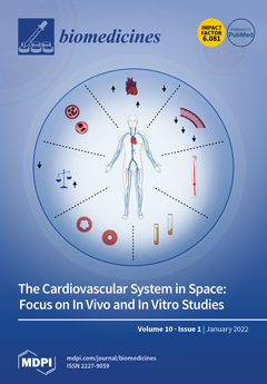
In space, astronauts are subjected to a prolonged state of microgravity that induces a myriad of physiological adaptations. Human spaceflight is associated with several cardiovascular risk factors that induce multiple structural and functional changes. Upon entering microgravity, cephalad fluid shift occurs and increases the stroke volume and cardiac output. Despite this increase, astronauts enter a state of hypovolemia. The absence of orthostatic pressure and a decrease in arterial pressures reduces the workload of the heart and is believed to be the underlying mechanism for the development of cardiac atrophy in space. In addition, parasympathetic overactivity and blood anemia are observed inflight, while orthostatic intolerance is a remarkable feature postflight. View this paper
  • Issues are regarded as officially published after their release is announced to the table of contents alert mailing list.
  • You may sign up for e-mail alerts to receive table of contents of newly released issues.
  • PDF is the official format for papers published in both, html and pdf forms. To view the papers in pdf format, click on the "PDF Full-text" link, and use the free Adobe Reader to open them.
Order results
Result details
Section
Select all
Export citation of selected articles as:
16 pages, 1161 KiB  
Review
Unveiling the Role of the Fatty Acid Binding Protein 4 in the Metabolic-Associated Fatty Liver Disease
by Juan Moreno-Vedia, Josefa Girona, Daiana Ibarretxe, Lluís Masana and Ricardo Rodríguez-Calvo
Biomedicines 2022, 10(1), 197; https://doi.org/10.3390/biomedicines10010197 - 17 Jan 2022
Cited by 11 | Viewed by 4321
Abstract
Metabolic-associated fatty liver disease (MAFLD), the main cause of chronic liver disease worldwide, is a progressive disease ranging from fatty liver to steatohepatitis (metabolic-associated steatohepatitis; MASH). Nevertheless, it remains underdiagnosed due to the lack of effective non-invasive methods for its diagnosis and staging. [...] Read more.
Metabolic-associated fatty liver disease (MAFLD), the main cause of chronic liver disease worldwide, is a progressive disease ranging from fatty liver to steatohepatitis (metabolic-associated steatohepatitis; MASH). Nevertheless, it remains underdiagnosed due to the lack of effective non-invasive methods for its diagnosis and staging. Although MAFLD has been found in lean individuals, it is closely associated with obesity-related conditions. Adipose tissue is the main source of liver triglycerides and adipocytes act as endocrine organs releasing a large number of adipokines and pro-inflammatory mediators involved in MAFLD progression into bloodstream. Among the adipocyte-derived molecules, fatty acid binding protein 4 (FABP4) has been recently associated with fatty liver and additional features of advanced stages of MAFLD. Additionally, emerging data from preclinical studies propose FABP4 as a causal actor involved in the disease progression, rather than a mere biomarker for the disease. Therefore, the FABP4 regulation could be considered as a potential therapeutic strategy to MAFLD. Here, we review the current knowledge of FABP4 in MAFLD, as well as its potential role as a therapeutic target for this disease. Full article
(This article belongs to the Special Issue NAFLD: From Mechanisms to Therapeutic Approaches)
Show Figures

Figure 1

21 pages, 2342 KiB  
Review
Multitasking Na+/Taurocholate Cotransporting Polypeptide (NTCP) as a Drug Target for HBV Infection: From Protein Engineering to Drug Discovery
by Dariusz Zakrzewicz and Joachim Geyer
Biomedicines 2022, 10(1), 196; https://doi.org/10.3390/biomedicines10010196 - 17 Jan 2022
Cited by 11 | Viewed by 3462
Abstract
Hepatitis B virus (HBV) infections are among the major public health concerns worldwide with more than 250 million of chronically ill individuals. Many of them are additionally infected with the Hepatitis D virus, a satellite virus to HBV. Chronic infection frequently leads to [...] Read more.
Hepatitis B virus (HBV) infections are among the major public health concerns worldwide with more than 250 million of chronically ill individuals. Many of them are additionally infected with the Hepatitis D virus, a satellite virus to HBV. Chronic infection frequently leads to serious liver diseases including cirrhosis and hepatocellular carcinoma, the most common type of liver cancer. Although current antiviral therapies can control HBV replication and slow down disease progress, there is an unmet medical need to identify therapies to cure this chronic infectious disease. Lately, a noteworthy progress in fighting against HBV has been made by identification of the high-affinity hepatic host receptor for HBV and HDV, namely Na+/taurocholate cotransporting polypeptide (NTCP, gene symbol SLC10A1). Next to its primary function as hepatic uptake transporter for bile acids, NTCP is essential for the cellular entry of HBV and HDV into hepatocytes. Due to this high-ranking discovery, NTCP has become a valuable target for drug development strategies for HBV/HDV-infected patients. In this review, we will focus on a newly predicted three-dimensional NTCP model that was generated using computational approaches and discuss its value in understanding the NTCP’s membrane topology, substrate and virus binding taking place in plasma membranes. We will review existing data on structural, functional, and biological consequences of amino acid residue changes and mutations that lead to loss of NTCP’s transport and virus receptor functions. Finally, we will discuss new directions for future investigations aiming at development of new NTCP-based HBV entry blockers that inhibit HBV tropism in human hepatocytes. Full article
Show Figures

Figure 1

16 pages, 1987 KiB  
Article
Selective Isolation of Liver-Derived Extracellular Vesicles Redefines Performance of miRNA Biomarkers for Non-Alcoholic Fatty Liver Disease
by Lauren A. Newman, Zivile Useckaite, Jillian Johnson, Michael J. Sorich, Ashley M. Hopkins and Andrew Rowland
Biomedicines 2022, 10(1), 195; https://doi.org/10.3390/biomedicines10010195 - 17 Jan 2022
Cited by 27 | Viewed by 4103
Abstract
Non-alcoholic fatty liver disease (NAFLD) is the most common chronic liver disease. Definitive diagnosis of the progressive form, non-alcoholic steatohepatitis (NASH), requires liver biopsy, which is highly invasive and unsuited to early disease or tracking changes. Inadequate performance of current minimally invasive tools [...] Read more.
Non-alcoholic fatty liver disease (NAFLD) is the most common chronic liver disease. Definitive diagnosis of the progressive form, non-alcoholic steatohepatitis (NASH), requires liver biopsy, which is highly invasive and unsuited to early disease or tracking changes. Inadequate performance of current minimally invasive tools is a critical barrier to managing NAFLD burden. Altered circulating miRNA profiles show potential for minimally invasive tracking of NAFLD. The selective isolation of the circulating extracellular vesicle subset that originates from hepatocytes presents an important opportunity for improving the performance of miRNA biomarkers of liver disease. The expressions of miR-122, -192, and -128-3p were quantified in total cell-free RNA, global EVs, and liver-specific EVs from control, NAFL, and NASH subjects. In ASGR1+ EVs, each miR biomarker trended positively with disease severity and expression was significantly higher in NASH subjects compared with controls. The c-statistic defining the performance of ASGR1+ EV derived miRNAs was invariably >0.78. This trend was not observed in the alternative sources. This study demonstrates the capacity for liver-specific isolation to transform the performance of EV-derived miRNA biomarkers for NAFLD, robustly distinguishing patients with NAFL and NASH. Full article
(This article belongs to the Special Issue NAFLD: From Mechanisms to Therapeutic Approaches)
Show Figures

Figure 1

33 pages, 2269 KiB  
Review
Detailed Molecular Mechanisms Involved in Drug-Induced Non-Alcoholic Fatty Liver Disease and Non-Alcoholic Steatohepatitis: An Update
by Laura Giuseppina Di Pasqua, Marta Cagna, Clarissa Berardo, Mariapia Vairetti and Andrea Ferrigno
Biomedicines 2022, 10(1), 194; https://doi.org/10.3390/biomedicines10010194 - 17 Jan 2022
Cited by 9 | Viewed by 6368
Abstract
Non-alcoholic fatty liver disease (NAFLD) and non-alcoholic steatohepatitis (NASH) are some of the biggest public health challenges due to their spread and increasing incidence around the world. NAFLD is characterized by intrahepatic lipid deposition, accompanied by dyslipidemia, hypertension, and insulin resistance, leading to [...] Read more.
Non-alcoholic fatty liver disease (NAFLD) and non-alcoholic steatohepatitis (NASH) are some of the biggest public health challenges due to their spread and increasing incidence around the world. NAFLD is characterized by intrahepatic lipid deposition, accompanied by dyslipidemia, hypertension, and insulin resistance, leading to more serious complications. Among the various causes, drug administration for the treatment of numerous kinds of diseases, such as antiarrhythmic and antihypertensive drugs, promotes the onset and progression of steatosis, causing drug-induced hepatic steatosis (DIHS). Here, we reviewed in detail the major classes of drugs that cause DIHS and the specific molecular mechanisms involved in these processes. Eight classes of drugs, among the most used for the treatment of common pathologies, were considered. The most diffused mechanism whereby drugs can induce NAFLD/NASH is interfering with mitochondrial activity, inhibiting fatty acid oxidation, but other pathways involved in lipid homeostasis are also affected. PubMed research was performed to obtain significant papers published up to November 2021. The key words included the class of drugs, or the specific compound, combined with steatosis, nonalcoholic steatohepatitis, fibrosis, fatty liver and hepatic lipid deposition. Additional information was found in the citations listed in other papers, when they were not displayed in the original search. Full article
Show Figures

Figure 1

13 pages, 759 KiB  
Article
Effects of Alirocumab on Triglyceride Metabolism: A Fat-Tolerance Test and Nuclear Magnetic Resonance Spectroscopy Study
by Thomas Metzner, Deborah R. Leitner, Karin Mellitzer, Andrea Beck, Harald Sourij, Tatjana Stojakovic, Gernot Reishofer, Winfried März, Ulf Landmesser, Hubert Scharnagl, Hermann Toplak and Günther Silbernagel
Biomedicines 2022, 10(1), 193; https://doi.org/10.3390/biomedicines10010193 - 17 Jan 2022
Cited by 4 | Viewed by 2707
Abstract
Background: PCSK9 antibodies strongly reduce LDL cholesterol. The effects of PCSK9 antibodies on triglyceride metabolism are less pronounced. The present study aimed to investigate in detail the effects of alirocumab on triglycerides, triglyceride-rich lipoproteins, and lipase regulators. Methods: A total of 24 patients [...] Read more.
Background: PCSK9 antibodies strongly reduce LDL cholesterol. The effects of PCSK9 antibodies on triglyceride metabolism are less pronounced. The present study aimed to investigate in detail the effects of alirocumab on triglycerides, triglyceride-rich lipoproteins, and lipase regulators. Methods: A total of 24 patients with an indication for treatment with PCSK9 antibodies were recruited. There were two visits at the study site: the first before initiation of treatment with alirocumab and the second after 10 weeks of treatment. Fat-tolerance tests, nuclear magnetic resonance spectroscopy, and enzyme-linked immunosorbent assays were performed to analyze lipid metabolism. Results: A total of 21 participants underwent the first and second investigation. Among these, two participants only received alirocumab twice and 19 patients completed the trial per protocol. All of them had atherosclerotic vascular disease. There was no significant effect of alirocumab treatment on fasting triglycerides, post-prandial triglycerides, or lipoprotein-lipase regulating proteins. Total, large, and small LDL particle concentrations decreased, while the HDL particle concentration increased (all p < 0.001). Mean total circulating PCSK9 markedly increased in response to alirocumab treatment (p < 0.001). Whereas PCSK9 increased more than three-fold in all 19 compliant patients, it remained unchanged in those two patients with two injections only. Conclusion: Significant effects of alirocumab on triglyceride metabolism were not detectable in the ALIROCKS trial. The total circulating PCSK9 concentration might be a useful biomarker to differentiate non-adherence from non-response to PCSK9 antibodies. Full article
Show Figures

Graphical abstract

12 pages, 2341 KiB  
Article
Neuroprotective Effects of Novel Treatments on Acute Optic Neuritis—A Meta-Analysis
by Tsung-Hsien Tsai, Chao-Wen Lin, Li-Wei Chan, Teck-Boon Tew and Ta-Ching Chen
Biomedicines 2022, 10(1), 192; https://doi.org/10.3390/biomedicines10010192 - 17 Jan 2022
Viewed by 1697
Abstract
Optic neuritis, inflammation of the optic nerve, can cause visual impairment through retinal nerve fiber layer (RNFL) degeneration. Optical coherence tomography could serve as a sensitive noninvasive tool for measuring RNFL thickness and evaluating the neuroprotective effects of treatment. We conducted a meta-analysis [...] Read more.
Optic neuritis, inflammation of the optic nerve, can cause visual impairment through retinal nerve fiber layer (RNFL) degeneration. Optical coherence tomography could serve as a sensitive noninvasive tool for measuring RNFL thickness and evaluating the neuroprotective effects of treatment. We conducted a meta-analysis to compare RNFL loss between novel add-on treatments and corticosteroid therapy at least 3 months after acute optic neuritis. The outcome measures were mean differences (MDs) in (1) RNFL thickness compared with the baseline in the affected and unaffected eye and (2) LogMAR visual acuity (VA). Seven studies involving five novel agents (memantine, erythropoietin, interferon-beta, phenytoin, and clemastine) were analyzed. When compared with the baseline RNFL thickness of the affected eye, the neuroprotective effects of novel add-on treatments could not be demonstrated. The difference in visual outcomes was also not significant between the two treatment groups. One study revealed that phenytoin has the potential to alleviate RNFL loss when the baseline thickness of the unaffected eye is considered. Larger randomized controlled trials with suitable outcome measures are warranted to evaluate the neuroprotective effects of novel treatments. Further studies should also tailor therapies to specific patient populations and investigate a more targeted treatment for acute optic neuritis. Full article
(This article belongs to the Section Neurobiology and Clinical Neuroscience)
Show Figures

Figure 1

8 pages, 761 KiB  
Article
Pretherapeutic Serum Albumin as an Outcome Prognosticator in Head and Neck Adenoid-Cystic Carcinoma
by Marlene Friedl, Stefan Stoiber, Faris F. Brkic and Lorenz Kadletz-Wanke
Biomedicines 2022, 10(1), 191; https://doi.org/10.3390/biomedicines10010191 - 17 Jan 2022
Cited by 4 | Viewed by 1727
Abstract
Background: A head and neck adenoid-cystic carcinoma is a rare malignant tumor arising from the salivary gland tissues. The long-term survival outcome is poor due to a high risk of recurrences and distant metastasis. The identification of prognostic markers could contribute to a [...] Read more.
Background: A head and neck adenoid-cystic carcinoma is a rare malignant tumor arising from the salivary gland tissues. The long-term survival outcome is poor due to a high risk of recurrences and distant metastasis. The identification of prognostic markers could contribute to a better risk assessment of each patient. The aim of this study is to assess the potential prognostic value of serum albumin in patients with head and neck adenoid-cystic carcinomas. Patients and Methods: This retrospective cohort study included all patients treated for a head and neck adenoid-cystic carcinoma between 1993 and 1 June 2019 with available pretherapeutic albumin values and clinical follow-up data. The cohort was stratified into a high and low group according to the median albumin value. The log-rank test was used for comparing overall and disease-free survival. Results: A total of 37 patients with complete follow-up data and available pretreatment albumin values were available. The overall mortality and recurrence rates were 21.6% (n = 8) and 45.9% (n = 17), respectively. Survival was shorter in the low albumin group. In particular, the mean overall survival for the low and high albumin groups were 121.0 months and 142.8 months, respectively. However, the difference was not statistically significant (p = 0.155). A statistically significant difference was observed in context with disease-free survival (45.2 months, 95% confidence interval 31.7–58.8 months vs. 114.8 months, 95% confidence interval 79.3–150.4 months; p = 0.029). Conclusion: Our study suggests a potential prognostic value of serum albumin in patients with a head and neck ACC. A further, external validation of our results is warranted. Full article
(This article belongs to the Special Issue Liquid Biopsies in Cancer Diagnosis, Monitoring and Prognosis)
Show Figures

Figure 1

8 pages, 581 KiB  
Article
Elevated Plasma Apurinic/Apyrimidinic Endonuclease 1/Redox Effector Factor-1 Levels in Refractory Kawasaki Disease
by Yu-Ran Lee, Eun Young Bae, Hong Ryang Kil, Byeong-Hwa Jeon and Geena Kim
Biomedicines 2022, 10(1), 190; https://doi.org/10.3390/biomedicines10010190 - 17 Jan 2022
Viewed by 1345
Abstract
Kawasaki disease (KD) refers to systemic vasculitis of medium-sized vessels accompanied by fever. The multifunctional protein apurinic/apyrimidinic endonuclease-1/redox factor-1 (APE1/Ref-1) is a new biomarker for vascular inflammation. Here, we investigated the association between APE1/Ref-1 and KD. Three groups, including 32 patients with KD [...] Read more.
Kawasaki disease (KD) refers to systemic vasculitis of medium-sized vessels accompanied by fever. The multifunctional protein apurinic/apyrimidinic endonuclease-1/redox factor-1 (APE1/Ref-1) is a new biomarker for vascular inflammation. Here, we investigated the association between APE1/Ref-1 and KD. Three groups, including 32 patients with KD (KD group), 33 patients with fever (Fever group), and 19 healthy individuals (Healthy group), were prospectively analyzed. APE1/Ref-1 levels were measured, and the clinical characteristics of KD were evaluated. The mean age of all patients was 2.7 ± 1.8 years, but the Healthy group participants were older than the other participants. Fever duration was longer in the KD group than in the fever group. APE1/Ref-1 levels were significantly higher in the KD group (p = 0.004) than in the other two groups, but there was no difference between the healthy and fever groups. APE1/Ref-1 levels did not differ according to fever duration or coronary arterial lesion but were higher in refractory KD cases than in non-refractory cases. APE1/Ref-1 levels were significantly higher during the acute phase of KD. We propose that APE1/Ref-1 could be a beneficial biological marker for the diagnosis and prognosis of KD, especially in refractory KD. Full article
Show Figures

Figure 1

17 pages, 2149 KiB  
Review
Activation of STAT and SMAD Signaling Induces Hepcidin Re-Expression as a Therapeutic Target for β-Thalassemia Patients
by Hanan Kamel M. Saad, Alawiyah Awang Abd Rahman, Azly Sumanty Ab Ghani, Wan Rohani Wan Taib, Imilia Ismail, Muhammad Farid Johan, Abdullah Saleh Al-Wajeeh and Hamid Ali Nagi Al-Jamal
Biomedicines 2022, 10(1), 189; https://doi.org/10.3390/biomedicines10010189 - 17 Jan 2022
Cited by 5 | Viewed by 4450
Abstract
Iron homeostasis is regulated by hepcidin, a hepatic hormone that controls dietary iron absorption and plasma iron concentration. Hepcidin binds to the only known iron export protein, ferroportin (FPN), which regulates its expression. The major factors that implicate hepcidin regulation include iron [...] Read more.
Iron homeostasis is regulated by hepcidin, a hepatic hormone that controls dietary iron absorption and plasma iron concentration. Hepcidin binds to the only known iron export protein, ferroportin (FPN), which regulates its expression. The major factors that implicate hepcidin regulation include iron stores, hypoxia, inflammation, and erythropoiesis. When erythropoietic activity is suppressed, hepcidin expression is hampered, leading to deficiency, thus causing an iron overload in iron-loading anemia, such as β-thalassemia. Iron overload is the principal cause of mortality and morbidity in β-thalassemia patients with or without blood transfusion dependence. In the case of thalassemia major, the primary cause of iron overload is blood transfusion. In contrast, iron overload is attributed to hepcidin deficiency and hyperabsorption of dietary iron in non-transfusion thalassemia. Beta-thalassemia patients showed marked hepcidin suppression, anemia, iron overload, and ineffective erythropoiesis (IE). Recent molecular research has prompted the discovery of new diagnostic markers and therapeutic targets for several diseases, including β-thalassemia. In this review, signal transducers and activators of transcription (STAT) and SMAD (structurally similar to the small mothers against decapentaplegic in Drosophila) pathways and their effects on hepcidin expression have been discussed as a therapeutic target for β-thalassemia patients. Therefore, re-expression of hepcidin could be a therapeutic target in the management of thalassemia patients. Data from 65 relevant published experimental articles on hepcidin and β-thalassemia between January 2016 and May 2021 were retrieved by using PubMed and Google Scholar search engines. Published articles in any language other than English, review articles, books, or book chapters were excluded. Full article
(This article belongs to the Section Cell Biology and Pathology)
Show Figures

Figure 1

13 pages, 2191 KiB  
Article
Penetration of the SARS-CoV-2 Spike Protein across the Blood–Brain Barrier, as Revealed by a Combination of a Human Cell Culture Model System and Optical Biosensing
by Dániel Petrovszki, Fruzsina R. Walter, Judit P. Vigh, Anna Kocsis, Sándor Valkai, Mária A. Deli and András Dér
Biomedicines 2022, 10(1), 188; https://doi.org/10.3390/biomedicines10010188 - 17 Jan 2022
Cited by 21 | Viewed by 8575
Abstract
Since the outbreak of the global pandemic caused by severe acute respiratory coronavirus 2 (SARS-CoV-2), several clinical aspects of the disease have come into attention. Besides its primary route of infection through the respiratory system, SARS-CoV-2 is known to have neuroinvasive capacity, causing [...] Read more.
Since the outbreak of the global pandemic caused by severe acute respiratory coronavirus 2 (SARS-CoV-2), several clinical aspects of the disease have come into attention. Besides its primary route of infection through the respiratory system, SARS-CoV-2 is known to have neuroinvasive capacity, causing multiple neurological symptoms with increased neuroinflammation and blood–brain barrier (BBB) damage. The viral spike protein disseminates via circulation during infection, and when reaching the brain could possibly cross the BBB, which was demonstrated in mice. Therefore, its medical relevance is of high importance. The aim of this study was to evaluate the barrier penetration of the S1 subunit of spike protein in model systems of human organs highly exposed to the infection. For this purpose, in vitro human BBB and intestinal barrier cell–culture systems were investigated by an optical biosensing method. We found that spike protein crossed the human brain endothelial cell barrier effectively. Additionally, spike protein passage was found in a lower amount for the intestinal barrier cell layer. These observations were corroborated with parallel specific ELISAs. The findings on the BBB model could provide a further basis for studies focusing on the mechanism and consequences of spike protein penetration across the BBB to the brain. Full article
(This article belongs to the Special Issue Biosensors at the Aid of Medicine)
Show Figures

Figure 1

12 pages, 2755 KiB  
Article
Effect of Ulinastatin on Syndecan-2-Mediated Vascular Damage in IDH2-Deficient Endothelial Cells
by Su-jeong Choi, Harsha Nagar, Jun Wan Lee, Seonhee Kim, Ikjun Lee, Shuyu Piao, Byeong Hwa Jeon and Cuk-Seong Kim
Biomedicines 2022, 10(1), 187; https://doi.org/10.3390/biomedicines10010187 - 17 Jan 2022
Cited by 3 | Viewed by 1667
Abstract
Syndecan-2 (SDC2), a cell-surface heparin sulfate proteoglycan of the glycocalyx, is mainly expressed in endothelial cells. Although oxidative stress and inflammatory mediators have been shown to mediate dysfunction of the glycocalyx, little is known about their role in vascular endothelial cells. In this [...] Read more.
Syndecan-2 (SDC2), a cell-surface heparin sulfate proteoglycan of the glycocalyx, is mainly expressed in endothelial cells. Although oxidative stress and inflammatory mediators have been shown to mediate dysfunction of the glycocalyx, little is known about their role in vascular endothelial cells. In this study, we aimed to identify the mechanism that regulates SDC2 expression in isocitrate dehydrogenase 2 (IDH2)-deficient endothelial cells, and to investigate the effect of ulinastatin (UTI) on this mechanism. We showed that knockdown of IDH2 induced SDC2 expression in human umbilical vein endothelial cells (HUVECs). Matrix metalloproteinase 7 (MMP7) influences SDC2 expression. When IDH2 was downregulated, MMP7 expression was increased, as was TGF-β signaling, which regulates MMP7. Inhibition of MMP7 activity using MMP inhibitor II significantly reduced SDC2, suggesting that IDH2 mediated SDC2 expression via MMP7. Moreover, expression of SDC2 and MMP7, as well as TGF-β signaling, increased in response to IDH2 deficiency, and treatment with UTI reversed this increase. Similarly, the increase in SDC2, MMP7, and TGF-β signaling in the aorta of IDH2 knockout mice was reversed by UTI treatment. These findings suggest that IDH2 deficiency induces SDC2 expression via TGF-β and MMP7 signaling in endothelial cells. Full article
(This article belongs to the Section Molecular and Translational Medicine)
Show Figures

Figure 1

4 pages, 214 KiB  
Editorial
Oral Microbiome, Oral Health and Systemic Health: A Multidirectional Link
by Elena Maria Varoni and Lia Rimondini
Biomedicines 2022, 10(1), 186; https://doi.org/10.3390/biomedicines10010186 - 17 Jan 2022
Cited by 10 | Viewed by 2347
Abstract
The oral cavity can be regarded as the mirror of systemic health, since many systemic diseases may have manifestations in the oral cavity, as in the case, among oral, potentially malignant disorders, of lupus erythematosus oral lichenoid lesions, and, vice-versa, oral diseases may [...] Read more.
The oral cavity can be regarded as the mirror of systemic health, since many systemic diseases may have manifestations in the oral cavity, as in the case, among oral, potentially malignant disorders, of lupus erythematosus oral lichenoid lesions, and, vice-versa, oral diseases may affect systemic health, impairing patient’s nutrition and wellbeing, reducing the quality of life and increasing stress and anxiety [...] Full article
15 pages, 4125 KiB  
Article
Ribosome Biogenesis Serves as a Therapeutic Target for Treating Endometriosis and the Associated Complications
by Cherry Yin-Yi Chang, An-Jen Chiang, Man-Ju Yan, Ming-Tsung Lai, Yun-Yi Su, Hsin-Yi Huang, Chan-Yu Chang, Ya-Hui Li, Pei-Fen Li, Chih-Mei Chen, Tritium Hwang, Chloe Hogg, Erin Greaves and Jim Jinn-Chyuan Sheu
Biomedicines 2022, 10(1), 185; https://doi.org/10.3390/biomedicines10010185 - 17 Jan 2022
Cited by 1 | Viewed by 3484
Abstract
Ribosome biogenesis is a cellular process critical for protein homeostasis during cell growth and multiplication. Our previous study confirmed up-regulation of ribosome biogenesis during endometriosis progression and malignant transition, thus anti-ribosome biogenesis may be effective for treating endometriosis and the associated complications. A [...] Read more.
Ribosome biogenesis is a cellular process critical for protein homeostasis during cell growth and multiplication. Our previous study confirmed up-regulation of ribosome biogenesis during endometriosis progression and malignant transition, thus anti-ribosome biogenesis may be effective for treating endometriosis and the associated complications. A mouse model with human endometriosis features was established and treated with three different drugs that can block ribosome biogenesis, including inhibitors against mTOR/PI3K (GSK2126458) and RNA polymerase I (CX5461 and BMH21). The average lesion numbers and disease frequencies were significantly reduced in treated mice as compared to controls treated with vehicle. Flow cytometry analyses confirmed the reduction of small peritoneal macrophage and neutrophil populations with increased large versus small macrophage ratios, suggesting inflammation suppression by drug treatments. Lesions in treated mice also showed lower nerve fiber density which can support the finding of pain-relief by behavioral studies. Our study therefore suggested ribosome biogenesis as a potential therapeutic target for treating endometriosis. Full article
(This article belongs to the Special Issue Advanced Research in Endometriosis 2.0)
Show Figures

Figure 1

12 pages, 1095 KiB  
Article
Plasma Proteomics in Healthy Subjects with Differences in Tissue Glucocorticoid Sensitivity Identifies A Novel Proteomic Signature
by Nicolas C. Nicolaides, Manousos Makridakis, Rafael Stroggilos, Vasiliki Lygirou, Eleni Koniari, Ifigeneia Papageorgiou, Amalia Sertedaki, Jerome Zoidakis and Evangelia Charmandari
Biomedicines 2022, 10(1), 184; https://doi.org/10.3390/biomedicines10010184 - 16 Jan 2022
Cited by 2 | Viewed by 1960
Abstract
Significant inter-individual variation in terms of susceptibility to several stress-related disorders, such as myocardial infarction and Alzheimer’s disease, and therapeutic response has been observed among healthy subjects. The molecular features responsible for this phenomenon have not been fully elucidated. Proteomics, in association with [...] Read more.
Significant inter-individual variation in terms of susceptibility to several stress-related disorders, such as myocardial infarction and Alzheimer’s disease, and therapeutic response has been observed among healthy subjects. The molecular features responsible for this phenomenon have not been fully elucidated. Proteomics, in association with bioinformatics analysis, offer a comprehensive description of molecular phenotypes with clear links to human disease pathophysiology. The aim of this study was to conduct a comparative plasma proteomics analysis of glucocorticoid resistant and glucocorticoid sensitive healthy subjects and provide clues of the underlying physiological differences. For this purpose, 101 healthy volunteers were given a very low dose (0.25 mg) of dexamethasone at midnight, and were stratified into the 10% most glucocorticoid sensitive (S) (n = 11) and 10% most glucocorticoid resistant (R) (n = 11) according to the 08:00 h serum cortisol concentrations determined the following morning. One month following the very-low dose dexamethasone suppression test, DNA and plasma samples were collected from the 22 selected individuals. Sequencing analysis did not reveal any genetic defects in the human glucocorticoid receptor (NR3C1) gene. To investigate the proteomic profile of plasma samples, we used Liquid Chromatography–Mass Spectrometry (LC-MS/MS) and found 110 up-regulated and 66 down-regulated proteins in the S compared to the R group. The majority of the up-regulated proteins in the S group were implicated in platelet activation. To predict response to cortisol prior to administration, a random forest classifier was developed by using the proteomics data in order to distinguish S from R individuals. Apolipoprotein A4 (APOA4) and gelsolin (GSN) were the most important variables in the classification, and warrant further investigation. Our results indicate that a proteomics signature may differentiate the S from the R healthy subjects, and may be useful in clinical practice. In addition, it may provide clues of the underlying molecular mechanisms of the chronic stress-related diseases, including myocardial infarction and Alzheimer’s disease. Full article
(This article belongs to the Section Molecular and Translational Medicine)
Show Figures

Figure 1

10 pages, 2019 KiB  
Article
Serpinin in the Skin
by Cristina Fraquelli, Jasmine Hauzinger, Christian Humpel, Maria Nolano, Vincenzo Provitera, Vinay Kumar Sharma, Peng Loh, Zenon Pidsudko, Georgios Blatsios and Josef Troger
Biomedicines 2022, 10(1), 183; https://doi.org/10.3390/biomedicines10010183 - 16 Jan 2022
Viewed by 1899
Abstract
The serpinins are relatively novel peptides generated by proteolytic processing of chromogranin A and they are comprised of free serpinin, serpinin-RRG and pGlu-serpinin. In this study, the presence and source of these peptides were studied in the skin. By Western blot analysis, a [...] Read more.
The serpinins are relatively novel peptides generated by proteolytic processing of chromogranin A and they are comprised of free serpinin, serpinin-RRG and pGlu-serpinin. In this study, the presence and source of these peptides were studied in the skin. By Western blot analysis, a 40 kDa and a 50 kDa protein containing the sequence of serpinin were detected in the trigeminal ganglion and dorsal root ganglia in rats but none in the skin. RP-HPLC followed by EIA revealed that the three serpinins are present in similar, moderate amounts in rat dorsal root ganglia, whereas in the rat skin, free serpinin represents the predominant molecular form. There were abundant serpinin-positive cells in rat dorsal root ganglia and colocalization with substance P was evident. However, much more widespread distribution of the serpinins was found in dorsal root ganglia when compared with substance P. In the skin, serpinin immunoreactivity was found in sensory nerves and showed colocalization with substance P; as well, some was present in autonomic nerves. Thus, although not exclusively, there is evidence that serpinin is a constituent of the sensory innervation of the skin. The serpinins are biologically highly active and might therefore be of functional significance in the skin. Full article
(This article belongs to the Special Issue Neuropeptides in Biomedicines)
Show Figures

Figure 1

21 pages, 754 KiB  
Review
Impact of Sarcopenia and Myosteatosis in Non-Cirrhotic Stages of Liver Diseases: Similarities and Differences across Aetiologies and Possible Therapeutic Strategies
by Annalisa Cespiati, Marica Meroni, Rosa Lombardi, Giovanna Oberti, Paola Dongiovanni and Anna Ludovica Fracanzani
Biomedicines 2022, 10(1), 182; https://doi.org/10.3390/biomedicines10010182 - 16 Jan 2022
Cited by 13 | Viewed by 2672
Abstract
Sarcopenia is defined as a loss of muscle strength, mass and function and it is a predictor of mortality. Sarcopenia is not only a geriatric disease, but it is related to several chronic conditions, including liver diseases in both its early and advanced [...] Read more.
Sarcopenia is defined as a loss of muscle strength, mass and function and it is a predictor of mortality. Sarcopenia is not only a geriatric disease, but it is related to several chronic conditions, including liver diseases in both its early and advanced stages. Despite the increasing number of studies exploring the role of sarcopenia in the early stages of chronic liver disease (CLD), its prevalence and the relationship between these two clinical entities are still controversial. Myosteatosis is characterized by fat accumulation in the muscles and it is related to advanced liver disease, although its role in the early stages is still under researched. Therefore, in this narrative review, we firstly aimed to evaluate the prevalence and the pathogenetic mechanisms underlying sarcopenia and myosteatosis in the early stage of CLD across different aetiologies (mainly non-alcoholic fatty liver disease, alcohol-related liver disease and viral hepatitis). Secondly, due to the increasing prevalence of sarcopenia worldwide, we aimed to revise the current and the future therapeutic approaches for the management of sarcopenia in CLD. Full article
Show Figures

Figure 1

15 pages, 4111 KiB  
Article
Differential Expression and Localization of EHBP1L1 during the First Wave of Rat Spermatogenesis Suggest Its Involvement in Acrosome Biogenesis
by Massimo Venditti and Sergio Minucci
Biomedicines 2022, 10(1), 181; https://doi.org/10.3390/biomedicines10010181 - 16 Jan 2022
Cited by 8 | Viewed by 1912
Abstract
The identification and characterization of new proteins involved in spermatogenesis is fundamental, considering that good-quality gametes are basic in ensuring proper reproduction. Here, we further analyzed the temporal and spatial localization during the first spermatogenic wave of rat testis of EHBP1L1, which is [...] Read more.
The identification and characterization of new proteins involved in spermatogenesis is fundamental, considering that good-quality gametes are basic in ensuring proper reproduction. Here, we further analyzed the temporal and spatial localization during the first spermatogenic wave of rat testis of EHBP1L1, which is involved in vesicular trafficking due to the CH and bMERB domains, which bind to actin and Rab8/10, respectively. Western blot and immunofluorescence analyses showed that EHBP1L1 protein expression started at 21 days post-partum (dpp) concomitantly with the appearance of primary spermatocytes (I SPC). In subsequent stages, EHBP1L1 specifically localized together with actin in the perinuclear cytoplasm close to the acrosomal and Golgian regions of spermatids (SPT) during the different phases of acrosome biogenesis (AB). Moreover, it was completely absent in elongated SPT and in mature spermatozoa, suggesting that its role was completed in previous stages. The combined data, also supported by our previous report demonstrating that EHBP1L1 mRNA was expressed by primary (I) and secondary (II) SPC, lead us to hypothesize its specific role during AB. Although these results are suggestive, further studies are needed to better clarify the underlying molecular mechanisms of AB, with the aim to use EHBP1L1 as a potential new marker for spermatogenesis. Full article
Show Figures

Figure 1

12 pages, 1667 KiB  
Article
A Unique Immune-Related Gene Signature Represents Advanced Liver Fibrosis and Reveals Potential Therapeutic Targets
by Pil-Soo Sung, Chang-Min Kim, Jung-Hoon Cha, Jin-Young Park, Yun-Suk Yu, Hee-Jung Wang, Jin-Kyeoung Kim and Si-Hyun Bae
Biomedicines 2022, 10(1), 180; https://doi.org/10.3390/biomedicines10010180 - 16 Jan 2022
Cited by 5 | Viewed by 2777
Abstract
Innate and adaptive immune responses are critically associated with the progression of fibrosis in chronic liver diseases. In this study, we aim to identify a unique immune-related gene signature representing advanced liver fibrosis and to reveal potential therapeutic targets. Seventy-seven snap-frozen liver tissues [...] Read more.
Innate and adaptive immune responses are critically associated with the progression of fibrosis in chronic liver diseases. In this study, we aim to identify a unique immune-related gene signature representing advanced liver fibrosis and to reveal potential therapeutic targets. Seventy-seven snap-frozen liver tissues with various chronic liver diseases at different fibrosis stages (1: n = 12, 2: n = 12, 3: n = 25, 4: n = 28) were subjected to expression analyses. Gene expression analysis was performed using the nCounter PanCancer Immune Profiling Panel (NanoString Technologies, Seattle, WA, USA). Biological meta-analysis was performed using the CBS Probe PINGSTM (CbsBioscience, Daejeon, Korea). Using non-tumor tissues from surgically resected specimens, we identified the immune-related, five-gene signature (CHIT1_FCER1G_OSM_VEGFA_ZAP70) that reliably differentiated patients with low- (F1 and F2) and high-grade fibrosis (F3 and F4; accuracy = 94.8%, specificity = 91.7%, sensitivity = 96.23%). The signature was independent of all pathological and clinical features and was independently associated with high-grade fibrosis using multivariate analysis. Among these genes, the expression of inflammation-associated FCER1G, OSM, VEGFA, and ZAP70 was lower in high-grade fibrosis than in low-grade fibrosis, whereas CHIT1 expression, which is associated with fibrogenic activity of macrophages, was higher in high-grade fibrosis. Meta-analysis revealed that STAT3, a potential druggable target, highly interacts with the five-gene signature. Overall, we identified an immune gene signature that reliably predicts advanced fibrosis in chronic liver disease. This signature revealed potential immune therapeutic targets to ameliorate liver fibrosis. Full article
Show Figures

Figure 1

11 pages, 1223 KiB  
Article
Predictive Factors for Skip Lymph Node Metastasis and Their Implication on Recurrence in Papillary Thyroid Carcinoma
by Young-Jae Ryu, Seong-Young Kwon, Soo-Young Lim, Yong-Min Na and Min-Ho Park
Biomedicines 2022, 10(1), 179; https://doi.org/10.3390/biomedicines10010179 - 16 Jan 2022
Cited by 7 | Viewed by 1892
Abstract
Skip lymph node (LN) metastases in papillary thyroid carcinoma (PTC) belong to N1b classification in the absence of central neck LN involvement. This study aimed to evaluate the predictive factors of skip metastases and their impact on recurrence in PTC patients with pN1b. [...] Read more.
Skip lymph node (LN) metastases in papillary thyroid carcinoma (PTC) belong to N1b classification in the absence of central neck LN involvement. This study aimed to evaluate the predictive factors of skip metastases and their impact on recurrence in PTC patients with pN1b. A total of 334 PTC patients who underwent total thyroidectomy with LN dissection (central and lateral neck compartment) followed by radioactive iodine ablation were included. Patients with skip metastases tended to have a small primary tumor (≤1 cm) and single lateral neck level involvement. Tumor size ≤ 1 cm was an important predictive factor for skip metastases. Univariate analysis for recurrence showed that patients with a central LN ratio > 0.68, lateral LN ratio > 0.21, and stimulated thyroglobulin (Tg) levels > 7.3 ng/mL had shorter RFS (recurrence-free survival). The stimulated Tg level was associated with shorter RFS on multivariate analysis (>7.3 vs. ≤7.3 ng/mL; hazard ratio, 4.226; 95% confidence interval, 2.226−8.022; p < 0.001). Although patients with skip metastases tended to have a small primary tumor and lower burden of lateral neck LN involvement, there was no association between skip metastases and RFS in PTC with pN1b. Stimulated Tg level was a strong predictor of recurrence. Full article
(This article belongs to the Special Issue Recent Advances in Thyroid Cancer: From Diagnosis to Treatment)
Show Figures

Figure 1

26 pages, 1480 KiB  
Review
Novel Insight into the Mechanisms of the Bidirectional Relationship between Diabetes and Periodontitis
by Federica Barutta, Stefania Bellini, Marilena Durazzo and Gabriella Gruden
Biomedicines 2022, 10(1), 178; https://doi.org/10.3390/biomedicines10010178 - 16 Jan 2022
Cited by 23 | Viewed by 6786
Abstract
Periodontitis and diabetes are two major global health problems despite their prevalence being significantly underreported and underestimated. Both epidemiological and intervention studies show a bidirectional relationship between periodontitis and diabetes. The hypothesis of a potential causal link between the two diseases is corroborated [...] Read more.
Periodontitis and diabetes are two major global health problems despite their prevalence being significantly underreported and underestimated. Both epidemiological and intervention studies show a bidirectional relationship between periodontitis and diabetes. The hypothesis of a potential causal link between the two diseases is corroborated by recent studies in experimental animals that identified mechanisms whereby periodontitis and diabetes can adversely affect each other. Herein, we will review clinical data on the existence of a two-way relationship between periodontitis and diabetes and discuss possible mechanistic interactions in both directions, focusing in particular on new data highlighting the importance of the host response. Moreover, we will address the hypothesis that trained immunity may represent the unifying mechanism explaining the intertwined association between diabetes and periodontitis. Achieving a better mechanistic insight on clustering of infectious, inflammatory, and metabolic diseases may provide new therapeutic options to reduce the risk of diabetes and diabetes-associated comorbidities. Full article
Show Figures

Figure 1

13 pages, 2354 KiB  
Article
Detection Rate and Clinical Impact of PET/CT with 18F-FACBC in Patients with Biochemical Recurrence of Prostate Cancer: A Retrospective Bicentric Study
by Luca Filippi, Oreste Bagni, Carmelo Crisafulli, Ivan Cerio, Gabriele Brunotti, Agostino Chiaravalloti, Orazio Schillaci and Franca Dore
Biomedicines 2022, 10(1), 177; https://doi.org/10.3390/biomedicines10010177 - 15 Jan 2022
Cited by 8 | Viewed by 1702
Abstract
Our aim was to assess the detection rate (DR) of positron emission computed tomography (PET/CT) with anti-1-amino-3-[18F]-flurocyclobutane-1-carboxylic acid (18F-FACBC) in patients with biochemical recurrence (BCR) from prostate cancer (PC). As a secondary endpoint, we evaluated 18F-FACBC PET/CT’s impact [...] Read more.
Our aim was to assess the detection rate (DR) of positron emission computed tomography (PET/CT) with anti-1-amino-3-[18F]-flurocyclobutane-1-carboxylic acid (18F-FACBC) in patients with biochemical recurrence (BCR) from prostate cancer (PC). As a secondary endpoint, we evaluated 18F-FACBC PET/CT’s impact on patients management. Clinical records of 81 patients submitted to 18F-FACBC PET/CT due to PC BCR in two Italian Nuclear Medicine Units were retrospectively assessed. DR was gauged in the whole cohort and stratifying patients by discrete intervals of PSA levels. PET/CT’s impact on clinical management was scored as (1) major if it entailed an intermodality change (e.g., from systemic to loco-regional therapy); (2) minor if it led to an intramodality change (e.g., modified radiotherapy field). PET/CT’s DR resulted in 76.9% in the whole cohort, with a positive predictive value of 96.7%. Stratified by PSA quartile intervals, PET/CT’s DR was 66.7%, 71.4%, 78.9% and 90% for PSA 0.2–0.57 ng/mL, 0.58–0.99 ng/mL, 1–1.5 ng/mL and >1.5 ng/mL without significant difference among groups (p = 0.81). The most common sites of relapse were prostate bed and pelvic lymph nodes (59.3%). PET/CT impacted on clinical management in 33/81 cases (40.7%), leading to a major change in 30 subjects (90.9%). 18F-FACBC PET/CT localized recurrence in patients with BCR, with meaningful DR also at low PSA levels and significantly impacted on clinical management. Full article
Show Figures

Figure 1

16 pages, 3892 KiB  
Article
Hypoxia Preconditioned Serum (HPS)-Hydrogel Can Accelerate Dermal Wound Healing in Mice—An In Vivo Pilot Study
by Jun Jiang, Ursula Kraneburg, Ulf Dornseifer, Arndt F. Schilling, Ektoras Hadjipanayi, Hans-Günther Machens and Philipp Moog
Biomedicines 2022, 10(1), 176; https://doi.org/10.3390/biomedicines10010176 - 14 Jan 2022
Cited by 5 | Viewed by 2328
Abstract
The ability to use the body’s resources to promote wound repair is increasingly becoming an interesting area of regenerative medicine research. Here, we tested the effect of topical application of blood-derived hypoxia preconditioned serum (HPS) on wound healing in a murine wound model. [...] Read more.
The ability to use the body’s resources to promote wound repair is increasingly becoming an interesting area of regenerative medicine research. Here, we tested the effect of topical application of blood-derived hypoxia preconditioned serum (HPS) on wound healing in a murine wound model. Alginate hydrogels loaded with two different HPS concentrations (10 and 40%) were applied topically on full-thickness wounds created on the back of immunocompromised mice. We achieved a significant dose-dependent wound area reduction after 5 days in HPS-treated groups compared with no treatment (NT). On average, both HPS-10% and HPS-40% -treated wounds healed 1.4 days faster than NT. Healed tissue samples were investigated on post-operative day 15 (POD 15) by immunohistology and showed an increase in lymphatic vessels (LYVE-1) up to 45% with HPS-40% application, while at this stage, vascularization (CD31) was comparable in the HPS-treated and NT groups. Furthermore, the expression of proliferation marker Ki67 was greater on POD 15 in the NT-group compared to HPS-treated groups, in accordance with the earlier completion of wound healing observed in the latter. Collagen deposition was similar in all groups, indicating lack of scar tissue hypertrophy as a result of HPS-hydrogel treatment. These findings show that topical HPS application is safe and can accelerate dermal wound healing in mice. Full article
(This article belongs to the Topic Animal Model in Biomedical Research)
Show Figures

Figure 1

15 pages, 4563 KiB  
Article
Puerarin Attenuates Obesity-Induced Inflammation and Dyslipidemia by Regulating Macrophages and TNF-Alpha in Obese Mice
by Ji-Won Noh, Hee-Kwon Yang, Min-Soo Jun and Byung-Cheol Lee
Biomedicines 2022, 10(1), 175; https://doi.org/10.3390/biomedicines10010175 - 14 Jan 2022
Cited by 14 | Viewed by 2548
Abstract
Obesity causes low-grade inflammation that results in dyslipidemia and insulin resistance. We evaluated the effect of puerarin on obesity and metabolic complications both in silico and in vivo and investigated the underlying immunological mechanisms. Twenty C57BL/6 mice were divided into four groups: normal [...] Read more.
Obesity causes low-grade inflammation that results in dyslipidemia and insulin resistance. We evaluated the effect of puerarin on obesity and metabolic complications both in silico and in vivo and investigated the underlying immunological mechanisms. Twenty C57BL/6 mice were divided into four groups: normal chow, control (HFD), HFD + puerarin (PUE) 200 mg/kg, and HFD + atorvastatin (ATO) 10 mg/kg groups. We examined bodyweight, oral glucose tolerance test, serum insulin, oral fat tolerance test, serum lipids, and adipocyte size. We also analyzed the percentage of total, M1, and M2 adipose tissue macrophages (ATMs) and the expression of F4/80, tumor necrosis factor-α (TNF-α), C-C motif chemokine ligand 2 (CCL2), CCL4, CCL5, and C-X-C motif chemokine receptor 4. In silico, we identified the treatment-targeted genes of puerarin and simulated molecular docking with puerarin and TNF, M1, and M2 macrophages based on functionally enriched pathways. Puerarin did not significantly change bodyweight but significantly improved fat pad weight, adipocyte size, fat area in the liver, free fatty acids, triglycerides, total cholesterol, and HDL-cholesterol in vivo. In addition, puerarin significantly decreased the ATM population and TNF-α expression. Therefore, puerarin is a potential anti-obesity treatment based on its anti-inflammatory effects in adipose tissue. Full article
(This article belongs to the Special Issue Macrophages in Health and Non-infectious Disease 3.0)
Show Figures

Figure 1

21 pages, 3173 KiB  
Article
A More Diverse Cervical Microbiome Associates with Better Clinical Outcomes in Patients with Endometriosis: A Pilot Study
by Cherry Yin-Yi Chang, An-Jen Chiang, Ming-Tsung Lai, Man-Ju Yan, Chung-Chen Tseng, Lun-Chien Lo, Lei Wan, Chia-Jung Li, Kuan-Hao Tsui, Chih-Mei Chen, Tritium Hwang, Fuu-Jen Tsai and Jim Jinn-Chyuan Sheu
Biomedicines 2022, 10(1), 174; https://doi.org/10.3390/biomedicines10010174 - 14 Jan 2022
Cited by 17 | Viewed by 5748
Abstract
Infection-induced chronic inflammation is common in patients with endometriosis. Although microbial communities in the reproductive tracts of patients have been reported, little was known about their dynamic profiles during disease progression and complication development. Microbial communities in cervical mucus were collected by cervical [...] Read more.
Infection-induced chronic inflammation is common in patients with endometriosis. Although microbial communities in the reproductive tracts of patients have been reported, little was known about their dynamic profiles during disease progression and complication development. Microbial communities in cervical mucus were collected by cervical swabs from 10 healthy women and 23 patients, and analyzed by 16S rRNA amplicon sequencing. The abundance, ecological relationships and functional networks of microbiota were characterized according to their prevalence, clinical stages, and clinical features including deeply infiltrating endometriosis (DIE), CA125, pain score and infertility. Cervical microbiome can be altered during endometriosis development and progression with a tendency of increased Firmicutes and decreased Actinobacteria and Bacteroidetes. Distinct from vaginal microbiome, upregulation of Lactobacillus, in combination with increased Streptococcus and decreased Dialister, was frequently associated with advanced endometriosis stages, DIE, higher CA125 levels, severe pain, and infertility. Significantly, reduced richness and diversity of cervical microbiome were detected in patients with more severe clinical symptoms. Clinical treatments against infertility can partially reverse the ecological balance of microbes through remodeling nutrition metabolism and transport and cell-cell/cell-matrix interaction. This study provides a new understanding on endometriosis development and a more diverse cervical microbiome may be beneficial for patients to have better clinical outcomes. Full article
(This article belongs to the Special Issue Microbial Ecology in Health and Disease 2.0)
Show Figures

Figure 1

16 pages, 4008 KiB  
Article
Systemic Injection of Oncolytic Vaccinia Virus Suppresses Primary Tumor Growth and Lung Metastasis in Metastatic Renal Cell Carcinoma by Remodeling Tumor Microenvironment
by Jee Soo Park, Myung Eun Lee, Won Sik Jang, Jongchan Kim, Se Mi Park, Keunhee Oh, Namhee Lee and Won Sik Ham
Biomedicines 2022, 10(1), 173; https://doi.org/10.3390/biomedicines10010173 - 14 Jan 2022
Cited by 5 | Viewed by 2721
Abstract
Immune checkpoint inhibitors and tyrosine kinase inhibitors are the first-line treatment for metastatic renal cell carcinoma (mRCC), but their benefits are limited to specific patient subsets. Here, we aimed to evaluate the therapeutic efficacy of JX-594 (pexastimogene devacirepvec, Pexa-vec) monotherapy by systemic injection [...] Read more.
Immune checkpoint inhibitors and tyrosine kinase inhibitors are the first-line treatment for metastatic renal cell carcinoma (mRCC), but their benefits are limited to specific patient subsets. Here, we aimed to evaluate the therapeutic efficacy of JX-594 (pexastimogene devacirepvec, Pexa-vec) monotherapy by systemic injection in comparison with sunitinib monotherapy in metastatic orthotopic RCC murine models. Two highly metastatic orthotopic RCC models were developed to compare the treatment efficacy in the International Metastatic RCC Database Consortium favorable-risk and intermediate- or poor-risk groups. JX-594 was systemically injected through the peritoneum, whereas sunitinib was orally administered. Post-treatment, tumor microenvironment (TME) remodeling was determined using immunofluorescence analysis. Systemic JX-594 monotherapy injection demonstrated therapeutic benefit in both early- and advanced-stage mRCC models. Sunitinib monotherapy significantly reduced the primary tumor burden and number of lung metastases in the early-stage, but not in the advanced-stage mRCC model. Systemic JX-594 delivery remodeled the primary TME and lung metastatic sites by increasing tumor-infiltrating CD4/8+ T cells and dendritic cells. Systemic JX-594 monotherapy demonstrated significantly better therapeutic outcomes compared with sunitinib monotherapy in both early- and advanced-stage mRCCs by converting cold tumors into hot tumors. Sunitinib monotherapy effectively suppressed primary tumor growth and lung metastasis in early-stage mRCC. Full article
(This article belongs to the Special Issue Oncolytic Viruses as a Novel Form of Immunotherapy for Cancer III)
Show Figures

Figure 1

13 pages, 1557 KiB  
Systematic Review
The Role of Neuropilin-2 in the Epithelial to Mesenchymal Transition of Colorectal Cancer: A Systematic Review
by Cristina Lungulescu, Valentina Ghimpau, Dan Ionut Gheonea, Daniel Sur and Cristian Virgil Lungulescu
Biomedicines 2022, 10(1), 172; https://doi.org/10.3390/biomedicines10010172 - 14 Jan 2022
Cited by 5 | Viewed by 2117
Abstract
Neuropilin-2 (NRP-2) expression has been found in various investigations on the expression and function of NRP-2 in colorectal cancer. The link between NRP-2 and colorectal cancer, as well as the mechanism that regulates it, is still mostly unclear. This systematic review was carried [...] Read more.
Neuropilin-2 (NRP-2) expression has been found in various investigations on the expression and function of NRP-2 in colorectal cancer. The link between NRP-2 and colorectal cancer, as well as the mechanism that regulates it, is still mostly unclear. This systematic review was carried out according to the Cochrane guidelines for systematic reviews. We searched PubMed, Embase®, MEDLINE, Allied & Complementary MedicineTM, Medical Toxicology & Environmental Health, DH-DATA: Health Administration for articles published before 1 October 2021. The following search terms were used: “neuropilin-2” “neuropilin 2”, “NRP2” and “NRP-2”, “colorectal cancer”, “colon cancer”. Ten articles researching either tumor tissue samples, cell lines, or mice models were included in this review. The majority of human primary and metastatic colon cancer cell lines expressed NRP-2 compared to the normal colonic mucosa. NRPs have been discovered in human cancers as well as neovasculature. The presence of NRP-2 appears to be connected to the epithelial–mesenchymal transition’s function in cancer dissemination and metastatic evolution. The studies were heterogeneous, but the data assessed indicates NRP-2 might have an impact on the metastatic potential of colorectal cancer cells. Nevertheless, further research is needed. Full article
Show Figures

Figure 1

14 pages, 5440 KiB  
Article
Humoral Immune Response in IBD Patients Three and Six Months after Vaccination with the SARS-CoV-2 mRNA Vaccines mRNA-1273 and BNT162b2
by Richard Vollenberg, Phil-Robin Tepasse, Joachim Ewald Kühn, Marc Hennies, Markus Strauss, Florian Rennebaum, Tina Schomacher, Göran Boeckel, Eva Lorentzen, Arne Bokemeyer and Tobias Max Nowacki
Biomedicines 2022, 10(1), 171; https://doi.org/10.3390/biomedicines10010171 - 13 Jan 2022
Cited by 20 | Viewed by 3381
Abstract
Severe acute respiratory syndrome coronovirus-2 (SARS-CoV-2) is the cause of the coronavirus disease 2019 (COVID-19) pandemic. Vaccination is considered the core approach to containing the pandemic. There is currently insufficient evidence on the efficacy of these vaccines in immunosuppressed inflammatory bowel disease (IBD) [...] Read more.
Severe acute respiratory syndrome coronovirus-2 (SARS-CoV-2) is the cause of the coronavirus disease 2019 (COVID-19) pandemic. Vaccination is considered the core approach to containing the pandemic. There is currently insufficient evidence on the efficacy of these vaccines in immunosuppressed inflammatory bowel disease (IBD) patients. The aim of this study was to investigate the humoral response in immunosuppressed IBD patients after COVID-19 mRNA vaccination. In this prospective study, IgG antibody levels (AB) against the SARS-CoV-2 receptor-binding domain (spike-protein) were quantitatively determined. For assessing the potential neutralizing capacity, a SARS-CoV-2 surrogate neutralization test (sVNT) was employed in IBD patients (n = 95) and healthy controls (n = 38). Sera were examined prior to the first/second vaccination and 3/6 months after second vaccination. Patients showed lower sVNT (%) and IgG-S (AU/mL) AB both before the second vaccination (sVNT p < 0.001; AB p < 0.001) and 3 (sVNT p = 0.002; AB p = 0.001) and 6 months (sVNT p = 0.062; AB p = 0.061) after the second vaccination. Although seroconversion rates (sVNT, IgG-S) did not differ between the two groups 3 months after second vaccination, a significant difference was seen 6 months after second vaccination (sVNT p = 0.045). Before and three months after the second vaccination, patients treated with anti-tumor necrosis factor (TNF) agents showed significantly lower AB than healthy subjects. In conclusion, an early booster shot vaccination should be discussed for IBD patients on anti-TNF therapy. Full article
(This article belongs to the Special Issue Next-Generation Vaccines and Antivirals against SARS-CoV-2)
Show Figures

Figure 1

33 pages, 4421 KiB  
Review
Epigenetic Mechanisms as Emerging Therapeutic Targets and Microfluidic Chips Application in Pulmonary Arterial Hypertension
by Linh Ho, Nazir Hossen, Trieu Nguyen, Au Vo and Fakhrul Ahsan
Biomedicines 2022, 10(1), 170; https://doi.org/10.3390/biomedicines10010170 - 13 Jan 2022
Cited by 7 | Viewed by 5040
Abstract
Pulmonary arterial hypertension (PAH) is a disease that progress over time and is defined as an increase in pulmonary arterial pressure and pulmonary vascular resistance that frequently leads to right-ventricular (RV) failure and death. Epigenetic modifications comprising DNA methylation, histone remodeling, and noncoding [...] Read more.
Pulmonary arterial hypertension (PAH) is a disease that progress over time and is defined as an increase in pulmonary arterial pressure and pulmonary vascular resistance that frequently leads to right-ventricular (RV) failure and death. Epigenetic modifications comprising DNA methylation, histone remodeling, and noncoding RNAs (ncRNAs) have been established to govern chromatin structure and transcriptional responses in various cell types during disease development. However, dysregulation of these epigenetic mechanisms has not yet been explored in detail in the pathology of pulmonary arterial hypertension and its progression with vascular remodeling and right-heart failure (RHF). Targeting epigenetic regulators including histone methylation, acetylation, or miRNAs offers many possible candidates for drug discovery and will no doubt be a tempting area to explore for PAH therapies. This review focuses on studies in epigenetic mechanisms including the writers, the readers, and the erasers of epigenetic marks and targeting epigenetic regulators or modifiers for treatment of PAH and its complications described as RHF. Data analyses from experimental cell models and animal induced PAH models have demonstrated that significant changes in the expression levels of multiple epigenetics modifiers such as HDMs, HDACs, sirtuins (Sirt1 and Sirt3), and BRD4 correlate strongly with proliferation, apoptosis, inflammation, and fibrosis linked to the pathological vascular remodeling during PAH development. The reversible characteristics of protein methylation and acetylation can be applied for exploring small-molecule modulators such as valproic acid (HDAC inhibitor) or resveratrol (Sirt1 activator) in different preclinical models for treatment of diseases including PAH and RHF. This review also presents to the readers the application of microfluidic devices to study sex differences in PAH pathophysiology, as well as for epigenetic analysis. Full article
(This article belongs to the Special Issue Druggability of Proteins/Enzymes)
Show Figures

Figure 1

15 pages, 1953 KiB  
Article
Diagnostic Blood Biomarkers in Alzheimer’s Disease
by Jung Eun Park, Tamil Iniyan Gunasekaran, Yeong Hee Cho, Seong-Min Choi, Min-Kyung Song, Soo Hyun Cho, Jahae Kim, Ho-Chun Song, Kyu Yeong Choi, Jang Jae Lee, Zee-Yong Park, Woo Keun Song, Han-Seong Jeong, Kun Ho Lee, Jung Sup Lee and Byeong C. Kim
Biomedicines 2022, 10(1), 169; https://doi.org/10.3390/biomedicines10010169 - 13 Jan 2022
Cited by 13 | Viewed by 3566
Abstract
Potential biomarkers for Alzheimer’s disease (AD) include amyloid β1–42 (Aβ1–42), t-Tau, p-Tau181, neurofilament light chain (NFL), and neuroimaging biomarkers. Their combined use is useful for diagnosing and monitoring the progress of AD. Therefore, further development of a combination [...] Read more.
Potential biomarkers for Alzheimer’s disease (AD) include amyloid β1–42 (Aβ1–42), t-Tau, p-Tau181, neurofilament light chain (NFL), and neuroimaging biomarkers. Their combined use is useful for diagnosing and monitoring the progress of AD. Therefore, further development of a combination of these biomarkers is essential. We investigated whether plasma NFL/Aβ1–42 can serve as a plasma-based primary screening biomarker reflecting brain neurodegeneration and amyloid pathology in AD for monitoring disease progression and early diagnosis. We measured the NFL and Aβ1–42 concentrations in the CSF and plasma samples and performed correlation analysis to evaluate the utility of these biomarkers in the early diagnosis and monitoring of AD spectrum disease progression. Pearson’s correlation analysis was used to analyse the associations between the fluid biomarkers and neuroimaging data. The study included 136 participants, classified into five groups: 28 cognitively normal individuals, 23 patients with preclinical AD, 22 amyloid-negative patients with amnestic mild cognitive impairment, 32 patients with prodromal AD, and 31 patients with AD dementia. With disease progression, the NFL concentrations increased and Aβ1–42 concentrations decreased. The plasma and CSF NFL/Aβ1–42 were strongly correlated (r = 0.558). Plasma NFL/Aβ1–42 was strongly correlated with hippocampal volume/intracranial volume (r = 0.409). In early AD, plasma NFL/Aβ1–42 was associated with higher diagnostic accuracy than the individual biomarkers. Moreover, in preclinical AD, plasma NFL/Aβ1–42 changed more rapidly than the CSF t-Tau or p-Tau181 concentrations. Our findings highlight the utility of plasma NFL/Aβ1–42 as a non-invasive plasma-based biomarker for early diagnosis and monitoring of AD spectrum disease progression. Full article
(This article belongs to the Special Issue Molecular Mechanisms and Treatments on Neurodegenerative Diseases)
Show Figures

Figure 1

20 pages, 9907 KiB  
Article
The Effect of Coenzyme Q10 on Liver Injury Induced by Valproic Acid and Its Antiepileptic Activity in Rats
by Fahad Alqarni, Hala S. Eweis, Ahmed Ali, Aziza Alrafiah, Mohammed Alsieni, Shahid Karim and Mosleh Ayed Alkathyri
Biomedicines 2022, 10(1), 168; https://doi.org/10.3390/biomedicines10010168 - 13 Jan 2022
Cited by 6 | Viewed by 2113
Abstract
Valproic acid (VPA) has toxic metabolites that can elevate oxidative stress markers, and the hepatotoxicity of VPA has been reported. Coenzyme Q10 (CoQ10) is one of the most widely used antioxidants. The effect of CoQ10 on epileptogenesis and VPA hepatotoxicity were examined. Rats [...] Read more.
Valproic acid (VPA) has toxic metabolites that can elevate oxidative stress markers, and the hepatotoxicity of VPA has been reported. Coenzyme Q10 (CoQ10) is one of the most widely used antioxidants. The effect of CoQ10 on epileptogenesis and VPA hepatotoxicity were examined. Rats were randomly divided into five groups: the control group received 0.5% methylcellulose by oral gavages daily and saline by intraperitoneal injection three times weekly. The PTZ group received 1% methylcellulose by gavages daily and 30 mg/kg PTZ by intraperitoneal injection three times weekly. The valproic acid group received 500 mg/kg valproic acid by gavage and 30 mg/kg PTZ, as above. The CoQ10 group received 200 mg/kg CoQ10 by gavages daily and 30 mg/kg PTZ, as above. The Valproic acid + CoQ10 group received valproic acid and CoQ10, as above. Results: CoQ10 exhibited anticonvulsant activity and potentiated the anticonvulsant effect of VPA. CoQ10 combined with VPA induced a more significant reduction in oxidative stress and improved the histopathological changes in the brain and liver compared to VPA treatment. In addition, CoQ10 reduced the level of toxic VPA metabolites. These findings suggest that the co-administration of CoQ10 with VPA in epilepsy might have therapeutic potential by increasing antiepileptic activity and reducing the hepatotoxicity of VPA. Full article
(This article belongs to the Special Issue Neuroinflammation and Neuroinflammation-Induced Symptoms)
Show Figures

Figure 1

Previous Issue
Next Issue
Back to TopTop